Cervical Spine Pathologies and Treatments Jorge D. Flechas MD, MPH, H(MD) 80 Doctors Dr Ste 3,...
54
Cervical Spine Pathologies and Treatments Jorge D. Flechas MD, MPH, H(MD) 80 Doctors Dr Ste 3, Hendersonville, NC 28792 (828-684-3233)
-
Upload
eustace-mitchell -
Category
Documents
-
view
216 -
download
0
Transcript of Cervical Spine Pathologies and Treatments Jorge D. Flechas MD, MPH, H(MD) 80 Doctors Dr Ste 3,...
Cervical Spine Pathologiesand Treatments
Jorge D. Flechas MD, MPH, H(MD)
80 Doctors Dr Ste 3, Hendersonville, NC 28792 (828-684-3233)

Cerebellum
• Anatomy• Hemispheres
• Functions• Compares intended movement with what is
actually happening• Smooth, coordinated movements• Posture/balance
Brain Stem
• Anatomy• Medulla oblongata• Pons• Midbrain
• Functions• Medulla
• Relays motor and sensory information• Regulates heartbeat, breathing and blood vessel dilation
• Pons• Control breathing


Cerebral Spinal Fluid
• Formed in the choroid plexus• Reabsorbed into saggittal sinus by arachnoid
villi• Protects brain against concussive trauma• removes waste products
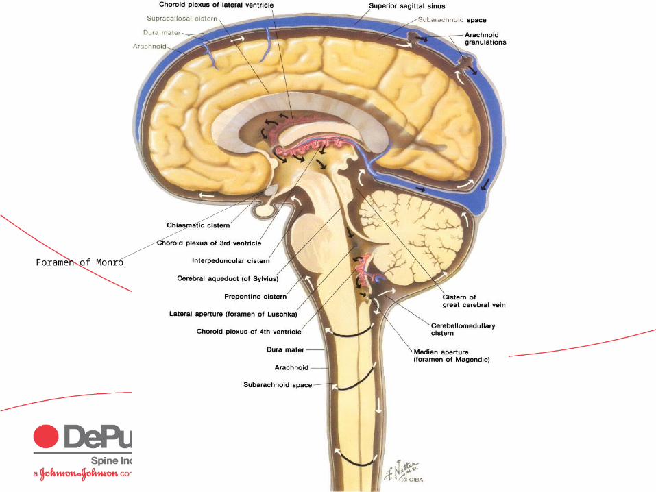
Foramen of Monro

Cranial Nerves
• I: Olfactory• II: Optic• III: Oculomotor• IV: Trochlear• V: Trigeminal• VI: Abducens
• VII: Facial• VIII:Vestibulocochlear
• Acoustic
• IX:Glossopharyngeal
• X: Vagus• XI: Accessory• XII: Hypoglossal
http://www.gwc.maricopa.edu/class/bio201/cn/cranial.htm
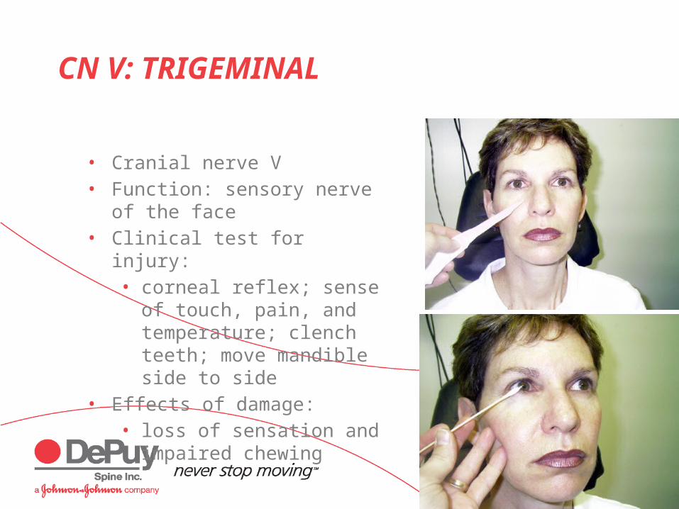
CN V: TRIGEMINAL
• Cranial nerve V• Function: sensory nerve of the
face• Clinical test for injury:
• corneal reflex; sense of touch, pain, and temperature; clench teeth; move mandible side to side
• Effects of damage: • loss of sensation and
impaired chewing

CN VI: ABDUCENS AND CN VII: FACIAL
• Cranial Nerve VI• Function: Eye
movements• Clinical test: lateral eye
movement• Effects of damage:
inability to rotate eye laterally; at rest – eye rotates medially because of action of antagonistic muscles
• Cranial Nerve VII• Function: facial
expression; sense of taste
• Clinical test: motor functions – close eyes, smile, whistle, frown, raise eyebrows; taste
• Effects of damage: inability to control facial muscles; distorted sense of taste
CN VIII: VESTIBULOCOCHLEAR
• Cranial Nerve VIII• Function: hearing and equilibrium• Clinical tests: test hearing, balance, and
ability to walk a straight line• Effects of damage: deafness, dizziness,
nausea, loss of balance, and nystagmus
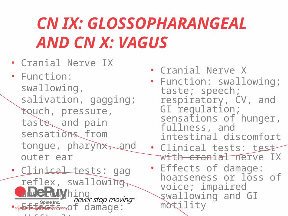
CN IX: GLOSSOPHARANGEAL AND CN X: VAGUS
• Cranial Nerve IX• Function: swallowing,
salivation, gagging; touch, pressure, taste, and pain sensations from tongue, pharynx, and outer ear
• Clinical tests: gag reflex, swallowing, and coughing
• Effects of damage: difficulty swallowing
• Cranial Nerve X• Function: swallowing; taste;
speech; respiratory, CV, and GI regulation; sensations of hunger, fullness, and intestinal discomfort
• Clinical tests: test with cranial nerve IX
• Effects of damage: hoarseness or loss of voice; impaired swallowing and GI motility
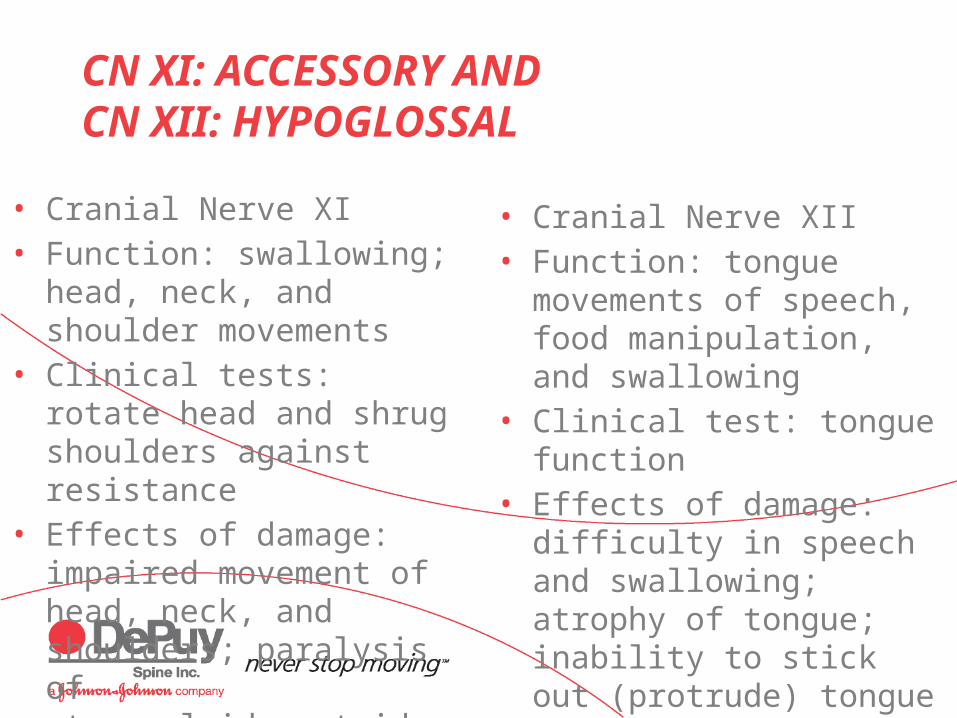
CN XI: ACCESSORY AND CN XII: HYPOGLOSSAL
• Cranial Nerve XI• Function: swallowing; head,
neck, and shoulder movements
• Clinical tests: rotate head and shrug shoulders against resistance
• Effects of damage: impaired movement of head, neck, and shoulders; paralysis of sternocleidomastoid
• Cranial Nerve XII• Function: tongue movements
of speech, food manipulation, and swallowing
• Clinical test: tongue function• Effects of damage: difficulty
in speech and swallowing; atrophy of tongue; inability to stick out (protrude) tongue
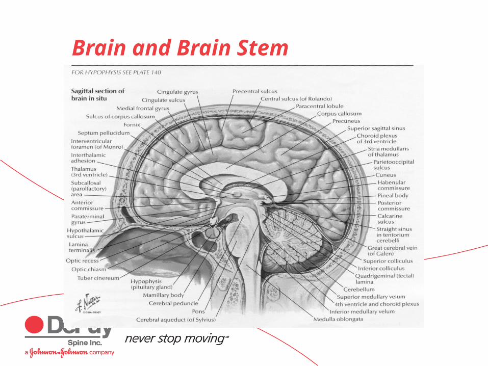
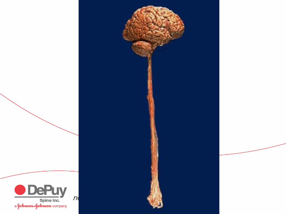
Brain and Brain Stem
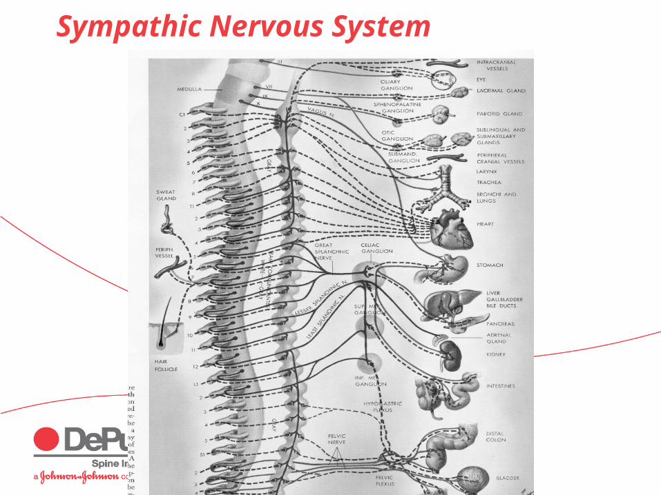
Sympathic Nervous System
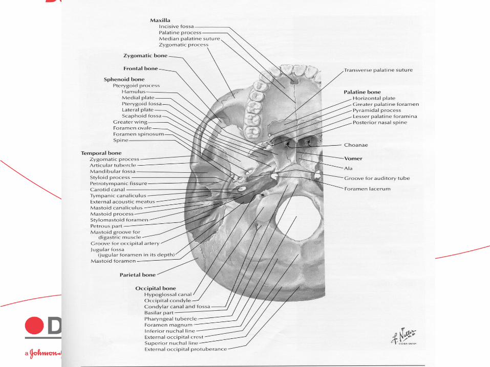
Base of Skull
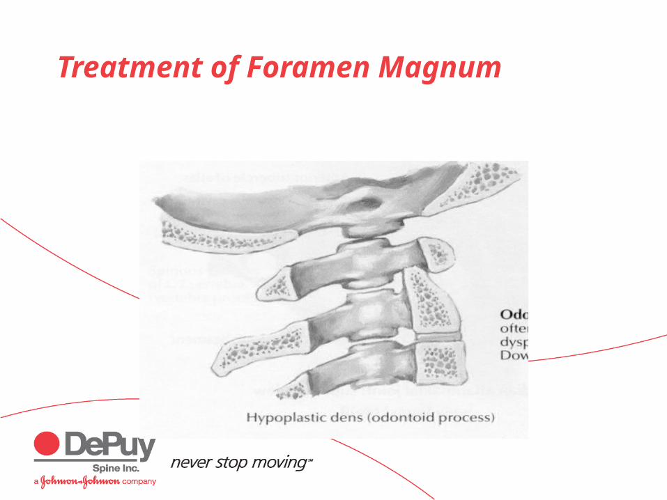
Treatment of Foramen Magnum
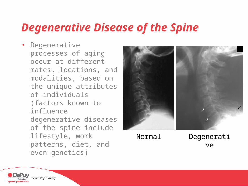
Degenerative Disease of the Spine
• Degenerative processes of aging occur at different rates, locations, and modalities, based on the unique attributes of individuals (factors known to influence degenerative diseases of the spine include lifestyle, work patterns, diet, and even genetics)
Normal Degenerative

Cervical Spondylosis
• Cervical spondylosis is a general term encompassing a number of degenerative conditions• Degenerative disc disease (DDD)• Spinal stenosis• With or without degenerative facet joints• With or without the formation of osteophytes• With or without a herniated disc
• One single component as a diagnosis is rare
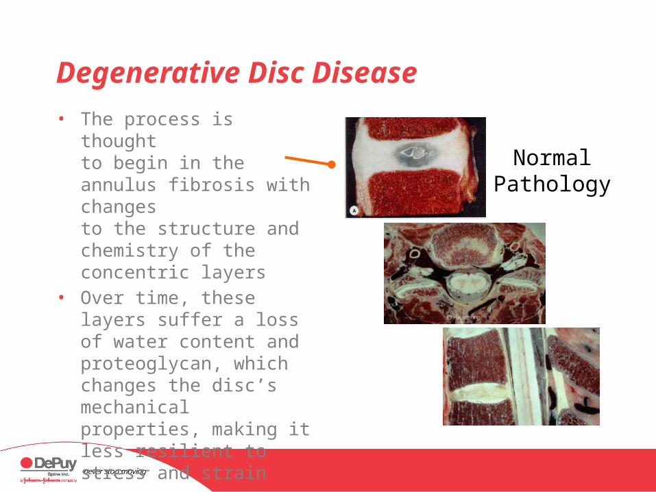
Degenerative Disc Disease
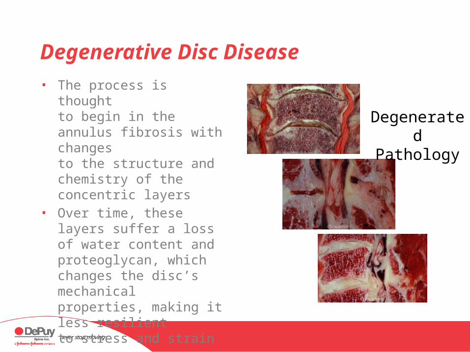
• The process is thought to begin in the annulus fibrosis with changes to the structure and chemistry of the concentric layers
• Over time, these layers suffer a loss of water content and proteoglycan, which changes the disc’s mechanical properties, making it less resilient to stress and strain
Normal Pathology
Degenerative Disc Disease
• The process is thought to begin in the annulus fibrosis with changes to the structure and chemistry of the concentric layers
• Over time, these layers suffer a loss of water content and proteoglycan, which changes the disc’s mechanical properties, making it less resilient to stress and strain
Degenerated Pathology

Degenerative Disease: Facet Joints
• Changes in disc structure and function can lead to changes in the articular facets, especially hypertrophy (overgrowth), resulting from the redirection of compressive loads from the anterior and middle columns to the posterior elements
Degenerative Disease: Osteophytes
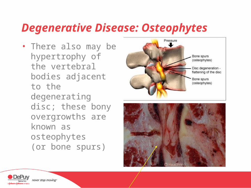
• There also may be hypertrophy of the vertebral bodies adjacent to the degenerating disc; these bony overgrowths are known as osteophytes (or bone spurs)
Herniated Nucleus Pulposus
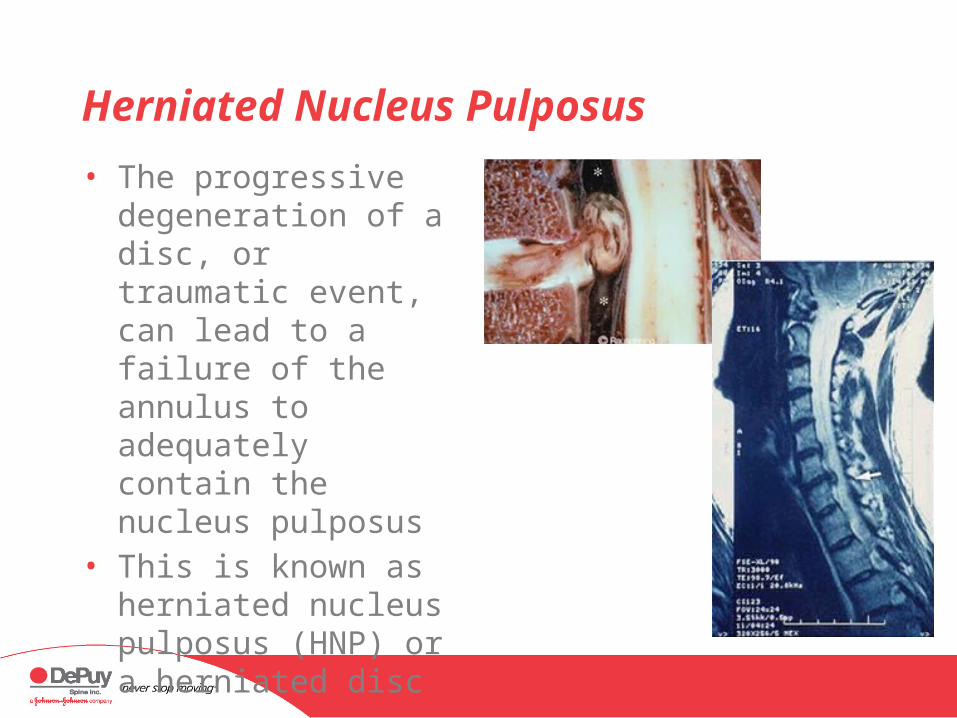
• The progressive degeneration of a disc, or traumatic event, can lead to a failure of the annulus to adequately contain the nucleus pulposus
• This is known as herniated nucleus pulposus (HNP) or a herniated disc

Herniated Nucleus Pulposus
• Varying degrees• Disc bulge
• Mild symptoms• Usually go away with
nonoperative treatment
• Rarely an indication for surgery
• Extrusion (herniation)• Moderate/severe
symptoms• Nonoperative treatment
Herniated Nucleus Pulposus
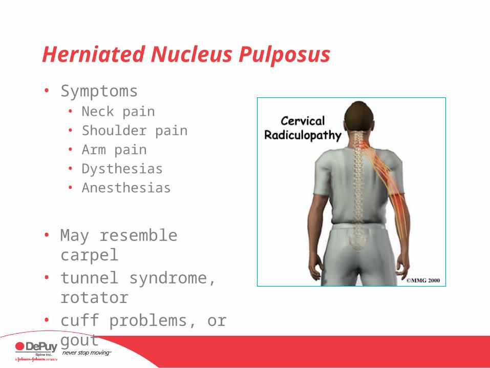
• Symptoms• Neck pain • Shoulder pain• Arm pain• Dysthesias• Anesthesias
• May resemble carpel • tunnel syndrome,
rotator • cuff problems, or gout
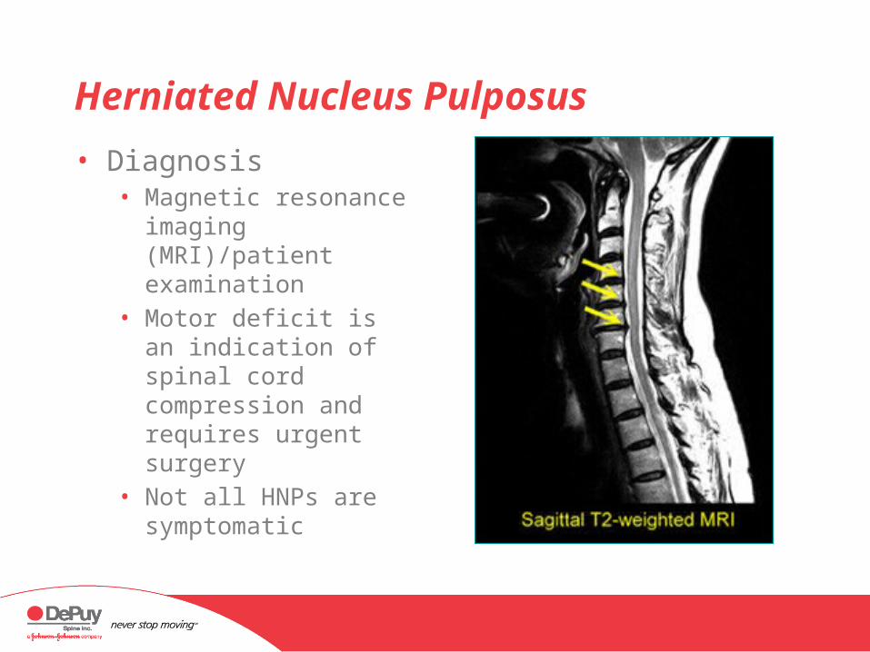
Herniated Nucleus Pulposus
• Diagnosis• Magnetic resonance
imaging (MRI)/patient examination
• Motor deficit is an indication of spinal cord compression and requires urgent surgery
• Not all HNPs are symptomatic

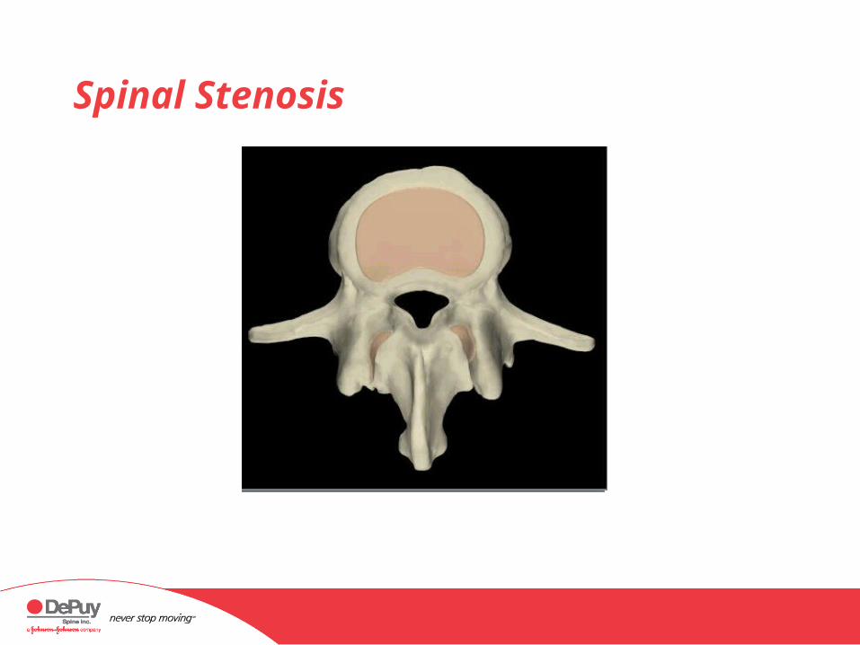
Spinal Stenosis
• Grouped as “spinal stenosis”• Central stenosis
• Narrowing of the central part of the spinal canal
• Foraminal stenosis• Narrowing of the
foramen resulting in pressure on the exiting nerve root
Spinal Stenosis
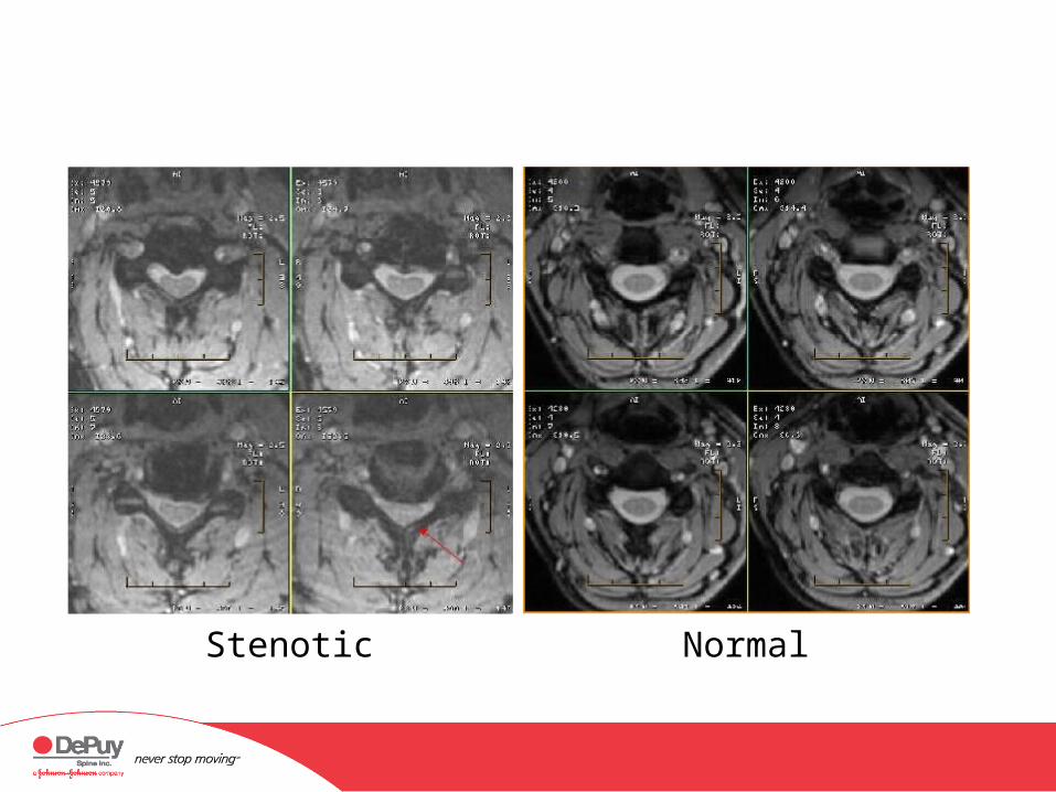
Stenotic Normal
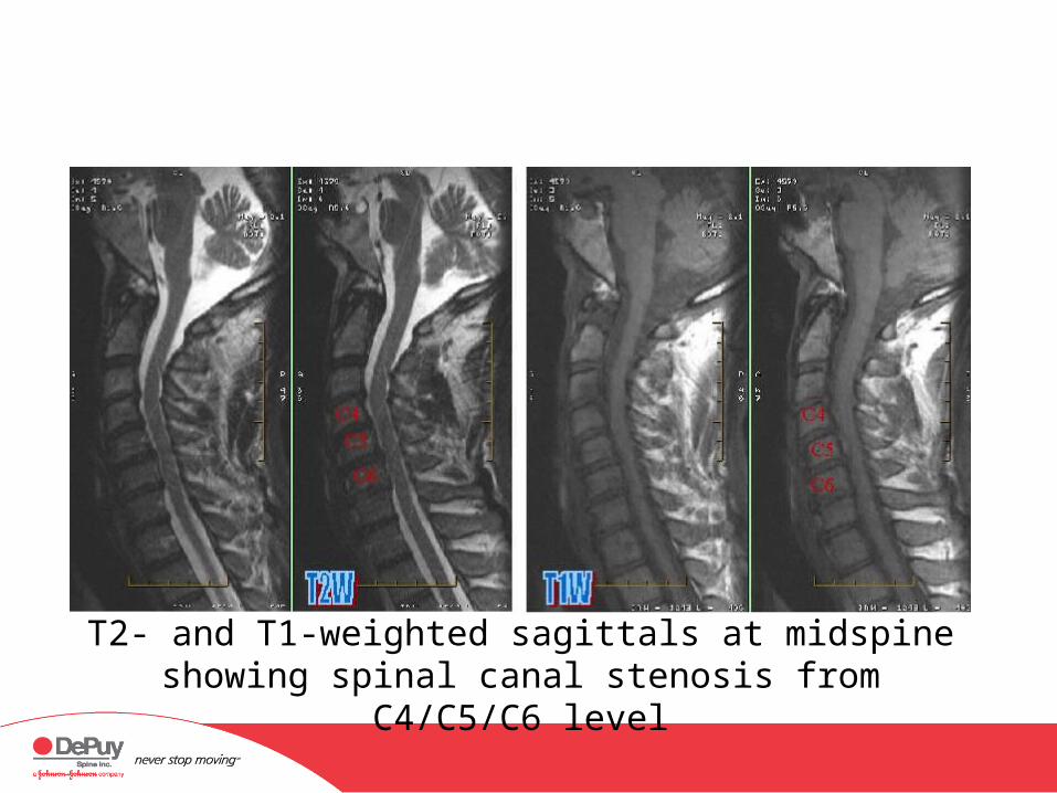
T2- and T1-weighted sagittals at midspine showing spinal canal stenosis from C4/C5/C6
level
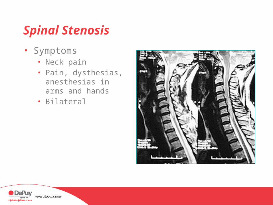
Spinal Stenosis
• Symptoms• Neck pain• Pain, dysthesias,
anesthesias in arms and hands
• Bilateral

Spinal Stenosis
• Diagnosis• MRI/computerized
tomography (CT)scan/patient examination
• Failure of nonoperative care —minimum 6 months• Rest, nonsteroidal anti-
inflammatory (NSAID) medication, physical therapy, epidural steroid injections
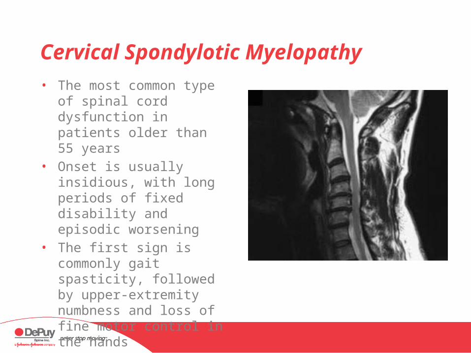
Cervical Spondylotic Myelopathy
• The most common type of spinal cord dysfunction in patients older than 55 years
• Onset is usually insidious, with long periods of fixed disability and episodic worsening
• The first sign is commonly gait spasticity, followed by upper-extremity numbness and loss of fine motor control in the hands
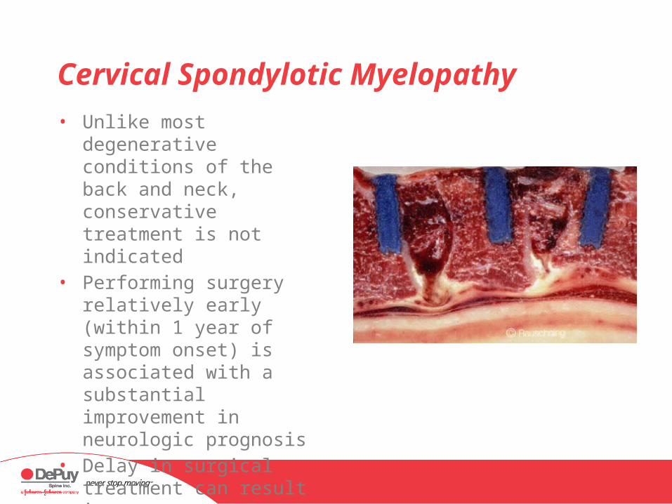
Cervical Spondylotic Myelopathy
• Unlike most degenerative conditions of the back and neck, conservative treatment is not indicated
• Performing surgery relatively early (within 1 year of symptom onset) is associated with a substantial improvement in neurologic prognosis
• Delay in surgical treatment can result in permanent impairment
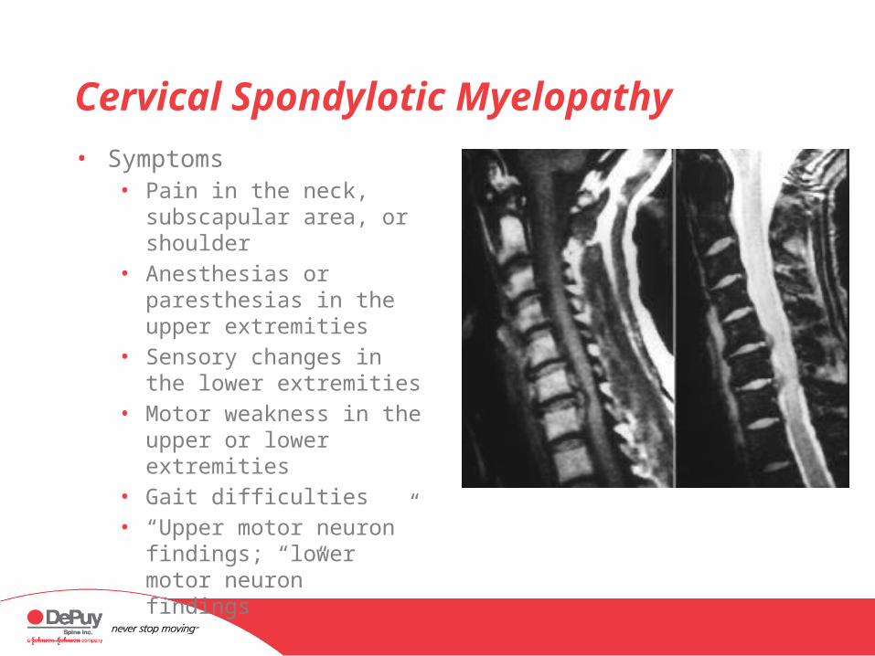
Cervical Spondylotic Myelopathy
• Symptoms• Pain in the neck,
subscapular area, or shoulder
• Anesthesias or paresthesias in the upper extremities
• Sensory changes in the lower extremities
• Motor weakness in the upper or lower extremities
• Gait difficulties• “Upper motor neuron”
findings; “lower motor neuron” findings

Cervical Spondylosis Without Myelopathy
• Surgical care• For radicular/neurologic symptoms• Not for axial neck pain• Dependent on the anatomy and the lordosis of the
affected segments, and surgeon preference• Anterior cervical discectomy and fusion• Anterior cervical corpectomy (multiple levels)• In some cases, adjunct posterior-instrumented fusion

Cervical Spondylotic Myelopathy
• Surgical care• Posterior cervical fusion—instrumented• Dependent upon the anatomy and the lordosis of
the affected segments, and surgeon preference• Posterior cervical fusion• Laminoplasty• May involve an adjunct anterior fusion procedure to
address spondylosis
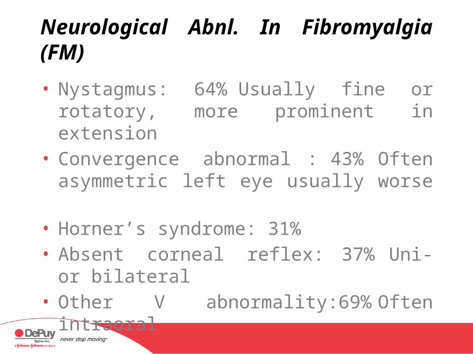
Neurological Abnl. In Fibromyalgia (FM)
• Nystagmus: 64% Usually fine or rotatory, more prominent in extension
• Convergence abnormal : 43%Often asymmetric left eye usually worse
• Horner’s syndrome: 31%• Absent corneal reflex: 37% Uni- or bilateral• Other V abnormality:69% Often intraoral
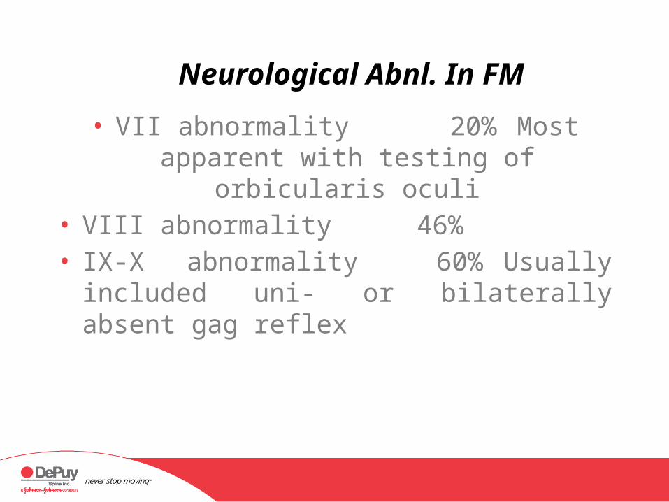
Neurological Abnl. In FM
• VII abnormality 20% Most apparent with testing of orbicularis oculi
• VIII abnormality 46%• IX-X abnormality 60% Usually included
uni- or bilaterally absent gag reflex
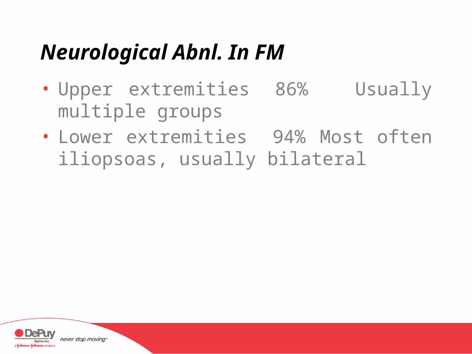
Neurological Abnl. In FM
• Upper extremities 86% Usually multiple groups
• Lower extremities 94% Most often iliopsoas, usually bilateral
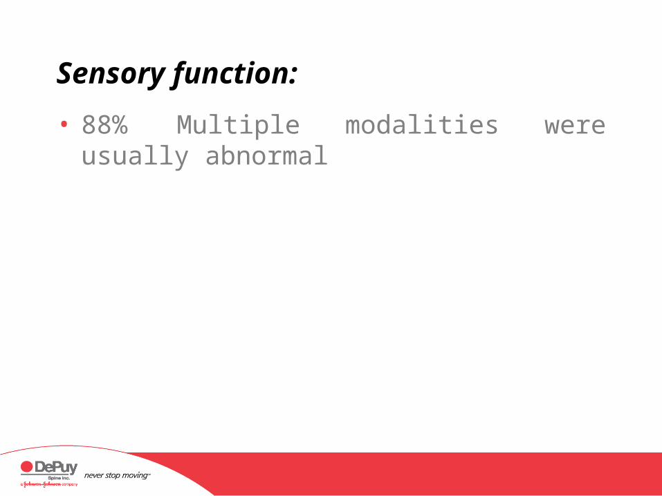
Sensory function:
• 88% Multiple modalities were usually abnormal
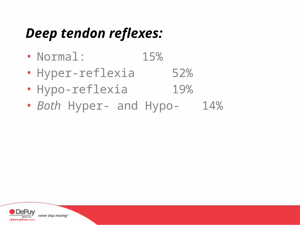
Deep tendon reflexes:
• Normal: 15%• Hyper-reflexia 52%• Hypo-reflexia 19%• Both Hyper- and Hypo- 14%

Pathological reflexes:
• Babinski’s response• Normal 40%• Equivocal
• Unilateral 14%• Bilateral 6%
• Present • Unilateral 15%• Bilateral 25%
Brainstem Summary
• The brainstem not only is adequate explanation for symptoms such as dysphagia, dysphonia, difficulty with balance and other, but it also harbors the centers for cardiovascular control, gastrointestinal control, respirations and the reticular activating system . The latter is particularly important with regard to sleep, arousal, attention and the subsequent incorporation of memory. The lower medulla also is the source of origin for descending nociceptive control fibers, which originate in the nucleus raphe magnus and paragigantocellularis .

FM and Neurological Deficit
• Fibromyalgia is not an explanation for neurological deficit, no matter how subtle it may be. Neurological abnormalities should invoke a search for treatable lesions: that search should focus on the upper spinal cord and brainstem based upon the observations presented here.
Conclusions:
• In patients with the previous diagnosis of Fibromyalgia who also had positive tilt table examinations for NMH/POTS, physical examination abnormalities were common.
• These abnormalities center about the lower • brainstem and upper spinal cord.• Fibromyalgia (and related syndromes) is not an
adequate explanation for neurological abnormality.
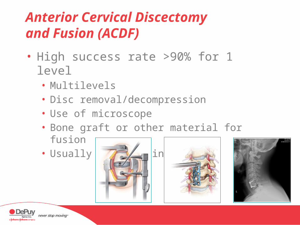
Anterior Cervical Discectomy and Fusion (ACDF)
• High success rate >90% for 1 level• Multilevels• Disc removal/decompression• Use of microscope• Bone graft or other material for fusion• Usually with plating

Anterior Cervical Corpectomy (and Fusion)
• Multilevel spondylosis/spondylotic myelopathy• Disc and vertebra removal• Decompression• Use of microscope• Bone graft or other
material for fusion• Always with plating
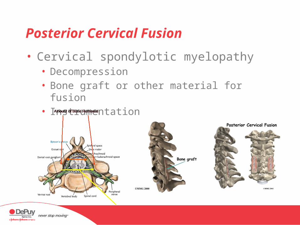
Posterior Cervical Fusion
• Cervical spondylotic myelopathy• Decompression• Bone graft or other material for fusion• Instrumentation
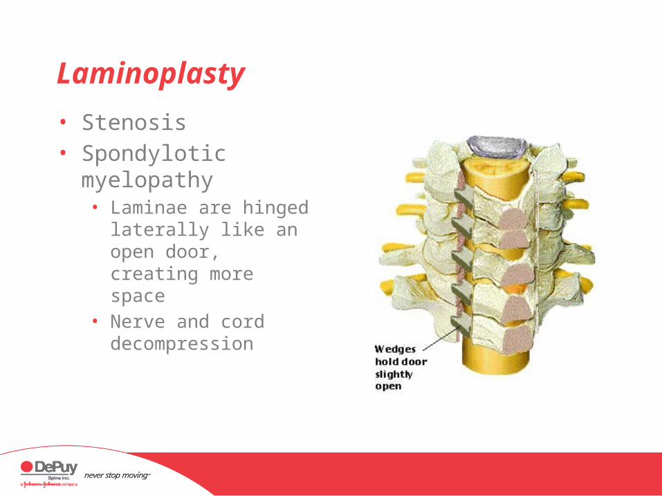
Laminoplasty
• Stenosis• Spondylotic myelopathy
• Laminae are hinged laterally like an open door, creating more space
• Nerve and cord decompression