Cerebrovascular accident is most common brain...
16
Transcript of Cerebrovascular accident is most common brain...


• Cerebrovascular accident is most common brain disorder also called a
stroke or brain attack.
• CVA is characterized by abrupt onset of persisting neurological
symptoms such as paralysis or loss of sensation that arise from
destruction of brain tissue.
• Causes for CVA-
• Intracranial cerebral haemorrage.
• Embolism.
• Atherosclerosis.

• Increased B.P.
• Increased blood cholesterol.
• Heart disease.
• Narrowed carotid arteries.
• Transient ischemic attacks.
• Diabetes.
• Smoking.
• Obesity.
• Excessive alcohol intake.

History
• Age- young patients consider-
• Cardiac disease- Infective endocarditis, Mitral valve stenosis.
• Vascular disease- HTN, vasculitis,arteritis of intracranial vessels.
• Aneurysm/ A-V Malformation.
• Intracranial space occupying lesion.
• Migraine.
• History of previous minor episodes-
may suggest emboli disease of arteries.
• Progressive cerebral arteriosclerosis.
• Migraine/ epilepsy- suggest intracranial A-v malformation.
• H/o intermittent claudication, bleeding tendency, diabetes- suggest intracranial tumour.

• H/o head injury- depressed fracture
subdural haematoma.
• H/o drugs- contraceptive pills.
Anti hypertensive drugs.
Anti coagulants.
• Family history- May give family history of stroke.
• Past history- DM, HTN or cardiac disease, anaemia or loss of
fluid

• Symptoms- • Mode of onset-
• Catastrophic- hemorrhage.
• Progressive- Thrombosis.
• Instantaneous- Embolism.
• Transient hemiplegia- focal neurological disturbances.
• Headache-
Cerebral haemorrhage- Intense with stiffness of neck.
Carotid insufficiency- temporal headache.
Basilar artery insufficiency- occipital or sub occipital.
Subaracnoid haemorrhage- sever.
Vomiting preceding a stroke favors diagnosis of haemorrhage.
• Chest pain- suggests associated myocardial infarction.

• Symptom suggesting hysterical hemiplegia-
• Hysterical gait.
• Onset after emotional shock.
• Hysterical type of rigidity.
• Contractions of platysma on affected side.
• Coma- subaracnoid haemorrhage & intra cerebral haemorrhage-
sudden or rapid loss of consciousness.
• Seizure- in tumor.
• Fever- meningitis, cerebral abscess, encephalitis.
• Involuntary movements- in encephalitis & chorea.
• Mental symptoms, abdominal pain & malena

• Neurological-
• State of consciousness- full conscious- stupor, semiconscious or coma.
• Speech- slurred dysarthria.
Dysphagic speech.
• neck rigidity.
• Eyes- nystagmus.
pupil-carotid thrombosis/brain stem disease- ipsilateral Horner’s syndrome.
Pupillary enlargement- early paralysis of 3rd CN.
• Focal neurological deficit- test for hemi paresis, hemi sensory loss.

• General-
• Blood pressure- for arterial HTN.
• Arterial pulse- for peripheral vascular disease.
• Bruits- over carotid & subclavian artery- due to stenosis.
• Signs of head injury.
• Opthalmodynamometry- to record ophthalmic artery pressure
(difference in two ophthalmic arteries would suggest a disease
in internal carotid artery on side of low pressure).

• CT Scan- Indication- To establish pathological diagnosis-
Infarction.
Haematoma.
Tumour.
• MRI- more sensitive to small area of ischemia than CT.
• MRA- for non invasive detection of carotid artery stenosis &
occlusion.
to image distal vertebral & intracranial vessels.
• Digital subtraction angiography-
to confirm occlusion.
to diagnose source of bleeding insubarachnoid &
intracranial
hemorrhage.
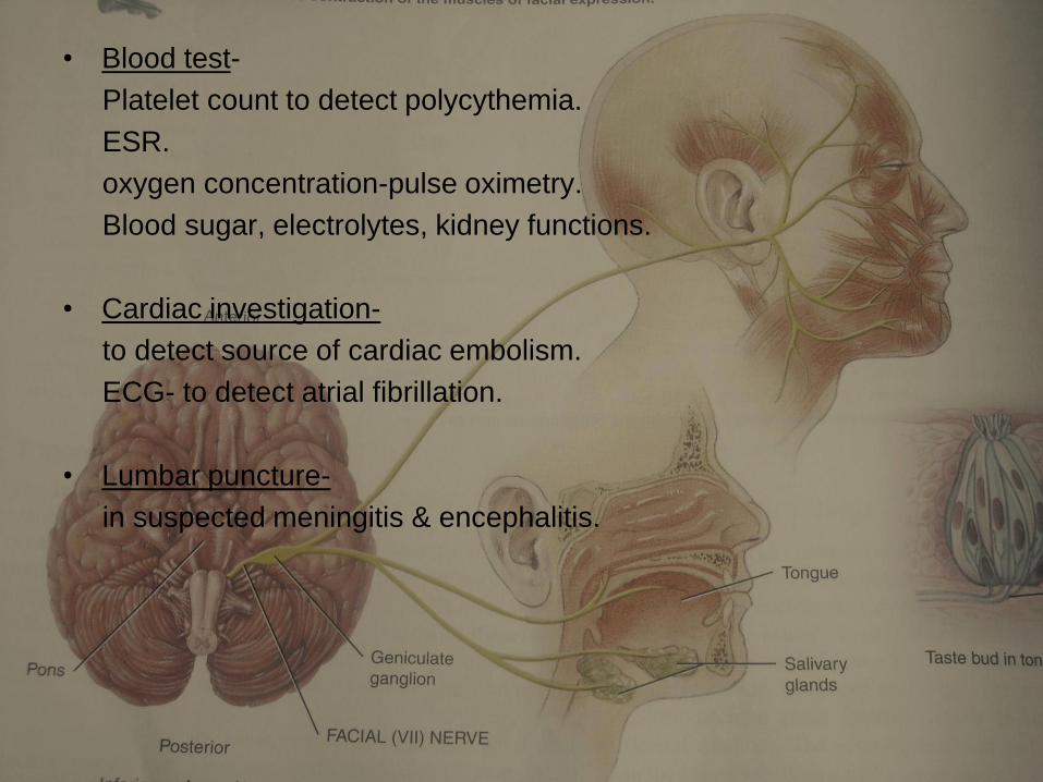
• Blood test-
Platelet count to detect polycythemia.
ESR.
oxygen concentration-pulse oximetry.
Blood sugar, electrolytes, kidney functions.
• Cardiac investigation-
to detect source of cardiac embolism.
ECG- to detect atrial fibrillation.
• Lumbar puncture-
in suspected meningitis & encephalitis.

Differential diagnosis of vascular causes-
Embolism Thrombosis Haemorrhage
Age young Middle/old Middle/old
Nature of onset instantaneous Sudden/progressiv
e
Catastrophic.
Premonitory
symptoms
Absent Difficulty in
speaking,
weakness of arm or
leg
Absent.
Common cause Mitral stenosis with
atrial fibrillation or
carotid stenosis.
Arteriosclerosis
with or without HTN
HTN almost
invariable.

Clinical feature.
headache
variable Slight or absent Severe.
Vomiting at onset rare rare common
Convulsions. common rare Common
coma Rarely deep Varies with extent of
thrombosis.
Deep
Cheyne strokes
breathing.
Not common seldom Common.
Stiff neck rare rare Frequent.
Conjugate deviation
of eyes
rare Seldom. Frequent.
Reaction of pupil to
light.
No changes May be impaired. Commonly impaired.
Blood pressure normal May be high. Usually high.
Bilateral extensor
plantar
rare,. May be present. Frequent.
CSF Usually normal Clear, pressure
slightly increased.
Usually bloody,
pressure increased
CT scan or MRI Infarction may not
appear for 2-4 days.
May not appear for
2-4 days.
Can be confirmed
within minutes.
Termination Recover usually Recover often Rapid deterioration
high mortality

Location of lesion-
• Cortex – Flaccid hemiplegia, Aphasia is common convulsions may occur.
• Internal capsule- commonest site,
Hemiplegia,
no loss of consciousness.
spasticity marked.
(Hemianaesthesia-post 1/3rd).
• Thalamus- Impairment of superficial & loss of deep sensation on opp. side of lesion, ataxia, tremors etc.
• Midbrain- upper level- 3rd nerve palsy.
lower level- 3rd nerve affection on side of lesion, ataxia &
hypertonia.-opp. Side.
• Pons- Millard- Gubbler syndrome, Foville’s Syndrome, Avellis’s syndrome, Horner’s syndrome.
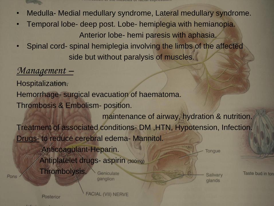
• Medulla- Medial medullary syndrome, Lateral medullary syndrome.
• Temporal lobe- deep post. Lobe- hemiplegia with hemianopia.
Anterior lobe- hemi paresis with aphasia.
• Spinal cord- spinal hemiplegia involving the limbs of the affected
side but without paralysis of muscles.
Management – Hospitalization.
Hemorrhage- surgical evacuation of haematoma.
Thrombosis & Embolism- position.
maintenance of airway, hydration & nutrition.
Treatment of associated conditions- DM ,HTN, Hypotension, Infection.
Drugs- to reduce cerebral edema- Mannitol.
Anticoagulant-Heparin.
Antiplatelet drugs- aspirin (300mg)
Thrombolysis.

• Ayurvedic correlation- Pakshaghaata
Pakshavadha
Ardhangavaata