![CEREBRAL CIRCULATION AND CEREBROSPINAL FLUID [CSF]](https://static.fdocuments.in/doc/165x107/56814ee4550346895dbc77ad/cerebral-circulation-and-cerebrospinal-fluid-csf.jpg)
Cerebrospinal fluid (CSF) - ECVN › edu › nc › 2014 › handouts › bc › files ›...
20
1 Cerebrospinal fluid (CSF) The CSF examination Andrea Tipold TiHo Hannover Review IVIS Cerebrospinal fluid - history Presence of fluid in the cavities: known to the ancients first report 17th century B.C. Hippocrates 4th century B.C. Galen 2nd century A.D.: description of ventricular cavities first examinations in animals: 1825 by Magendie Cerebrospinal fluid - history 2 1891 Quincke: diagnostic and therapeutic aid 1900 cytology 2011: nearly the same techniques: CYTOLOGY, PROTEIN-CONTENT
Transcript of Cerebrospinal fluid (CSF) - ECVN › edu › nc › 2014 › handouts › bc › files ›...
1
Cerebrospinal fluid (CSF)
The CSF examination
Andrea TipoldTiHo Hannover
Review IVIS
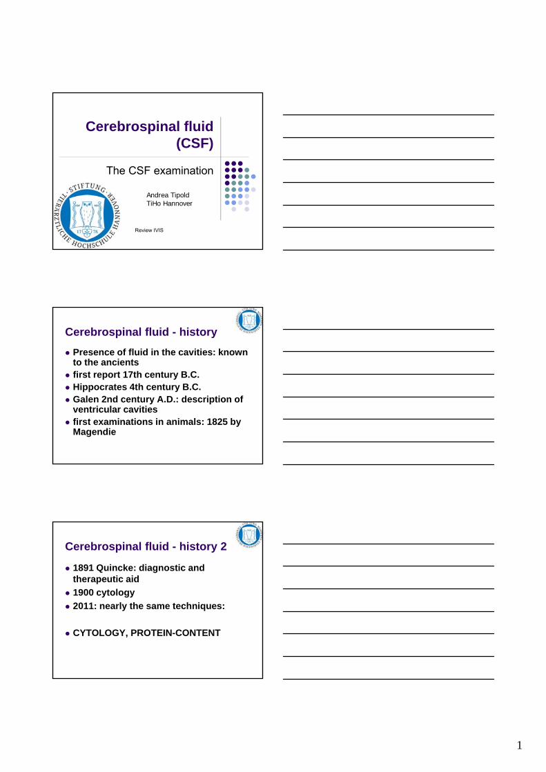
Cerebrospinal fluid - history
Presence of fluid in the cavities: knownto the ancients
first report 17th century B.C. Hippocrates 4th century B.C. Galen 2nd century A.D.: description of
ventricular cavities first examinations in animals: 1825 by
Magendie
Cerebrospinal fluid - history 2
1891 Quincke: diagnostic andtherapeutic aid
1900 cytology
2011: nearly the same techniques:
CYTOLOGY, PROTEIN-CONTENT

2
CSF – what do you know about the physiology ?
Cerebrospinal fluid - anatomy
CSF: ventricular system, subarachnoid space
cranial cavity – closed space, continuous adjustment of the intracranial pressure
pressure: brain parenchyma, CSF, blood
Intracranial pressure
Brain-tissue
Blood
CSF

3
1. Plexus choroideus2. ependymal lining3. pia-glial membrane4. blood vessels in the
pia-arachnoid
Production of CSF
Fig Vandevelde
Cerebrospinal fluid -physiology
Formation of the CSF: formation rate: 0,002 ml/minute in rats; 0,35
ml/minute in man various species: 0,2 to 0,5 ml/minute/gm choroid plexus: secretion directly related to
sodium transport (ATPase), filtration, active transport (Vitamin C,B...)
transependymal formation
Tight junction
ventricles
Active transport
Vitamin CRibonucleosides etcfolatesVitamin B6
Ion exchange (facilitated or active)
Na+
Cl-
H+CO2 and OH-

4
Cerebrospinal fluid -physiology
CSF circulates from the ventricular system to the subarachnoid space
caudal flow (spinal cord, central canal)
circulating flow
cranial flow
Cerebrospinal fluid -physiology
Choroid plexus: „ectopic renal tubular epithelium“ active transport of metabolites (e.g. organic
acids) from CSF to blood absorption: equals to formation (dependent
upon hydrostatic pressure) arachnoid villi (major place), choroid plexus,
diffusion into brain and brain capillaries
Cerebrospinal fluid - functions
physical support (brain - water content 80%)
protection of the brain from acute changes in central venous pressure
excretory function (lactate, hydrogen ions....)
intracerebral transport (hormones, cytokines - research)

5
Cerebrospinal fluid -composition
watery solution (99% water)
ions (different concentration than plasma)
nutrients, neuroendocrine substances and neurotransmitter
Osmolality: same as plasma (289 mOsm/L)
glucose 80% of plasma
protein: < 25mg/dl (mostly albumin)
0-3 cells/ul, mostly lymphocytes
Cerebrospinal fluid -acquisition
Cisternal puncture
22 gauge, 1,5 inch
Fig. S.Wheeler
Cerebrospinal fluid -acquisition
cisternal puncture pressure – spinal manometer ? amount of CSF: 1 ml / 5 kg 1-2 ml cell count + protein measurement: 100
– 200 ul CSF puncture of radicular vessels: blood
contamination
6
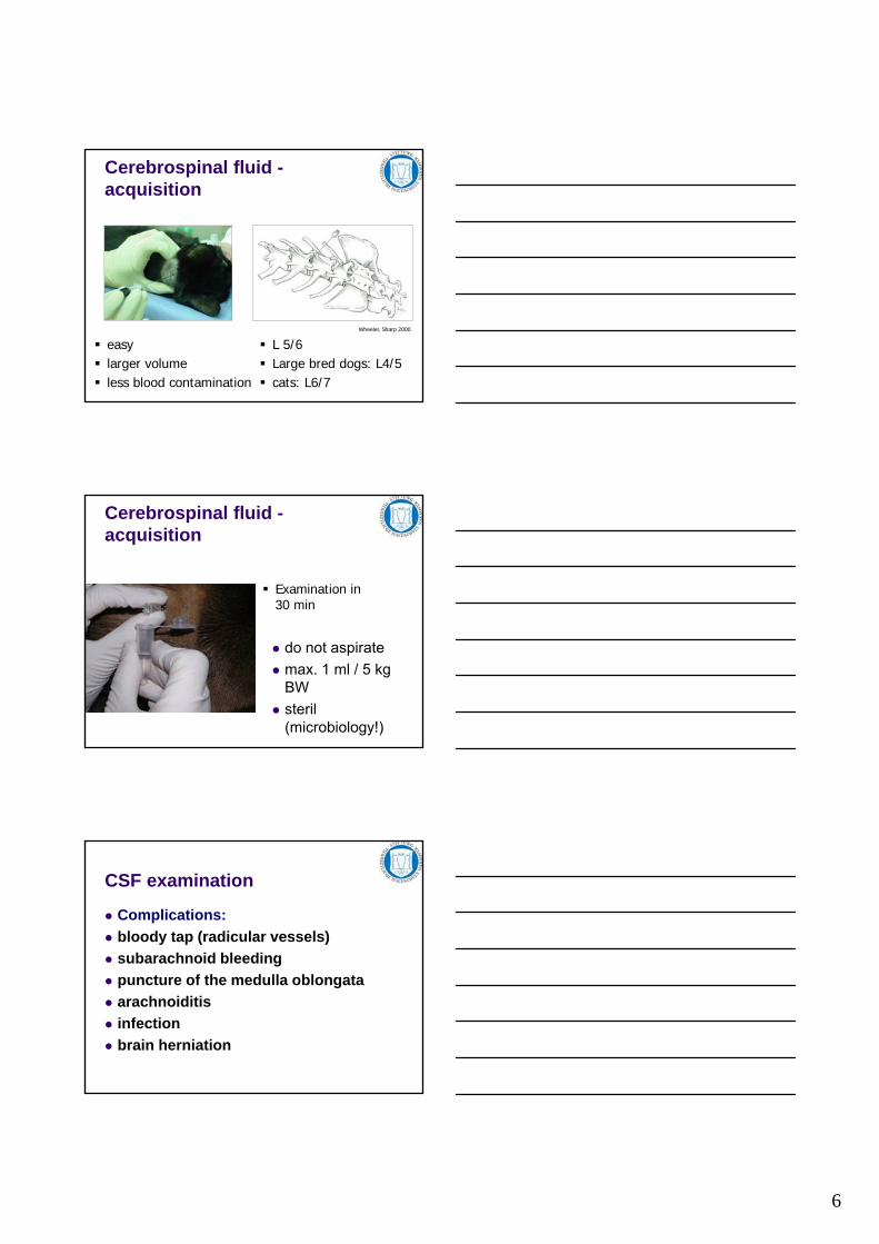
Cerebrospinal fluid -acquisition
easy larger volume less blood contamination
L 5/6 Large bred dogs: L4/5 cats: L6/7
Wheeler, Sharp 2000
Cerebrospinal fluid -acquisition
do not aspirate
max. 1 ml / 5 kg BW
steril (microbiology!)
Examination in30 min
CSF examination
Complications:
bloody tap (radicular vessels)
subarachnoid bleeding
puncture of the medulla oblongata
arachnoiditis
infection
brain herniation

7
CSF examination
Pressure (dogs 5-12 mm Hg under general anaesthesia)
colour and viscosity
cell count
differential cell count
protein
CSF colour
• watery, clear, colourless• cloudy (> 500 cells / ul)• viscous: high protein content• red• Xanthochromia
Protein content
< 25 mg / dl
protein entry: mainly pinocytosis (albumin ca. 20 hours for equilibration)
protein exit: 200 times the entry rate
Pandy reaction (10% carbolic acid)
turbidometric methods: trichloracetic acid, benzethonium chloride
nephelometry

8
Pandy reaction
Pandy reaction
Protein
normal: < 25 mg/dl occipital< 40 mg/dl lumbal
Blood contamination:– 1 mg/dl / 1000 erythrocytes
9
Elevated protein
nonspecific indicator of CNS disease
damaged blood-brain barrier
increased local IgG production
inflammatory/infectious
toxic/metabolic
vascular
neoplastic
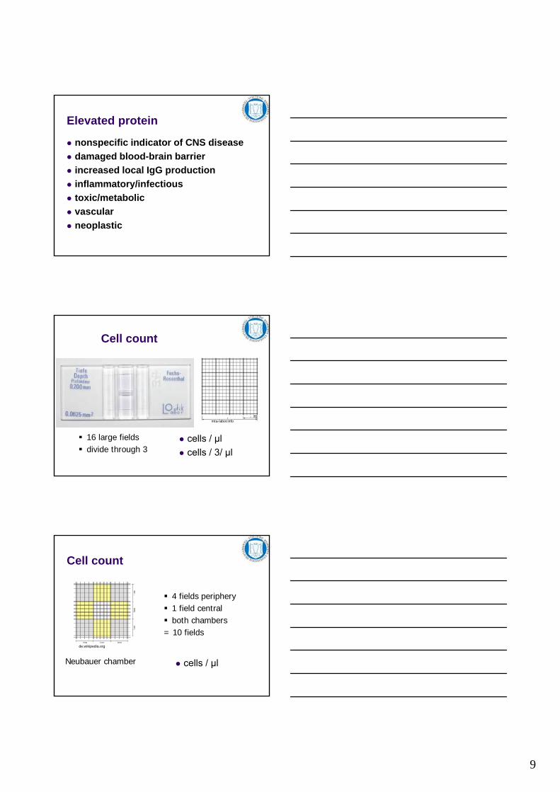
Cell count
cells / µl
cells / 3/ µl
16 large fields divide through 3
mta-labor.info
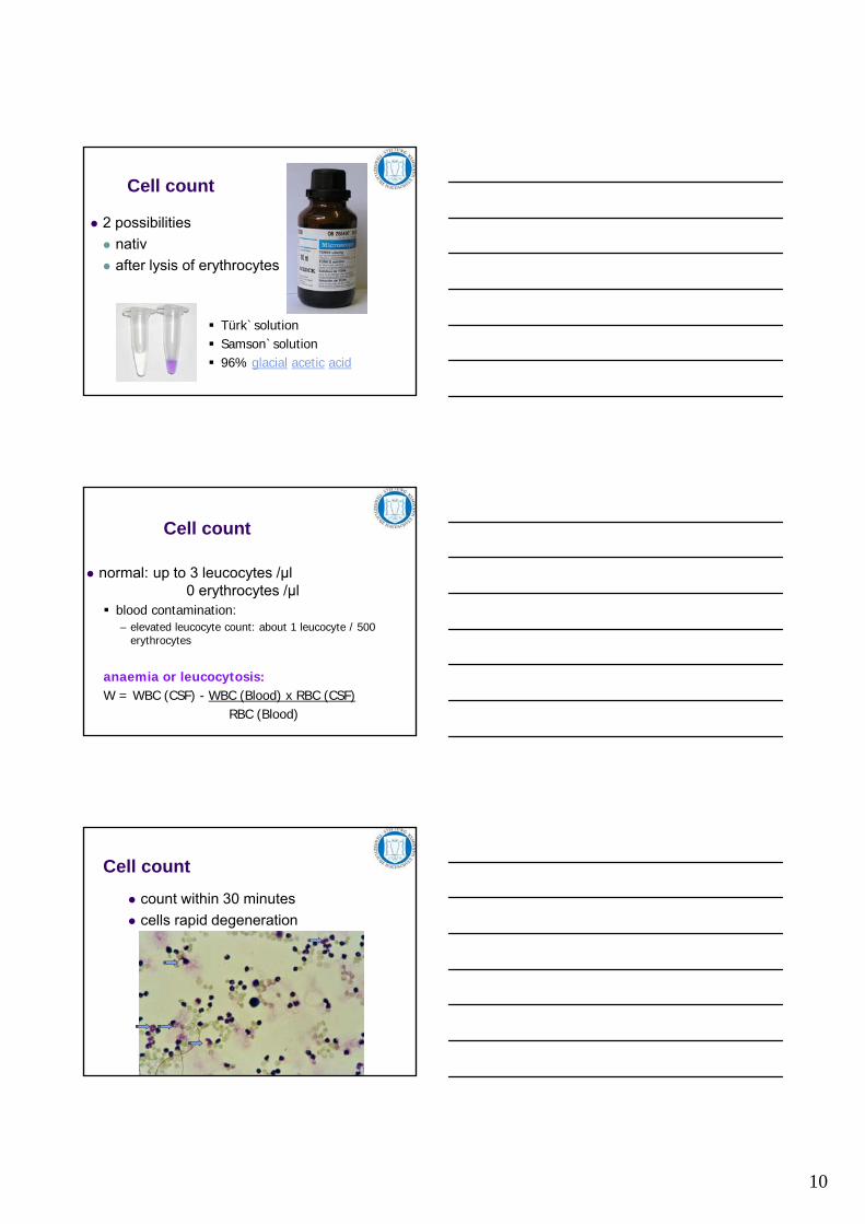
Cell count
cells / µl
4 fields periphery 1 field central both chambers= 10 fields
de.wikipedia.org
Neubauer chamber
10
Cell count
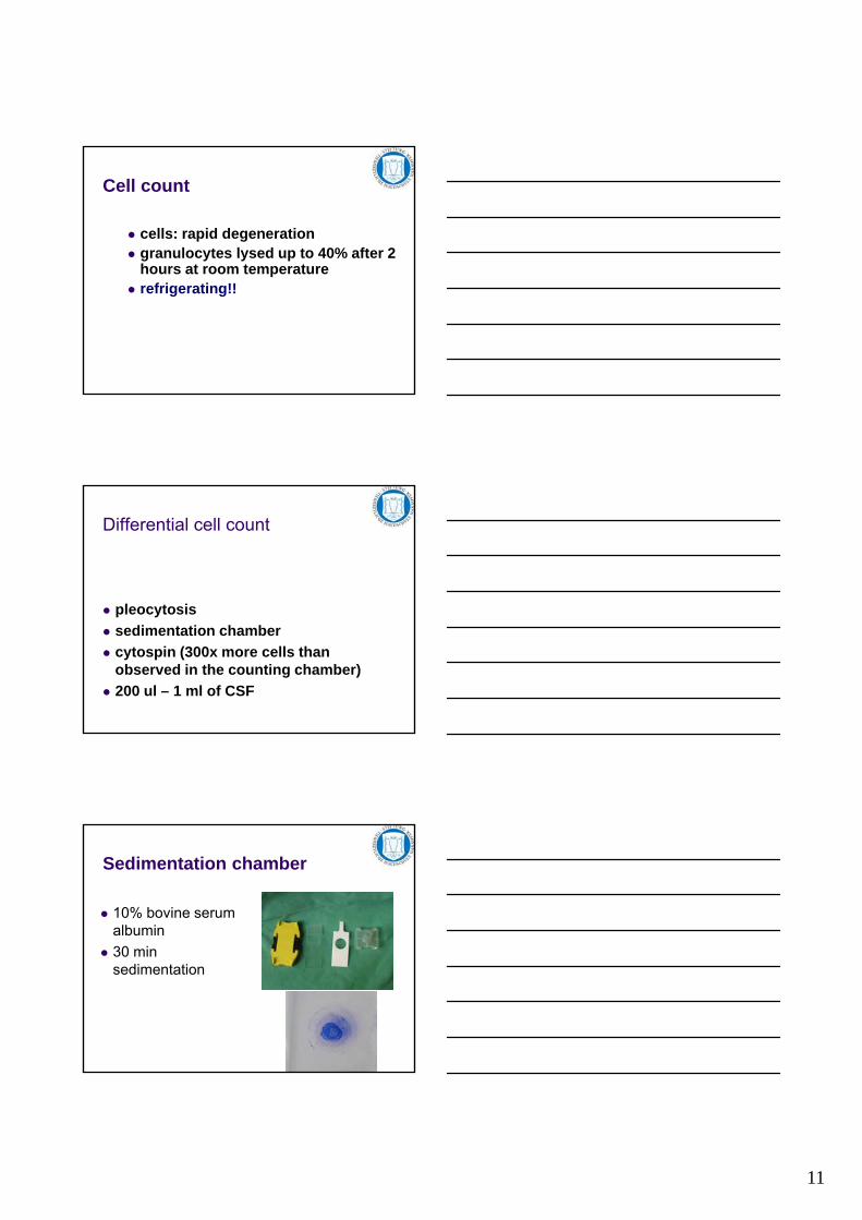
2 possibilities
nativ
after lysis of erythrocytes
Türk`solution Samson`solution 96% glacial acetic acid
Cell count
normal: up to 3 leucocytes /µl0 erythrocytes /µl
blood contamination: – elevated leucocyte count: about 1 leucocyte / 500
erythrocytes
anaemia or leucocytosis:W = WBC (CSF) - WBC (Blood) x RBC (CSF)
RBC (Blood)
Cell count
count within 30 minutes
cells rapid degeneration
11
Cell count
cells: rapid degeneration granulocytes lysed up to 40% after 2
hours at room temperature refrigerating!!
Differential cell count
pleocytosis
sedimentation chamber
cytospin (300x more cells than observed in the counting chamber)
200 ul – 1 ml of CSF
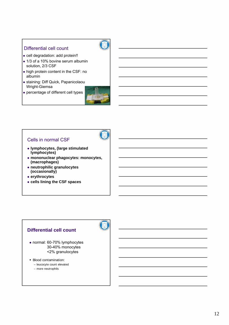
Sedimentation chamber
10% bovine serum albumin
30 min sedimentation
12
Differential cell count
cell degradation: add protein!!
1/3 of a 10% bovine serum albumin solution, 2/3 CSF
high protein content in the CSF: no albumin
staining: Diff Quick, Papanicolaou Wright-Giemsa
percentage of different cell types
Cells in normal CSF
lymphocytes, (large stimulated lymphocytes)
mononuclear phagocytes: monocytes, (macrophages)
neutrophilic granulocytes (occasionally)
erythrocytes cells lining the CSF spaces
Differential cell count
normal: 60-70% lymphocytes30-40% monocytes<2% granulocytes
Blood contamination:– leucocyte count elevated– more neutrophils
13
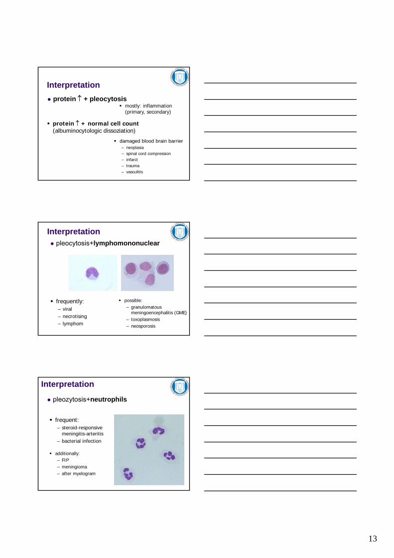
Interpretation
protein + pleocytosis
protein + normal cell count(albuminocytologic dissoziation)
mostly: inflammation (primary, secondary)
damaged blood brain barrier– neoplasia– spinal cord compression– infarct– trauma– vasculitis
Interpretation pleocytosis+lymphomononuclear
frequently:– viral– necrotising– lymphom
possible:– granulomatous
meningoencephalitis (GME)– toxoplasmosis– neosporosis
Interpretation
pleozytosis+neutrophils
frequent:– steroid-responsive
meningitis-arteritis– bacterial infection
additionally:– FIP– meningioma– after myelogram

14
Interpretation
pleozytosis+eosinophils
mostly:– parasitic infection– protocoal infection– idiopathic(eosinophilic encephalitis)
rare finding
Interpretation
pleocytosis + mixed cell population
every cell typ < 50%
frequently:– GME– FIP– protocoal infection– rickettsia– fungal infection
additionally:– infarct– myelomalacia
• elevated number of macrophages: spinal cord injury(Srugo et al, 2011, JVIM)
CSF examination (2)
Additional examinations:
glucose
CK
IgG-Index
IgA
lactate and pyruvate
15
Special examinations
Glucose in CSF
normal: about 2/3 of glucose level in serum
low levels:– high cell count in CSF– bacteria
Weber et al, 2012
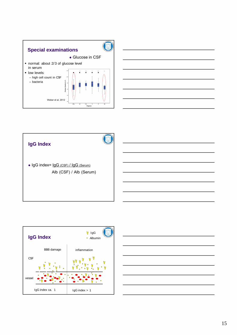
IgG Index
IgG index= IgG (CSF) / IgG (Serum)
Alb (CSF) / Alb (Serum)
IgG Index
CSF
vessel
IgG
Albumin
BBB damage inflammation
IgG index ca. 1 IgG index > 1

16
What else
Myelin basic protein: tissue destruction
S-100 protein: unspecific
C-reactive protein: acute reactant increased barrier permeability
search for biomarkers
Tau protein (intracellular protein, microtubuli, nerve cells - axons)
Tau protein spinal cord injury
control grade 2/3 grade 4/50
100
200
300
400
500
500600700800
*
*
Tau
in p
g/m
l
neurologic improvement no or slower improvement0
100
200
300
400
500
600
700
800
*
Tau
in p
g/m
l
p = 0,016
p = 0.025 p =0.033
What else
numerous metabolites
enzymes
neurotransmitter (GABA, glutamate, acetylcholinesterase)
neuropeptides (orexin, hypocretin –narcolepsy)
cytokines

17
50
results comparison
IVDD
Macrophage inflammatory protein 3-β (MIP-3β) /CCL19
(chemokine)
Asterisks: statistically significant differences* P < 0.05 ** P < 0.01 *** P < 0.005
Log 10 of MIP-3b (pg/ml)
CSF concentrations
Bartels et al 2014
antigen detection
etiology
bacterial or fungal organism –microscopic evaluation
Culture
PCR

18
antigen detection
Viral encephalitis
PCR
Staining techniques
Antibody detection
specific antibodies in the CSF
mostly not diagnostic
serial serum determinations
evaluation of specific indices
Antibody detection example
Central European tick borne encephalitis
Most experience in men
75 % pos IgM
100 % pos IgG
10/12 pos IgG with other diseases

19
Antibody detection FIP (Böttcher, Fischer 2003)
• 67 CSF samples• 12 IgG pos• 6 FIP with CNS involvement• 4 FIP without any CNS involvement• 2 with other CNS diseases• 4 false negativ
Immunphenotyping of lymphocytes
Research
Diagnosis?

20
CD3 CD21