Cerebral Angiography Jessica Birt CIT Noah’s Ark-----
17
Cerebral Angiography Jessica Birt CIT Noah’s Ark-----
-
Upload
alfred-tooke -
Category
Documents
-
view
230 -
download
2
Transcript of Cerebral Angiography Jessica Birt CIT Noah’s Ark-----

Cerebral Angiography
Jessica Birt
CIT
Noah’s Ark-----

Introduction
• Angiography: study of blood vessels, by the use of contrast to make the vessels visible under fluoroscopic procedures.
• Cerebral angiography: specifically looking at the vessels of the brain– Done to detect abnormalities, blockages, or
for therapeutic reasons

Indications
• Narrowing or blockage• Acute stroke• Therapeutic reasons• “Map” of the brain prior to
neurosurgery• Bleeding within the skull• Inflammation
• Bulges in arterial walls/ Aneurysms
• Blood clots in the brain• Intracerebral hemorrhage• Arteriovenous & fistulae
malformations• Highly vascular extra-
and intra cranial tumors

Bleeding Brain

Cerebral Aneurysm

Risks
• Internal bleeding and hemorrhage– Involves puncturing an artery
• Stroke or heart attack– Blood clots or plaque dislodge forming a
blockage
• Allergic reaction– To contrast

Contraindications
–Pregnancy

–Coagulation ability
–Kidney disease

AnatomyMajor vessels involved in cerebral angiography
include:
–Carotid arteries: supply blood to the anterior and middle portions of the brain
–Vertebral arteries: supply blood to the posterior portions of the brain
–Jugular veins: venous drainage of the brain
–Femoral artery: accessed site for arterial puncture

Preprocedural Care and Preprocedural Care and CautionsCautions
• Review patients medical history and lab resultsReview patients medical history and lab results• This will give the physician a better understanding of the This will give the physician a better understanding of the
condition of the patientcondition of the patient
• Establish and understandingEstablish and understanding• Patient needs to know what to expect, risks, complications Patient needs to know what to expect, risks, complications
etc.etc.
• Must have permission in the form of written Must have permission in the form of written informed consentinformed consent
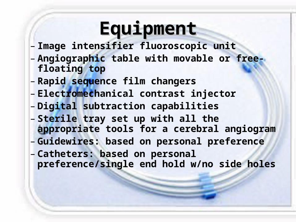
EquipmentEquipment– Image intensifier fluoroscopic unit– Angiographic table with movable or free-
floating top– Rapid sequence film changers– Electromechanical contrast injector– Digital subtraction capabilities– Sterile tray set up with all the appropriate
tools for a cerebral angiogram– Guidewires: based on personal preference– Catheters: based on personal
preference/single end hold w/no side holes

Contrast
• Past: used ionic iodinated contrastPast: used ionic iodinated contrast
• Today: use nonionic iodinated contrastToday: use nonionic iodinated contrast– Iohexol, iopamidol, ioversal, imagopaqueIohexol, iopamidol, ioversal, imagopaque
Amount injected depends on condition, Amount injected depends on condition, age, weight and pathology of vesselsage, weight and pathology of vessels
Procedure Details
• Local anesthetic at puncture site
• Small incision over the femoral artery
• Needle placed into vessel using Seldinger Technique
• Guidewire placement• Catheter placement• Contrast injected

Common Filming PositionsCommon Filming Positions
• Carotid Arteries: Carotid Arteries: anteroposterior axial anteroposterior axial laterallateral
anteroposterior anteroposterior obliqueoblique
• Vertebral Arteries:Vertebral Arteries:
anteroposterior axialanteroposterior axial
laterallateral
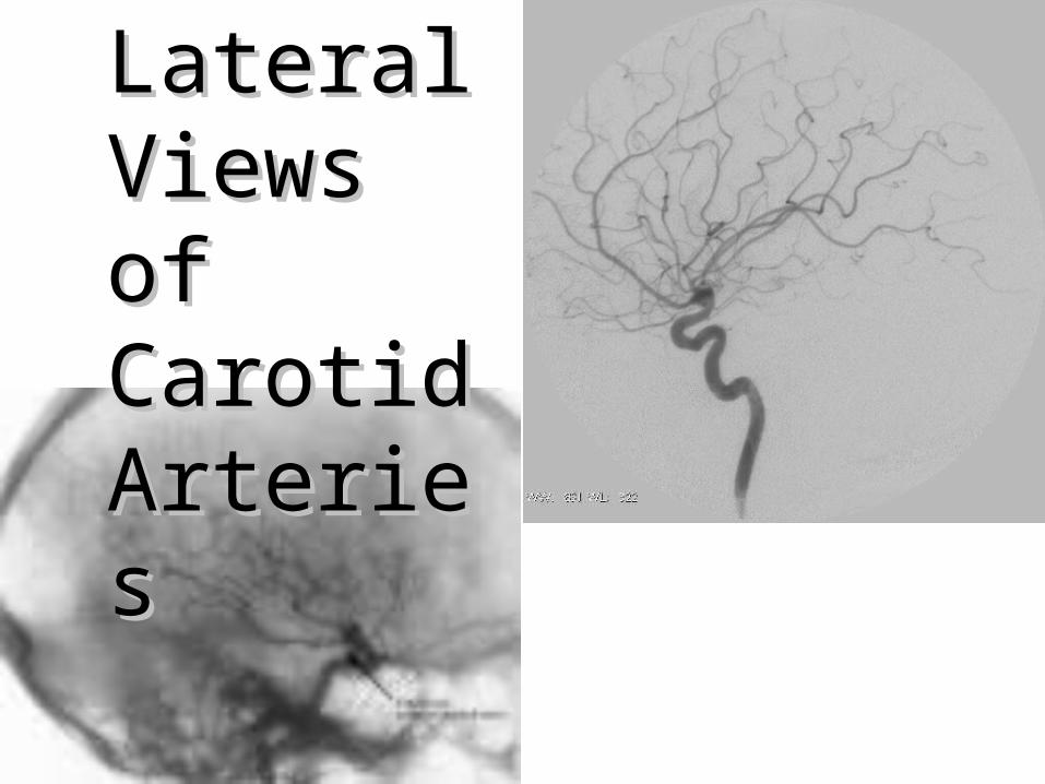
Lateral Lateral Views of Views of Carotid Carotid ArteriesArteries

Digital SubtractionDigital Subtraction• Useful for vessels that Useful for vessels that
cannot be seen due to cannot be seen due to bony structure bony structure overlapping.overlapping.
• Arterial, capillary, venous Arterial, capillary, venous phase subtraction films phase subtraction films are routine in cerebral are routine in cerebral angiography.angiography.
• Small lesions, that are Small lesions, that are virtually undetectable on virtually undetectable on routine films are often routine films are often clearly visible.clearly visible.
ConclusionConclusion• No longer primary No longer primary means of diagnosing means of diagnosing pathological pathological conditions of the conditions of the cerebrovascular cerebrovascular circulatory systemcirculatory system
• Its use in conjunction Its use in conjunction with neurovascular with neurovascular interventional interventional studies has not studies has not diminisheddiminished
• Still a very effective tool Still a very effective tool in diagnosing cerebral in diagnosing cerebral blood vessels because blood vessels because it determines the extent it determines the extent of vascular involvement of vascular involvement of a problem of a problem
• Can be risky, but its Can be risky, but its accuracy and benefits accuracy and benefits usually outweigh the usually outweigh the risks making it a risks making it a valuablevaluable tooltool