Differential regulation of centrosome integrity by DNA damage ...

Centrosome-Kinase Fusions Promote OncogenicSignaling and Disrupt Centrosome Function inMyeloproliferative NeoplasmsJoanna Y. Lee1, Wan-Jen Hong2,3, Ravindra Majeti2,3, Tim Stearns1,4*
1 Department of Biology, Stanford University, Stanford, California, United States of America, 2 Stanford Institute for Stem Cell Biology and Regenerative Medicine and
Cancer Institute, Stanford University School of Medicine, Stanford, California, United States of America, 3 Department of Medicine, Division of Hematology, Stanford
University School of Medicine, Stanford, California, United States of America, 4 Department of Genetics, Stanford School of Medicine, Stanford, California, United States of
America
Abstract
Chromosomal translocations observed in myeloproliferative neoplasms (MPNs) frequently fuse genes that encodecentrosome proteins and tyrosine kinases. This causes constitutive activation of the kinase resulting in aberrant, proliferativesignaling. The function of centrosome proteins in these fusions is not well understood. Among others, kinase centrosomelocalization and constitutive kinase dimerization are possible consequences of centrosome protein-kinase fusions. To testthe relative contributions of localization and dimerization on kinase signaling, we targeted inducibly dimerizable FGFR1 tothe centrosome and other subcellular locations and generated a mutant of the FOP-FGFR1 MPN fusion defective incentrosome localization. Expression in mammalian cells followed by western blot analysis revealed a significant decrease inkinase signaling upon loss of FOP-FGFR1 centrosome localization. Kinase dimerization alone resulted in phosphorylation ofthe FGFR1 signaling target PLCc, however levels comparable to FOP-FGFR1 required subcellular targeting in addition tokinase dimerization. Expression of MPN fusion proteins also resulted in centrosome disruption in epithelial cells andtransformed patient cells. Primary human MPN cells showed masses of modified tubulin that colocalized with centrin,Smoothened (Smo), IFT88, and Arl13b. This is distinct from acute myeloid leukemia (AML) cells, which are not associatedwith centrosome-kinase fusions and had normal centrosomes. Our results suggest that effective proliferative MPN signalingrequires both subcellular localization and dimerization of MPN kinases, both of which may be provided by centrosomeprotein fusion partners. Furthermore, centrosome disruption may contribute to the MPN transformation phenotype.
Citation: Lee JY, Hong W-J, Majeti R, Stearns T (2014) Centrosome-Kinase Fusions Promote Oncogenic Signaling and Disrupt Centrosome Function inMyeloproliferative Neoplasms. PLoS ONE 9(3): e92641. doi:10.1371/journal.pone.0092641
Editor: Claude Prigent, Institut de Genetique et Developpement de Rennes, France
Received December 11, 2013; Accepted February 17, 2014; Published March 21, 2014
This is an open-access article, free of all copyright, and may be freely reproduced, distributed, transmitted, modified, built upon, or otherwise used by anyone forany lawful purpose. The work is made available under the Creative Commons CC0 public domain dedication.
Funding: This work was supported by an NIH Cell and Molecular Biology Training Grant to J.Y.L. (http://www.nigms.nih.gov/Training/InstPredoc/PredocTrainingDescription.htm), an ARCS Foundation Award to J.Y.L. (https://www.arcsfoundation.org), and NIH Grant GM52022 to T.S. The funders had norole in study design, data collection and analysis, decision to publish, or preparation of the manuscript.
Competing Interests: The authors have declared that no competing interests exist.
* E-mail: [email protected]
Introduction
Myeloproliferative neoplasms are a class of chronic leukemias
and malignant bone marrow disorders characterized by abnormal
proliferation of one or more of the myeloid lineages. One of the
molecular mechanisms underlying the transformation of a normal
blood cell to a malignant cell involves chromosomal translocation
events which join segments of two otherwise separated genes,
creating at least one new fusion gene whose function is associated
with the transformed phenotype. The resulting leukemia-associat-
ed fusion proteins provide growth and survival advantages by
interfering with regulation of differentiation, apoptosis, and
proliferation [1]. The leukemia-associated translocations are
classified by the type of regulatory protein making up one of the
pairs in the fusion: fusions with transcriptional regulator genes are
associated with acute myeloid leukemia (AML), whereas fusions
with tyrosine kinase genes are associated with myeloproliferative
neoplasm (MPN), formerly known as myeloproliferative disease
(MPD) [2,3].
The proteins identified as partners in the fusions with tyrosine
kinases are varied in function, including proteins involved in
intracellular trafficking, nuclear functions, and regulatory process-
es [4,5]. However, one common theme is that many of the MPN
fusion partners are proteins that localize to the centrosome [6].
The centrosome is the main microtubule-organizing center of
animal cells. Each centrosome consists of two centrioles and
associated pericentriolar material. The centrosome is involved in
cell cycle progression, possibly by serving as a scaffold for signaling
proteins [7,8]. Additionally, the centrosome templates the growth
of a primary cilium, which is found in many cell types in mammals
and is required for several important signaling pathways.
Mutations in ciliary signaling pathways such as Hedgehog (Hh)
and PDGFRa are commonly found in cancers [9,10]. Although
blood cells have not been reported to form cilia, chronic
myelogenous leukemia (CML), a form of MPN, has been shown
to require Hedgehog signaling for survival of the leukemic stem
cell population [11,12]. Therefore, these cells must either possess
some form of primary cilium or perform Hh signaling in the
PLOS ONE | www.plosone.org 1 March 2014 | Volume 9 | Issue 3 | e92641

absence of a cilium; a process shown to require a cilium in other
mammalian cell types [13].
What functions might a centrosome protein impart upon the
leukemia-associated fusion protein? In all identified cases an N-
terminal segment of the centrosome protein is fused to a C-
terminal segment of a receptor tyrosine kinase (RTK) [6,14].
RTKs typically contain an N-terminal extracellular regulatory
domain, a transmembrane domain, and a C-terminal intracellular
kinase catalytic domain. The leukemia-associated fusions retain
the kinase domain but lack extracellular and transmembrane
domains [4,15,16]. Upon ligand binding, receptors dimerize,
resulting in kinase activation. Many of the partner proteins,
including centrosomal partners, contain protein-protein interac-
tion domains, which are thought to promote kinase dimerization
and activation in the absence of regulatory domains [4,5,17].
Indeed, the presence of oligomerization domains in virtually every
MPN fusion partner has been considered as evidence that
dimerization is the only crucial role of the partner protein.
However, centrosome proteins make up only 3.6% of total coiled-
coil proteins (Marcoil prediction [18]) while they appear in almost
half of MPN fusions. MPNs caused by protein fusions with FGFR1
have clinical presentations of disease that vary depending on the
fusion partner [14], suggesting that partners play a role in
generation of phenotype that is independent of presence of
dimerization domains. One such role may be disruption of normal
centrosome function. Centrosomes aberrations are frequently
observed in cancers and abnormal c–tubulin staining has been
reported in CML patient cells [19].
Given the disproportionate number of partner proteins that
share a common subcellular localization at the centrosome, the
importance of this localization has previously been tested.
Targeting of the PDGFRa and PDGFRb catalytic domains to
the centrosome using the PACT domain [20] did not enhance
oncogenicity as assayed by IL-3 independent growth of BaF3 cells,
a mouse bone marrow-derived cell line [21]. However, it is
possible that dimerization is required in concert with localization.
In one type of MPN, the FGFR1 tyrosine kinase is fused with
the centrosome protein FOP [15]. When the FOP-FGFR1 fusion
protein is expressed in BaF3 cells, it localizes to the centrosome
where it recruits and phosphorylates its signaling substrates
[22,23,24]. Retroviral transduction of FOP-FGFR1 in primary
blood cells reproduces MPN in mice [23]. In this study, we test the
functional significance of centrosome fusion partners in MPN. We
assay the contribution of centrosome partner proteins in transfor-
mative MPN fusion signaling. Additionally, we assay centrosome
disruption in MPN fusion expressing patient samples and RPE-1
cells, both of which show centrosome defects.
Results
Separation of function mutations in FOP-FGFR1To test the importance of centrosome localization in MPNs, we
used the centrosome-localizing MPN fusion FOP-FGFR1 as a
system. FOP-FGFR1 localizes to centrosomes, where it leads to
increased phosphotyrosine (PY) labeling [22]. To test the
importance of centrosome localization on MPN fusion function,
we made V74F/E97K mutations in the FOP portion of FOP-
FGFR1 that were previously shown to disrupt centrosome
localization of FOP (Fig. 1A) [25]. A kinase-dead (KD) version
of FOP-FGFR1, FOP-FGFR1K259A, has been previously described
and was generated as a control (Fig. 1A) [22]. All constructs were
expressed in RPE-1 cells, an hTERT-immortalized retinal
pigment epithelial cell line. As previously reported, Myc-FOP-
FGFR1 and Myc-FOP-FGFR1K259A both showed centrosome
localization, co-localizing with glutamylated tubulin at centrioles
(Fig. S1). Although we focus on FOP-FGFR1 at the centrosome
proper, we note that a subset of cells also showed FOP-FGFR1
localization and PY labeling at centriolar satellites, small
cytoplasmic particles associated with the centrosome, as we
previously reported [26]. In contrast, Myc-FOP-FGFR1V74F/E97K
localized to the cytoplasm with no detectable concentration at the
centrosome (Fig. S1), demonstrating that the V74F/E97K muta-
tions successfully disrupt centrosome localization of the FOP-
FGFR1 fusion.
We then assayed the kinase activity of Myc-FOP-FGFR1V74F/
E97K. The FOPV74F/E97K mutant protein was previously described
as defective in dimerization as well as localization [25,27]. As
kinase fusion partners are thought to aid kinase activation by
facilitating dimerization, FOP-FGFR1V74F/E97K could cause loss of
fusion activity by disrupting dimerization. Co-immunoprecipita-
tion (co-IP) experiments showed that the V74F/E97K mutations
disrupt FOP-FGFR1 dimerization in vitro as reported for FOPV74F/
E97K, however FOP-FGFR1V74F/E97K was able to co-IP differen-
tially tagged FOP-FGFR1V74F/E97K in vivo (Fig. S2), suggesting that
it is capable of interaction in this context. The kinase activity of
FOP-FGFR1V74F/E97K was tested directly by assaying for PY signal
by western blot (WB) and immunofluorescence (IF) following
expression and serum starvation in RPE-1 cells (Fig. 1B & 1C).
Antibodies against PY recognize the autophosphorylated FGFR1
fusion proteins in addition to their tyrosine-phosphorylated
substrates. Although RPE-1 cells normally express RTKs, PY by
endogenous RTKs is low in the absence of growth-factor-
containing serum. For this reason all PY assays are carried out
under low serum conditions (0.5% serum) to reduce background
from endogenous RTK signaling. WB analysis showed that Myc-
FOP-FGFR1V74F/E97K has an amount of PY similar to that of WT
Myc-FOP-FGFR1, which is absent for Myc-FOP-FGFR1K259A
(Fig. 1B). In cells expressing FOP-FGFR1V74F/E97K there was
diffuse cytoplasmic PY staining that corresponded with the
cytoplasmic staining of the Myc-tagged construct, which was
absent from Myc-FOP-FGFR1K259A controls (Fig. 1C). These
results indicate that FOP-FGFR1V74F/E97K localizes to the
cytoplasm as an active kinase.
The previous constructs allowed us to test the importance of
centrosome localization alone. To test the importance of
centrosome localization in concert with kinase dimerization, we
generated an inducibly dimerizable version of FGFR1 (idFGFR1),
using the ARGENT regulated homodimerization system (ARIAD
Pharmaceuticals, Inc.). idFGFR1 contains the truncated portion of
FGFR1 retained in MPN FGFR1 fusions, with the addition of
FKBP domains on the N-terminus (Fig. 2A). Intermolecular
dimerization of FKBP domains is achieved by addition of
dimerization ligand AP20187. Targeted localization of idFGFR1
was achieved through addition of localization tags.
We considered that whatever function provided to the FOP-
FGFR1 fusion protein by centrosome localization might be similarly
fulfilled by localization to cell structures other than the centrosome.
To account for this possibility, constructs were made in which either
FGFR1 or dimerizeable idFGFR1 would be targeted to the
centrosome, mitochondrial membrane, and plasma membrane
through addition of the PACT domain [20], a mitochondrial
targeting sequence (MTS) [28], and a myristoylation sequence
(MYR) [29], respectively (Fig. 2B). Expression of these constructs
showed that each localized as expected from the localization module
in the construct, and that the amount of PY was markedly increased
at those locations in the presence of AP20187 (Fig. 2C). The
dimerizable idFGFR1s were also tested for kinase activation by
assaying the amount of kinase autophosphorylation, measured by
Centrosome-Kinase Fusions Oncogenic Signaling
PLOS ONE | www.plosone.org 2 March 2014 | Volume 9 | Issue 3 | e92641

the amount of PY signal at the position of the Myc-tagged construct
on a WB (Fig. 2D). These results suggest that targeted constructs
localize properly and that addition of dimerization ligand promotes
construct kinase activation.
We considered that increased local concentration could
promote spontaneous kinase dimerization even in the absence of
intrinsic dimerization ability. We tested this by assaying kinase
activation of targeted constructs in the absence of drug. In each
case, a band corresponding to the idFGFR1-bearing construct
showed an increase in PY labeling upon the addition of AP20187
(Fig. 2D), suggesting that construct targeting alone does not
generate appreciable kinase dimerization. Importantly, Myc-
PACT-idFGFR1 showed moderate amounts of PY even in the
absence of AP20187. This is likely a result of the intrinsic
dimerization ability of the PACT domain. In agreement, co-IP
experiments using in vitro translated protein showed dimerization
of PACT-cFGFR1, a construct containing PACT fused to the
truncated portion of FGFR1 found in MPN without the addition
of FKBP dimerization domains (Fig. S3). However, experiments
using in vitro translated PACT-idFGFR1 showed that addition of
AP20187 resulted in an increase in dimerization (Fig. S3)
consistent with the increased kinase activity in in vivo experiments
(Fig. 2D). This suggests that increasing local kinase concentration
by targeting does not promote kinase dimerization, however
addition of the PACT domain alone does result in some kinase
activation due to the intrinsic dimerization ability of PACT.
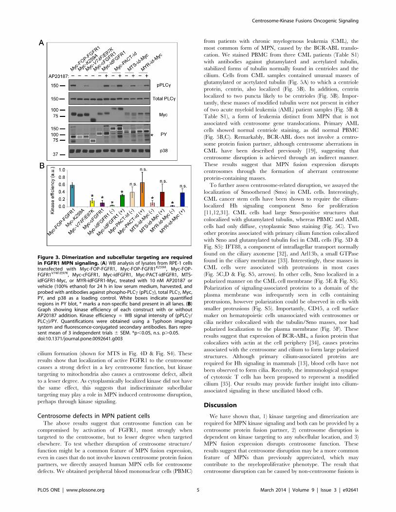
Efficient PLCc phosphorylation requires FGFR1dimerization and localization
In the experiments above we generated the tools to allow us to
test the relative contributions of centrosome localization, localiza-
tion more generally, and dimerization to fusion-FGFR1 signaling.
As a readout for the function of each construct we chose to assess
the phosphorylation of PLCc, a signaling substrate required in
FOP-FGFR1-induced transformation (Fig. 3) [24,30]. A construct
containing the truncated portion of FGFR1 (cFGFR1) found in
MPN fusions, without localization or dimerization domains, was
included as a control. Constructs were expressed in RPE-1 cells
and exposed to AP20187 or vehicle for 24 h before harvesting for
WB analysis (Fig. 3A). The relative signaling effectiveness of each
construct was measured by the fluorescence intensities of phospho-
PLCc (pPLCc), total PLCc, and PY of the fusion protein (Fig. 3A,
white boxes) for each sample; the ratio of (pPLCc/total PLCc)/PY
was used to calculate kinase signaling efficiency (Fig. 3B). This
shows the percent of total PLCc phosphorylated per unit of active
kinase, thus accounting for variation in the total available PLCcand differences in dimerization strength as a result of subcellular
targeting. Construct expression level was determined by the Myc
epitope tag on each construct and p38 was used as a loading
control.
To test the relative contributions of localization and dimeriza-
tion to kinase signaling, we compared the kinase signaling
efficiencies of the various FOP-FGFR1 and targeted idFGFR1
constructs. Combination of induced dimerization with subcellular
Figure 1. FOP-FGFR1V74F/E97K mutant lacks centrosome localization but not kinase activity. (A) Schematic of fusion between FOP andFGFR1 resulting in the FOP-FGFR1 oncogenic fusion and subsequent point mutations to produce KD, FOP-FGFR1K259A, and centrosome localizationmutant, FOP-FGFR1V74F/E97K. (B) WB analysis of lysates from RPE-1 cells transfected with WT Myc-FOP-FGFR1, Myc-FOP-FGFR1K259A, or Myc-FOP-FGFR1V74F/E97K, harvested, and probed with antibodies against phosphotyrosine (PY), Myc, and p38 as a loading control. (C) RPE-1 cells transfectedwith Myc-FOP-FGFR1, Myc-FOP-FGFR1K259A, or Myc-FOP-FGFR1V74F/E97K, fixed, and stained with antibodies against Myc (green) and PY (red). DNA isstained using DAPI (blue). Scale bars: 10 mm; insets: 106magnification.doi:10.1371/journal.pone.0092641.g001
Centrosome-Kinase Fusions Oncogenic Signaling
PLOS ONE | www.plosone.org 3 March 2014 | Volume 9 | Issue 3 | e92641

targeting to any site tested was sufficient to generate a signaling
efficiency statistically indistinguishable from WT (Fig. 3). Un-
dimerized idFGFR1 consistently had a decrease in kinase
efficiency, regardless of targeted localization, indicating the prime
importance of dimerization. However, localization to a discrete
subcellular site also had an effect; cytoplasmically localized,
dimerized cFGFR1 (both FOP-FGFR1V74F/E97K and dimerized
idFGFR1) showed a statistically significant decrease in kinase
efficiency. Importantly, addition of AP20187 to cFGFR1 lacking a
dimerization domain had no effect on signaling efficiency. These
results suggest that dimerization alone, but not localization, is
sufficient to produce limited FGFR1 kinase signaling efficiency.
However recapitulation of WT FOP-FGFR1 kinase efficiency
requires localization to a surface or structure in addition to
dimerization. Our results also show that the benefits of localization
are not limited to the centrosome, as dimerized constructs targeted
to the mitochondrial membrane or plasma membrane had a
similar effect.
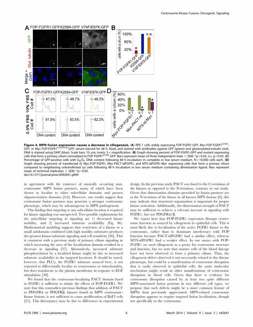
FOP-FGFR1 expression in RPE-1 cells causes a defect incentrosome function
In the assay above testing signaling efficiency of the fusion
kinases, localization was shown to be important, but there was
little difference between localization of active kinase to the
centrosome and localization to other sites. Another possibility to
explain the prevalence of centrosome protein fusion partners in
MPN is that localization of the active FGFR1 kinase to the
centrosome interferes with centrosome function in a manner
beneficial to the cancer cell. We examined several aspects of
centrosome function including microtubule nucleation, centro-
some duplication and primary cilium formation. Although
nucleation and duplication were unaffected, primary cilium
formation was strongly decreased in FOP-FGFR1 expressing cells
(Fig. 4A). RPE-1 cells stably expressing FOP-FGFR1-GFP, Myc-
FOP-FGFR1K259A-GFP, or FOP-FGFR1V74F/E97K–GFP were
serum-starved and assayed by IF for cilium formation. Expression
of FOP-FGFR1-GFP resulted in an 83% decrease in the number
of cells that formed a cilium when compared to KD FOP-
FGFR1K259A-GFP. In contrast, cells expressing FOP-FGFR1V74F/
E97K–GFP, which does not localize to the centrosome, showed only
an 18% decrease in ciliogenesis (Fig. 4B).
As the ciliation assay was performed under low serum to enrich
for cells in G0/G1, a decrease in ciliation may reflect defects in cell
cycle arrest rather than ciliogenesis. To determine if FOP-FGFR1
expressing cells are responsive to low serum, DNA content was
assayed by flow cytometry under normal and low serum conditions
(Fig. 4C). Cells expressing FOP-FGFR1-GFP, FOP-FGFR1K259A-
GFP, or FOP-FGFR1V74F/E97K–GFP arrested in G0/G1 equally
well upon serum starvation, thus, the ciliogenesis defect is not due
to a general cell cycle defect. RPE-1 cells transfected with Myc-
PACT-idFGFR1 and incubated in low serum medium containing
dimerization ligand also showed a substantial decrease in
ciliogenesis (Fig. 4D & Fig. S4). Interestingly, kinase targeting to
other subcellular locations had a smaller, but significant, effect on
Figure 2. Subcellular targeting of inducibly dimerizable FGFR1 (idFGFR1). (A) Schematic of normal FGFR dimerization and idFGFR1dimerization induced with dimerization ligand AP20187. (B) Table of idFGFR1 constructs targeted to subcellular domains by the addition oflocalization tags. (C) RPE-1 cells transfected with Myc-idFGFR1, Myc-PACT-idFGFR1, MTS-idFGFR1-Myc, or MYR-idFGFR1-Myc, treated with 10 nMAP20187 for 24 h, fixed, and stained with antibodies against Myc (green) and PY (red). DNA is stained using DAPI (blue). Scale bars: 10 mm; insets: 106magnification. (D) WB analysis of lysates from RPE-1 cells transfected with Myc-idFGFR1, Myc-PACT-idFGFR1, MTS-idFGFR1-Myc, or MYR-idFGFR1-Myc,treated with 10 nM AP20187 or vehicle (100% ethanol) for 24 h in low serum medium, harvested, and probed with antibodies against PY, Myc, andp38 as a loading control. Quantification of PY increase relative to control, normalized by Myc, for each construct is indicated.doi:10.1371/journal.pone.0092641.g002
Centrosome-Kinase Fusions Oncogenic Signaling
PLOS ONE | www.plosone.org 4 March 2014 | Volume 9 | Issue 3 | e92641
cilium formation (shown for MTS in Fig. 4D & Fig. S4). These
results show that localization of active FGFR1 to the centrosome
causes a strong defect in a key centrosome function, but kinase
targeting to mitochondria also causes a centrosome defect, albeit
to a lesser degree. As cytoplasmically localized kinase did not have
the same effect, this suggests that indiscriminate subcellular
targeting may play a role in MPN induced centrosome disruption,
perhaps through kinase signaling.
Centrosome defects in MPN patient cellsThe above results suggest that centrosome function can be
compromised by activation of FGFR1, most strongly when
targeted to the centrosome, but to lesser degree when targeted
elsewhere. To test whether disruption of centrosome structure/
function might be a common feature of MPN fusion expression,
even in cases that do not involve known centrosome protein fusion
partners, we directly assayed human MPN cells for centrosome
defects. We obtained peripheral blood mononuclear cells (PBMC)
from patients with chronic myelogenous leukemia (CML), the
most common form of MPN, caused by the BCR-ABL translo-
cation. We stained PBMC from three CML patients (Table S1)
with antibodies against glutamylated and acetylated tubulin,
stabilized forms of tubulin normally found in centrioles and the
cilium. Cells from CML samples contained unusual masses of
glutamylated or acetylated tubulin (Fig. 5A) to which a centriole
protein, centrin, also localized (Fig. 5B). In addition, centrin
localized to two puncta likely to be centrioles (Fig. 5B). Impor-
tantly, these masses of modified tubulin were not present in either
of two acute myeloid leukemia (AML) patient samples (Fig. 5B &
Table S1), a form of leukemia distinct from MPN that is not
associated with centrosome gene translocations. Primary AML
cells showed normal centriole staining, as did normal PBMC
(Fig. 5B,C). Remarkably, BCR-ABL does not involve a centro-
some protein fusion partner, although centrosome aberrations in
CML have been described previously [19], suggesting that
centrosome disruption is achieved through an indirect manner.
These results suggest that MPN fusion expression disrupts
centrosomes through the formation of aberrant centrosome
protein-containing masses.
To further assess centrosome-related disruption, we assayed the
localization of Smoothened (Smo) in CML cells. Interestingly,
CML cancer stem cells have been shown to require the cilium-
localized Hh signaling component Smo for proliferation
[11,12,31]. CML cells had large Smo-positive structures that
colocalized with glutamylated tubulin, whereas PBMC and AML
cells had only diffuse, cytoplasmic Smo staining (Fig. 5C). Two
other proteins associated with primary cilium function colocalized
with Smo and glutamylated tubulin foci in CML cells (Fig. 5D &
Fig. S5): IFT88, a component of intraflagellar transport normally
found on the ciliary axoneme [32], and Arl13b, a small GTPase
found in the ciliary membrane [33]. Interestingly, these masses in
CML cells were associated with protrusions in most cases
(Fig. 5C,D & Fig. S5, arrows). In other cells, Smo localized in a
polarized manner on the CML cell membrane (Fig. 5E & Fig. S5).
Polarization of signaling-associated proteins to a domain of the
plasma membrane was infrequently seen in cells containing
protrusions, however polarization could be observed in cells with
smaller protrusions (Fig. S5). Importantly, CD45, a cell surface
maker on hematopoietic cells unassociated with centrosomes or
cilia neither colocalized with the tubulin/Smo masses, nor had
polarized localization to the plasma membrane (Fig. 5F). These
results suggest that expression of BCR-ABL, a fusion protein that
colocalizes with actin at the cell periphery [34], causes proteins
associated with the centrosome and cilium to form large polarized
structures. Although primary cilium-associated proteins are
required for Hh signaling in mammals [13], blood cells have not
been observed to form cilia. Recently, the immunological synapse
of cytotoxic T cells has been proposed to represent a modified
cilium [35]. Our results may provide further insight into cilium-
associated signaling in these unciliated blood cells.
Discussion
We have shown that, 1) kinase targeting and dimerization are
required for MPN kinase signaling and both can be provided by a
centrosome protein fusion partner, 2) centrosome disruption is
dependent on kinase targeting to any subcellular location, and 3)
MPN fusion expression disrupts centrosome function. These
results suggest that centrosome disruption may be a more common
feature of MPNs than previously appreciated, which may
contribute to the myeloproliferative phenotype. The result that
centrosome disruption can be caused by non-centrosome fusions is
Figure 3. Dimerization and subcellular targeting are requiredin FGFR1 MPN signaling. (A) WB analysis of lysates from RPE-1 cellstransfected with Myc-FOP-FGFR1, Myc-FOP-FGFR1K259A, Myc-FOP-FGFR1V74F/E97K, Myc-cFGFR1, Myc-idFGFR1, Myc-PACT-idFGFR1, MTS-idFGFR1-Myc, or MYR-idFGFR1-Myc, treated with 10 nM AP20187 orvehicle (100% ethanol) for 24 h in low serum medium, harvested, andprobed with antibodies against phospho-PLCc (pPLCc), total PLCc, Myc,PY, and p38 as a loading control. White boxes indicate quantifiedregions in PY blot, * marks a non-specific band present in all lanes. (B)Graph showing kinase efficiency of each construct with or withoutAP20187 addition. Kinase efficiency = WB signal intensity of (pPLCc/PLCc)/PY. Quantifications were obtained using a Typhoon imagingsystem and fluorescence-conjugated secondary antibodies. Bars repre-sent mean of 3 independent trials 6 SEM. *p,0.05, n.s. p.0.05.doi:10.1371/journal.pone.0092641.g003
Centrosome-Kinase Fusions Oncogenic Signaling
PLOS ONE | www.plosone.org 5 March 2014 | Volume 9 | Issue 3 | e92641
in agreement with the existence of naturally occurring non-
centrosome MPN fusion partners, many of which have been
shown to localize to other subcellular domains and possess
oligomerization domains [4,6]. However, our results suggest that
centrosome fusion partners may generate a stronger centrosome
phenotype, which may be advantageous in MPN pathogenesis.
Our finding that targeting to any subcellular location is required
for kinase signaling was unexpected. Two possible explanations for
the subcellular targeting in signaling are 1) decreased kinase
mobility, and 2) increased substrate availability (Fig. S6).
Mathematical modeling suggests that restriction of a kinase to a
small subdomain combined with high motility substrates produces
the greatest kinase-substrate signaling and cell sensitivity [36]. This
is consistent with a previous study of primary cilium signaling in
which increasing the area of the localization domain resulted in a
decrease in signaling [37]. Alternatively, increased substrate
phosphorylation by a localized kinase might be due to increased
substrate availability in the targeted locations. It should be noted,
however, that PLCc, the FGFR1 substrate assayed here, is not
reported to differentially localize to centrosomes or mitochondria,
but does translocate to the plasma membrane in response to EGF
stimulation [38].
We found that the centrosome-localizing PACT domain fused
to FGFR1 is sufficient to mimic the effects of FOP-FGFR1. We
note that this contradicts previous findings that addition of PACT
to PDGFRa or PDGFRb, kinases found in MPN centrosome-
kinase fusions, is not sufficient to cause proliferation of BaF3 cells
[21]. This discrepancy may be due to differences in experimental
design. In the previous study PACT was fused to the C-terminus of
the kinases as opposed to the N-terminus, contrary to our study.
Given that dimerization domains provided by fusion partners are
at the N-terminus of the kinase in all known MPN fusions [6], this
may indicate that structural organization is important for proper
kinase activation. Additionally, the dimerization strength of PACT
may be sufficient to achieve a relevant increase in signaling with
FGFR1, but not PDGFRa/b.
We report here that FOP-FGFR1 expression disrupts centro-
some function as assayed by ciliogenesis in epithelial cells. This is
most likely due to localization of the active FGFR1 kinase to the
centrosome, rather than to dominant interference with FOP
function because PACT-idFGFR1 had a similar effect, whereas
MTS-idFGFR1 had a weaker effect. In our assays with FOP-
FGFR1 we used ciliogenesis as a proxy for centrosome structure
and function, but we note that mature cells of the blood lineage
have not been observed to form a primary cilium. Thus, the
ciliogenesis defect observed is not necessarily related to the disease
phenotype, but could be a manifestation of centrosome disruption
that is easily observed in epithelial cells; the same molecular
mechanism might result in other manifestations of centrosome
disruption in blood cells. Given that there is evidence for
centrosome disruption caused by at least two quite different
MPN-associated fusion proteins in two different cell types, we
propose that such defects might be a more common feature of
MPNs than previously appreciated. MPN-related centrosome
disruption appears to require targeted fusion localization, though
not specifically to the centrosome.
Figure 4. MPN fusion expression causes a decrease in ciliogenesis. (A) RPE-1 cells stably expressing FOP-FGFR1-GFP, Myc-FOP-FGFR1K259A-GFP, or Myc-FOP-FGFR1V74F/E97K-GFP, serum-starved for 48 h, fixed, and stained with antibodies against GFP (green) and glutamylated-tubulin (red).DNA is stained using DAPI (blue). Scale bars: 10 mm; insets: 56magnification. (B) Graph showing percent of FOP-FGFR1-GFP and mutant expressingcells that form a primary cilium normalized to FOP-FGFR1K259A-GFP. Bars represent mean of three independent trials 6 SEM. *p,0.05, n.s. p.0.05. (C)Percentage of GFP-positive cells with G0/G1 DNA content following 48 h incubation in complete or low serum medium. N = 10,000 cells each. (D)Graph showing percent of transfected (t) Myc-FOP-FGFR1, Myc-PACT-idFGFR1, and MTS-idFGFR1-Myc expressing cells that form a primary ciliumcompared to neighboring untransfected (u) cells following 48 h incubation in low serum medium containing dimerization ligand. Bars representmean of technical triplicates 6 SEM. *p,0.05.doi:10.1371/journal.pone.0092641.g004
Centrosome-Kinase Fusions Oncogenic Signaling
PLOS ONE | www.plosone.org 6 March 2014 | Volume 9 | Issue 3 | e92641

Remarkably, disruption of centrosome structure was observed
in human CML cells expressing the BCR-ABL fusion protein, in
the form of large centrosome and cilium protein-containing
structures not seen in control or AML cells. Interestingly, CML
cancer stem cells have been shown to require Hh signaling
[11,12]. Primary cilium-associated proteins are required for Hh
signaling in mammals [13]; although blood cells have not been
observed to form cilia, we report evidence for organized
localization of ciliary proteins in CML cells. Given that there is
evidence for centrosome disruption caused by at least two quite
different MPN-associated fusion proteins, we propose that such
defects might be a common feature of MPNs, unique to this type
of leukemia. This observation might be relevant to future therapies
for CML and other MPNs.
Materials and Methods
Ethics StatementHuman samples were obtained from patients at Stanford
University Medical Center according to the Institutional Review
Board (IRB) approved protocols (Stanford IRB no. 6453).
Participants provided written consent to participate in this study.
The IRB approved this consent procedure.
Human SamplesHuman CML and AML PBMC were cryopreserved in liquid
nitrogen in 90% FBS and 10% DMSO. Freshly thawed cells were
fixed in 4% PFA, spun onto polyethyleneimine coated coverslips,
and blocked for 1 h with guinea pig IgG in 3% BSA (Sigma) in
PBS +0.1% Triton (PBS-BT) at 1:500 for immunostaining.
PlasmidscDNAs for human FOP (GenBank: BC011902.2) and FGFR1
(GenBank: BC015035.1) were obtained from Open Biosystems.
The FOP-FGFR1 fusion was generated using precise gene fusion
by PCR [39], joining the first 519 nucleotides of FOP with the last
1,185 nucleotides of FGFR1 (cFGFR1). PCR products were
cloned into pDONR221 using the Invitrogen Gateway system.
The FOP-FGFR1K259A mutant was generated using site-directed
mutagenesis and FOP-FGFR1V74F/E97K generated using site-
directed mutagenesis and overlapping PCR following the V74F/
E97K mutations reported to disrupt centrosome localization in
FOP [25]. Synthetic MPN constructs were cloned using the PACT
domain from pericentrin [20], FKBP domains (26 FKBP36V)
PCR-amplified from pC4-Fv1E (ARGENT Regulated Homo-
dimerization Kit Version 2.0; ARIAD Pharmaceuticals, Inc.), the
MTS of human TOMM20 (NM_014765), and the MYR
sequence: ATGGGGAGTAGCAAGAGCAAGCCTAAGGAC-
CCCAGCCAGCGC, cloned into pENTR1A w48-1 (Eric Cam-
peau). Gateway recombination using pCS2+6xMyc DEST and
pLenti6.2 DEST cLAP provided by M. Nachury (Stanford
University, Stanford, CA), and pcDNA C-term 6xMyc DEST
(pTS2608) were used to produce Myc-FOP-FGFR1 (pTS2305),
Myc-FOP-FGFR1K259A (pTS2505), Myc-FOP-FGFR1V74F/E97K
(pTS2419), FOP-FGFR1-LAP (pTS2306), FOP-FGFR1K259A-
LAP (pTS2807), FOP-FGFR1V74F/E97K-LAP (pTS2808), Myc-
cFGFR1 (pTS2319), Myc-idFGFR1 (pTS2345), Myc-PACT-
idFGFR1 (pTS2347), MTS-idFGFR1-Myc (pTS2682), and
MYR-idFGFR1-Myc (pTS2642).
AntibodiesMouse (M) a–polyglutamylated tubulin (GT335; C. Janke,
Centre de Recherches de Biochemie Macromoleculaire) was used
at 1:5000 and M a–c-tubulin (GTU-88; Sigma-Aldrich) at 1:1000.
Figure 5. Localization of ciliary proteins in human CML cells. (A)Human CML cells stained with antibodies against acetylated (green)and glutamylated tubulin (red). (B) CML and AML cells stained withantibodies against centrin (green) and glutamylated tubulin (red). (C)Normal PBMC, CML, and AML cells stained with antibodies againstSmoothened (Smo) (green) and glutamylated tubulin (red). (D) CMLcells stained with antibodies against Smo (green) and either IFT88 (red).(E) CML cells stained with antibodies against Smo (green) and Arl13b(red). DNA is stained using DAPI (blue), scale bars: 5 mm, insets: 106magnification, white arrows: cell protrusions. In each case .100 cellswere imaged, and the phenotype represented in the images shown wasseen in greater than 50% of cells.doi:10.1371/journal.pone.0092641.g005
Centrosome-Kinase Fusions Oncogenic Signaling
PLOS ONE | www.plosone.org 7 March 2014 | Volume 9 | Issue 3 | e92641
Rabbit (Rb) a-GFP antibody was previously described [40]. M a-
Myc (9E10; Sigma-Aldrich): 1:500 for IF, 1:2000 for WBx. Mouse
a-PY (4G10; Millipore): 1:1000. Rb a-p38 (C-20; Santa Cruz
Biotechnology, Inc.): 1:5000. Rb a-phospho-PLCc1 (cat. #2821;
Cell Signaling Technology, Inc.) and rb a-PLCc1 (cat. #2822; Cell
Signaling Technology, Inc.): 1:1000. Rb a-Smo (cat. #38686,
Abcam): 1:1,000. Rb a-Arl13b (cat. # 17711-1-AP; ProteinTech):
1:500. Rb a-IFT88 (gift from Bradley Yoder, University of Alabama
at Birmingham, AL): 1:1,000. CD45-FITC (cat. # 347463; BD
Biosciences): 1:100. Double labeling using primary antibodies from
the same host species was previously described [41].
Lentivirus productionLentiviruses expressing GFP-tagged WT, KD, and localization
mutant FOP-FGFR1 were made using the lentiviral transfer vectors
described above. Recombinant lentivirus was produced by
cotransfection of HEK293T cells with the transfer vector, packaging
vector (pCMVDR8.74) and envelope vector (pMD2.VSVG) using
the calcium phosphate coprecipitation method [42].
Cell culture, transfection, and cell lineshTERT-RPE-1 cells (ATCC CRL-4000) were cultured in
DMEM/F12 50/50 medium (Cellgro) + 10% fetal bovine serum
(Atlanta Biologicals). RPE-1 cells were transfected with Lipofecta-
mine LTX (Invitrogen) for 24 h followed by 24 h incubation in
DMEM/F12 50/50 medium +0.5% fetal bovine +10 nM
AP20187 (ARGENT Regulated Homodimerization Kit Version
2.0; ARIAD Pharmaceuticals, Inc.), or the equivalent volume of
100% ethanol (vehicle). For stable cell lines, RPE-1 cells were
infected with lentiviral supernatant for 24 h and expanded. FACS,
described below, was used to isolate the GFP-positive cells.
Western blotting, Immunofluorescence, and FACSCells were lysed in triton buffer (1% triton, 150 mM NaCl,
50 mM Tris pH 8) supplemented with Protease Inhibitor Cocktail
Tablets (cat. #11836170001; Roche) and PhosSTOP Phosphatase
Inhibitor Cocktail Tablets (cat. #04906845001; Roche). Insoluble
material was pelleted for 5 min at 3.36kg (6,000 rpm) followed by
Bradford analysis and 25 mg protein loaded. For kinase efficiency
quantifications, blots were visualized using fluorescence conjugat-
ed secondary antibodies and the Typhoon 9210 imaging system
(GE Life Sciences). Analysis was performed using ImageQuant TL
v2003.01 (GE Life Sciences) with Local Average background
correction and background subtraction of non-specific bands. For
WB images, blots were visualized using HRP conjugated
secondary antibodies and exposure to film. For IF experiments,
cells were grown on poly-L-lysine coated coverslips and fixed with
220uC methanol. Coverslips were blocked in PBS-BT. Coverslips
were incubated in primary antibodies diluted in PBS-BT then
fluorescence-conjugated secondary antibodies (Invitrogen) diluted
1:1000 in PBS-BT. Coverslips were imaged using OpenLab 4.0.4
on an Axiovert 200M microscope (Carl Zeiss MicroImaging, Inc.)
with a Plan-NEOFLUAR 1006 (1.3 NA) objective. Images were
captured using an Orca-ER cooled CCD camera (Hamamatsu)
and processed using Photoshop (Adobe Systems). Flow cytometry
and FACS were performed at the Stanford Shared FACS Facility
using the Scanford and Vantoo instruments, respectively, and
analyzed using FlowJo (Tree Star, Inc.).
Statistical AnalysisAll statistical analyses were conducted with unpaired, two-tailed,
Student’s t tests using three independent trials. Values with p,
0.05 were considered statistically significant.
Supporting Information
Figure S1 Localization of WT FOP-FGFR1 and mutants.
RPE-1 cells transfected with WT Myc-FOP-FGFR1, kinase-dead
Myc-FOP-FGFR1K259A, or centrosome localization mutant Myc-
FOP-FGFR1V74F/E97K, fixed, and stained with antibodies against
Myc (green) and glutamylated-tubulin (red). Scale bars: 10 mm;
insets: 106magnification.
(TIF)
Figure S2 FOP-FGFR1V74F/E97K dimerizes in vivo, butnot in vitro. (A) Table showing combinations of constructs used
in co-expression, co-immunoprecipitation (co-IP) experiments. (B)In vitro translation of constructs in reticulocyte lysate followed by
assessment of Myc-tagged FOP-FGFR1 constructs in immuno-
precipitates (IP) of GFP-tagged FOP-FGFR1 constructs. (C)Expression of constructs in RPE-1 cells followed by assessment of
Myc-tagged FOP-FGFR1 constructs in IP of GFP-tagged FOP-
FGFR1 constructs.
(TIF)
Figure S3 PACT dimerization. Myc- and GFP-tagged PACT
fused to truncated FGFR1 (PACT-cFGFR1) or PACT fused to
idFGFR1 with the addition of dimerization ligand AP20187
(PACT-idFGFR1) were in vitro translated followed by assessment of
Myc-tagged PACT constructs in immunoprecipitates (IP) of GFP-
tagged PACT constructs.
(TIF)
Figure S4 Ciliogenesis in cells expressing targetedidFGFR1. (A) RPE-1 cells transfected with Myc-FOP-FGFR1,
Myc-PACT-idFGFR1, or MTS-idFGFR1-Myc, incubated in low
serum medium with dimerization ligand for 48 h, fixed, and
stained with antibodies against Myc (green) and glutamylated
tubulin (red). DNA is stained using DAPI (blue). Scale bars:
10 mm; insets: 106magnification.
(TIF)
Figure S5 Arl13b and IFT88 localization in CML cellscontaining protrusions. (A) Primary human CML cells or
normal PBMCs stained with antibodies against Arl13b (green) and
glutamylated tubulin (red). (B) Primary human CML cells or
normal PBMCs stained with antibodies against IFT88 (green) and
glutamylated tubulin (red). (C) Primary human CML cells stained
with antibodies against Smo (green) and Arl13b (red). DNA is
stained using DAPI (blue), scale bars: 5 mm, white arrows: cell
protrusions.
(TIF)
Figure S6 Models for effect of centrosome proteinfusion partner on kinase signaling. (A) Targeting of kinases
to the centrosome results in decreased mobility of the kinase,
which can more effectively interact with diffusing substrate
resulting in greater phosphorylation of normal kinase substrates
and increased downstream signaling. (B) If kinases substrates are
themselves concentrated at the centrosome, localization of the
kinase results in increased substrate availability, resulting in
increased phosphorylation and increased downstream signaling.
(TIF)
Table S1 Clinical characteristics of CML and AMLpatient samples.
(DOCX)
Acknowledgments
We thank the Stanford Hematology Division Tissue Bank and our patients
for their samples. We gratefully acknowledge Jonathan Van Dyke (Stanford
Centrosome-Kinase Fusions Oncogenic Signaling
PLOS ONE | www.plosone.org 8 March 2014 | Volume 9 | Issue 3 | e92641

University), Tim Knaak (Stanford University), Cathy Crumpton (Stanford
University), and the Stanford Shared FACS Facility for assistance with flow
cytometry and FACS. We thank W. James Nelson (Stanford University) for
careful reading of the manuscript and helpful discussions.
Author Contributions
Conceived and designed the experiments: JYL WH RM TS. Performed
the experiments: JYL. Analyzed the data: JYL TS. Contributed reagents/
materials/analysis tools: JYL WH RM TS. Wrote the paper: JYL TS.
References
1. Nussenzweig A, Nussenzweig MC (2010) Origin of chromosomal translocations
in lymphoid cancer. Cell 141: 27–38.
2. Tefferi A, Thiele J, Vardiman JW (2009) The 2008 World Health Organizationclassification system for myeloproliferative neoplasms: order out of chaos.
Cancer 115: 3842–3847.
3. Gilliland DG (2002) Molecular genetics of human leukemias: new insights into
therapy. Seminars in Hematology 39: 6–11.
4. Rosnet O, Birnbaum D (2007) Myeloproliferative disorders: let the partnerguide! Haematologica 92: 728–730.
5. Xiao S, McCarthy JG, Aster JC, Fletcher JA (2000) ZNF198-FGFR1
transforming activity depends on a novel proline-rich ZNF198 oligomerization
domain. Blood 96: 699–704.
6. Delaval B, Lelievre H, Birnbaum D (2005) Myeloproliferative disorders: thecentrosome connection. Leukemia 19: 1739–1744.
7. Rieder CL, Faruki S, Khodjakov A (2001) The centrosome in vertebrates: more
than a microtubule-organizing center. Trends in Cell Biology 11: 413–419.
8. Doxsey S, Zimmerman W, Mikule K (2005) Centrosome control of the cell
cycle. Trends in Cell Biology 15: 303–311.
9. Michaud EJ, Yoder BK (2006) The primary cilium in cell signaling and cancer.Cancer Res 66: 6463–6467.
10. Christensen ST, Clement CA, Satir P, Pedersen LB (2012) Primary cilia and
coordination of receptor tyrosine kinase (RTK) signalling. J Pathol 226: 172–
184.
11. Dierks C, Beigi R, Guo GR, Zirlik K, Stegert MR, et al. (2008) Expansion ofBcr-Abl-positive leukemic stem cells is dependent on Hedgehog pathway
activation. Cancer Cell 14: 238–249.
12. Zhao C, Chen A, Jamieson CH, Fereshteh M, Abrahamsson A, et al. (2009)
Hedgehog signalling is essential for maintenance of cancer stem cells in myeloidleukaemia. Nature 458: 776–779.
13. Huangfu D, Anderson KV (2005) Cilia and Hedgehog responsiveness in the
mouse. Proc Natl Acad Sci USA 102: 11325–11330.
14. Cross NCP, Reiter A (2008) Fibroblast growth factor receptor and platelet-
derived growth factor receptor abnormalities in eosinophilic myeloproliferativedisorders. Acta Haematol 119: 199–206.
15. Popovici C, Zhang B, Gregoire MJ, Jonveaux P, Lafage-Pochitaloff M, et al.
(1999) The t(6;8)(q27;p11) translocation in a stem cell myeloproliferativedisorder fuses a novel gene, FOP, to fibroblast growth factor receptor 1. Blood
93: 1381–1389.
16. Walz C, Metzgeroth G, Haferlach C, Schmitt-Graeff A, Fabarius A, et al. (2007)
Characterization of three new imatinib-responsive fusion genes in chronicmyeloproliferative disorders generated by disruption of the platelet-derived
growth factor receptor beta gene. Haematologica 92: 163–169.
17. Zhao X, Ghaffari S, Lodish H, Malashkevich VN, Kim PS (2002) Structure of
the Bcr-Abl oncoprotein oligomerization domain. Nat Struct Biol 9: 117–120.
18. Delorenzi M, Speed T (2002) An HMM model for coiled-coil domains and acomparison with PSSM-based predictions. Bioinformatics 18: 617–625.
19. Giehl M, Fabarius A, Frank O, Hochhaus A, Hafner M, et al. (2005)
Centrosome aberrations in chronic myeloid leukemia correlate with stage of
disease and chromosomal instability. Leukemia 19: 1192–1197.
20. Gillingham AK, Munro S (2000) The PACT domain, a conserved centrosomaltargeting motif in the coiled-coil proteins AKAP450 and pericentrin. EMBO
Rep 1: 524–529.
21. Bochtler T, Kirsch M, Maier B, Bachmann J, Klingmuller U, et al. (2011)
Centrosomal targeting of tyrosine kinase activity does not enhance oncogenicityin chronic myeloproliferative disorders. Leukemia 26: 728–735.
22. Delaval B, Letard S, Lelievre H, Chevrier V, Daviet L, et al. (2005) Oncogenic
tyrosine kinase of malignant hemopathy targets the centrosome. Cancer Res 65:7231–7240.
23. Guasch G, Delaval B, Arnoulet C, Xie MJ, Xerri L, et al. (2004) FOP-FGFR1tyrosine kinase, the product of a t(6;8) translocation, induces a fatal
myeloproliferative disease in mice. Blood 103: 309–312.24. Lelievre H, Chevrier V, Tassin AM, Birnbaum D (2008) Myeloproliferative
disorder FOP-FGFR1 fusion kinase recruits phosphoinositide-3 kinase and
phospholipase Cgamma at the centrosome. Mol Cancer 7: 30.25. Yan X, Habedanck R, Nigg EA (2006) A complex of two centrosomal proteins,
CAP350 and FOP, cooperates with EB1 in microtubule anchoring. Mol BiolCell 17: 634–644.
26. Lee JY, Stearns T (2013) FOP is a centriolar satellite protein involved in
ciliogenesis. PLoS ONE. pp. In press.27. Mikolajka A, Yan X, Popowicz GM, Smialowski P, Nigg EA, et al. (2006)
Structure of the N-terminal domain of the FOP (FGFR1OP) protein andimplications for its dimerization and centrosomal localization. J Mol Biol 359:
863–875.28. Kanaji S, Iwahashi J, Kida Y, Sakaguchi M, Mihara K (2000) Characterization
of the signal that directs Tom20 to the mitochondrial outer membrane. J Cell
Biol 151: 277–288.29. Towler DA, Eubanks SR, Towery DS, Adams SP, Glaser L (1987) Amino-
terminal processing of proteins by N-myristoylation. Substrate specificity of N-myristoyl transferase. J Biol Chem 262: 1030–1036.
30. Guasch G, Ollendorff V, Borg JP, Birnbaum D, Pebusque MJ (2001) 8p12 stem
cell myeloproliferative disorder: the FOP-fibroblast growth factor receptor 1fusion protein of the t(6;8) translocation induces cell survival mediated by
mitogen-activated protein kinase and phosphatidylinositol 3-kinase/Akt/mTORpathways. Mol Cell Biol 21: 8129–8142.
31. Rohatgi R, Milenkovic L, Scott MP (2007) Patched1 regulates hedgehogsignaling at the primary cilium. Science 317: 372–376.
32. Pazour GJ, Dickert BL, Vucica Y, Seeley ES, Rosenbaum JL, et al. (2000)
Chlamydomonas IFT88 and its mouse homologue, polycystic kidney diseasegene tg737, are required for assembly of cilia and flagella. J Cell Biol 151: 709–
718.33. Larkins CE, Aviles GDG, East MP, Kahn RA, Caspary T (2011) Arl13b
regulates ciliogenesis and the dynamic localization of Shh signaling proteins.
Molecular Biology of the Cell 22: 4694–4703.34. McWhirter JR, Wang JY (1993) An actin-binding function contributes to
transformation by the Bcr-Abl oncoprotein of Philadelphia chromosome-positivehuman leukemias. EMBO J 12: 1533–1546.
35. de la Roche M, Ritter AT, Angus KL, Dinsmore C, Earnshaw CH, et al. (2013)Hedgehog signaling controls T cell killing at the immunological synapse. Science
342: 1247–1250.
36. Kazmierczak B, Lipniacki T (2010) Spatial gradients in kinase cascaderegulation. IET Syst Biol 4: 348–355.
37. Mahjoub MR, Stearns T (2012) Supernumerary centrosomes nucleate extra ciliaand compromise primary cilium signaling. Curr Biol 22: 1628–1634.
38. Matsuda M, Paterson HF, Rodriguez R, Fensome AC, Ellis MV, et al. (2001)
Real time fluorescence imaging of PLC gamma translocation and its interactionwith the epidermal growth factor receptor. J Cell Biol 153: 599–612.
39. Yon J, Fried M (1989) Precise gene fusion by PCR. Nucleic Acids Res 17: 4895.40. Hatch EM, Kulukian A, Holland AJ, Cleveland DW, Stearns T (2010) Cep152
interacts with Plk4 and is required for centriole duplication. J Cell Biol 191: 721–
729.41. Negoescu A, Labat-Moleur F, Lorimier P, Lamarcq L, Guillermet C, et al.
(1994) F(ab) secondary antibodies: a general method for double immunolabelingwith primary antisera from the same species. Efficiency control by chemilumi-
nescence. J Histochem Cytochem 42: 433–437.42. Dull T, Zufferey R, Kelly M, Mandel RJ, Nguyen M, et al. (1998) A third-
generation lentivirus vector with a conditional packaging system. J Virol 72:
8463–8471.
Centrosome-Kinase Fusions Oncogenic Signaling
PLOS ONE | www.plosone.org 9 March 2014 | Volume 9 | Issue 3 | e92641