Central Venous Catheters and CVP Monitoring Nursing Competency Presented by: Jonna Bobeck BSN, RN,...
33
Central Venous Catheters and CVP Monitoring Nursing Competency Presented by: Jonna Bobeck BSN, RN, CEN
-
Upload
dangelo-drover -
Category
Documents
-
view
233 -
download
1
Transcript of Central Venous Catheters and CVP Monitoring Nursing Competency Presented by: Jonna Bobeck BSN, RN,...

Central Venous Catheters and CVP Monitoring Nursing CompetencyPresented by:Jonna Bobeck BSN, RN, CEN

Objectives
•Define indications and contraindications•Discuss complications•Articulate nursing management and care •Discuss prevention of intravascular
catheter related blood stream infections per CDC

Introduction
•Common modality•Typical sites•CVC devices

Insertion Site Selection
•Subclavian•Internal jugular•Femoral

Indications
•Monitoring of the central venous pressure (CVP)
•Long term medications•Parenteral nutrition•Caustic medications•Dialysis•Need for frequent intravenous access

General
Distorted local anatomy
Extremes of weight
Vasculitis
Prior long-term venous cannulation
Prior injection of sclerosis agents
Suspected proximal vascular injury
Previous radiation therapy
Bleeding disorders
Anticoagulation or thrombolytic therapy
Combative patients
Inexperienced, unsupervised physician
Subclavian Vein
Chest wall deformities
Pneumothorax on the contralateral side
Chronic obstructive pulmonary disease
Jugular Vein
Intravenous drug abuse via the jugular system
Femoral Vein
Need for patient mobility

Contraindications for Central Line Subclavian Placement Include?
Pneumothorax on contralateral side
Need for normal saline infusion
A patient who requires CVP readings
The need for frequent blood sampling

Correct

Sorry, Try Again

Technique Advantages Disadvantages
Basilic (peripheral puncture)
Low incidence of complicationsPerformed under direct visualizationAllow large quantities of fluid at a rapid rate.
Greater incidence of minor complicationsHinders movement of armIncrease difficulty for CVP monitoring
Internal jugular Good external landmarksMalposition is rareNearly a straight course to superior vena cava on right sideUseful alternative to cutdown on children <2 years of age
Slightly higher of failure compared to subclavianMore difficult to securePossibly higher infection risk than subclavian

Technique Advantages Disadvantages
Femoral puncture Good external landmarks.Useful alternative to other supradiaphragmatic approaches in patients with coagulopathies or superior vena trauma.
Difficult to secure in ambulatory patients.Generally not reliable for CVP measurement.Potentially a “dirty” siteHigher risk of thrombus.
Subclavian Good external landmarks.Practical method of inserting a central line in cardiorespiratory arrest
Unable to compress bleeding vessels.“Blind” procedure.Should not be attempted in children younger than 2 yr.

Anatomy of Internal Jugular

Anatomy of Femoral Vein

Anatomy of the Subclavian

Hand Washing
•Fingernails•Hand hygiene

Patient Positioning
•Jugular access•Subclavian access

The Central Line Bundle
•Hand hygiene•Maximal barrier precautions•Chlorhexadine skin antisepsis•Optimal site selection•Prompt removal

Skin Prep
•Chlorhexadine side to side scrub x 2•Clip hair, do not shave

Maximal Barrier Precautions
•Strict sterile conditions•Maximum Barrier Precautions
▫Full bed drape▫Sterile Gown▫Sterile Gloves▫Surgical mask for anyone with in three 3
feet of insertion site▫Head cover for anyone with in 3 feet of
insertion site

Equipment

Procedure
•Wash hands•Prepare IV flush•Prepare pressure tubing•Monitor patient•Obtain appropriate equipment•15-25 degree Trendelenberg•Flush ports•Obtain CXR

Post Procedure
•CONFIRM placement•Distal lumen•Adjust scale

Documentation
•Patient and family education•Vital signs•Hard copy of waveform•Catheter location•Date and time•Nursing interventions•Patient tolerance•dressing

Ports
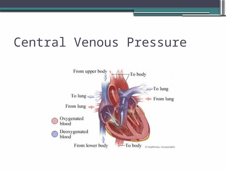
Central Venous Pressure

Measuring CVP
•Gather equipment•Plug transducer cable into red port•Prepare pressure tubing - Use heparin 1000 units/500 ml


The central venous waveform seen on the monitor reflects the events of cardiac contraction; the central venous catheter “sees” these slight variations in pressure that occur during the cardiac cycle and transmits them as a characteristic waveform. There are three positive waves (a, c, and v) and two negative waves (x and y), and these correlate with different phases of the cardiac cycle and EKG.

CVP Assessment
•2-6 mm Hg•Elevated CVP•Low CVP

Port a Cath and PICC Lines
•Click link to review PRH policy-IV Therapy Policy

Reading CVP Waveforms is Fun!!

References
•Lynne-MChaleWiegard, D., Carlson, K., Initials. (Ed.). (2001). Aacn procedure manual for critical care. United States: Elsevier Saunders.
•Proehl, J. (Ed.). (2004). Emergency nursing procedures. United States: Elsevier Saunders