CENTRAL RESPONSE TO INFRA-RED …jeb.biologists.org/content/jexbio/58/1/59.full.pdf · J. Exp. Bid....
18
J. Exp. Bid. (1973), 58, 59-76 59 With 10 text-figures Frinted in Great Britain CENTRAL RESPONSE TO INFRA-RED STIMULATION OF THE PIT RECEPTORS IN A CROTALINE SNAKE, TRIMERESURUS FLAVOVIRIDIS BY RICHARD C. GORIS AND SHIN-ICHI TERASHIMA Department of Physiology, School of Medicine, Tokyo Medical and Dental University (Received 13 June 1972) INTRODUCTION The facial pits of boid and crotaline snakes are unique in the animal kingdom as infra-red receptors. The pits of the crotaline snakes, in particular, have excited the interest and imagination of naturalists from the time that rattlesnakes were first encountered in the early stages of colonization of the American continents (Klauber, The snake family Viperidae is divided into two subfamilies by the presence or absence of pit receptors. The Viperinae, found throughout Europe, Africa and Asia, apparently lack any form of infra-red receptor (Bullock & Barrett, 1968). The Crotalinae, instead, are characterized by the presence of a highly specialized pit receptor in the loreal region of either side of the face. Five principal genera make up this subfamily: Crotalus (the rattlesnakes),* Bothrops and Lachesis, all confined to North and South America; Trimeresurus, distributed widely in Asia; and Agkistrodon, which is found both in America and throughout Asia as far west as the eastern part of Europe. There are no crotalines in Africa or Australia. The pits are found in a much more primitive form in the family Boidae; in fact, some boids possess thermoreception without any external pits at all (Bullock & Barrett, 1968). The function of the boid pit is essentially the same as that of the crotaline pit (Noble & Schmidt, 1937; Warren & Proske, 1968). However, most research on snake thermoreceptors has been confined to crotaline snakes. The structure and innervation of these pits have been described in considerable detail by the following workers: Lynn (1931), Noble & Schmidt (1937), Bullock & Fox (1957), Bleichmar & De Robertis (1962), Terashima, Goris & Katsuki (1970). That these pits are thermoreceptors responding to changes in background heat flux has been demonstrated amply by Noble & Schmidt (1937), using behavioural experi- mental techniques, and by Bullock & Cowles (1952), Bullock & Diecke (1956) and Goris & Nomoto (1967) using electro-physiological recording techniques. Terashima, Goris & Katsuki (1968) recorded the generator potential from the sensory membrane of crotaline receptors. Terashima et al. (1970) also determined the terminal nerve structure, and Meszler (1970) showed how changes in the densely packed mitochondria of the nerve endings indicate that these mitochondria are involved as transducers of heat energy to electrical potentials. • The pigmy rattlesnakes of North America, genus Sistrurus, are now included in the genus Crotalus (Underwood, 1967).
Transcript of CENTRAL RESPONSE TO INFRA-RED …jeb.biologists.org/content/jexbio/58/1/59.full.pdf · J. Exp. Bid....

J. Exp. Bid. (1973), 58, 59-76 5 9With 10 text-figures
Frinted in Great Britain
CENTRAL RESPONSE TOINFRA-RED STIMULATION OF THE PIT RECEPTORS IN A
CROTALINE SNAKE, TRIMERESURUS FLAVOVIRIDIS
BY RICHARD C. GORIS AND SHIN-ICHI TERASHIMA
Department of Physiology, School of Medicine, Tokyo Medical and Dental University
(Received 13 June 1972)
INTRODUCTION
The facial pits of boid and crotaline snakes are unique in the animal kingdom asinfra-red receptors. The pits of the crotaline snakes, in particular, have excited theinterest and imagination of naturalists from the time that rattlesnakes were firstencountered in the early stages of colonization of the American continents (Klauber,
The snake family Viperidae is divided into two subfamilies by the presence orabsence of pit receptors. The Viperinae, found throughout Europe, Africa and Asia,apparently lack any form of infra-red receptor (Bullock & Barrett, 1968). TheCrotalinae, instead, are characterized by the presence of a highly specialized pitreceptor in the loreal region of either side of the face. Five principal genera make upthis subfamily: Crotalus (the rattlesnakes),* Bothrops and Lachesis, all confined toNorth and South America; Trimeresurus, distributed widely in Asia; and Agkistrodon,which is found both in America and throughout Asia as far west as the eastern part ofEurope. There are no crotalines in Africa or Australia.
The pits are found in a much more primitive form in the family Boidae; in fact,some boids possess thermoreception without any external pits at all (Bullock &Barrett, 1968).
The function of the boid pit is essentially the same as that of the crotaline pit(Noble & Schmidt, 1937; Warren & Proske, 1968). However, most research on snakethermoreceptors has been confined to crotaline snakes. The structure and innervationof these pits have been described in considerable detail by the following workers:Lynn (1931), Noble & Schmidt (1937), Bullock & Fox (1957), Bleichmar & De Robertis(1962), Terashima, Goris & Katsuki (1970).
That these pits are thermoreceptors responding to changes in background heat fluxhas been demonstrated amply by Noble & Schmidt (1937), using behavioural experi-mental techniques, and by Bullock & Cowles (1952), Bullock & Diecke (1956) andGoris & Nomoto (1967) using electro-physiological recording techniques. Terashima,Goris & Katsuki (1968) recorded the generator potential from the sensory membraneof crotaline receptors. Terashima et al. (1970) also determined the terminal nervestructure, and Meszler (1970) showed how changes in the densely packed mitochondriaof the nerve endings indicate that these mitochondria are involved as transducers ofheat energy to electrical potentials.
• The pigmy rattlesnakes of North America, genus Sistrurus, are now included in the genusCrotalus (Underwood, 1967).

60 R. C. GORIS AND SHIN-ICHI TERASHIMA
To date, however, all published works, with the exception of Harris & Gamo\^(1971), who recorded evoked potentials from the boid brain, have been concernedwith peripheral responses. Much obscurity remains as to how the central nervoussystem receives these responses and how it utilizes them.
The work reported here was undertaken to study the response of the crotalinecentral nervous system to stimulation of the infra-red receptors; and to utilize thisresponse to elucidate further the function of these receptors.
Three problems were posed:(1) To discover an area of the snake brain from which response potentials to infra-
red stimulation could be recorded reliably and consistently.(2) To investigate the vertical and horizontal field of response of the pit receptors.(3) To investigate the possibility that the snake has stereoscopic perception of an
infra-red stimulus source.
MATERIALS AND METHODS
The habu, Trimeresurus flavoviridis, was chosen as the most suitable experimentalanimal. It is the largest pit viper in Asia, and was available in quantity at moderatecost. Trimeresurus ofunavensis and Agkistrodon halys were also considered, but weredeemed unsuitable because of their high resistance to the immobilizing agent curare.
About 100 snakes were utilized, averaging ca. 130 cm in total length and 300 g inweight. The snakes were immobilized with tubocurarine chloride injected intra-muscularly. Usually 0-3-0-6 mg was sufficient to immobilize snakes of the size usedwithin 30 min. The amount did not seem to be critical, but we tried to obtain thelightest degree of immobilization possible. With this amount of curare the snakeretained muscle tone and was capable of tail movements and other slight muscletwitches, but was unable to right itself when placed upside down. Under bright lightthe pupils contracted completely, although sluggishly. Respiration, evinced byopening and closing of the glottis, and by partial inflation of the lungs, took placeabout once a minute. We noted that as long as good pupil contraction persisted, goodresponse could be obtained; but when immobilization progressed to the point thatpupil contraction ceased, nerve potentials also became erratic or ceased. Thereforewe used the pupil reflex as an indicator of the degree of immobilization and of thereliability of the recordings: once the pupil reflex became weak, the animal was nolonger used.
Recordings were made with vinyl-coated tungsten electrodes and with glass micro-pipettes filled with 3 M-KC1. The tungsten electrode tips averaged about 5 fim indiameter. The micropipettes were uniformly of 0-5 /im in diameter, with a resistancevarying between 20 and 50 MD. The potentials obtained were amplified in a con-ventional manner, displayed on a dual-beam oscilloscope, and recorded both on filmand on magnetic tape. Infra-red stimulus was obtained from a small incandescentlamp and camera shutter, according to methods described by Terashima et al. (1968).The lamp was lighted by a 6 V direct current, and delivered, at a distance of 20 cm,approximately 9-74 mW/cm2 with an energy peak at a wavelength of 1 -2 fim(Terashima et al. 1968). This will be referred to below as the 'standard stimulus'.The lamp and shutter were mounted on a pivoting brass arm which could be swung1800 in either a horizontal or a vertical plane (Fig. 1). A phototransistor attached to

Infra-red stimulation of pit receptors of snake 61
180°
Fig. i. Position of recording electrode and movement of stimulus course.(A) Bilateral stimulation, arc indicates horizontal movement of stimulus. The o—1800 line
bisects the pit openings. The 90° line is an extension of the midline body axis. E, electrode;P, P, pit openings.
(B) Contralateral stimulation: arc indicates movement of stimulus for recording horizontalreceptive field. The c—1800 line is parallel to the plane of the pit opening. E, Electrode; P, P,pit openings.
(C) Arrangement for measurement of vertical response field: arc indicates vertical move-ment of stimulus. This arc represents a 900 vertical rotation of the arc in (A). P, Pit opening;N, nostril.
the front side of the shutter and connected to one beam of the oscilloscope indicatedonset and duration of the stimulus. The head of the snake was mounted on a beam ofbalsa wood and centred directly above the pivot of the arm, and the lamp was adjustedon the arm so that the bulb was approximately 20 cm from this point, with the fila-ment centred at the level of the pits.
Recordings were attempted from the trigeminal ganglion and the region of thetectum opticum. The trigeminal ganglion was approached dorsally, ventrally andlaterally. This proved extremely difficult. The ganglion is well protected on all sides

62 R. C. GORIS AND SHIN-ICHI TERASHIMA
by bony projections of the skull, and the removal of these was complicated by tharelatively large blood vessels associated with the ganglion or passing through foraminain the bone. This difficulty was overcome by drilling very precise holes with fine dentalburrs, but then these holes were too small to allow removal of the hard connectivetissues surrounding the ganglion. This tissue was tough enough to break glass capil-laries, and the tips of the tungsten electrodes usually bent instead of penetrating.
The trigeminal ganglion was also approached by entering from the occipital region.The neck muscles were cut with an electric scalpel at their point of attachment to theskull. A hole was opened in the occipital bones, and a slit was made in the dura. Thecerebro-spinal fluid was then carefully blotted with absorbent cotton until the surfaceof the brain was clearly visible. Electrodes were inserted deeply at an angle of about150 to the horizontal plane in hopes of penetrating to the neighbourhood of thetrigeminal ganglion.
The most successful operation was exposure of the dorsal surface of the tectumopticum. The snake was fixed in a natural position on the board, with the snout pro-truding over the front edge as far as the region of the pits. The snake was held lightlyto the board by a piece of tape at the nuchal region and a pin through the upperlabials on each side. Since saliva often clogged the glottis, a plastic catheter wasinserted to facilitate breathing. The skin of the head above the parietal bones was thenexcised. For a few moments after this operation the tectum opticum could be seenclearly through the bone, as well as the sinus venosus between the two lobes of thetectum. The bone became opaque as soon as it dried, but there was time to delineateprecisely the area of bone to be removed. This area came extremely close to the sinusvenosus, but the bone over the sinus had to be scrupulously avoided. The membranesover the sinus adhere to the bone, and if this part of the bone was drilled or moved thesinus would rupture and the snake would soon die. It was found impossible to stemthe haemorrhage once this sinus had been ruptured. Using the finest available dentalburr a series of cavities was drilled in a circle above one of the tectal lobes. The thick-ness of the bone in this area varies, so that considerable skill was necessary. The burrcould not be permitted to penetrate to the dura, as this would tear the dura andrupture the blood vessels within, spoiling the preparation. Thus the holes had to bedrilled down to, but not quite touching, the dura. All work was done under a dis-secting microscope at a magnification of x 15-20.
When the circle of holes was complete, the bone inside the circle was carefully andslowly chipped away with tweezers. Again great care had to be taken to avoid rupturingthe dura. Once the dura was exposed, the course of the blood vessels through it couldeasily be seen. The dura was slit with fine scissors between the blood vessels andparallel to them. Thus, with a minimum of trauma and practically no haemorrhagean area of cortex 1-2 mm in diameter was exposed. Into this both tungsten andmicrocapillary electrodes could be easily inserted. With the passage of time the surfaceof the cortex became hardened, and it was increasingly difficult to insert the electrode.However, this did not cause any great problems, since good response could usuallybe obtained from the beginning of the experiment.

Infra-red stimulation of pit receptors of snake 63
RESULTS
A. Site of recording
No satisfactory results were obtained from the trigeminal ganglion directly. Whenelectrodes were inserted into the neighbourhood of the ganglion from the occipitalregion, some response to infra-red stimulation was obtained. However, the locationof the electrode tip was imprecise, and it was difficult to obtain consistent results.Therefore data from these recordings will not be considered in this paper.
Response to infra-red stimulation was obtained clearly and repeatedly from elec-trodes inserted into the tectum opticum, or in contact with its surface. Consistentlygood results were obtained from 17 snakes. A total of approximately 100 units wereobserved. Of these, 50 preparations lasted long enough to make satisfactory recordings.Recordings were made with both glass electrodes and tungsten electrodes. Excellentaction potentials were obtained with the glass electrodes, while tungsten electrodes,being of a greater tip diameter, recorded mostly evoked potentials, together with afew multi-unit action potentials. Similar potentials were recorded from both rightand left tectal lobes, but for convenience of manipulation most recordings were madefrom the left lobe.
B. Potentials recorded
Single-unit action potentials were recorded with glass microcapillary electrodespenetrating the tectum opticum. The precise depth of penetration was difficult todetermine because the brain surface was depressed when the electrode penetrated.However, the best results were obtained when the electrode was advanced about 1 mmafter touching the surface at the centre of the tectum. Recordings were also made atother depths, both shallower and deeper, and throughout the entire exposed regionof the tectal lobe.
A few of the recorded units responded to optical stimulation, or to vibration, orto touch and vibration. Continuously firing units of unknown affiliation were also seen.However, the vast majority of single units encountered responded only to infra-redstimulation of the pits. This was confirmed by covering the pits, whereupon responseimmediately ceased. Covering the eyes, stroking the facial region with a brush, tappingthe table, pinching the body, etc., did not affect the response in any way. Further con-firmation was obtained by comparison with optical, vibration, and touch units,which manifested entirely different modes of discharge.
When unilateral stimulation was being used, care was taken to shield the pit notbeing stimulated from the stimulus source.
The following discharge patterns were observed:Background discharge (Fig. 2 A). By background discharge we mean discharge ob-
served when no special stimulus was present in the receptive field of the pit. All theinfra-red units exhibited background discharge to some degree. In some units it wasbarely 1-2 spikes or even less per minute; in others it reached a frequency of 5-7impulses per second, which approximates the background discharge of primary fibres(Goris & Nomoto, 1967). No completely silent fibres were encountered. Some fibreswere relatively silent, but resumed background discharge after receiving a stimulus.The background discharge frequency of any one unit also showed considerablefluctuation, increasing and decreasing from time to time.

R. C. GORIS AND SHIN-ICHI TERASHIMA
Bfliiiiimmmniii
D
Fig. z. Various types of central response: A, background discharge; B, tonic response; C,On-Off phasic response; D, phasic-tonic response. Lower line indicates stimulus. Stimulusduration i sec.
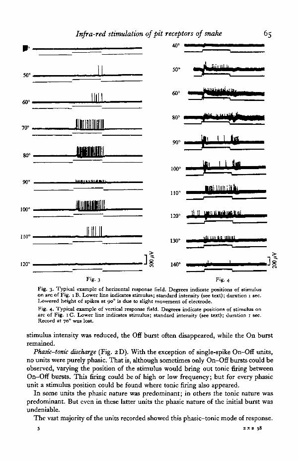
Tonic discharge (Fig. 2B). We recorded units which exhibited rapidly increasingfiring after onset of stimulus up to a certain peak, then a gradual adaptation. For anygiven unit the frequency increased and decreased in proportion to the intensity of thestimulus. However, no two units showed the same frequency of discharge in responseto the same stimulus. In response to the standard stimulus we recorded frequenciesof from a few impulses per second to a maximum of 83 impulses/sec, each unit beingdifferent. Intervals between spikes were always highly irregular. There was no recog-nizable recurring pattern. There was also some fluctuation in frequency and intervalseach time the stimulus was repeated. In some units the firing was more regular than inothers; but all were characterized by a certain irregularity. When the stimulus wasprolonged for several seconds, adaptation rapidly took place, eventually reaching theoriginal background firing level. When the stimulus source was moved in a horizontalor vertical arc within the receptive field of the pit, frequency and firing pattern variedconsiderably with each new position of the stimulus (Figs. 3, 4).
Phasic discharge (Fig. 2C). A large number of neurones exhibited On-Off phasicdischarge. In some of these both On and Off were represented by a single spike,regardless of the stimulus intensity or position. In these cases all possibilities ofstimulus artifact were carefully excluded.
Other units showed an On burst and an Off burst. The number of spikes in thebursts and the duration of each burst differed from unit to unit. Changing the intensityor position of the stimulus also caused a change in the number of spikes per buist.As in tonic response, the number of spikes per burst was directly proportional to thestimulus intensity. On bursts generally contained more spikes than Off bursts. When
Infra-red stimulation of pit receptors of snake 65
40° •*>^—•••—•••
50°50° 1 U
60°I l l l I
60°
80°iiinigniiiiiin
70°
90
100
•aimiii'iillki1100 ^Bn+nHmmmm
100° — — —120°
110°
120° • " -I £ 140° JlFig- 3 Fig. 4
Fig. 3. Typical example of horizontal response field. Degrees indicate positions of stimuluson arc of Fig. i B. Lower line indicates stimulus; standard intensity (see text); duration i sec.Lowered height of spikes at 90° is due to slight movement of electrode.Fig. 4. Typical example of vertical response field. Degrees indicate positions of stimulus onarc of Fig. 1 C. Lower line indicates stimulus; standard intensity (see text); duration 1 sec.Record at 70° was lost.
stimulus intensity was reduced, the Off burst often disappeared, while the On burstremained.
Phasic-tonic discharge (Fig. 2D). With the exception of single-spike On-Off units,no units were purely phasic. That is, although sometimes only On-Off bursts could beobserved, varying the position of the stimulus would bring out tonic firing betweenOn-Off bursts. This firing could be of high or low frequency; but for every phasicunit a stimulus position could be found where tonic firing also appeared.
In some units the phasic nature was predominant; in others the tonic nature waspredominant. But even in these latter units the phasic nature of the initial burst wasundeniable.
The vast majority of the units recorded showed this phasic—tonic mode of response.5 EXB 58
66 R. C. GORIS AND SHIN-ICHI TERASHIMA
Standardstimulus ^
i standard
\ standard
^standard
standard
standard .]l
Fig. 5. Evoked response from the surface of the tectum. Tungsten electrode.Lower line indicates stimulus. Stimulus duration 1 sec.
Purely tonic units were relatively few; and it is possible that if the stimulus for theseunits had been moved sufficiently, phasic-tonic response would have been recorded.In fact, with some units where the stimulus was moved in a 180° arc, the responsewas tonic in some positions and phasic-tonic in other positions (Fig. 4). It was alsopossible for bilaterally responding units (see § E below, also Fig. 6 A) to be, for example,tonic in response to ipsilateral stimulation and phasic-tonic in response to contra-lateral stimulation. In many phasic-tonic units there was no Off burst observable.
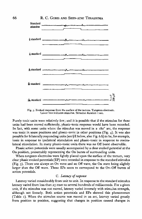
Phasic action potentials were usually accompanied by a clear evoked potential at theOn position, presumably representing the On bursts of surrounding units.
When tungsten electrodes were lightly placed upon the surface of the tectum, veryclear phasic evoked potentials (EP) were recorded in response to the standard stimulus(Fig. 5). There was always an On wave and an Off wave, the On wave being slightlylarger than the Off wave. These EPs seem to correspond to the On-Off bursts ofaction potentials.
C. Latency of response
Latency varied considerably from unit to unit. In response to the standard stimuluslatency varied from less than 25 msec to several hundreds of milliseconds. For a givenunit, if the stimulus was not moved, latency varied inversely with stimulus strength,although not linearly. Both action potentials and EPs showed this phenomenon(Table 1). When the stimulus source was moved in an arc, latency varied greatlyfrom position to position, suggesting that changes in position caused changes in

Infra-red stimulation of pit receptors of snake 67
Table 1. Latency of response with decreasing stimulus strength;single preparation; wide-band heat filters used
Stimulus
StandardStandard reduced by
5 0 %7 5 %9 0 %9 5 %9 9 %
Actionpotential(msec)
5°
6575
II2-5No responseNo response
Evokedpotential
(msec)
25
4050
5°60
65
stimulus intensity at the level of the receptor membrane. These changes were thenmanifested by changes in latency as well as in firing pattern.
D. Sensitivity
It was not the purpose of the present series of experiments to make sensitivitymeasurements. However, in contrast to the apparatus used when recording fromperipheral nerves (Goris & Nomoto, 1967), recording from the brain left the receptivefield of the pits relatively unencumbered. Hence some observations could be made onsensitivity, judged from the maximum distance at which a given stimulus would pro-duce a response. The experiments were carried on inside a wire-mesh shield box toreduce external magnetic fields. The box was about 1 metre cube. Inside the boxresponse was still obtained from the standard stimulus at the maximum distancepossible within the box, about 90 cm from the snake's head. A hand also was detectedat this distance. When the stimulus source was outside the shield box, response wasseverely attenuated or non-existent for most units recorded. This could be due to theinterposed wire mesh, which was extremely fine and possibly acted as a heat sink.However, despite the wire mesh, several units were encountered which responded tothe presence of a man standing outside the box at a maximum distance of 5 m. Thiswas the maximum distance possible without leaving the room. Possibly response wouldhave occurred at even greater distances if the room had been larger. With a manstanding at 1 or 2 m response was tonic, with a few spikes per second, which graduallyadapted. At greater distances adaptation was almost immediate and complete. How-ever, any movement on the part of the man at these distances produced a response ofone or two spikes. Waving the hands, moving the head from side to side, moving thebody from the waist up all produced an initial response, which ceased immediately,even though the movement continued. When the type of movement changed orceased, another response took place.
To exclude the possibility that the units thus responding were optical units, theexperiment was repeated in total darkness. Response was unchanged. The eyes werecovered and the experiment was again repeated; and again response was unchanged,leaving little doubt that a highly sensitive infra-red response was being observed.
E. Ipsilateral-contralateral response
The ipsilateral and contralateral pits were shielded from stimuli given to one or theother pit, and the responses to ipsilateral and contralateral stimuli were observed. It
5-2

68 R. C. GORIS AND SHIN-ICHI TERASHIMA
A
30°
90°
150°iimi ]l
B
30'
90'
Fig. 6. Two types of bilaterally responding units: A, ipsilateral type; B, contralateral type.30°, 900 and 1500 indicate positions of stimulus on arc of Fig. 1 (A). Lower line indicates stimulus.Stimulus intensity: standard (see text); duration 1 sec.
was immediately noted (1) that most units responded to contralateral stimulus only,and (2) that some units responded to both ipsilateral and contralateral stimulation(Fig. 6). No purely ipsilateral units were encountered. When a bilateral unit wasencountered, the shield was removed and the stimulus was adjusted so that thediameter of its arc was horizontal and perpendicular to the long axis of the snake'sbody, with the pivot centred between the pits, as in Fig. 1 A. The stimulus was thenmoved in an arc of 1800 in steps of 50 or io°, and the response was recorded.
With respect to the stimulus position at which maximum firing occurred, two typesof bilateral units were encountered: (1) ipsilateral type-maximum firing occurredwhen the stimulus was in the field of the ipsilateral pit (Fig. 6 A), and (2) contralateraltype — maximum firing occurred when the stimulus was in the receptive field of thecontralateral pit (Fig. 6B).
In most, but not all, of the bilateral units encountered, when the stimulus was inposition to illuminate both pits equally, firing was reinforced to a frequency superiorto that of either pit alone.

Infra-red stimulation of pit receptors of snake 69
Table 2. Horizontal width of receptive field; contralateral stimulation
Width Maximumof field Position firing
Unit no. (deg. of arc) of field point
1 50 From 6o° to no" 8o°2 40 From 300 to 70° 40°3 50 From 400 to 90° 700
4 80 From 50° to 1300 oo°Av. 55
Table 3. Vertical width of receptive field; contralateral stimulation
Width of Maximumof field Position firing
Unit no. (deg. of arc) of field point
5 40 From 400 to 8oc 700
6 70 From 400 to 110° 700
7 25 From 300 to 550 550
8 80 From 400 to 1200 700
9 70 From 200 to oo° 50-600
10 40 From 70° to 11 o° ioo°11 80 From 500 to 1300 no"12 60 From 400 to iooc 60—700
Av. 58
F. Effects of a cold stimulus
While observing the background firing of a unit, ice was introduced into the re-ceptive field. This caused immediate suppression of all discharge. When the ice waswithdrawn, an On burst was produced which was similar to the On burst producedby a 'hot' stimulus. If a warm metal plate was introduced into the receptivefield, it at first caused a phasic-tonic response. If the metal plate was allowed toremain in position, the unit rapidly adapted and firing returned to background levels.If at this point a hand was brought between the plate and the pit, the same effect asice was produced, i.e. suppression of background firing, and an On burst when thehand was removed.
G. Receptive field
The receptive field was measured both horizontally and vertically. However, ourapparatus was movable in only one plane, so that horizontal and vertical measurementshad to be made on separate units.
Moving the stimulus source as shown in Fig. 1B and C, the measurements ofTables 2 and 3 were obtained.
In brief, horizontal field width averaged 550 (extremes 40-800), and vertical widthaveraged 5 8° (extremes 25-800) of arc.
DISCUSSION
The histology of the tectum opticum of the reptile brain has been studied in detailby Senn (1968) (Lacerta sicula), and by Leghissa (1962) (all classes of reptiles).According to these authors the surface of the tectum is formed by bundles of opticfibres, while the layers beneath the surface contain vast amounts of other fibres, many

70 R. C. Gows AND SHEN-ICHI TERASHIMA
of which derive directly from, or communicate with, the trigeminal nerve. Many ofthese fibres communicate with the contralateral side across the intertectal com-missure on the dorsal side of the tectum. There is another commissure on the ventralside of the tectum, the tegumental commissure, but it is not clear whether fibres ofthe trigeminal system communicate across this commissure. In addition, at about thelevel of the ' stratum griseum et fibrosum periventriculare' of Leghissa, there are anumber of large nuclei. These are the nuclei of the mesencephalic tract of thetrigeminal nerve.
According to Legissa (1962) 'the optic tectum represents the centre of arrival anddeparture of impulses that control the general nervous activity and the behaviour ofnon-mammiferous animals, which do not have a differentiated hemispherical neo-cortex... The movement of an animal towards its prey, the swallowing of acceptablefood and the rejection of unacceptable food, the reactions of attack and defence, andfinally the presence or absence, in an animal, of an associative memory all find in theoptic lobe a most favourable morphological basis for realization. In the most highlyevolved tetrapods (reptiles and especially birds) the tectum opticum acquires also anintegrative function, analogous to that possessed by the mammals in the cortex of thehemispheres' (pp. 344-5 passim; translation by Goris).
Masai & Sato (1965, 1971), Sato (1971), Wells, Smith & Spaur (1971) and Masai (inpreparation) have demonstrated how the relative degree of development of a givenpart of the brain (e.g. the olfactory lobes) in related species of lower vertebrates isdirectly related to the degree or extent that each species makes use, in its daily life,of the senses directly connected with that part of the brain. Thus, to give an example,a snake which depends primarily on the sense of smell to detect food will show a highdegree of development of the olfactory lobes. In contrast, there will be a lesser degreeof development of these lobes in a snake which feeds primarily by sight (Wells, Smith &Spaur, 1971).
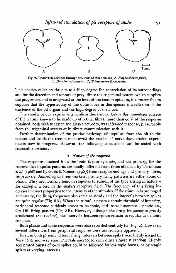
Masai (1972) has examined the tectum opticum of a number of typical snakes,mostly colubrids and natricids, and has shown that the degree of development of thetectum is directly related to the diurnal or nocturnal habits of the species. In Elapheclimacophora, an almost entirely diurnal species, the tectal lobes are very highly de-veloped (Fig. 7 A) and present a distinct swelling in a dorsal view. In frontal sectionthe fibrous layers of these lobes are seen to be very highly developed. In contrast tothis diurnal snake, Dinodon rufozonatus, of strictly nocturnal habits, has very poorlydeveloped tectal lobes (Fig. 7 B), and in section the fibrous layers are less developedthan E. climacophora.
Trimeresurus flavoviridis, the subject of the present experiments, is active primarilyat night, with a few periods of activity in early morning and evening during coolerweather. However, its tectal lobes show just as high a degree of development as thediurnal E. climacophora (Fig. 7C). Compared with D. rufozonatus, the eyes of T. flavo-viridis are larger and probably used to a greater extent. One would expect, therefore,a degree of development of the tectum opticum intermediate between D. rufozonatusand E. climacophora. However, this is not the case. The tectal lobes of this speciesshow a degree of development nearly equal to that of E. climacophora. Now, the onlyother difference between T. flavoviridis and D. rufozonatus is the possession of theinfra-red sensory pits, supplied by highly developed branches of the trigeminal nerve.
Infra-red stimulation of pit receptors of snake 71
Fig. 7. Transverse sections through the tecta of three snakes; A, Elaphe dimacophora,B, Dinodon rufozonatus, C, Trimeresunu flavoviridis.
This species relies on the pits to a high degree for appreciation of its surroundingsand for the detection and capture of prey. Since the trigeminal system, which suppliesthe pits, enters and is integrated at the level of the tectum opticum, it is reasonable tosuppose that the hypertrophy of the optic lobes in this species is a reflexion of theexistence of the pit organs and the high degree of their use.
The results of our experiments confirm this theory. Below the immediate surfaceof the tectum known to be made up of retinal fibres, more than 90 % of the responseobtained, both with tungsten and glass electrodes, was infra-red response, presumablyfrom the trigeminal system or in direct communication with it.
Further determination of the precise pathways of impulses from the pit to thetectum and inside the tectum must await the results of nerve degeneration experi-ments now in progress. However, the following conclusions can be stated withreasonable certainty.
A. Nature of the response
The response obtained from the brain is post-synaptic, and not primary, for thereasons that impulse patterns are totally different from those obtained by Terashimaet al. (1968) and by Goris & Nomoto (1967) from receptor endings and primary fibres,respectively. According to these workers, primary firing patterns are either tonic orphasic. They are normally tonic in response to stimuli of the type arising in nature —for example, a bird in the snake's receptive field. The frequency of this firing in-creases in direct proportion to the intensity of the stimulus. If the stimulus i3 prolongedand steady, the firing frequency also remains steady and the intervals between spikesare quite regular (Fig. 8 A). When the stimulus passes a certain threshold of intensity,peripheral response suddenly ceases to be tonic, and instead assumes a phasic i.e.,On-Off, firing pattern (Fig. 8B). However, although the firing frequency is greatlyaccelerated (80-100/sec), the intervals between spikes remain as regular as in tonicresponse.
Both phasic and tonic responses were also recorded centrally (cf. Fig. 2). However,several differences from peripheral response were immediately apparent.
First, in both phasic and tonic firing, intervals between spikes were highly irregular.Very long and very short intervals succeeded each other almost at random. Highlyaccelerated bursts of 5-10 spikes could be followed by less rapid bursts, or by singlespikes at varying intervals.

R. C. GORIS AND SHIN-ICHI TERASHIMA
lsec
Fig. 8. Typical peripheral response: A, tonic; B, phasic.Lower line indicates stimulus.
Standardstimulus i
i standard
standard
standard
& standard .31Fig. 9. Effect of reduction in stimulus intensity. Lower line indicates stimulus.
Stimulus duration 1 sec.
Secondly, in peripheral response phasic response occurred only when a certainintensity of stimulus had been reached. It disappeared again when the stimulus in-tensity was reduced. Centrally, phasic response had no direct relation to the intensityof the stimulus. Units which showed phasic response retained that pattern no matterhow much the stimulus was reduced. The spike frequency in each phasic burst variedwith the intensity of the stimulus, just as in tonic response. However, the phasicnature of the response remained, as long as the stimulus source remained unmoved(Fig- 9)-
A third difference was the appearance of phasic-tonic units. In these units firing
Infra-red stimulation of pit receptors of snake
i u i i m i i i i i i B i i i i i n i m i n i 111 m i l l i i i i l l I l i m i i 1 M i l I II73
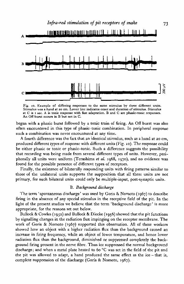
=3.
Fig. io. Example of differing responses to the same stimulus by three different units.Stimulus was a hand at 20 cm. Lower line indicates onset and duration of stimulus. Stimulusat C is 1 sec. A is tonic response with fast adaptation. B and C are phasic—tonic responses.An Off burst occurs in B but not in C.
began with a phasic burst followed by a tonic train of firing. An Off burst was alsooften encountered in this type of phasic-tonic combination. In peripheral responsesuch a combination was never encountered at any time.
A fourth difference was the fact that an identical stimulus, such as a hand at 20 cm,produced different types of response with different units (Fig. 10). The response couldbe either phasic or tonic or phasic-tonic. Such a difference suggests the possibilitythat recording was being made from several different types of units. However, peri-pherally all units were uniform (Terashima et al. 1968, 1970), and no evidence wasfound for the possible presence of different types of receptors.
Finally, the existence of bilaterally responding units with firing patterns similar tothose of the unilateral units supports the supposition that all these units are notprimary, for such bilateral units could only be multiple-input, post-synaptic units.
B. Background discharge
The term 'spontaneous discharge* was used by Goris & Nomoto (1967) to describefiring in the absence of any special stimulus in the receptive field of the pit. In thelight of the present studies we believe that the term ' background discharge' is moreappropriate, for the reasons set out below.
Bullock & Cowles (1952) and Bullock & Diecke (1956) showed that the pit functionsby signalling changes in the radiation flux impinging on the receptor membrane. Thework of Goris & Nomoto (1967) supported this observation. All of these workersshowed how an object with a higher radiation flux than the background caused anincrease in firing frequency, while an object of lower temperature, and hence lowerradiation flux than the background, diminished or suppressed completely the back-ground firing present in the nerve fibre. Thus ice suppressed the normal backgrounddischarge; and when a metal plate heated to 60 °C was set in the field of the pit andthe pit was allowed to adapt, a hand produced the same effect as the ice - that is,complete suppression of the discharge (Goris & Nomoto, 1967).

74 R- C. GORIS AND SHIN-ICHI TERASHIMA
We repeated the experiments of Goris & Nomoto (1967) while recording from thatectum, with identical results. Background discharge was present, in varying degrees,in every unit that we recorded. The firing pattern was essentially identical with that ofperipheral units - that is, random firing, without any recognizable recurring pattern.It differed from peripheral background firing in that firing frequency was often con-siderably lower at central levels, being sometimes only 1-2 spikes/min; some units,however, approached the peripheral frequency of 5-6 impulses/sec. Adaptation -that is, a return, during stimulation, to pre-stimulus levels of discharge - was alsomuch faster at central levels. Several minutes were required for adaptation at the peri-pheral level when the stimulus was a hand at 20 cm. At central levels tonically firingunits adapted to the same stimulus in a matter of seconds (Fig. 10 A).
We propose the theory that background firing represents a level of adaptation to theflux of background radiation. Any change in the flux causes a change in the firingpattern. Thus central units adapted quickly to a human body which entered the re-ceptive field, but any movement of the limbs or trunk produced a noticeable, thoughtransient, response.
We propose that there would be no background discharge if there were no back-ground radiation, a condition which normally cannot exist. A similar condition iscreated temporarily when ice blocks the receptive field and acts as a heat sink, absorb-ing the background radiation. In this case background discharge disappears. When theice is removed, there is a sudden On burst of firing similar to the On burst in responseto a sudden stimulus. This is because the membrane receptors, now no longer adaptedto the background radiation because of the ice, suddenly are confronted with thisbackground radiation and react accordingly.
C. Field of response
The configuration of the field response seems to be determined not so much by thenature of the receptors themselves but by the physical configurations of the pit - thatis, the shape and width of the opening, the distance from opening to sensory membrane,the orientation of the plane of the opening, etc. These factors vary widely from speciesto species, and slightly even from individual to individual. Thus no absolute figurescan be given.
In our experiments, as long as the stimulus was not moved, repeated stimuli pro-duced repetitions of the same firing pattern. However, when the stimulus was moved,the firing pattern was different for each pdsition (Figs. 3, 4). Assuming that all thereceptors in the receptor membrane are identical and produce a response proportionalto the amount of radiation received, the variation in firing pattern with a movingstimulus can be explained by a combination of the following two factors:
(1) The unit being recorded from is a multiple-input unit in the integration centreof the brain, possibly several synapses removed from the peripheral nerves.
(2) The amount of radiation striking the various receptors in the membrane is notthe same for each receptor. This is caused by the physical configurations of the pit.In the first place, the membrane is not taut and flat but hangs loosely inside the pitin a more or less concave fashion, so that no two points on the membrane are exactlyequidistant from the plane of the opening. In addition to this, the opening itself isquite irregular in shape, with considerable overhang of the scales lining the edge of

Infra-red stimulation of pit receptors of snake 75
khe pit. Thus with the stimulus at a given position, a considerable pattern of light,shadow and reflexion is formed on the surface of the membrane, with each individualreceptor receiving different amounts of radiation. When the stimulus is moved, thepattern of light and shadow shifts across the membrane, so that the amount of radia-tion received by each individual receptor, and consequently the firing pattern whichreaches the central nervous system also shifts accordingly. The effect is analogousto that of a pattern moving across the compound eye of an insect, although the pitreceptors seem far too simple to even remotely approach the image-resolving powerof an insect eye. Not only does the pit lack a lens, but the receptor population iscompletely homogeneous, as has been noted by Terashima et al. (1970), without anyknown inter-reactions such as facilitation or inhibition. However, it is apparent fromour results that the general direction of a stimulus, as well as its movement and directionof movement, can be easily perceived even by a single pit.
Our results also support the supposition that the crotaline snakes have stereoscopicperception of stimulus objects. The existence in the tectum of bilaterally respondingunits is cogent proof of this. All of the bilateral units recorded showed a change ofresponse pattern when the stimulus was moved from ipsilateral to contralateral pit.Some units showed maximum firing with ipsilateral stimulation, others with contra-lateral stimulation. Most showed reinforcement (increase) of firing when the stimuluswas in a position to irradiate both pits at once (Fig. 6). Thus it is evident that thesnake not only perceives an object as being simply to its right or to its left, but is alsoable to determine the position of an object within the overlapping receptive field ofthe two pits by comparing the discharge between right and left pits. The scanningbehaviour noted by Goris & Nomoto (1967) is evidence that precisely this type ofcomparison is being carried out. The accuracy of this orientation was shown long agoby the behavioural experiments of Noble & Schmidt (1937).
SUMMARY
1. Both action potentials and evoked potentials were recorded from the tectumopticum of a crotaline snake, Trimeresurus flavoviridis, in response to infra-redstimulation of the facial pit organs. Action potentials from single units were recordedthroughout the tectum.
2. Most units responded to contralateral stimulation, while some responded to bothipsi- and contralateral stimulation.
3. Firing patterns were tonic, phasic, or phasic-tonic, depending on the positionof the stimulus and the type of unit being recorded.
4. Sensitivity to stimulus movement was observed.5. All potentials differed from peripheral potentials in firing patterns.6. Firing frequency was directly proportional to stimulus intensity.7. Measurements were made of the vertical and horizontal response fields of single
units.8. Background discharge was noted in all units and its nature discussed.9. The integrative function of the tectum in regard to infra-red perception was also
discussed, as well as the possibility of stereoscopic perception.

76 R. C. GORIS AND SHIN-ICHI TERASHIMA
We wish to thank the following persons: Dr Y. Katsuki, now of the Department oiPhysiology, School of Dentistry, Tsurumi Women's University, for his sage guidance;and Dr H. Masai, Dr T. Kusunoki and Dr Y. Sato of the Department of Anatomy,School of Medicine, Yokohama City University, for making available to us theirlaboratory facilities, their reptile brain preparations, and their wide knowledge andexperience regarding the brains of lower vertebrates.
REFERENCES
BLEICHMAR, H. & DE ROBERTIS, E. (1962). Submicroscopic morphology of the infrared receptor of pitvipers. Z. Zellforsch. mikrosk. Anat. 56, 748-61.
BULLOCK, T. H. & BARRETT, R. (1968). Radiant heat reception in snakes. Cornm. Behav. Biol. (A)i,19-29.
BULLOCK, T. H. & COWLES, R. B. (1952). Physiology of an infrared receptor - the facial pit of pit vipers.Science, N.Y. 115, 541-3.
BULLOCK, T. H. & DIECKE, P. J. (1956). Properties of an infrared receptor. J. Pkysiol, Lond. 134,47-87-
BULLOCK, T. H. & Fox, W. (1957). The anatomy of the infrared sense organ in the facial pit of pitvipers. Q. jfl microic. Sci. 98, 219-34.
GORIS, R. C. & NOMOTO, M. (1967). Infrared reception in oriental crotaline snakes. Comp. Biochem.Pkytiol. 23, 879-92.
HARRIS, J. F. & GAMOW, I. R. (1971). Snake infrared receptors: thermal or photochemical mechanism.Science, N.Y. 173, 1252-3.
KLAUBER, L. M. (1956). Rattlesnakes. Their Habits, Life Histories, and Influence on Mankind, vol. 1,p. 369. University of California Press.
LEGHISSA, S. (1962). L'evoluzione del tetto ottico nei bassi Vertebrati. Archo. ital. Anat. Embriol. 67, (4),343-413-
LYNN, W. G. (I 931). Structure and function of the facial pit of the pit vipers. Am. J. Anat. 49, 97-139.MASAI, H. (1972). Structure patterns of the optic tectum of Japanese snakes of the family Colubridae
in relatipn to habit. (In preparation.)MASAI, H. & SATO, Y. (1965). The brain patterns in relation to behaviour in fish hybrids. Natunoissen-
shaften 52, 43-4.MASAI, H. & SATO, Y. (1971). The variation of brain patterns through the domestication in the goldfish.
Acta anat. nippon. 46, no. 1.MESZLER, R. M. (1970). Correlation of ultrastructure and function. In The Biology of the Reptilia, vol. 2.
New York: Academic Press.NOBLE, G. K. & SCHMIDT, A. (1937). Structure and function of the facial and labial pits of snakes.
Proc. Am. Phil. Soc. 77, 263-88.SATO, Y. (1971). A morpho-ethological study on the brain - the cytoarchitectonics of the endobrain in
relation to habit. Zool. Mag. 80, 303-15 (in Japanese).SENN, D. G. (1968). Bau und Ontogenese von Zwischen- und Mittelhirn bei Lacerta sicula (Rafin-
esque). Acta anat., supplementum 55, vol. 71.TERASHIMA, S., GORIS, R. C. & KATSUKI, Y. (1968). Generator potential of Crotaline snake infrared
receptor. J. Neurophysiol. 31, 682-8.TERASHIMA, S., GORIS, R. C. & KATSUKI, Y. (1970). Structure of warm fiber terminals in the pit mem-
brane of vipers. J. Ultrastr. Res. 31, 494-506.UNDERWOOD, G. (1967). A Contribution to the Classification of Snakes. London: Trustees of the British
Museum (Natural History).WARREN, J. W. & PROSKE, U. (1968). Infrared receptors in the facial pits of the Australian python
Morelia spilotes. Science, N.Y. 159, 439-41.WELLS, K. E., SMITH, H. M. & SPAUR, R. C. (1971). Correlation of certain ophidian sensory modali-
ties with gross brain proportions. Journal of Herpetology 5 (3-4), 200-4.