Cellular/Molecular NerveGrowthFactor ...
12
Cellular/Molecular Nerve Growth Factor-Regulated Emergence of Functional -Opioid Receptors Bihua Bie, 1 Zhi Zhang, 1 You-Qing Cai, 1 Wei Zhu, 1 Yong Zhang, 1 Jaile Dai, 2 Charles J. Lowenstein, 3,4 Edward J. Weinman, 5 and Zhizhong Z. Pan 1 1 Department of Anesthesiology and Pain Medicine, The University of Texas-MD Anderson Cancer Center, Houston, Texas 77030, 2 Department of Molecular Pathology, The University of Texas–MD Anderson Cancer Center, Houston, Texas 77030, Departments of 3 Medicine and 4 Pathology, The Johns Hopkins University School of Medicine, Baltimore, Maryland 21205, and 5 Division of Nephrology, University of Maryland Hospital, Baltimore, Maryland 21201 Sorting of intracellular G-protein-coupled receptors (GPCRs) either to lysosomes for degradation or to plasma membrane for surface insertion and functional expression is a key process regulating signaling strength of GPCRs across the plasma membrane in adult mammalian cells. However, little is known about the molecular mechanisms governing the dynamic process of receptor sorting to the plasma membrane for functional expression under normal and pathological conditions. In this study, we demonstrate that -opioid receptor (DOPr), a GPCR constitutively targeted to intracellular compartments, is driven to the surface membrane of central synaptic terminals and becomes functional by the neurotrophin nerve growth factor (NGF) in native brainstem neurons. The NGF-triggered DOPr translocation is predominantly mediated by the signaling pathway involving the tyrosine receptor kinase A, Ca 2 -mobilizing phospho- lipase C, and Ca 2 /calmodulin-dependent protein kinase II. Importantly, it requires interactions with the cytoplasmic sorting protein NHERF-1 (Na /H exchange regulatory factor-1) and N-ethyl-maleimide-sensitive factor-regulated exocytosis. In addition, this NGF- mediated mechanism is likely responsible for the emergence of functional DOPr induced by chronic opioids. Thus, NGF may function as a key molecular switch that redirects the sorting of intracellularly targeted DOPr to plasma membrane, resulting in new functional DOPr on central synapses under chronic opioid conditions. Introduction Surface density and signaling strength of G-protein-coupled receptors (GPCRs) on mammalian cell membrane are actively regulated by receptor trafficking through endocytosis (internal- ization) and exocytosis, two trafficking processes dynamically determined by agonist stimulation and molecular properties of the receptors (Hanyaloglu and von Zastrow, 2008). The mecha- nisms of GPCR internalization mediated by receptor phosphor- ylation and -arrestin association upon agonist binding have been well characterized and are consistent among many different types of GPCRs, whereas the cellular fates of internalized GPCRs are rather diverse. Constitutively, GPCRs can be sorted to the lysosomal pathway for degradation or to the recycling pathway for surface membrane insertion through exocytosis (Tan et al., 2004; Hanyaloglu and von Zastrow, 2008). A typical example of this constitutive sorting of GPCRs is opioid receptors: while in- ternalized -opioid receptor (MOPr) is mostly directed to the recycling pathway for surface insertion, intracellular -opioid re- ceptor (DOPr) is normally sorted to the lysosomal pathway for degradation or is constitutively retained in intracellular compart- ments, resulting in a low level of functional DOPr on plasma membrane in many types of central neurons (Tsao and von Zastrow, 2000; Finn and Whistler, 2001; Wang and Pickel, 2001; Ma et al., 2006). Recent studies show that the lysosomal sorting of DOPr is regulated by a GPCR-associated sorting protein (Whistler et al., 2002) and the predominant intracellular targeting of DOPr is retained by a specific domain in the cytoplasmic tail of DOPr, which is lacking in MOPr (Kim and von Zastrow, 2003). How- ever, certain pathological conditions can alter the normal sorting of GPCRs and induce GPCR mislocalization with abnormal GPCR signaling, leading to a variety of diseases (Tan et al., 2004). For example, prolonged exposure to opioids induces exocytotic membrane trafficking of intracellular DOPr to surface mem- brane, resulting in new functional DOPr in central neurons in- volved in pain modulation and in drug addiction (Morinville et al., 2004; Hack et al., 2005; Ma et al., 2006; Zhang et al., 2006; Cahill et al., 2007; Bie et al., 2009). This mechanism of DOPr membrane trafficking has also been demonstrated in our previ- ous confocal immunocytochemical study on brainstem neurons, which shows that chronic morphine recruits new functional DOPr on presynaptic terminals through DOPr translocation, with increased DOPr protein on plasma membrane and an in- creased number of DOPr-immunoreactive varicosities (presumably on presynaptic terminals) that appose postsynaptic membrane of cell bodies (Ma et al., 2006). In contrast to the well characterized mechanism of GPCR internalization, little is known about the molecular mechanisms Received Oct. 26, 2009; revised Feb. 11, 2010; accepted March 9, 2010. This work was supported by the National Institute on Drug Abuse Grants DA023069 and DA025826. We thank Dr. Moses Chao for his generous gift of the p75 NTR -blocking antibody. Correspondence should be addressed to Dr. Zhizhong Z. Pan, Department of Anesthesiology and Pain Medicine, Unit 110, The University of Texas–MD Anderson Cancer Center, 1515 Holcombe Boulevard, Houston, TX 77030. E-mail: [email protected]. DOI:10.1523/JNEUROSCI.5296-09.2010 Copyright © 2010 the authors 0270-6474/10/305617-12$15.00/0 The Journal of Neuroscience, April 21, 2010 • 30(16):5617–5628 • 5617
Transcript of Cellular/Molecular NerveGrowthFactor ...
Cellular/Molecular
Nerve Growth Factor-Regulated Emergence of Functional�-Opioid Receptors
Bihua Bie,1 Zhi Zhang,1 You-Qing Cai,1 Wei Zhu,1 Yong Zhang,1 Jaile Dai,2 Charles J. Lowenstein,3,4
Edward J. Weinman,5 and Zhizhong Z. Pan1
1Department of Anesthesiology and Pain Medicine, The University of Texas-MD Anderson Cancer Center, Houston, Texas 77030, 2Department of MolecularPathology, The University of Texas–MD Anderson Cancer Center, Houston, Texas 77030, Departments of 3Medicine and 4Pathology, The Johns HopkinsUniversity School of Medicine, Baltimore, Maryland 21205, and 5Division of Nephrology, University of Maryland Hospital, Baltimore, Maryland 21201
Sorting of intracellular G-protein-coupled receptors (GPCRs) either to lysosomes for degradation or to plasma membrane for surfaceinsertion and functional expression is a key process regulating signaling strength of GPCRs across the plasma membrane in adultmammalian cells. However, little is known about the molecular mechanisms governing the dynamic process of receptor sorting to theplasma membrane for functional expression under normal and pathological conditions. In this study, we demonstrate that �-opioidreceptor (DOPr), a GPCR constitutively targeted to intracellular compartments, is driven to the surface membrane of central synapticterminals and becomes functional by the neurotrophin nerve growth factor (NGF) in native brainstem neurons. The NGF-triggered DOPrtranslocation is predominantly mediated by the signaling pathway involving the tyrosine receptor kinase A, Ca 2�-mobilizing phospho-lipase C, and Ca 2�/calmodulin-dependent protein kinase II. Importantly, it requires interactions with the cytoplasmic sorting proteinNHERF-1 (Na �/H � exchange regulatory factor-1) and N-ethyl-maleimide-sensitive factor-regulated exocytosis. In addition, this NGF-mediated mechanism is likely responsible for the emergence of functional DOPr induced by chronic opioids. Thus, NGF may function asa key molecular switch that redirects the sorting of intracellularly targeted DOPr to plasma membrane, resulting in new functional DOPron central synapses under chronic opioid conditions.
IntroductionSurface density and signaling strength of G-protein-coupledreceptors (GPCRs) on mammalian cell membrane are activelyregulated by receptor trafficking through endocytosis (internal-ization) and exocytosis, two trafficking processes dynamicallydetermined by agonist stimulation and molecular properties ofthe receptors (Hanyaloglu and von Zastrow, 2008). The mecha-nisms of GPCR internalization mediated by receptor phosphor-ylation and �-arrestin association upon agonist binding havebeen well characterized and are consistent among many differenttypes of GPCRs, whereas the cellular fates of internalized GPCRsare rather diverse. Constitutively, GPCRs can be sorted to thelysosomal pathway for degradation or to the recycling pathwayfor surface membrane insertion through exocytosis (Tan et al.,2004; Hanyaloglu and von Zastrow, 2008). A typical example ofthis constitutive sorting of GPCRs is opioid receptors: while in-ternalized �-opioid receptor (MOPr) is mostly directed to therecycling pathway for surface insertion, intracellular �-opioid re-ceptor (DOPr) is normally sorted to the lysosomal pathway fordegradation or is constitutively retained in intracellular compart-
ments, resulting in a low level of functional DOPr on plasmamembrane in many types of central neurons (Tsao and vonZastrow, 2000; Finn and Whistler, 2001; Wang and Pickel, 2001;Ma et al., 2006).
Recent studies show that the lysosomal sorting of DOPr isregulated by a GPCR-associated sorting protein (Whistler et al.,2002) and the predominant intracellular targeting of DOPr isretained by a specific domain in the cytoplasmic tail of DOPr,which is lacking in MOPr (Kim and von Zastrow, 2003). How-ever, certain pathological conditions can alter the normal sortingof GPCRs and induce GPCR mislocalization with abnormalGPCR signaling, leading to a variety of diseases (Tan et al., 2004).For example, prolonged exposure to opioids induces exocytoticmembrane trafficking of intracellular DOPr to surface mem-brane, resulting in new functional DOPr in central neurons in-volved in pain modulation and in drug addiction (Morinville etal., 2004; Hack et al., 2005; Ma et al., 2006; Zhang et al., 2006;Cahill et al., 2007; Bie et al., 2009). This mechanism of DOPrmembrane trafficking has also been demonstrated in our previ-ous confocal immunocytochemical study on brainstem neurons,which shows that chronic morphine recruits new functionalDOPr on presynaptic terminals through DOPr translocation,with increased DOPr protein on plasma membrane and an in-creased number of DOPr-immunoreactive varicosities (presumablyon presynaptic terminals) that appose postsynaptic membrane ofcell bodies (Ma et al., 2006).
In contrast to the well characterized mechanism of GPCRinternalization, little is known about the molecular mechanisms
Received Oct. 26, 2009; revised Feb. 11, 2010; accepted March 9, 2010.This work was supported by the National Institute on Drug Abuse Grants DA023069 and DA025826. We thank Dr.
Moses Chao for his generous gift of the p75 NTR-blocking antibody.Correspondence should be addressed to Dr. Zhizhong Z. Pan, Department of Anesthesiology and Pain Medicine,
Unit 110, The University of Texas–MD Anderson Cancer Center, 1515 Holcombe Boulevard, Houston, TX 77030.E-mail: [email protected].
DOI:10.1523/JNEUROSCI.5296-09.2010Copyright © 2010 the authors 0270-6474/10/305617-12$15.00/0
The Journal of Neuroscience, April 21, 2010 • 30(16):5617–5628 • 5617

regulating the diverse sorting and membrane trafficking of intra-cellular GPCRs. Particularly, the molecular determinants thatmay alter the normal sorting destination of GPCRs and theircellular signaling under certain pathological conditions are un-known at present (Cahill et al., 2007; Hanyaloglu and von Zastrow,2008). In this study, we used this morphine-induced DOPr as afunctional measure of DOPr trafficking to investigate the mech-anisms of its intracellular sorting and surface expression.
Materials and MethodsAll procedures involving the use of animals conformed to the guidelinesby the Institutional Animal Care and Use Committee of MD AndersonCancer Center.
Animals and chronic morphine treatment. Male Wistar rats were treatedwith chronic morphine for 6 d by twice-daily injections of morphinesolution with escalating doses (10 –30 mg/kg, i.p.) as reported before (Bieet al., 2005; Ma et al., 2006). Saline was similarly injected in a separategroup of rats as controls. Na �/H � exchange regulatory factor-1 knock-out (KO) (NHERF-1 �/�) mice, originally generated and reported pre-viously (Shenolikar et al., 2002), were inbred and genotyped onsite, andsimilarly treated with chronic morphine. DOPr �/� mice were purchasedfrom The Jackson Laboratory.
Brain slice preparations. Neonatal rats (9 –14 d), or NHERF-1 knock-out and corresponding wild-type (WT) mice, were used for preparationsof brainstem slices (250 �m thick) containing the nucleus raphe magus(NRM) for whole-cell recording as previously described (Bie et al., 2005).A single slice was submerged in a shallow recording chamber and per-fused with preheated (35°C) physiological saline. Neonatal rats were usedfor better visualization of NRM neurons, and we have shown that thephysiological and pharmacological properties of neurons from theseyoung rats are indistinguishable from those of adult rats (Pan et al., 1997;Bie et al., 2005). The slice was generally treated with a specific inhibitor invitro for a long (4 h) period, or a short (30 min) period for controls. Thetarget specificity of those inhibitors at the concentration used has beendocumented previously (see supplemental material, available at www.jneurosci.org). Other drugs were applied through the bath solution.
Whole-cell recording and synaptic currents. Visualized whole-cellvoltage-clamp recordings were obtained from identified NRM neuronswith a glass pipette (resistance 3–5 M�) filled with a solution containingthe following (in mM): KCl, 126; NaCl, 10; MgCl2, 1; EGTA, 11; HEPES,10; ATP, 2; GTP, 0.25; pH adjusted to 7.3 with KOH; osmolarity 280 –290mOsm/L. Electrical stimuli of constant current (0.25 ms, 0.2– 0.4 mA)were used to evoke glutamate EPSCs or GABA IPSCs, as described indetails previously (Bie et al., 2005; Ma et al., 2006). An inhibition by adrug was defined as a reduction of �10% of control in the amplitude ofsynaptic currents. Miniature EPSCs were obtained in 60 s epochs in thepresence of tetrodotoxin (1 �M), and a sliding EPSC template customdefined with the acquisition software was used to detect and analyze thefrequency and amplitude of miniature EPSCs.
Western blots. NRM tissues of rats (200 –250 g) were harvested andstandard Western blot experiments were performed as in our previousreport (Ma et al., 2006). After tissue homogenization and lysis, sampleswere centrifuged and the supernatant was used for SDS-PAGE. Proteinswere separated and electrotransferred onto nitrocellulose membrane.Samples were incubated overnight at 4°C with a primary antibody fornerve growth factor (NGF, 1:1000, Sigma, N6655, mouse), Ca 2�/calmodulin-dependent protein kinase II (CaMKII, 1:200, Santa CruzBiotechnology, sc-9035, rabbit), phosphorylated CaMKII (p-CaMKII)(1:200, Santa Cruz Biotechnology, sc-32289, mouse), or NHERF-1 (1:250, Santa Cruz Biotechnology, sc-71698, mouse), and for actin (1:200,Santa Cruz Biotechnology, sc-81178, mouse) or GAPDH (1:1000, SantaCruz Biotechnology, sc-20357, goat) as controls (references to antibodyspecificity are available on the manufacturer’s website). After washes, theblots were incubated with horseradish peroxidase-conjugated secondaryantibody (1:10,000) (GE Healthcare) for 1 h and stained with 3,3�-diaminobenzidine tetrahydrochloride. The amount of protein loaded ineach lane is 20 �g. Immunoreactive proteins were detected by the en-hanced chemiluminescence (ECL) Advance Kit (GE Healthcare). The
intensity of bands was digitally captured and quantitatively analyzed withthe software Kodak 1D (Eastman Kodak). The immunoreactivity of tar-get proteins was normalized to that of actin or GAPDH.
Coimmunoprecipitation. NRM tissues were homogenized in a lysisbuffer (0.1% Triton X-100, 150 mM NaCl, 25 mM KCl, and 10 mM
Tris�HCl, pH 7.4, with protease inhibitors and phosphatase inhibitors).Cleared lysate was incubated overnight at 4°C with rabbit polyclonalanti-DOPr antibody (1:100, Millipore Bioscience Research Reagents,AB1560) and precipitated for 5 h by protein A–agarose beads (Sigma).Precipitates were washed extensively, and the proteins were separated ona 10% SDS-polyacrylamide gel and blotted to nitrocellulose membrane.The blots were incubated overnight at 4°C with monoclonal anti-NHERF-1 antibody (1:250, Santa Cruz Biotechnology). The membranewas washed with Tris-buffered saline and incubated with anti-mouse IgGhorseradish peroxidase (HRP) (1:10,000, GE Healthcare). The membranewas then reprobed with goat polyclonal anti-DOPr primary antibody(1:250, Santa Cruz Biotechnology, sc-7492) to detect immunoprecipi-tated DOPr. The DOPr-blocking peptide (Millipore) had the sequenceLVPSARAELQSSPLV and was incubated with the DOPr antibody for 45min before use. For serine/threonine phosphorylation of DOPr, themembrane was incubated with monoclonal anti-phosphoserine/threo-nine antibody (1:2000; BD Biosciences, 612548; mouse), extensivelywashed, and then incubated with anti-mouse IgG HRP (1:10,000) beforeECL detection was performed. The total amount of DOPr was detectedby incubating the stripped membranes with goat polyclonal anti-DOPrantibody (1:250, Santa Cruz Biotechnology).
Synaptosome preparations. As in our previous report (Bie et al., 2009),NRM tissues from saline- and morphine-treated rats were gently homog-enized in ice-cold 0.32 M sucrose buffer and centrifuged at 1000 � g(4°C). The supernatant was collected and centrifuged at 10,000 � g(4°C), and the synaptosomal pellet was resuspended in a lysis buffer withprotease inhibitors. For total protein preparations (Ma et al., 2006),NRM tissues from saline- and morphine-treated rats were homogenizedin the lysis buffer, the lysates were centrifuged at 14,000 rpm, and thesupernatant was used for SDS-PAGE. Protein concentrations were deter-mined by using the Bio-Rad protein assay kit.
Immunohistochemistry. Rats pretreated with morphine (n � 6) or sa-line (n � 6) were deeply anesthetized with 60 mg/kg sodium pentobar-bital and perfused transcardially with 0.1 M PBS followed by 4% formalin.The brainstem was collected, postfixed in the same fixative for 4 h, andthen cryoprotected in 30% sucrose in PBS for 3 d. Serial sections (30 �m,15–20/rat) containing the NRM were cut from the fixed brainstem. Thesections were incubated for at least 1 h in 0.01 M PBS with 0.3% TritonX-100 plus 5% normal donkey serum. Primary and secondary antibodieswere diluted in 0.01 M PBS with 0.3% Triton X-100 plus 1% bovineserum. Sections were processed overnight at 4°C for double-labelingimmunofluorescence using rabbit antibody directed against NGF (1:100,Santa Cruz Biotechnology, sc-548) and mouse monoclonal antibodyagainst the neuronal marker NeuN (1:500, Millipore Bioscience ResearchReagents, MAB377). Then, the sections were incubated with a mixture ofFITC- (1:500, Jackson ImmunoResearch, Lot No. 87017) and Cy3-conjugated secondary antibodies (1:500, Jackson ImmunoResearch, LotNo. 88464) for 1 h. The stained sections were examined with a NikonE600 fluorescence microscope (Nikon Instech), and images were cap-tured with a CCD spot camera. Quantitative comparison of NGF-positive cells within the NRM between the two groups of rats was mademanually in randomly selected sections.
Microinjection. As we described before (Bie et al., 2005; Ma et al.,2006), a rat was anesthetized with sodium pentobarbital (50 mg/kg i.p.)and restrained in a stereotaxic apparatus. A 26 gauge double-guide can-nula (Plastics One) was inserted into the brain, aiming at the NRM(anteroposterior, �10.0 mm from the bregma; lateral, 0; dorsoventral,�10.5 mm from the dura) (Paxinos and Watson, 1986). The guide can-nula was then cemented in place to the skull and securely capped. The ratwas allowed to recover for at least 5 d before drug treatment. For in vivotreatment by microinjection, NGF (0.05 �g/0.5 �l), K252a (47 �g/1 �l),or saline was delivered into the NRM twice daily for 2 d through a 33gauge double injector with an infusion pump at a rate of 0.2 �l/min. The
5618 • J. Neurosci., April 21, 2010 • 30(16):5617–5628 Bie et al. • NGF and Emergence of �-Opioid Receptors

injection sites for the NRM were histologically verified afterward by in-jecting 0.5 �l of a blue dye.
Statistical analyses and materials. General numerical data were statis-tically analyzed with paired or unpaired Students’ t tests, or ANOVAwhere appropriate, and presented as mean � SEM. Statistical analysis ofminiature EPSCs was performed using the Statview software with theKolmogorov–Smirnov test. Morphine sulfate, deltorphin, and naltribenwere supplied by the Drug Program of the National Institute on DrugAbuse. The p75 NTR-blocking antibody was kindly provided by Dr. MosesChao at New York University School of Medicine (New York, NY). Otherdrugs were purchased from Sigma-Aldrich or Tocris Bioscience.
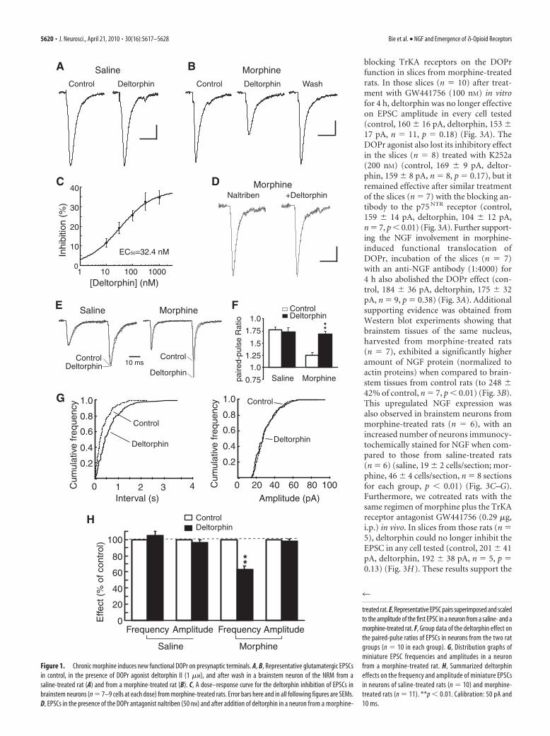
ResultsChronic morphine induces new functional DOPr onpresynaptic terminals of glutamate synapsesWe first examined morphine-induced emergence of functionalDOPr on glutamate synapses in brainstem neurons of the nucleusraphe magnus, which, under normal conditions, steadily expressfunctional MOPr, but lack functional DOPr due to its predomi-nant intracellular localization (Pan et al., 1997; Ma et al., 2006).In these neurons under whole-cell voltage-clamp recording incontrol slices taken from saline-treated rats (n � 12), the selectiveDOPr agonist deltorphin II (1 �M) had no significant effect onthe amplitude of evoked glutamatergic EPSC in any neurontested (control, 136 � 11 pA, deltorphin, 132 � 11 pA, n � 21,p � 0.30) (Fig. 1A), indicating that functional DOPr is lacking onnormal glutamate synapses in these neurons. By contrast, in neu-rons from chronic morphine-treated rats (n � 15), deltorphin (1�M) produced a reversible inhibition in the EPSC amplitude inevery neuron tested (control, 170 � 12 pA, deltorphin, 112 � 8pA, n � 21, p 0.01) (Fig. 1B). The deltorphin inhibition wasdose dependent, with a near-maximum inhibition of 34 � 2% at1 �M and an EC50 value of 32.4 nM (Fig. 1C), and it was com-pletely abolished by the selective DOPr antagonist naltriben (50nM) (control in naltriben, 183 � 19 pA, �deltorphin, 172 � 19pA, n � 9, p � 0.15) (Fig. 1D). This indicates a selective DOPr-mediated effect, as we reported previously (Ma et al., 2006).
To determine the synaptic site of this newly emerged DOPr,we first used the paradigm of paired-pulse ratio (PPR), which hasan inverse relationship with the probability of presynaptic trans-mitter release and has been widely used to determine a presynap-tic involvement in a synaptic change (Dobrunz and Stevens,1997; Bie et al., 2005). In those brainstem neurons of controlslices, deltorphin failed to alter the PPR of glutamate EPSCs (con-trol, 1.77 � 0.06, deltorphin, 1.73 � 0.09, n � 10, p � 0.48) (Fig.1E,F). In neurons from morphine-treated rats, the baseline PPRwas significantly decreased when compared to that of controlneurons (1.25 � 0.05 vs 1.77 � 0.06 of controls, p 0.01),indicating a significantly increased activity of glutamate synapsesby the morphine treatment, an observation consistent with ourprevious study on the same neurons (Bie et al., 2005). In theseneurons of morphine-treated rats, however, deltorphin signifi-cantly increased the EPSC PPR (control, 1.25 � 0.05, deltorphin,1.68 � 0.06, n � 10, p 0.01) (Fig. 1E,F), indicating a likelydecrease in presynaptic glutamate release. To further confirm thismechanism, we analyzed the properties of spontaneous minia-ture EPSCs in the presence of tetrodotoxin (1 �M). We found thatthe DOPr agonist, while having no effect on either the frequencyor amplitude of miniature EPSCs in neurons of control rats, sig-nificantly decreased the frequency of miniature EPSCs withoutaltering the amplitude in neurons from morphine-treated rats(frequency: control, 9.80 � 0.51 Hz, deltorphin, 6.08 � 0.42 Hz,p 0.01; amplitude: control, 23.7 � 1.4 pA, deltorphin, 23.3 �1.6 pA, p � 0.51, n � 11) (Fig. 1G,H). As we reported previously
(Ma et al., 2006), deltorphin remained ineffective on the postsyn-aptic membrane conductance in those neurons as well as in controlneurons. These results suggest that chronic morphine induces newfunctional DOPr that appears on presynaptic glutamate terminals,and DOPr activation inhibits glutamate release.
NGF induces new functional DOPr on glutamate synapsesRecent studies, including ours, have shown that this morphine-induced functional DOPr results from exocytotic membranetrafficking of intracellularly localized DOPr (Ma et al., 2006;Zhang et al., 2006; Cahill et al., 2007). We began our search for themolecular determinant that might be responsible for thismorphine-induced DOPr translocation to surface membrane bytesting a number of compounds that activate the signaling path-ways known to be upregulated by chronic opioids (Williams etal., 2001), in an attempt to trigger similar DOPr translocation innaive, normal neurons in vitro. Treatment of control slices withthe adenylyl cyclase activator forskolin (10 �M), the cAMP ana-log/protein kinase A activator 8-bromo-cAMP (1 mM), the pro-tein kinase C (PKC) activator phorbol 12-myristate 13-acetate (1�M), or neuropeptide cholecystokinin (300 nM) for up to 5 h allfailed to induce deltorphin-mediated inhibition of glutamate EPSCs(data not shown).
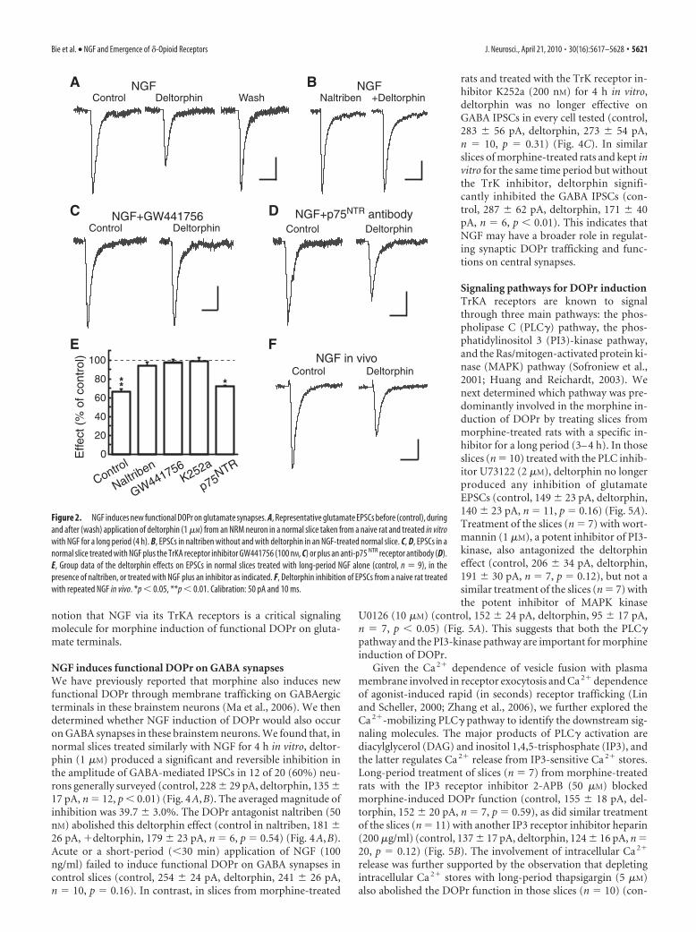
However, in control slices (n � 10) treated with NGF (100ng/ml) for a long period (4 h), deltorphin (1 �M) produced asignificant and reversible inhibition in the EPSC amplitude in 9 of11 (82%) neurons generally surveyed (control, 207 � 27 pA,deltorphin, 147 � 19 pA, wash, 195 � 33 pA, n � 9, p 0.01; nosignificant effect on the remaining 2 cells) (Fig. 2A,E). The inhi-bition was reversed by the DOPr antagonist naltriben (50 nM)(control in naltriben, 180 � 17 pA, �deltorphin, 171 � 19 pA,n � 5 of a separate group, p � 0.20) (Fig. 2B,E). This NGFtreatment itself did not appear to have a significant effect on basalEPSC amplitude when compared between control and NGF-treated slices ( p � 0.28). Cotreatment of control slices (n � 5)with NGF (100 ng/ml) and the tyrosine receptor kinase (TrK)inhibitor K252a (200 nM) for the same time period blocked thedeltorphin effect (control, 191 � 39 pA, deltorphin, 186 � 34 pA,n � 5, p � 0.50) (Fig. 2E); so did the cotreatment with NGF plusthe specific TrKA receptor antagonist GW441756 (100 nM) (con-trol, 124 � 11 pA, deltorphin, 121 � 10 pA, n � 7 from 7 slices,p � 0.43), but not with a blocking antibody (1:2000) to the otherNGF receptor p75 NTR (control, 188 � 26 pA, deltorphin, 127 �21 pA, n � 5 from 5 slices, p 0.01) (Fig. 2C–E). In contrast,treatment of control slices (n � 5) with NGF (100 ng/ml) for ashort period (30 min) was unable to induce functional DOPr onglutamate synapses (control, 157 � 22 pA, deltorphin, 150 � 25pA, n � 5, p � 0.45). We also determined whether NGF admin-istered in intact rats in vivo would induce similar appearance offunctional DOPr on glutamate synapses. In brainstem slices fromnaive rats (n � 6) treated with twice-daily NGF (100 ng, i.p.) for2 d, deltorphin (1 �M) significantly and reversibly inhibited theEPSC in every neuron tested (control, 183 � 27 pA, deltorphin,118 � 14 pA, wash, 171 � 15 pA, n � 6, p 0.01) (Fig. 2F).
Thus, it appears that long-term (at least hours) NGF treat-ment of both normal brainstem slices in vitro and naive rats invivo, similar to the morphine treatment in vivo, is able to bringout functional DOPr on glutamate synaptic terminals by activat-ing TrKA receptors.
NGF is responsible for morphine induction of DOPrTo determine whether NGF via TrKA receptors was responsiblefor morphine induction of DOPr, we first examined the effect of
Bie et al. • NGF and Emergence of �-Opioid Receptors J. Neurosci., April 21, 2010 • 30(16):5617–5628 • 5619
blocking TrKA receptors on the DOPrfunction in slices from morphine-treatedrats. In those slices (n � 10) after treat-ment with GW441756 (100 nM) in vitrofor 4 h, deltorphin was no longer effectiveon EPSC amplitude in every cell tested(control, 160 � 16 pA, deltorphin, 153 �17 pA, n � 11, p � 0.18) (Fig. 3A). TheDOPr agonist also lost its inhibitory effectin the slices (n � 8) treated with K252a(200 nM) (control, 169 � 9 pA, deltor-phin, 159 � 8 pA, n � 8, p � 0.17), but itremained effective after similar treatmentof the slices (n � 7) with the blocking an-tibody to the p75 NTR receptor (control,159 � 14 pA, deltorphin, 104 � 12 pA,n � 7, p 0.01) (Fig. 3A). Further support-ing the NGF involvement in morphine-induced functional translocation ofDOPr, incubation of the slices (n � 7)with an anti-NGF antibody (1:4000) for4 h also abolished the DOPr effect (con-trol, 184 � 36 pA, deltorphin, 175 � 32pA, n � 9, p � 0.38) (Fig. 3A). Additionalsupporting evidence was obtained fromWestern blot experiments showing thatbrainstem tissues of the same nucleus,harvested from morphine-treated rats(n � 7), exhibited a significantly higheramount of NGF protein (normalized toactin proteins) when compared to brain-stem tissues from control rats (to 248 �42% of control, n � 7, p 0.01) (Fig. 3B).This upregulated NGF expression wasalso observed in brainstem neurons frommorphine-treated rats (n � 6), with anincreased number of neurons immunocy-tochemically stained for NGF when com-pared to those from saline-treated rats(n � 6) (saline, 19 � 2 cells/section; mor-phine, 46 � 4 cells/section, n � 8 sectionsfor each group, p 0.01) (Fig. 3C–G).Furthermore, we cotreated rats with thesame regimen of morphine plus the TrKAreceptor antagonist GW441756 (0.29 �g,i.p.) in vivo. In slices from those rats (n �5), deltorphin could no longer inhibit theEPSC in any cell tested (control, 201 � 41pA, deltorphin, 192 � 38 pA, n � 5, p �0.13) (Fig. 3H). These results support the
10
0
20
30
40
10 100 1000
Inhi
bitio
n (%
)
[Deltorphin] (nM)1
EC50=32.4 nM
CNaltriben +Deltorphin
D Morphine
H
4 20 40 60 80 1000
0.2
0.4
0.6
0.8
1.0
Cum
ulat
ive
freq
uenc
y
Amplitude (pA)
Control
Deltorphin
100
80
60
40
20
0Frequency Amplitude
Saline Morphine
AmplitudeFrequency
**
ControlDeltorphin
Effe
ct (
% o
f con
trol
)
0 1 2 3
0.2
0.4
0.6
0.8
1.0
Cum
ulat
ive
freq
uenc
y
Interval (s)
Control
Deltorphin
G
F
paire
d-pu
lse
Rat
io
0.75
1.0
1.25
1.5
1.75
1.0
Saline Morphine
ControlDeltorphin
**
E Saline Morphine
ControlDeltorphin
Control
Deltorphin10 ms
Saline
Control Deltorphin
A Morphine
WashControl Deltorphin
B
Figure 1. Chronic morphine induces new functional DOPr on presynaptic terminals. A, B, Representative glutamatergic EPSCsin control, in the presence of DOPr agonist deltorphin II (1 �M), and after wash in a brainstem neuron of the NRM from asaline-treated rat (A) and from a morphine-treated rat (B). C, A dose–response curve for the deltorphin inhibition of EPSCs inbrainstem neurons (n � 7–9 cells at each dose) from morphine-treated rats. Error bars here and in all following figures are SEMs.D, EPSCs in the presence of the DOPr antagonist naltriben (50 nM) and after addition of deltorphin in a neuron from a morphine-
4
treated rat. E, Representative EPSC pairs superimposed and scaledto the amplitude of the first EPSC in a neuron from a saline- and amorphine-treated rat. F, Group data of the deltorphin effect onthe paired-pulse ratios of EPSCs in neurons from the two ratgroups (n � 10 in each group). G, Distribution graphs ofminiature EPSC frequencies and amplitudes in a neuronfrom a morphine-treated rat. H, Summarized deltorphineffects on the frequency and amplitude of miniature EPSCsin neurons of saline-treated rats (n � 10) and morphine-treated rats (n � 11). **p 0.01. Calibration: 50 pA and10 ms.
5620 • J. Neurosci., April 21, 2010 • 30(16):5617–5628 Bie et al. • NGF and Emergence of �-Opioid Receptors
notion that NGF via its TrKA receptors is a critical signalingmolecule for morphine induction of functional DOPr on gluta-mate terminals.
NGF induces functional DOPr on GABA synapsesWe have previously reported that morphine also induces newfunctional DOPr through membrane trafficking on GABAergicterminals in these brainstem neurons (Ma et al., 2006). We thendetermined whether NGF induction of DOPr would also occuron GABA synapses in these brainstem neurons. We found that, innormal slices treated similarly with NGF for 4 h in vitro, deltor-phin (1 �M) produced a significant and reversible inhibition inthe amplitude of GABA-mediated IPSCs in 12 of 20 (60%) neu-rons generally surveyed (control, 228 � 29 pA, deltorphin, 135 �17 pA, n � 12, p 0.01) (Fig. 4A,B). The averaged magnitude ofinhibition was 39.7 � 3.0%. The DOPr antagonist naltriben (50nM) abolished this deltorphin effect (control in naltriben, 181 �26 pA, �deltorphin, 179 � 23 pA, n � 6, p � 0.54) (Fig. 4A,B).Acute or a short-period (30 min) application of NGF (100ng/ml) failed to induce functional DOPr on GABA synapses incontrol slices (control, 254 � 24 pA, deltorphin, 241 � 26 pA,n � 10, p � 0.16). In contrast, in slices from morphine-treated
rats and treated with the TrK receptor in-hibitor K252a (200 nM) for 4 h in vitro,deltorphin was no longer effective onGABA IPSCs in every cell tested (control,283 � 56 pA, deltorphin, 273 � 54 pA,n � 10, p � 0.31) (Fig. 4C). In similarslices of morphine-treated rats and kept invitro for the same time period but withoutthe TrK inhibitor, deltorphin signifi-cantly inhibited the GABA IPSCs (con-trol, 287 � 62 pA, deltorphin, 171 � 40pA, n � 6, p 0.01). This indicates thatNGF may have a broader role in regulat-ing synaptic DOPr trafficking and func-tions on central synapses.
Signaling pathways for DOPr inductionTrKA receptors are known to signalthrough three main pathways: the phos-pholipase C (PLC�) pathway, the phos-phatidylinositol 3 (PI3)-kinase pathway,and the Ras/mitogen-activated protein ki-nase (MAPK) pathway (Sofroniew et al.,2001; Huang and Reichardt, 2003). Wenext determined which pathway was pre-dominantly involved in the morphine in-duction of DOPr by treating slices frommorphine-treated rats with a specific in-hibitor for a long period (3– 4 h). In thoseslices (n � 10) treated with the PLC inhib-itor U73122 (2 �M), deltorphin no longerproduced any inhibition of glutamateEPSCs (control, 149 � 23 pA, deltorphin,140 � 23 pA, n � 11, p � 0.16) (Fig. 5A).Treatment of the slices (n � 7) with wort-mannin (1 �M), a potent inhibitor of PI3-kinase, also antagonized the deltorphineffect (control, 206 � 34 pA, deltorphin,191 � 30 pA, n � 7, p � 0.12), but not asimilar treatment of the slices (n � 7) withthe potent inhibitor of MAPK kinase
U0126 (10 �M) (control, 152 � 24 pA, deltorphin, 95 � 17 pA,n � 7, p 0.05) (Fig. 5A). This suggests that both the PLC�pathway and the PI3-kinase pathway are important for morphineinduction of DOPr.
Given the Ca 2� dependence of vesicle fusion with plasmamembrane involved in receptor exocytosis and Ca 2� dependenceof agonist-induced rapid (in seconds) receptor trafficking (Linand Scheller, 2000; Zhang et al., 2006), we further explored theCa 2�-mobilizing PLC� pathway to identify the downstream sig-naling molecules. The major products of PLC� activation arediacylglycerol (DAG) and inositol 1,4,5-trisphosphate (IP3), andthe latter regulates Ca 2� release from IP3-sensitive Ca 2� stores.Long-period treatment of slices (n � 7) from morphine-treatedrats with the IP3 receptor inhibitor 2-APB (50 �M) blockedmorphine-induced DOPr function (control, 155 � 18 pA, del-torphin, 152 � 20 pA, n � 7, p � 0.59), as did similar treatmentof the slices (n � 11) with another IP3 receptor inhibitor heparin(200 �g/ml) (control, 137 � 17 pA, deltorphin, 124 � 16 pA, n �20, p � 0.12) (Fig. 5B). The involvement of intracellular Ca 2�
release was further supported by the observation that depletingintracellular Ca 2� stores with long-period thapsigargin (5 �M)also abolished the DOPr function in those slices (n � 10) (con-
Naltriben
D
E
0
20
40
60
80
100
Control
Naltriben
GW441756K252a
p75NTR
***
Effe
ct (
% o
f con
trol
)NGFA
DeltorphinControl Wash +DeltorphinNGF
NGF+p75NTR antibody
DeltorphinControl
NGF in vivo
j
DeltorphinControl
B
NGF+GW441756DeltorphinControl
C
F
Figure 2. NGF induces new functional DOPr on glutamate synapses. A, Representative glutamate EPSCs before (control), duringand after (wash) application of deltorphin (1 �M) from an NRM neuron in a normal slice taken from a naive rat and treated in vitrowith NGF for a long period (4 h). B, EPSCs in naltriben without and with deltorphin in an NGF-treated normal slice. C, D, EPSCs in anormal slice treated with NGF plus the TrKA receptor inhibitor GW441756 (100 nM, C) or plus an anti-p75 NTR receptor antibody (D).E, Group data of the deltorphin effects on EPSCs in normal slices treated with long-period NGF alone (control, n � 9), in thepresence of naltriben, or treated with NGF plus an inhibitor as indicated. F, Deltorphin inhibition of EPSCs from a naive rat treatedwith repeated NGF in vivo. *p 0.05, **p 0.01. Calibration: 50 pA and 10 ms.
Bie et al. • NGF and Emergence of �-Opioid Receptors J. Neurosci., April 21, 2010 • 30(16):5617–5628 • 5621

trol, 154 � 20 pA, deltorphin, 147 � 18 pA, n � 14, p � 0.17)(Fig. 5B). In addition, morphine induction of DOPr appeared tobe also dependent on Ca 2� entry through store-operated Ca 2�
channels (SOCCs), as long-period treatment of the slices (n � 11)with MRS1845 (5 �M), a potent SOCC blocker, eliminated thedeltorphin effect (control, 140 � 13 pA, deltorphin, 127 � 13 pA,n � 13, p � 0.15) (Fig. 5B). In contrast, in the slices (n � 6)similarly treated with long-period GF109203X (3 �M), a selectiveinhibitor of PKC, which is the substrate of DAG, deltorphin re-tained its inhibition of glutamate EPSCs (control, 256 � 76 pA,deltorphin, 164 � 57 pA, n � 6, p 0.05) (Fig. 5B).
Increased activity of CaMKII, a serine/threonine kinase, has beenshown to drive rapid (in seconds) membrane insertion of AMPAglutamate receptors into synapses through interactions betweenGluR1 subunits of AMPA receptors and cytoplasmic proteins con-taining the PSD-95-Dlg-ZO1 (PDZ) domains (Hayashi et al.,
2000; Collingridge et al., 2004). Considering the Ca 2� depen-dence of this DOPr induction, we were wondering whetherCa 2�-dependent activation of CaMKII was also required, al-though unlike the ligand-gated ion channel AMPA receptor, in-duction of membrane trafficking of DOPr, a GPCR, is a muchslower (at least hours) process (Cahill et al., 2007). Nevertheless,in slices (n � 10) from morphine-treated rats and treated with thespecific CaMKII inhibitor KN-93 (5 �M) for a long period, theDOPr agonist was no longer effective (control, 191 � 16 pA,deltorphin, 183 � 18 pA, n � 11, p � 0.45) (Fig. 5C), whereas itinhibited EPSCs after similar treatment with the inactive analogKN-92 (5 �M) (control, 164 � 20 pA, deltorphin, 110 � 13 pA,n � 12 from 10 slices, p 0.01) (Fig. 5C). In further support ofthis CaMKII role, brainstem tissues taken from morphine-treated rats contained a significantly higher level of phosphory-lated CaMKII proteins, the activated form of CaMKII, with thetotal CaMKII protein unchanged from controls (Fig. 5D,E).
As controls for the effects of all inhibitors we used, a short-period (30 min) treatment of slices from morphine-treated ratswith those inhibitors failed to significantly alter morphine-induced DOPr function (see supplemental material, available atwww.jneurosci.org).
Sorting of DOPrNext, we sought other signaling proteins, downstream of CaMKIIactivation, which might account for altering the normal, intracel-
MorSal
A
0
20
40
60
80
100
Control
GW441756K252a
NGF antibody
p75NTR
** **
Effe
ct (
% o
f con
trol
)
Control DeltorphinGW441756 in vivo
Morphine Saline
Actin
NGF
B
H
C D
NGFE NeuNF
MergedG
Figure 3. NGF is responsible for morphine induction of functional DOPr. A, Summarizeddeltorphin effects on EPSCs in slices of morphine-treated rats in control or after treatment invitro with the indicated inhibitor. B, Representative Western blot lanes of NGF and actin pro-teins in NRM tissues taken from saline- and morphine-treated rats. C, D, Microscopic images ofimmunofluorescence labeling for NGF in NRM sections from saline (Sal)- and morphine (Mor)-treated rats. Calibration: 100 �m. E–G, Images of labeling for NGF (red) and for neurons withthe neuronal marker NeuN (green), and a merged image in NRM slices from a morphine-treatedrat. Calibration: 20 �m. H, EPSCs from a rat treated with morphine plus GW441756 in vivo.**p 0.01. Calibration: 50 pA and 10 ms.
Control
Deltorphin
NGF
DeltorphinControl
NGF+Naltriben
0
20
40
60
80
100
NGF +Naltriben
% o
f con
trol
B ControlDeltorphin
**
ControlDeltorphin
NormalA
CControl Deltorphin
K252a
Figure 4. NGF mediates morphine induction of DOPr on GABA synapses. A, SuperimposedGABA-mediated IPSCs in control and in deltorphin (1 �M) in NRM neurons in a normal slice andin normal slices treated with NGF (100 ng/ml) alone or with NGF plus naltriben (50 nM) for 4 h invitro. B, Group data of the deltorphin effects after treatment of normal slices with NGF or NGFplus naltriben. C, Representative GABA IPSCs in slices from morphine-treated rats and treatedwith the TrK receptor inhibitor K252a (200 nM) for 4 h in vitro. Note that use of KCl in theintrapipette solution with a holding potential of �60 mV resulted in a downward direction ofthe IPSCs. **p 0.01. Calibration: 50 pA and 20 ms.
5622 • J. Neurosci., April 21, 2010 • 30(16):5617–5628 Bie et al. • NGF and Emergence of �-Opioid Receptors

lular sorting fate of DOPr after prolonged exposure to morphineand NGF. One of the few such candidate proteins currentlyknown is NHERF-1, also known as EBP50 (ezrin-radixin-moesin-binding phosphoprotein-50), which is a PDZ domain-containing scaffolding protein implicated in the assembly ofprotein complexes and in sorting of internalized �2-adrenergicreceptors and �-opioid receptor (KOPr) to the recycling pathwayfor receptor resensitization (Liu-Chen, 2004; Weinman et al.,2006; Hanyaloglu and von Zastrow, 2008). We first examinedwhether the morphine treatment would change NHERF-1 ex-pression. In brainstem tissues taken from morphine-treated rats(n � 8), we found that the protein level of NHERF-1, normalizedto GAPDH, was significantly increased to 152 � 18% of control( p 0.05) (Fig. 6A), raising the possibility that upregulatedNHERF-1 protein changes the sorting fate of DOPr from normallyintracellular targeting to exocytotic trafficking for membrane inser-tion and functional expression. This hypothesis was supported byour following experiments using coimmunoprecipitation (co-IP) toassess interaction between DOPr and NHERF-1 proteins. In controlbrainstem tissues, there was a low level of NHERF-1 protein thatcoprecipitated with DOPr (Fig. 6B,C), consistent with previous re-ports that NHERF-1 normally has low affinity to or interaction withthe c-terminal of DOPr (Heydorn et al., 2004; Huang et al., 2004).However, in brainstem tissues from morphine-treated rats (n � 10),the amount of NHERF-1 protein that coprecipitated with DOPr wassignificantly increased [Fig. 6B (top two panels),C]. In contrast, afterincubation of the DOPr antibody with a sequence-specific DOPr-blocking peptide (Patwardhan et al., 2005), no DOPr or NHERF-1protein was detected in the precipitated preparations following thesame IP procedure (Fig. 6B, bottom two panels), indicating asequence-specific binding of the DOPr antibody used for DOPr co-IP. Additionally, this specific binding of DOPr by the DOPr antibod-ies is supported by the loss of DOPr immunostaining in DOPr KOmice (see supplemental material, available at www.jneurosci.org).
Demonstrating a specific role of TrK receptors in this in-creased NHERF-1-DOPr interaction, cotreatment of rats (n � 5)with morphine and the TrK receptor antagonist K252a in vivo
significantly inhibited the morphine-induced increase in NHERF-1-DOPr co-precipitation (Fig. 6C). In addition,morphine treatment also increased theserine/threonine phosphorylation of DOPr(n � 13 rats), which was blocked by cotreat-ment in vivo with morphine and K252a (n�5 rats) (Fig. 6D). A similar increase inNHERF-1-DOPr coprecipitation was alsoobserved in brainstem tissues from naiverats (n � 6) treated with NGF (50 ng/0.5 �l)in vivo through microinjections into thebrainstem nucleus (NHERF/DOPr ratio:saline, 0.36 � 0.07, NGF, 0.75 � 0.07, p 0.01) (Fig. 6E). The NGF treatment of naiverats in vivo (n � 6 rats) also increased theamount of phosphorylated DOPr (pDOPr/DOPr ratio: saline, 0.40 � 0.04, NGF,0.61 � 0.04, p 0.01) (Fig. 6F).
DOPr in NHERF-1 knock-out miceTo further validate the specificity ofDOPr–NHERF-1 interaction and its rolein the DOPr trafficking, we examined theeffects of morphine and NGF on DOPrinduction in NHERF-1 KO mice we origi-
nally reported (Shenolikar et al., 2002). In saline-treated NHERF-1�/� mice and corresponding WT mice, deltorphin (1 �M) wasineffective on the amplitude of glutamate EPSCs in the brainstemneurons (WT: control, 209 � 14 pA, deltorphin, 198 � 14 pA,n � 6, p � 0.38; KO: control, 292 � 8 pA, deltorphin, 285 � 7 pA,n � 7, p � 0.37) (Fig. 7A,E). In morphine-treated WT mice,functional DOPr, similar to that in morphine-treated rats,emerged as shown by deltorphin inhibition of EPSCs (control,173 � 11 pA, deltorphin, 109 � 7 pA, n � 6, p 0.01), but it wasabsent in the KO mice treated similarly with morphine (control,234 � 7 pA, deltorphin, 253 � 8 pA, n � 11, p � 0.29) (Fig.7B,E). Moreover, the same long-period NGF treatment of brain-stem slices from naive WT mice induced functional DOPr onglutamate synapses in 6 of 8 cells (control, 201 � 18 pA, deltor-phin, 130 � 13 pA, n � 6, p 0.01), but failed to do so in any celltested in slices from naive KO mice (control, 257 � 8 pA, deltor-phin, 252 � 8 pA, n � 10, p � 0.46) (Fig. 7C,E). In addition,morphine-induced DOPr function in WT mice was also abol-ished by long-period treatment of the slices in vitro with the TrKreceptor antagonist K252a (200 nM) (control, 138 � 6 pA, del-torphin, 133 � 7 pA, n � 5, p � 0.36) (Fig. 7D,E). The expressionof DOPr protein in the NHERF-1 KO mice was not significantlychanged from that in the WT mice (DOPr/actin ratio: WT,0.75 � 0.04, n � 5; KO, 0.72 � 0.06, n � 6, p � 0.72) (Fig. 7F).These findings from NHERF-1-lacking mice further support ourresults from rats by showing that NHERF-1 is required for bothmorphine and NGF induction of synaptic DOPr.
Translocation of DOPr to surface membraneTo determine whether the emerged DOPr on synaptic terminalswas indeed due to translocation of intracellular DOPr to surfacemembrane, we next examined the amount of DOPr protein onterminal membrane using brainstem preparations of synapto-somes, which contain mostly membrane structures of synapticterminals with nearly no cell body contents and greatly reducedintraterminal contents (Ghijsen et al., 2003; Dunkley et al., 2008).In a previous study (Ma and Pan, 2006), we have shown that the
A
C D E
*
B
Control
U73122
Effe
ct (
% o
f con
trol
)
Wortmannin
U0126
** *
0
20
40
60
80
100
0
20
40
60
80
100
Control2-APB
HeparinThapsig.
MRS1845
GF109203X
CaMKII
p-CaMKII
GAPDH
GAPDH
MorphineSaline
**
chan
ge (
%)
SalineMorphine
0
60
120
180
CaMKII p-CaMKII
**E
ffect
(%
of c
ontr
ol)
020406080
100
Control KN-93 KN-92
** **
Effe
ct (
% o
f con
trol
)
Figure 5. Morphine induction of DOPr involves the PLC–CaMKII pathway. A–C, Group data of deltorphin effects on EPSCsin slices from morphine-treated rats in control or treated in vitro with the indicated inhibitor for a long period. D, Repre-sentative lanes of CaMKII, p-CaMKII, and GAPDH proteins in NRM tissues from saline- and morphine-treated rats. E,Summarized Western blot results of percentage changes in CaMKII and p-CaMKII proteins normalized to GAPDH. Thapsig,Thapsigargin. *p 0.05, **p 0.01.
Bie et al. • NGF and Emergence of �-Opioid Receptors J. Neurosci., April 21, 2010 • 30(16):5617–5628 • 5623

morphine treatment does not signifi-cantly change the total DOPr protein inthe brainstem tissue. However, we foundthat, in brainstem synaptosomes frommorphine-treated rats, the amount ofDOPr protein, normalized to the specificsynaptic terminal marker synaptophysin,was significantly increased when com-pared to controls, as revealed by Westernblot analysis; this increase in synaptoso-mal DOPr was blocked by cotreatment ofrats in vivo with morphine plus the TrKreceptor antagonist K252a (Fig. 8A).Moreover, in rats treated with NGF invivo that induced functional DOPr, theamount of synaptosomal DOPr proteinwas also significantly increased (Fig. 8A).Thus, it appears that the morphine andNGF induction of functional DOPr onsynaptic terminals is likely due, at leastpartially, to DOPr translocation to surfacemembrane of synaptic terminals, consis-tent with our previous anatomical studyof labeled DOPr with confocal micros-copy on the same brainstem neurons (Maand Pan, 2006) and with our recent reportof morphine-induced increase in synap-tosomal DOPr of amygdala neurons (Bieet al., 2009). This notion is also in line withthe mechanism for membrane traffickingof postsynaptic DOPr on dorsal root gan-glion cells (Zhang et al., 2006; Cahill et al.,2007).
If the appearance of functional DOPrwas indeed due to translocation or traf-ficking of intracellular DOPr to surfacemembrane rather than other mecha-nisms, it should be blocked in conditionsunder which known processes required for exocytotic receptortrafficking and membrane insertion were disrupted. The endo-plasmic reticulum (ER) and the Golgi apparatus are importantmembrane network for packing and transporting of cytoplasmicproteins in receptor trafficking (Tan et al., 2004). In slices (n � 9)from morphine-treated rats and treated for 4 h with brefeldin A(5 �M), an inhibitor of protein transportation from ER to theGolgi apparatus (Donaldson et al., 1992), deltorphin (1 �M)could no longer inhibit the EPSC (control, 165 � 12 pA, deltor-phin, 159 � 13 pA, n � 11, p � 0.22) (Fig. 8B,G).
Furthermore, to inhibit receptor exocytosis, we used a fusionpolypeptide, TAT-NSF81 (5 �M), which we have previouslyshown to block exocytosis by inhibiting N-ethyl-maleimide-sensitive factor (NSF)-mediated disassembly of soluble NSFattachment protein receptor (SNARE) molecules (Matsushita etal., 2005). After the TAT-NSF81 treatment of slices frommorphine-treated rats for 3 h in vitro, the slices were left in nor-mal bath solution without the polypeptide for at least 20 minbefore EPSC recording. We found that TAT-NSF81 preventeddeltorphin-induced inhibition of EPSCs in these slices (n � 8)(control, 153 � 20 pA, deltorphin, 142 � 17 pA, n � 10, p � 0.47)(Fig. 8C,G). By contrast, similar treatment with the correspond-ing but scrambled control peptide TAT-NSF81scr (5 �M) failedto alter the deltorphin inhibition of EPSCs (control, 198 � 23 pA,deltorphin, 135 � 24 pA, n � 7, p 0.01) (Fig. 8D,G). Next, we
confirmed that TAT-NSF81 did not inhibit signaling by otherneuronal receptors constitutively localized on the plasmamembrane, using GABAB receptor-mediated presynaptic in-hibition as an example. In slices from morphine-treated rats,baclofen (10 �M), a potent GABAB receptor agonist, produceda large inhibition in the EPSC amplitude (control, 289 � 57pA, baclofen, 141 � 39 pA, n � 7, p 0.01) (Fig. 8 E, G). Thisbaclofen inhibition was largely due to activation of presynap-tic GABAB receptors on glutamate synaptic terminals (B. Bieand Z. Z. Pan, unpublished observation). In slices that werecomparable but pretreated similarly with TAT-NSF81 (5 �M),baclofen (10 �M) induced a similar EPSC inhibition (control,201 � 23 pA, baclofen, 89 � 12 pA, n � 8, p 0.01) (Fig.8 F, G). There was no statistical difference ( p � 0.45) in thebaclofen-induced EPSC inhibitions with and without thepolypeptide treatment, indicating that TAT-NSF81 does nothave a significant effect on the synaptic function of preexistingGABAB receptors on glutamate synapses. Thus, the recruit-ment of functional DOPr appears to require DOPr transpor-tation from ER to Golgi and NSF-regulated vesicle exocytosisto the plasma membrane, two key processes for membranetrafficking of GPCRs (Tan et al., 2004). Figure 8 H illustratesthe proposed signaling pathways for chronic opioid-inducedexocytotic trafficking and functional expression of DOPr on acentral synaptic terminal.
C D
E
BA
NHERF
GAPDH
Saline Morphine
DOPr
NHERF
MorphineSaline
NH
ER
F/D
OP
r R
atio
0
0.2
0.4
0.6
0.8
1.0
SalineMorphine
+K252a
** **
Saline Mor +K252a
DOPr
NHERF
NHERF
DOPr
NGF in vivoSaline
pDOPr
Saline Mor +K252a
0
0.2
0.4
0.6
0.8
SalineMorphine
+K252apDO
Pr/
DO
Pr
Rat
io
** **
F Saline NGF in vivo
DOPr
pDOPr
DOPrNHERF
DOPr
Figure 6. Chronic morphine increases interaction between DOPr and NHERF-1 proteins. A, Western blot lanes of NHERF-1 andGAPDH proteins in NRM tissues from saline- and morphine-treated rats. B, DOPr immunoprecipitates (IP) immunoblotted forNHERF-1 and for DOPr in NRM tissues from saline- and morphine-treated rats in control (top two panels) and after preincubationof the DOPr antibody with a DOPr-blocking peptide (bottom two panels). C, D, Similar IP data of NHERF-1 and DOPr (C) and DOPrimmunoprecipitates with immunoblotted phosphorylated DOPr (pDOPr) and total DOPr (D) in tissues from saline-treated rats andrats treated with morphine without or with K252a cotreatment in vivo. Also shown are changes in the ratios of NHERF-1 over DOPrand pDOPr over total DOPr proteins in the indicated three groups of rats. E, F, Similar DOPr precipitates immunoblotted for NHERF-1and DOPr (E) and for pDOPr and total DOPr (F) in NRM tissues from naive rats treated with saline or NGF in vivo. Mor, Morphine.**p 0.01.
5624 • J. Neurosci., April 21, 2010 • 30(16):5617–5628 Bie et al. • NGF and Emergence of �-Opioid Receptors

DiscussionWe have shown that long-period NGF, applied to central neuronsboth in vitro and in animals in vivo, causes the emergence of newfunctional DOPr on central synaptic terminals likely by redirect-ing constitutively intracellular DOPr to surface membrane, amechanism that may account for chronic opioid-induced synap-tic DOPr function. In addition, we have identified the prominentsignaling pathway for the NGF effect, from TrKA receptors toexocytosis for surface insertion. Thus, NGF may function as a key
molecular switch that alters the normalsorting of intracellular DOPr, redirectingintracellular DOPr to plasma membranefor functional expression under sustainedopioid stimulation.
Classically, NGF has well known rolesin neuronal survival and cell differentia-tion in the developing brain. Recent evi-dence has shown diverse trophic effects ofNGF on the adult brain as well, particularlyon structural modifications and synaptic ef-ficacy in central synapses (Poo, 2001;Sofroniew et al., 2001; Chao and Bothwell,2002). In cultured HEK293 cells, NGF orepidermal growth factor rapidly (withinminutes) increases membrane insertion ofTRP channels by PI3 kinase-dependentphosphorylation (Zhang et al., 2005). Thecurrent study shows that, in native brainneurons, NGF causes translocation and sur-face expression of intracellular DOPr, amember of the GPCR superfamily repre-senting the largest population of membranereceptors responsible for most of signalingacross plasma membrane in mammaliancells (Tan et al., 2004). Thus, NGF may havemuch broader regulating functions in re-ceptor signaling of the adult brain underboth normal and pathological conditions.
Recent studies increasingly suggest thatsorting of intracellular GPCRs to either ly-sosomes for degradation or endosomes forrecycling is diverse and highly regulated bycytoplasmic sorting proteins. However, ourunderstanding of GPCR sorting is still in itsinfancy, as only a few GPCR sorting proteinshave been identified to date, in contrast tothe vast number of GPCRs in brain neurons.For constitutive recycling under normalconditions, the PDZ domain-containingNHERF-1 is responsible for recycling of �2-adrenergic receptors and KOPr (Liu-Chen,2004; Hanyaloglu and von Zastrow, 2008),but has little interaction with DOPr as ob-served in heterologously expressing cell sys-tems (Huang et al., 2004). Consistently, littleNHERF-1-DOPr co-IP was observed incontrol, opioid-naive tissues in the presentstudy. In contrast to constitutive sortingof GPCRs, little is known about the mo-lecular mechanisms by which the nor-mal sorting of GPCRs is altered underpathological conditions. Our results ofsignificant NHERF-1-DOPr interaction,
observed only after chronic morphine treatment and supported byresults from NHERF-1 KO mice, indicate that NHERF-1 may pos-sess a broader binding capacities and functional diversity in GPCRsorting and recycling under various conditions. It remains to beinvestigated how chronic opioids change NHERF-1-DOPr bindingproperties for their increased interaction, whether the increasedDOPr phosphorylation we observed plays a role, and what specificC-terminal sequence of DOPr with certain phosphorylated sites isinvolved in the interaction.
Control Deltorphin
Wild Type Knockout
Control Deltorphin
A Saline
B MorphineControl Deltorphin Control Deltorphin
C NGFControl Deltorphin Control Deltorphin
D Morphine+K252aControl Deltorphin
0
20
4060
80
100
WT KO
Effe
cts
(% o
f con
trol
)
SalineMorphine
NGFMor+K252a
E
****
DOPr
Actin
F WT KO
Figure 7. NHERF-1 is required for morphine and NGF induction of functional DOPr. A–D, Representative glutamate EPSCsin control and in deltorphin (1 �M) in an NRM neuron from WT mice (left column) and NHERF-1 KO mice (right column)treated with saline (A), morphine (B), from normal slices treated with long-period NGF (C), or from a K252a-treated slice ofa morphine-treated WT mouse (D). E, Summarized data of the deltorphin effects in the four treatment groups of WT and KO mice.F, Western blot lanes of DOPr and actin proteins in NRM tissues from WT (n � 5) and NHERF-1 KO mice (n � 6). **p 0.01.Calibration: 50 pA and 10 ms.
Bie et al. • NGF and Emergence of �-Opioid Receptors J. Neurosci., April 21, 2010 • 30(16):5617–5628 • 5625

The intracellular retention of DOPrwas shown to be maintained by DOPrbinding to vesicle luminal substance P(SP), and upon agonist stimulation, SP-bound DOPr in vesicles was rapidly (inseconds) inserted into plasma membranein peripheral sensory neurons (Guan etal., 2005). However, that notion has beenargued against by a recent study, usingknock-in mice with a functional DOPr-eGFP fusion receptor in the same spinalneurons, showing that DOPr had no over-lap with SP in expression and DOPr wastrafficked independently of SP (Scherrer etal., 2009). Interestingly, NGF acutely (60min) increases the intracellular pool of ER-derived, Golgi-associated DOPr in culturedsecretory PC12 cells in vitro (Kim and vonZastrow, 2003). This relatively acute NGFeffect is in contrast to the long-term (4 h)NGF effect of DOPr translocation to surfacemembrane described in this study, andmight serve as a parallel NGF effect thatprimes and replenishes the intracellularDOPr pool for surface trafficking. While wedid not observe a significant increase in totalDOPr protein after morphine treatment(Ma et al., 2006), upregulated NGF may in-crease the expression of many other down-stream signaling proteins given its broadstimulating effect in neural protein expres-sion, contributing to the NGF effect (Huangand Reichardt, 2003). For example, NGFtreatment of PC12 cells increased expres-sion of DOPr gene through epigenetic mod-ifications (Chen et al., 2008). Previouslyreported, agonist- or depolarization-in-duced membrane trafficking of DOPr andTRP channels share a characteristic of rapidoccurrence (within seconds to minutes)(Guan et al., 2005; Odell et al., 2005; Zhanget al., 2005), but clearly differ from thepresent finding of NGF-induced, slowly oc-curring (at least hours) translocation of syn-aptic DOPr. The latter may be indicative ofextra layers of adaptive regulations at tran-scriptional and translational levels, and inthe forward transportation of DOPrthrough the ER–Golgi–surface membranesecretory pathway.
Receptor exocytosis and insertion intosurface membrane involve membrane fu-sion between plasma membrane and themembrane of receptor-containing vesi-cles (Pfeffer, 2007; Wojcik and Brose,2007). The SNARE complex is a core execu-tion component in the membrane fusionmachinery. While much of our current un-derstanding of SNARE-mediated fusionmechanisms has been derived from studieson fast (millisecond) synaptic release ofneurotransmitters (Wojcik and Brose,2007), emerging evidence suggests that re-
Control Deltorphin
TAT-NSF81
Control Deltorphin
BrefeldinB CA
Control Deltorphin
TAT-NSF81scrD
Saline Mor NGF +K252a
DOPr
Synapsin
0.50.40.3
0.10
0.2
DO
Pr/
Syn
apsi
n ra
tio
SalineMor
NGF+K252a
** **
Control BaclofenNormal
Control BaclofenTAT-NSF81FE
G100
80
60
20
0
40
Effe
ct (
% o
f con
trol
)
** **
Control
Brefeldin
TAT-NSF81
TAT-NSF81scr
** **
Control
TAT-NSF81
Deltorphin Baclofen
PIP2
DAG
TrKA
Ca++
SOCC
Golgi/ER
NGF
NGF
PLCγ
IP3
CaCa
CaMKIIIP3R
δ
δ
δp
δδ
NHERFδ
Morphine
PI3K
NSF
SNARE δ
1
2
3
45
6
78
9
NHERF
H
Figure 8. Morphine induction of DOPr involves DOPr translocation to surface membrane of synaptic terminals. A, Western blotlanes of DOPr protein and the synaptic terminal marker synaptophysin (synapsin), and their ratio changes in NRM synaptosomesfrom rats treated with saline, morphine, NGF, or morphine plus K252a in vivo. B, Representative EPSCs in a slice from a morphine-treated rat and treated in vitro with long-period brefeldin A. C, D, Deltorphin actions on EPSCs in slices from morphine-treated ratsand treated in vitro with the exocytosis-inhibiting peptide TAT-NSF 81 (C) or with the corresponding but scrambled peptideTAT-NSF81scr (D) for 3 h. E, F, Effects of the GABAB receptor agonist baclofen (10 �M) on EPSCs in NRM slices from morphine-treated rats without (normal) or with the TAT-NSF81 treatment in vitro. G, Group data showing the effects of deltorphin andbaclofen in NRM slices of morphine-treated rats in control or after long-period treatment with the agents indicated. **p 0.01.Calibration: 50 pA and 10 ms. H, Diagram of the proposed signaling pathways for DOPr trafficking on a central synaptic terminal.Chronic morphine upregulates NGF (1), which activates the TrKA receptor (2) and its coupled PLC� pathway (3) and PI3K pathway(downstream components to be investigated). PLC� activation leads to production of IP3 and increased intracellular Ca 2�
through Ca 2� release from the IP3 receptor (IP3R)-controlled Ca 2� store (4) and by Ca 2� influx through SOCC. DOPr is normallypacked and transported through the ER/Golgi network and constitutively targeted to an intracellular pool (5). Increased intracel-lular Ca 2� activates CaMKII (6), which would lead to downstream changes in the expression of DOPr and NHERF-1 genes andproteins through yet unknown mechanisms of transcription and translation (7), resulting in an increased interaction betweenupregulated NHERF-1 and phosphorylated DOPr, and exocytotic translocation of intracellular DOPr to surface membrane. Interac-tion of DOPr-containing vesicles with the SNARE complex, whose function is regulated by NSF (8), causes membrane fusion forexocytosis and surface expression of functional DOPr (9).
5626 • J. Neurosci., April 21, 2010 • 30(16):5617–5628 Bie et al. • NGF and Emergence of �-Opioid Receptors

ceptor trafficking and surface delivery also involve SNARE-dependent exocytosis (Lan et al., 2001). NSF, an ATPase associatedwith the SNARE complex, regulates exocytosis by disassembling theSNARE complex in the SNARE assemble/disassemble cycle neces-sary for normal SNARE functions (Zhao et al., 2007). We have de-veloped the cell-penetrating peptide TAT-NSF81 that interruptsexocytosis by inhibiting NSF activity (Matsushita et al., 2005). Ourobservation of TAT-NSF81 blocking the surface recruitment offunctional DOPr indicates that the DOPr recruitment likely re-quires Ca2�-dependent, NSF/SNARE-mediated exocytosis. Wenoticed a reduction in EPSC amplitude after the TAT-NSF81pretreatment, which possibly reflects its inhibitory effect on SNARE-dependent membrane fusion of synaptic vesicles for glutamate re-lease. A short period of wash of the peptide appeared to partiallyrecover its inhibition of glutamate release, but not the already trans-located DOPr. In addition to regulating SNARE disassembly, NSFdirectly interacts with specific recycling sequences on the cytoplas-mic terminal of receptors to regulate their membrane trafficking,including �2-adrenergic receptors and AMPA glutamate receptors(Zhao et al., 2007; Hanyaloglu and von Zastrow, 2008). Therefore, itis also possible that TAT-NSF81 blocks DOPr translocation by dis-rupting direct NSF–DOPr interaction.
Functional interaction between DOPr and MOPr has beenwell documented in previous studies using behavioral and ge-netic approaches (Kieffer and Gaveriaux-Ruff, 2002). It likelyinvolves diverse mechanisms at multiple levels, including syner-gistic interaction at the receptor level as DOPr-MOPr het-erodimers in enhancing morphine analgesia (Gomes et al., 2004).Our current finding provides another potential mechanism bywhich NGF functions as a link between sustained MOPr stimu-lation and increased function of synaptic DOPr at the cellularlevel.
Proper GPCR localization and signaling are required for nor-mal cell functions, and GPCR mislocalization and resultant ab-normal signaling cause diseases (Tan et al., 2004). For example,NGF-mediated rapid membrane insertion of TRPV1 channels byPI3 kinase-dependent phosphorylation likely accounts for hyper-sensitivity of peripheral sensory neurons under inflammatorypain sensitization (Ji et al., 2002; Zhang et al., 2005). Given thewidespread DOPr distribution and its membrane traffickingfound in pain-related central neurons and recently in drugaddiction-related brain areas (Ma et al., 2006; Zhang et al., 2006;Cahill et al., 2007; Bie et al., 2009), NGF-regulated membranetrafficking of DOPr may represent another leap in the ever-diversifying functions of this neurotrophin for GPCR signaling inthe adult brain. In view of wide NGF involvement in many patho-logical conditions, such as tissue inflammation, nerve injury,drug addiction, behavioral stress, degenerative changes, and im-mune response-related diseases (Sofroniew et al., 2001), NGFcontrol of GPCR trafficking may have broader functional impli-cations as a potential cellular mechanism for synaptic alterations,abnormal cellular signaling, and system changes under thoseconditions (Poo, 2001; Collingridge et al., 2004; Hefti et al.,2006).
ReferencesBie B, Peng Y, Zhang Y, Pan ZZ (2005) cAMP-mediated mechanisms for
pain sensitization during opioid withdrawal. J Neurosci 25:3824 –3832.Bie B, Zhu W, Pan ZZ (2009) Rewarding morphine-induced synaptic func-
tion of delta-opioid receptors on central glutamate synapses. J PharmacolExp Ther 329:290 –296.
Cahill CM, Holdridge SV, Morinville A (2007) Trafficking of delta-opioidreceptors and other G-protein-coupled receptors: implications for painand analgesia. Trends Pharmacol Sci 28:23–31.
Chao MV, Bothwell M (2002) Neurotrophins: to cleave or not to cleave.Neuron 33:9 –12.
Chen YL, Law PY, Loh HH (2008) NGF/PI3K signaling-mediated epige-netic regulation of delta opioid receptor gene expression. Biochem Bio-phys Res Commun 368:755–760.
Collingridge GL, Isaac JT, Wang YT (2004) Receptor trafficking and synap-tic plasticity. Nat Rev Neurosci 5:952–962.
Dobrunz LE, Stevens CF (1997) Heterogeneity of release probability, facili-tation, and depletion at central synapses. Neuron 18:995–1008.
Donaldson JG, Finazzi D, Klausner RD (1992) Brefeldin A inhibits Golgimembrane-catalysed exchange of guanine nucleotide onto ARF protein.Nature 360:350 –352.
Dunkley PR, Jarvie PE, Robinson PJ (2008) A rapid Percoll gradient proce-dure for preparation of synaptosomes. Nat Protoc 3:1718 –1728.
Finn AK, Whistler JL (2001) Endocytosis of the mu opioid receptor reducestolerance and a cellular hallmark of opiate withdrawal. Neuron 32:829–839.
Ghijsen WE, Leenders AG, Lopes da Silva FH (2003) Regulation of vesicletraffic and neurotransmitter release in isolated nerve terminals. Neuro-chem Res 28:1443–1452.
Gomes I, Gupta A, Filipovska J, Szeto HH, Pintar JE, Devi LA (2004) A rolefor heterodimerization of mu and delta opiate receptors in enhancingmorphine analgesia. Proc Natl Acad Sci U S A 101:5135–5139.
Guan JS, Xu ZZ, Gao H, He SQ, Ma GQ, Sun T, Wang LH, Zhang ZN, Lena I,Kitchen I, Elde R, Zimmer A, He C, Pei G, Bao L, Zhang X (2005) Inter-action with vesicle luminal protachykinin regulates surface expression ofdelta-opioid receptors and opioid analgesia. Cell 122:619 – 631.
Hack SP, Bagley EE, Chieng BC, Christie MJ (2005) Induction of �-opioidreceptor function in the midbrain after chronic morphine treatment.J Neurosci 25:3192–3198.
Hanyaloglu AC, von Zastrow M (2008) Regulation of GPCRs by endocyticmembrane trafficking and its potential implications. Annu Rev Pharma-col Toxicol 48:537–568.
Hayashi Y, Shi SH, Esteban JA, Piccini A, Poncer JC, Malinow R (2000)Driving AMPA receptors into synapses by LTP and CaMKII: requirementfor GluR1 and PDZ domain interaction. Science 287:2262–2267.
Hefti FF, Rosenthal A, Walicke PA, Wyatt S, Vergara G, Shelton DL, DaviesAM (2006) Novel class of pain drugs based on antagonism of NGF.Trends Pharmacol Sci 27:85–91.
Heydorn A, Søndergaard BP, Ersbøll B, Holst B, Nielsen FC, Haft CR, Whistler J,Schwartz TW (2004) A library of 7TM receptor C-terminal tails. Interac-tions with the proposed post-endocytic sorting proteins ERM-binding phos-phoprotein 50 (EBP50), N-ethylmaleimide-sensitive factor (NSF), sortingnexin 1 (SNX1), and G protein-coupled receptor-associated sorting protein(GASP). J Biol Chem 279:54291–54303.
Huang EJ, Reichardt LF (2003) Trk receptors: roles in neuronal signal trans-duction. Annu Rev Biochem 72:609 – 642.
Huang P, Steplock D, Weinman EJ, Hall RA, Ding Z, Li J, Wang Y, Liu-Chen LY(2004) kappa Opioid receptor interacts with Na(�)/H(�)-exchanger regu-latory factor-1/Ezrin-radixin-moesin-binding phosphoprotein-50 (NHERF-1/EBP50) to stimulate Na(�)/H(�) exchange independent of G(i)/G(o)proteins. J Biol Chem 279:25002–25009.
Ji RR, Samad TA, Jin SX, Schmoll R, Woolf CJ (2002) p38 MAPK activationby NGF in primary sensory neurons after inflammation increases TRPV1levels and maintains heat hyperalgesia. Neuron 36:57– 68.
Kieffer BL, Gaveriaux-Ruff C (2002) Exploring the opioid system by geneknockout. Prog Neurobiol 66:285–306.
Kim KA, von Zastrow M (2003) Neurotrophin-regulated sorting of opioidreceptors in the biosynthetic pathway of neurosecretory cells. J Neurosci23:2075–2085.
Lan JY, Skeberdis VA, Jover T, Grooms SY, Lin Y, Araneda RC, Zheng X,Bennett MV, Zukin RS (2001) Protein kinase C modulates NMDA re-ceptor trafficking and gating. Nat Neurosci 4:382–390.
Lin RC, Scheller RH (2000) Mechanisms of synaptic vesicle exocytosis.Annu Rev Cell Dev Biol 16:19 – 49.
Liu-Chen LY (2004) Agonist-induced regulation and trafficking of kappaopioid receptors. Life Sci 75:511–536.
Ma J, Pan ZZ (2006) Contribution of brainstem GABA(A) synaptic trans-mission to morphine analgesic tolerance. Pain 122:163–173.
Ma J, Zhang Y, Kalyuzhny AE, Pan ZZ (2006) Emergence of functional{delta}-opioid receptors induced by chronic morphine. Mol Pharmacol69:1137–1145.
Bie et al. • NGF and Emergence of �-Opioid Receptors J. Neurosci., April 21, 2010 • 30(16):5617–5628 • 5627

Matsushita K, Morrell CN, Lowenstein CJ (2005) A novel class of fusionpolypeptides inhibits exocytosis. Mol Pharmacol 67:1137–1144.
Morinville A, Cahill CM, Aibak H, Rymar VV, Pradhan A, Hoffert C,Mennicken F, Stroh T, Sadikot AF, O’Donnell D, Clarke PB, Collier B,Henry JL, Vincent JP, Beaudet A (2004) Morphine-induced changes in �opioid receptor trafficking are linked to somatosensory processing in therat spinal cord. J Neurosci 24:5549 –5559.
Odell AF, Scott JL, Van Helden DF (2005) Epidermal growth factor inducestyrosine phosphorylation, membrane insertion, and activation of tran-sient receptor potential channel 4. J Biol Chem 280:37974 –37987.
Pan ZZ, Tershner SA, Fields HL (1997) Cellular mechanism for anti-analgesic action of agonists of the kappa-opioid receptor. Nature389:382–385.
Patwardhan AM, Berg KA, Akopain AN, Jeske NA, Gamper N, Clarke WP,Hargreaves KM (2005) Bradykinin-induced functional competence andtrafficking of the �-opioid receptor in trigeminal nociceptors. J Neurosci25:8825– 8832.
Paxinos G, Watson C (1986) The rat brain in stereotaxic coordinates, Ed 2.Sydney: Academic.
Pfeffer SR (2007) Unsolved mysteries in membrane traffic. Annu Rev Bio-chem 76:629 – 645.
Poo MM (2001) Neurotrophins as synaptic modulators. Nat Rev Neurosci2:24 –32.
Scherrer G, Imamachi N, Cao YQ, Contet C, Mennicken F, O’Donnell D,Kieffer BL, Basbaum AI (2009) Dissociation of the opioid receptormechanisms that control mechanical and heat pain. Cell 137:1148 –1159.
Shenolikar S, Voltz JW, Minkoff CM, Wade JB, Weinman EJ (2002) Tar-geted disruption of the mouse NHERF-1 gene promotes internalization ofproximal tubule sodium-phosphate cotransporter type IIa and renalphosphate wasting. Proc Natl Acad Sci U S A 99:11470 –11475.
Sofroniew MV, Howe CL, Mobley WC (2001) Nerve growth factor signaling,neuroprotection, and neural repair. Annu Rev Neurosci 24:1217–1281.
Tan CM, Brady AE, Nickols HH, Wang Q, Limbird LE (2004) Membranetrafficking of G protein-coupled receptors. Annu Rev Pharmacol Toxicol44:559 – 609.
Tsao PI, von Zastrow M (2000) Type-specific sorting of G protein-coupledreceptors after endocytosis. J Biol Chem 275:11130 –11140.
Wang H, Pickel VM (2001) Preferential cytoplasmic localization of �-opioidreceptors in rat striatal patches: comparison with plasmalemmal mu-opioidreceptors. J Neurosci 21:3242–3250.
Weinman EJ, Hall RA, Friedman PA, Liu-Chen LY, Shenolikar S (2006) Theassociation of NHERF adaptor proteins with g protein-coupled receptorsand receptor tyrosine kinases. Annu Rev Physiol 68:491–505.
Whistler JL, Enquist J, Marley A, Fong J, Gladher F, Tsuruda P, Murray SR,Von Zastrow M (2002) Modulation of postendocytic sorting of Gprotein-coupled receptors. Science 297:615– 620.
Williams JT, Christie MJ, Manzoni O (2001) Cellular and synaptic adapta-tions mediating opioid dependence. Physiol Rev 81:299 –343.
Wojcik SM, Brose N (2007) Regulation of membrane fusion in synapticexcitation-secretion coupling: speed and accuracy matter. Neuron55:11–24.
Zhang X, Huang J, McNaughton PA (2005) NGF rapidly increases mem-brane expression of TRPV1 heat-gated ion channels. EMBO J 24:4211–4223.
Zhang X, Bao L, Guan JS (2006) Role of delivery and trafficking of delta-opioid peptide receptors in opioid analgesia and tolerance. Trends Phar-macol Sci 27:324 –329.
Zhao C, Slevin JT, Whiteheart SW (2007) Cellular functions of NSF: not justSNAPs and SNAREs. FEBS Lett 581:2140 –2149.
5628 • J. Neurosci., April 21, 2010 • 30(16):5617–5628 Bie et al. • NGF and Emergence of �-Opioid Receptors