Cellular and developmental control of O2 homeostasis by...
15
Cellular and developmental control of O 2 homeostasis by hypoxia-inducible factor 1a Narayan V. Iyer, 1,5 Lori E. Kotch, 1,5 Faton Agani, 1 Sandra W. Leung, 1 Erik Laughner, 1 Roland H. Wenger, 2 Max Gassmann, 2 John D. Gearhart, 3 Ann M. Lawler, 3 Aimee Y. Yu, 1 and Gregg L. Semenza 1,4 1 Center for Medical Genetics, Departments of Pediatrics and Medicine, Johns Hopkins University School of Medicine, Baltimore, Maryland 21287-3914 USA; 2 Institute of Physiology, University of Zurich-Irchel, 8057 Zurich, Switzerland; 3 Department of Gynecology and Obstetrics, Johns Hopkins University School of Medicine, Baltimore, Maryland 21205 USA Hypoxia is an essential developmental and physiological stimulus that plays a key role in the pathophysiology of cancer, heart attack, stroke, and other major causes of mortality. Hypoxia-inducible factor 1 (HIF-1) is the only known mammalian transcription factor expressed uniquely in response to physiologically relevant levels of hypoxia. We now report that in Hif1a -/- embryonic stem cells that did not express the O 2 -regulated HIF-1a subunit, levels of mRNAs encoding glucose transporters and glycolytic enzymes were reduced, and cellular proliferation was impaired. Vascular endothelial growth factor mRNA expression was also markedly decreased in hypoxic Hif1a -/- embryonic stem cells and cystic embryoid bodies. Complete deficiency of HIF-1a resulted in developmental arrest and lethality by E11 of Hif1a -/- embryos that manifested neural tube defects, cardiovascular malformations, and marked cell death within the cephalic mesenchyme. In Hif1a +/+ embryos, HIF-1a expression increased between E8.5 and E9.5, coincident with the onset of developmental defects and cell death in Hif1a -/- embryos. These results demonstrate that HIF-1a is a master regulator of cellular and developmental O 2 homeostasis. [Key Words: Cardiovascular development; glycolysis; knockout; oxygen; VEGF] Received October 2, 1997; revised version accepted November 18, 1997. Among the most extensively developed physiologic sys- tems are those devoted to O 2 homeostasis, which re- flects the human body’s constant and absolute require- ment for this essential molecule. In particular, mammals have evolved circulatory systems of considerable com- plexity to ensure that every cell of these large organisms receives sufficient O 2 for normal metabolic function. Circulation of blood via the heart and vasculature repre- sents the first physiologic system that is established within the developing embryo. Failure of cardiac, ery- throid, or vascular development results in lethality of mouse embryos at mid-gestation, suggesting that O 2 de- livery may become diffusion limited at this stage, but the degree to which early embryonic development is in- fluenced by tissue hypoxia is unknown. In contrast, there is abundant evidence that in postnatal life hypoxia elicits multiple cellular and systemic physiologic re- sponses, including angiogenesis, erythropoiesis, and gly- colysis (for review, see Guillemin and Krasnow 1997). In addition, hypoxia is a pathophysiologic component of many disorders, including heart attack, stroke, and can- cer, the major causes of mortality in Western societies. Understanding the molecular mechanisms by which cells sense and respond to hypoxia may provide the basis for novel therapeutic approaches to these disorders. A growing number of adaptive responses to hypoxia that are understood at the molecular level involve tran- scriptional activation of gene expression by hypoxia-in- ducible factor 1 (HIF-1), a heterodimeric basic helix– loop–helix–PAS domain (bHLH–PAS) transcription fac- tor (Wang et al. 1995). mRNAs encoding the HIF-1a and HIF-1b subunits were detected in all human and rodent tissues assayed (Wenger et al. 1996; Wiener et al. 1996). HIF-1 is unique among mammalian transcription factors with respect to the demonstrated specificity and sensi- tivity of its induction by hypoxia. Expression of the HIF- 1a subunit, which is unique to HIF-1, is precisely regu- lated by cellular O 2 concentration such that levels of HIF-1a protein and HIF-1 DNA-binding activity in- creased exponentially as O 2 concentration decreased (Jiang et al. 1996b). HIF-1a transactivation domain func- tion was also hypoxia inducible, an effect that was inde- pendent of HIF-1a protein levels (Jiang et al. 1997b; Pugh et al. 1997). In contrast to the specificity of HIF-1a, the HIF-1b subunit, which was identified previously as the aryl hydrocarbon receptor nuclear translocator (ARNT) 4 Corresponding author. E-MAIL [email protected]; FAX (410) 955-0484. 5 These authors contributed equally to this work. GENES & DEVELOPMENT 12:149–162 © 1998 by Cold Spring Harbor Laboratory Press ISSN 0890-9369/98 $5.00; www.genesdev.org 149 Cold Spring Harbor Laboratory Press on March 29, 2019 - Published by genesdev.cshlp.org Downloaded from
Transcript of Cellular and developmental control of O2 homeostasis by...

Cellular and developmental control of O2homeostasis by hypoxia-induciblefactor 1a
Narayan V. Iyer,1,5 Lori E. Kotch,1,5 Faton Agani,1 Sandra W. Leung,1 Erik Laughner,1
Roland H. Wenger,2 Max Gassmann,2 John D. Gearhart,3 Ann M. Lawler,3 Aimee Y. Yu,1
and Gregg L. Semenza1,4
1Center for Medical Genetics, Departments of Pediatrics and Medicine, Johns Hopkins University School of Medicine,Baltimore, Maryland 21287-3914 USA; 2Institute of Physiology, University of Zurich-Irchel, 8057 Zurich, Switzerland;3Department of Gynecology and Obstetrics, Johns Hopkins University School of Medicine, Baltimore, Maryland 21205 USA
Hypoxia is an essential developmental and physiological stimulus that plays a key role in the pathophysiologyof cancer, heart attack, stroke, and other major causes of mortality. Hypoxia-inducible factor 1 (HIF-1) is theonly known mammalian transcription factor expressed uniquely in response to physiologically relevant levelsof hypoxia. We now report that in Hif1a−/− embryonic stem cells that did not express the O2-regulated HIF-1asubunit, levels of mRNAs encoding glucose transporters and glycolytic enzymes were reduced, and cellularproliferation was impaired. Vascular endothelial growth factor mRNA expression was also markedly decreasedin hypoxic Hif1a−/− embryonic stem cells and cystic embryoid bodies. Complete deficiency of HIF-1a resultedin developmental arrest and lethality by E11 of Hif1a−/− embryos that manifested neural tube defects,cardiovascular malformations, and marked cell death within the cephalic mesenchyme. In Hif1a+/+ embryos,HIF-1a expression increased between E8.5 and E9.5, coincident with the onset of developmental defects andcell death in Hif1a−/− embryos. These results demonstrate that HIF-1a is a master regulator of cellular anddevelopmental O2 homeostasis.
[Key Words: Cardiovascular development; glycolysis; knockout; oxygen; VEGF]
Received October 2, 1997; revised version accepted November 18, 1997.
Among the most extensively developed physiologic sys-tems are those devoted to O2 homeostasis, which re-flects the human body’s constant and absolute require-ment for this essential molecule. In particular, mammalshave evolved circulatory systems of considerable com-plexity to ensure that every cell of these large organismsreceives sufficient O2 for normal metabolic function.Circulation of blood via the heart and vasculature repre-sents the first physiologic system that is establishedwithin the developing embryo. Failure of cardiac, ery-throid, or vascular development results in lethality ofmouse embryos at mid-gestation, suggesting that O2 de-livery may become diffusion limited at this stage, butthe degree to which early embryonic development is in-fluenced by tissue hypoxia is unknown. In contrast,there is abundant evidence that in postnatal life hypoxiaelicits multiple cellular and systemic physiologic re-sponses, including angiogenesis, erythropoiesis, and gly-colysis (for review, see Guillemin and Krasnow 1997). Inaddition, hypoxia is a pathophysiologic component ofmany disorders, including heart attack, stroke, and can-
cer, the major causes of mortality in Western societies.Understanding the molecular mechanisms by whichcells sense and respond to hypoxia may provide the basisfor novel therapeutic approaches to these disorders.
A growing number of adaptive responses to hypoxiathat are understood at the molecular level involve tran-scriptional activation of gene expression by hypoxia-in-ducible factor 1 (HIF-1), a heterodimeric basic helix–loop–helix–PAS domain (bHLH–PAS) transcription fac-tor (Wang et al. 1995). mRNAs encoding the HIF-1a andHIF-1b subunits were detected in all human and rodenttissues assayed (Wenger et al. 1996; Wiener et al. 1996).HIF-1 is unique among mammalian transcription factorswith respect to the demonstrated specificity and sensi-tivity of its induction by hypoxia. Expression of the HIF-1a subunit, which is unique to HIF-1, is precisely regu-lated by cellular O2 concentration such that levels ofHIF-1a protein and HIF-1 DNA-binding activity in-creased exponentially as O2 concentration decreased(Jiang et al. 1996b). HIF-1a transactivation domain func-tion was also hypoxia inducible, an effect that was inde-pendent of HIF-1a protein levels (Jiang et al. 1997b; Pughet al. 1997). In contrast to the specificity of HIF-1a, theHIF-1b subunit, which was identified previously as thearyl hydrocarbon receptor nuclear translocator (ARNT)
4Corresponding author.E-MAIL [email protected]; FAX (410) 955-0484.5These authors contributed equally to this work.
GENES & DEVELOPMENT 12:149–162 © 1998 by Cold Spring Harbor Laboratory Press ISSN 0890-9369/98 $5.00; www.genesdev.org 149
Cold Spring Harbor Laboratory Press on March 29, 2019 - Published by genesdev.cshlp.orgDownloaded from

(Hoffman et al. 1991), also heterodimerizes with severalother mammalian bHLH–PAS proteins including theAryl hydrocarbon receptor (Dolwick et al. 1993), Single-minded-2 (Moffett et al. 1997), and a protein that hasbeen designated EPAS1 (Tian et al. 1997), HLF (Ema et al.1997), HRF (Flamme et al. 1997), MOP2 (Hogenesch etal. 1997), or HIF-2a (Wenger and Gassmann 1997).
HIF-1 was identified originally by its binding to a hyp-oxia response element in the human erythropoietin(EPO) gene that was required for transcriptional activa-tion in response to reduced cellular O2 concentration(Semenza and Wang 1992). Subsequently, hypoxia re-sponse elements containing functionally essential HIF-1binding sites with the consensus sequence 58-RCGTG-38(Semenza et al. 1996) were identified in genes encodingtransferrin, vascular endothelial growth factor (VEGF),inducible nitric oxide synthase (iNOS), heme oxygenase1 (HO1), glucose transporter 1 (GLUT1), and the glyco-lytic enzymes aldolase A, enolase 1, lactate dehydroge-nase A, phosphofructokinase L, and phosphoglycerate ki-nase 1 (for review, see Wenger and Gassmann 1997).Each of these proteins plays an important role in sys-temic, local, or intracellular O2 homeostasis: EPO in-creases blood O2-carrying capacity by stimulating eryth-ropoiesis; transferrin delivers iron to the bone marrowfor incorporation into hemoglobin; VEGF mediates vas-cularization; iNOS and HO1 synthesize NO and CO, re-spectively, which modulate vascular tone; and inductionof GLUT1 and glycolytic enzymes allows for increasedanaerobic ATP synthesis.
Although the evidence supporting a role for HIF-1a inthe transcriptional activation of EPO, VEGF, and otherhypoxia-inducible genes in cultured cells is compelling,its involvement in embryonic development has not beendetermined. The recent identification of the related pro-tein HIF-2a (EPAS/HLF/HRF/MOP2), which can dimer-ize with HIF-1b and recognize HIF-1 binding sites, hasalso called into question the unique role of HIF-1a, es-pecially with respect to control of VEGF expression andembryonic vascularization (Ema et al. 1997; Flamme etal. 1997; Hogenesch et al. 1997; Tian et al. 1997). Thedefects reported in ARNT-deficient mouse embryos(Maltepe et al. 1997) are also difficult to interpret at thetranscriptional level because ARNT (HIF-1b) has mul-tiple potential dimerization partners, including HIF-2a,and because the related ARNT2 protein (Hirose et al.1996) is a potential alternative dimerization partner forHIF-1a. To definitively establish the role of HIF-1 in O2
homeostasis, we have generated Hif1a−/− embryonicstem (ES) cells and mice that lack expression of HIF-1a.
Results
Generation of HIF-1a-deficient ES cells
A targeting vector was designed to disrupt the mouseHif1a gene by homologous recombination resulting inthe replacement of exon 2 by a PGK promoter–neomycinresistance (neo) gene (Fig. 1A). Recombination results indeletion of Hif1a sequences encoding the bHLH domain,which is essential for dimerization of HIF-1a with HIF-
1b and binding of HIF-1 to DNA (Jiang et al. 1996a), andintroduces termination codons in all three readingframes. Splicing of exon 1 to exon 3 would result in aframeshift and disruption of the coding sequence, thuspredicting the generation of a null allele. After electro-poration and selection of ES cells in the presence of G418and gancyclovir, three independent ES cell clones, 7, 19,and H7, were established that were heterozygous for therecombinant allele (Hif1a+/−) as determined by DNAblot hybridization using a 38 probe not included withinthe targeting vector (Fig. 1B, lane 2). Hybridization witha neo probe indicated a single homologous recombina-tion event, and karyotype analysis revealed a normalchromosome constitution within clone 7 and 19 cells,whereas H7 cells contained an additional chromosome(data not shown). Clone 7 and 19 cells were utilized forin vivo studies, whereas in tissue culture experiments,clone H7 and 19 cells were analyzed and gave identicalresults. ES cell clones were subjected to increased G418selection, and subclones were generated that had con-verted to homozygosity (Hif1a−/−) for the recombinantallele (Fig. 1B, lane 3).
Immunoblot analysis of ES cell nuclear extracts wasperformed to determine HIF-1a and HIF-1b protein lev-els. Wild-type (Hif1a+/+) ES cells differed from Hep3Bhuman hepatoblastoma cells and other cell lines ana-lyzed previously by the presence of abundant HIF-1a pro-tein under nonhypoxic conditions (Fig. 1C, top panel). Inresponse to hypoxia (exposure to 1% O2 for 4 hr), therewas only a modest increase in HIF-1a protein levels in EScells, but a mobility shift suggestive of a post-transla-tional modification such as phosphorylation was appar-ent. In contrast, HIF-1a protein levels increased dramati-cally in hypoxic Hep3B cells. HIF-1b protein levels in-creased modestly in response to hypoxia in both Hep3Band ES cells (Fig. 1C, bottom panel). Changes in HIF-1blevels may reflect greater protein stability in the contextof a HIF-1a:HIF-1b heterodimer, as forced expression ofHIF-1a is sufficient to increase HIF-1b levels (Jiang et al.1996a). Compared with Hif1a+/+ ES cells, HIF-1a proteinlevels were reduced in Hif1a+/− cells, and no HIF-1a pro-tein was detectable in Hif1a−/− cells (Fig. 1D, top panel).HIF-1b levels progressively decreased in Hif1a+/− andHif1a−/− cells (Fig. 1D, middle panel). Analysis of a con-trol nuclear protein, topoisomerase I, revealed constantlevels of expression under all conditions (Fig. 1D, bottompanel).
To analyze the effect of differences in Hif1a genotypeon HIF-1 DNA-binding activity, aliquots of the samenuclear extracts were analyzed by electrophoretic mobil-ity-shift assay using a double-stranded oligonucleotideprobe containing the HIF-1 binding site from the EPOgene (Semenza and Wang 1992). HIF-1 DNA-binding ac-tivity was present in nuclear extracts prepared from non-hypoxic Hif1a+/+ ES cells but not Hep3B cells (Fig. 1E). Inresponse to hypoxia, high levels of HIF-1 activity weredetected in Hif1a+/+ ES and Hep3B cells. Hif1a+/− EScells manifested diminished HIF-1 activity, and no HIF-1activity was detected in Hif1a−/− cells. In contrast, a con-stitutively expressed DNA-binding activity was present
Iyer et al.
150 GENES & DEVELOPMENT
Cold Spring Harbor Laboratory Press on March 29, 2019 - Published by genesdev.cshlp.orgDownloaded from
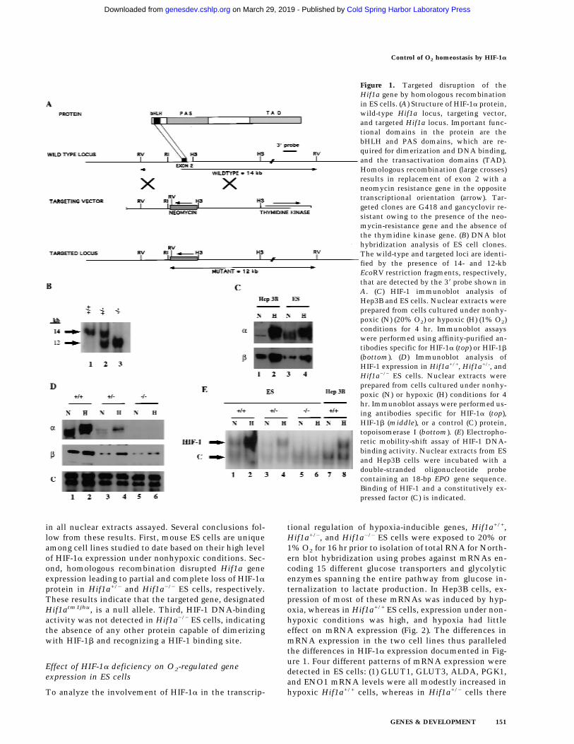
in all nuclear extracts assayed. Several conclusions fol-low from these results. First, mouse ES cells are uniqueamong cell lines studied to date based on their high levelof HIF-1a expression under nonhypoxic conditions. Sec-ond, homologous recombination disrupted Hif1a geneexpression leading to partial and complete loss of HIF-1aprotein in Hif1a+/− and Hif1a−/− ES cells, respectively.These results indicate that the targeted gene, designatedHif1atm1jhu, is a null allele. Third, HIF-1 DNA-bindingactivity was not detected in Hif1a−/− ES cells, indicatingthe absence of any other protein capable of dimerizingwith HIF-1b and recognizing a HIF-1 binding site.
Effect of HIF-1a deficiency on O2-regulated geneexpression in ES cells
To analyze the involvement of HIF-1a in the transcrip-
tional regulation of hypoxia-inducible genes, Hif1a+/+,Hif1a+/−, and Hif1a−/− ES cells were exposed to 20% or1% O2 for 16 hr prior to isolation of total RNA for North-ern blot hybridization using probes against mRNAs en-coding 15 different glucose transporters and glycolyticenzymes spanning the entire pathway from glucose in-ternalization to lactate production. In Hep3B cells, ex-pression of most of these mRNAs was induced by hyp-oxia, whereas in Hif1a+/+ ES cells, expression under non-hypoxic conditions was high, and hypoxia had littleeffect on mRNA expression (Fig. 2). The differences inmRNA expression in the two cell lines thus paralleledthe differences in HIF-1a expression documented in Fig-ure 1. Four different patterns of mRNA expression weredetected in ES cells: (1) GLUT1, GLUT3, ALDA, PGK1,and ENO1 mRNA levels were all modestly increased inhypoxic Hif1a+/+ cells, whereas in Hif1a+/− cells there
Figure 1. Targeted disruption of theHif1a gene by homologous recombinationin ES cells. (A) Structure of HIF-1a protein,wild-type Hif1a locus, targeting vector,and targeted Hif1a locus. Important func-tional domains in the protein are thebHLH and PAS domains, which are re-quired for dimerization and DNA binding,and the transactivation domains (TAD).Homologous recombination (large crosses)results in replacement of exon 2 with aneomycin resistance gene in the oppositetranscriptional orientation (arrow). Tar-geted clones are G418 and gancyclovir re-sistant owing to the presence of the neo-mycin-resistance gene and the absence ofthe thymidine kinase gene. (B) DNA blothybridization analysis of ES cell clones.The wild-type and targeted loci are identi-fied by the presence of 14- and 12-kbEcoRV restriction fragments, respectively,that are detected by the 38 probe shown inA. (C) HIF-1 immunoblot analysis ofHep3B and ES cells. Nuclear extracts wereprepared from cells cultured under nonhy-poxic (N) (20% O2) or hypoxic (H) (1% O2)conditions for 4 hr. Immunoblot assayswere performed using affinity-purified an-tibodies specific for HIF-1a (top) or HIF-1b
(bottom). (D) Immunoblot analysis ofHIF-1 expression in Hif1a+/+, Hif1a+/-, andHif1a−/− ES cells. Nuclear extracts wereprepared from cells cultured under nonhy-poxic (N) or hypoxic (H) conditions for 4hr. Immunoblot assays were performed us-ing antibodies specific for HIF-1a (top),HIF-1b (middle), or a control (C) protein,topoisomerase I (bottom). (E) Electropho-retic mobility-shift assay of HIF-1 DNA-binding activity. Nuclear extracts from ESand Hep3B cells were incubated with adouble-stranded oligonucleotide probecontaining an 18-bp EPO gene sequence.Binding of HIF-1 and a constitutively ex-pressed factor (C) is indicated.
Control of O2 homeostasis by HIF-1a
GENES & DEVELOPMENT 151
Cold Spring Harbor Laboratory Press on March 29, 2019 - Published by genesdev.cshlp.orgDownloaded from

was no hypoxic induction, and in Hif1a−/− cells mRNAlevels were decreased under both hypoxic and nonhy-poxic conditions. (2) Hypoxia did not affect the levels ofHK1, HK2, and PKM mRNA in Hif1a+/+ cells, but inHif1a+/− and Hif1a−/− cells mRNA levels decreased inresponse to hypoxia. (3) PFKL, ALDC, TPI, GAPDH, andLDHA mRNA levels also did not increase in hypoxicHif1a+/+ cells, but mRNA levels were decreased inHif1a−/− cells under both hypoxic and nonhypoxic con-ditions. (4) GPI and PGM mRNA levels were not inducedby hypoxia in Hif1a+/+ cells, and HIF-1a deficiency hadno reproducible effect on expression.
Each filter was subsequently stripped and rehybridizedwith an 18S rRNA probe to demonstrate the presence ofequal amounts of RNA in each lane (data not shown). Inaddition, autoradiographs were serially hybridized withdifferent probes. For example, the LDHA blot shown wasstripped and rehybridized to generate the PKM blotshown. Therefore, the different patterns of expressionreflect varying effects of HIF-1a deficiency on the expres-sion of genes encoding glycolytic enzymes. These resultsindicate that HIF-1 coordinately regulates the expressionof at least 13 genes encoding proteins involved in theanaerobic synthesis of ATP by conversion of extracellu-lar glucose to intracellular lactate. Furthermore, thisregulation is complex because in some cases (groups 2and 3), it appears that the level of gene expression repre-sents the net effect of multiple positive and/or negativeregulators such that HIF-1 is required to maintain, ratherthan to induce, gene expression in hypoxic ES cells. Asfurther evidence of the specific effects of HIF-1a defi-ciency, expression of ODC mRNA (encoding ornithinedecarboxylase) was induced to the same degree in hyp-oxic Hif1a+/+, Hif1a+/−, and Hif1a−/− ES cells (data notshown). Therefore, hypoxia-inducible ODC gene expres-sion is not HIF-1a-dependent. Expression of other hyp-oxia-inducible genes (encoding adrenomedullin, cyclo-oxygenase-2, 58-ecto-nucleotidase, endothelin-1, EPO,HO1, iNOS, platelet-derived growth factor-B, transfer-rin, and transforming growth factor-b1) was demon-strated in Hep3B cells but could not be detected in wild-type or HIF-1a-deficient ES cells (data not shown).
VEGF mRNA expression in ES cells was detected byblot hybridization and quantitated by densitometricanalysis of autoradiographs. The results were correctedfor differences in sample preparation or loading by nor-malizing to results obtained by analysis of 18S rRNA.VEGF mRNA expression was induced by hypoxia inHif1a+/+ but not in Hif1a−/− ES cells (Fig. 3A). Comparedwith Hif1a+/+ cells, normalized VEGF mRNA levelswere 4.4- and 10.2-fold lower in nonhypoxic and hypoxicHif1a−/− cells, respectively. In contrast, VEGF mRNAexpression increased 2.9- and 4.4-fold in glucose-de-prived Hif1a+/+ and Hif1a−/− cells, respectively, indicat-ing that this response is not HIF-1a dependent. Thesedata are consistent with the finding that neither HIF-1aprotein nor HIF-1 DNA-binding activity was induced byglucose deprivation of Hif1a+/+ ES cells (data not shown).ES cells were also differentiated into cystic embryoidbodies by suspension culture in methylcellulose for 5
Figure 2. Expression of genes encoding glucose transportersand glycolytic enzymes. The glycolytic pathway is shown atleft. Symbols for genes encoding the respective enzymes arecoded by font according to the mRNA expression pattern (nor-malized to 18S rRNA) in ES cells cultured under nonhypoxic (N)or hypoxic (H) conditions for 16 hr (lanes 1–6 at right) as follows:(1) (bold) increased expression in hypoxic Hif1a+/+ cells, loss ofinduction in Hif1a+/− cells, and loss of basal and induced ex-pression in Hif1a−/− cells; (2) (bold and italicized) no effect ofhypoxia on expression in Hif1a+/+ cells but decreased expressionin hypoxic Hif1a+/− and Hif1a−/− cells; (3) (italicized) no effect ofhypoxia on expression in Hif1a+/+ cells but decreased expressionin hypoxic and nonhypoxic Hif1a−/− cells; (4) (plain) no effect ofhypoxia or HIF-1a deficiency on expression. mRNA expressionin Hep3B cells was also assayed (lanes 7,8). The indicated genesencode the following proteins: (GLUT1 and GLUT3) glucosetransporter 1 and 3; (HK1 and HK2) hexokinase 1 and 2; (GPI)glucosephosphate isomerase; (PFKL) phosphofructokinase L;(ALDA and ALDC) aldolase A and C; (TPI) triosephosphateisomerase; (GAPDH) glyceraldehyde-3-phosphate dehydroge-nase; (PGK1) phosphoglycerate kinase 1; (PGM) phosphogluco-mutase; (ENO1) enolase 1; (PKM) pyruvate kinase M; (LDHA)lactate dehydrogenase A. GLUCOSE (EXT) and GLUCOSE(INT) refer to extracellular and intracellular glucose, respec-tively.
Iyer et al.
152 GENES & DEVELOPMENT
Cold Spring Harbor Laboratory Press on March 29, 2019 - Published by genesdev.cshlp.orgDownloaded from
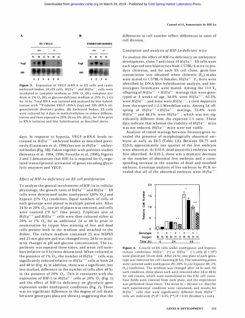
days. In response to hypoxia, VEGF mRNA levels in-creased in Hif1a+/+ embryoid bodies as described previ-ously (Gassmann et al. 1996) but not in Hif1a−/− embry-oid bodies (Fig. 3B). Taken together with previous studies(Semenza et al. 1994, 1996; Forsythe et al. 1996), Figures2 and 3 demonstrate that HIF-1a is required for O2-regu-lated transcriptional activation of genes encoding glyco-lytic enzymes and VEGF.
Effect of HIF-1a deficiency on ES cell proliferation
To analyze the general involvement of HIF-1a in cellularphysiology, the growth rates of Hif1a+/+ and Hif1a−/− EScells were determined under nonhypoxic (20% O2) andhypoxic (1% O2) conditions. Equal numbers of cells ofeach genotype were plated in multiple paired sets. After24 hr at 20% O2, one set of plates was removed, and cellswere counted (‘‘0 hr’’ time point). Triplicate sets ofHif1a+/+ and Hif1a−/− cells were then cultured either at20% or 1% O2 for an additional 24 or 48 hr prior toenumeration by trypan blue staining of live and deadcells present both in the medium and attached to thedishes. The culture medium contained 25 mM HEPESand 25 mM glucose and was changed every 24 hr to mini-mize changes in pH and glucose concentration. The ex-periment was repeated three times, and mean cell num-bers (relative to 0 hr) were determined. When cultured inthe presence of 1% O2, the number of Hif1a−/− cells wassignificantly reduced relative to Hif1a+/+ cells at both 24and 48 hr (Fig. 4). In addition, there was a significant, butless marked, difference in the number of cells after 48 hrin the presence of 20% O2. This is consistent with theexpression of HIF-1 in Hif1a+/+ cells at 20% O2 (Fig. 1)and the effect of HIF-1a deficiency on glycolytic geneexpression under nonhypoxic conditions (Fig. 2). Therewas no significant difference in the degree of cell deathbetween genotypes (data not shown), suggesting that the
differences in cell number reflect differences in rates ofcell division.
Generation and analysis of HIF-1a-deficient mice
To analyze the effect of HIF-1a deficiency on embryonicdevelopment, clone 7 and clone 19 Hif1a+/− ES cells wereeach injected into blastocysts from C57BL/6 mice to pro-duce chimeras, and for each ES cell clone, germ-linetransmission was obtained when chimeric (F0) maleswere mated to C57BL/6 females. Hif1a+/− F1 mice wereidentified by DNA blot hybridization analysis, and het-erozygous littermates were mated. Among the 113 F2
offspring of Hif1a+/− × Hif1a+/− matings that were geno-typed at 4 weeks of age, 34.4% were Hif1a+/+, 65.5%were Hif1a+/−, and none were Hif1a−/−, a clear departurefrom the expected 1:2:1 Mendelian ratio. Among 54 off-spring of Hif1a+/+ × Hif1a+/− matings, 51.8% wereHif1a+/+ and 48.1% were Hif1a+/−, which was not sig-nificantly different from the expected 1:1 ratio. Thesedata indicate that whereas the viability of Hif1a+/− micewas not reduced, Hif1a−/− mice were not viable.
Analysis of timed matings between heterozygotes re-vealed the presence of morphologically abnormal em-bryos as early as E8.5 (Table 1A). Between E8.75 andE10.0, approximately one quarter of the live embryoswere abnormal. At E10.0, dead (asystolic) embryos werefirst identified. At E10.5, there was a dramatic decreasein the number of abnormal live embryos and a corre-sponding increase in the number of dead and resorbedembryos. Genotype analysis of live embryos by PCR re-vealed that all of the abnormal embryos were Hif1a−/−
Figure 3. Expression of VEGF mRNA in ES cells and cysticembryoid bodies. (A) ES cells. Hif1a+/+ and Hif1a−/− cells wereincubated in complete medium at 20% O2 (N), complete me-dium at 1% O2 (H), or glucose-deficient medium at 20% O2 (−G)for 16 hr. Total RNA was isolated and analyzed by blot hybrid-ization with 32P-labeled VEGF cDNA (top) and 18S rRNA oli-gonucleotide (bottom) probes. (B) Embryoid bodies. ES cellswere cultured for 4 days in methylcellulose to induce differen-tiation and then exposed to 20% (N) or 0% (H) O2 for 16 hr priorto RNA isolation and blot hybridization as described above.
Figure 4. Growth of ES cells under nonhypoxic and hypoxicculture conditions. Hif1a+/+ (+) or Hif1a−/− (−) cells (6 × 10⁵)were plated per 10-cm dish. After 24 hr, one plate of each geno-type was removed for cell counting (0 hr). The remaining plateswere cultured under nonhypoxic (N; 20% O2) or hypoxic (H; 1%O2) conditions. The medium was changed after 24 hr and, foreach condition, three plates each were removed after 24 or 48 hrfor cell counts, which were normalized to the 0-hr cell count.Four fields were counted from each plate, and the experimentwas performed three times. The mean (n = 36) and S.D. (bar) foreach experimental condition were calculated, and results forHif1a−/− cells that were significantly different from Hif1a+/+
cells are indicated: (*) P < 0.05; (**) P < 0.01 (Student’s t-test).
Control of O2 homeostasis by HIF-1a
GENES & DEVELOPMENT 153
Cold Spring Harbor Laboratory Press on March 29, 2019 - Published by genesdev.cshlp.orgDownloaded from
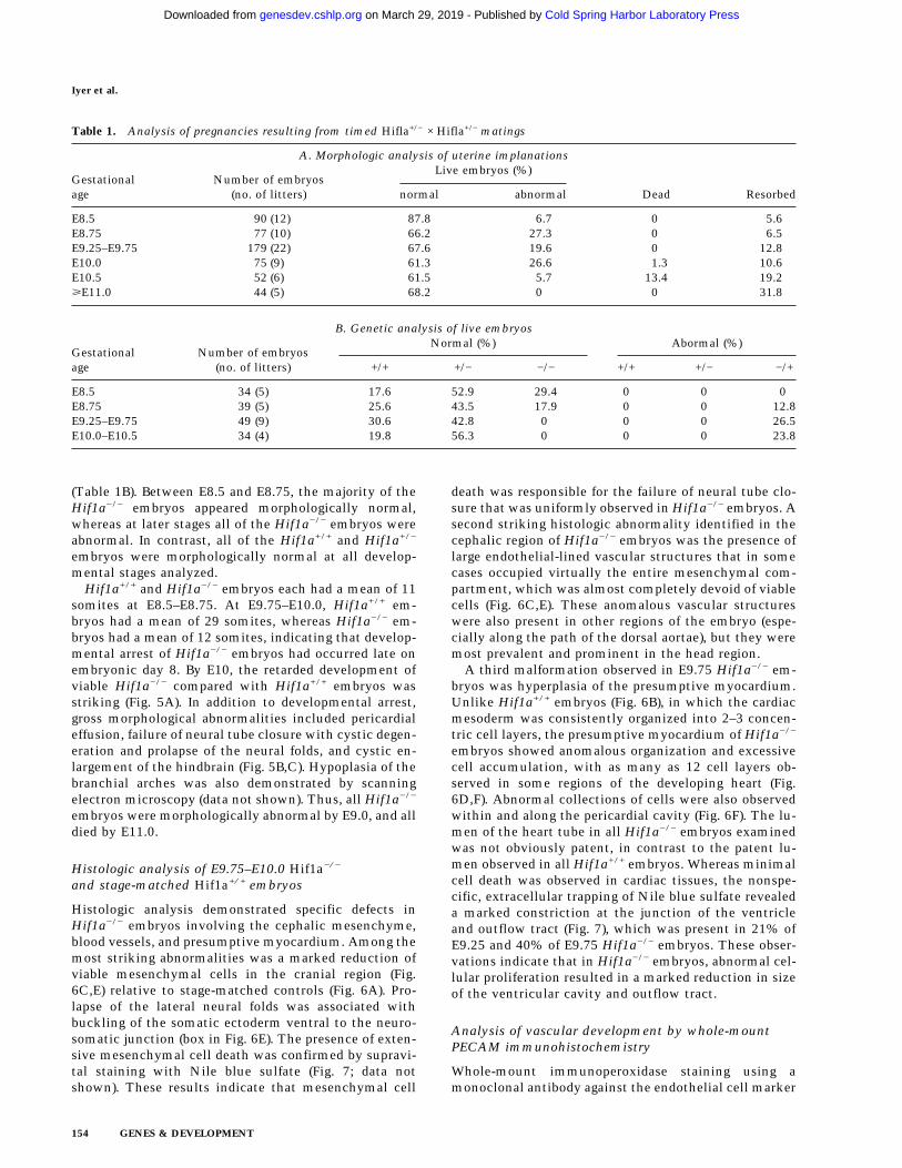
(Table 1B). Between E8.5 and E8.75, the majority of theHif1a−/− embryos appeared morphologically normal,whereas at later stages all of the Hif1a−/− embryos wereabnormal. In contrast, all of the Hif1a+/+ and Hif1a+/−
embryos were morphologically normal at all develop-mental stages analyzed.
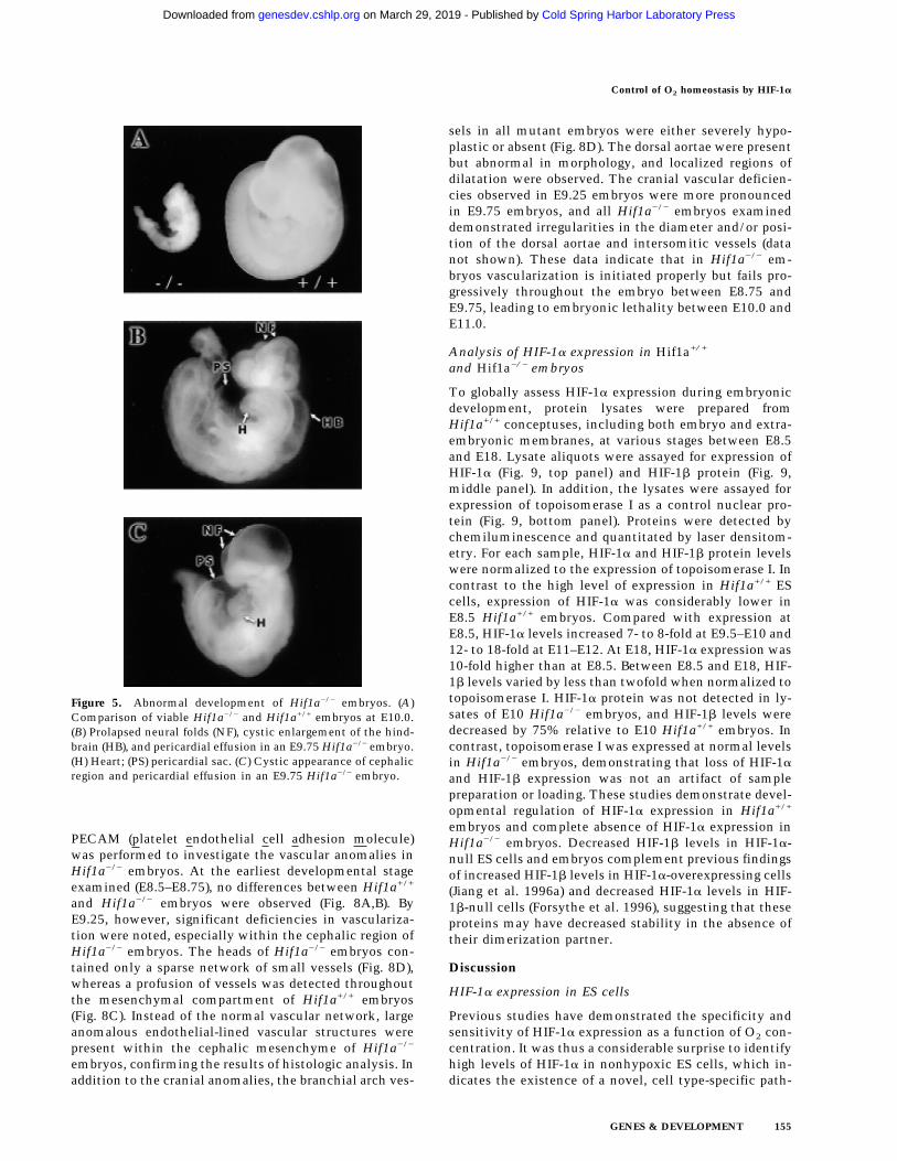
Hif1a+/+ and Hif1a−/− embryos each had a mean of 11somites at E8.5–E8.75. At E9.75–E10.0, Hif1a+/+ em-bryos had a mean of 29 somites, whereas Hif1a−/− em-bryos had a mean of 12 somites, indicating that develop-mental arrest of Hif1a−/− embryos had occurred late onembryonic day 8. By E10, the retarded development ofviable Hif1a−/− compared with Hif1a+/+ embryos wasstriking (Fig. 5A). In addition to developmental arrest,gross morphological abnormalities included pericardialeffusion, failure of neural tube closure with cystic degen-eration and prolapse of the neural folds, and cystic en-largement of the hindbrain (Fig. 5B,C). Hypoplasia of thebranchial arches was also demonstrated by scanningelectron microscopy (data not shown). Thus, all Hif1a−/−
embryos were morphologically abnormal by E9.0, and alldied by E11.0.
Histologic analysis of E9.75–E10.0 Hif1a−/−
and stage-matched Hif1a+/+ embryos
Histologic analysis demonstrated specific defects inHif1a−/− embryos involving the cephalic mesenchyme,blood vessels, and presumptive myocardium. Among themost striking abnormalities was a marked reduction ofviable mesenchymal cells in the cranial region (Fig.6C,E) relative to stage-matched controls (Fig. 6A). Pro-lapse of the lateral neural folds was associated withbuckling of the somatic ectoderm ventral to the neuro-somatic junction (box in Fig. 6E). The presence of exten-sive mesenchymal cell death was confirmed by supravi-tal staining with Nile blue sulfate (Fig. 7; data notshown). These results indicate that mesenchymal cell
death was responsible for the failure of neural tube clo-sure that was uniformly observed in Hif1a−/− embryos. Asecond striking histologic abnormality identified in thecephalic region of Hif1a−/− embryos was the presence oflarge endothelial-lined vascular structures that in somecases occupied virtually the entire mesenchymal com-partment, which was almost completely devoid of viablecells (Fig. 6C,E). These anomalous vascular structureswere also present in other regions of the embryo (espe-cially along the path of the dorsal aortae), but they weremost prevalent and prominent in the head region.
A third malformation observed in E9.75 Hif1a−/− em-bryos was hyperplasia of the presumptive myocardium.Unlike Hif1a+/+ embryos (Fig. 6B), in which the cardiacmesoderm was consistently organized into 2–3 concen-tric cell layers, the presumptive myocardium of Hif1a−/−
embryos showed anomalous organization and excessivecell accumulation, with as many as 12 cell layers ob-served in some regions of the developing heart (Fig.6D,F). Abnormal collections of cells were also observedwithin and along the pericardial cavity (Fig. 6F). The lu-men of the heart tube in all Hif1a−/− embryos examinedwas not obviously patent, in contrast to the patent lu-men observed in all Hif1a+/+ embryos. Whereas minimalcell death was observed in cardiac tissues, the nonspe-cific, extracellular trapping of Nile blue sulfate revealeda marked constriction at the junction of the ventricleand outflow tract (Fig. 7), which was present in 21% ofE9.25 and 40% of E9.75 Hif1a−/− embryos. These obser-vations indicate that in Hif1a−/− embryos, abnormal cel-lular proliferation resulted in a marked reduction in sizeof the ventricular cavity and outflow tract.
Analysis of vascular development by whole-mountPECAM immunohistochemistry
Whole-mount immunoperoxidase staining using amonoclonal antibody against the endothelial cell marker
Table 1. Analysis of pregnancies resulting from timed Hifla+/− × Hifla+/− matings
A. Morphologic analysis of uterine implanations
Gestationalage
Number of embryos(no. of litters)
Live embryos (%)
Dead Resorbednormal abnormal
E8.5 90 (12) 87.8 6.7 0 5.6E8.75 77 (10) 66.2 27.3 0 6.5E9.25–E9.75 179 (22) 67.6 19.6 0 12.8E10.0 75 (9) 61.3 26.6 1.3 10.6E10.5 52 (6) 61.5 5.7 13.4 19.2ùE11.0 44 (5) 68.2 0 0 31.8
B. Genetic analysis of live embryos
Gestationalage
Number of embryos(no. of litters)
Normal (%) Abormal (%)
+/+ +/− −/− +/+ +/− −/+
E8.5 34 (5) 17.6 52.9 29.4 0 0 0E8.75 39 (5) 25.6 43.5 17.9 0 0 12.8E9.25–E9.75 49 (9) 30.6 42.8 0 0 0 26.5E10.0–E10.5 34 (4) 19.8 56.3 0 0 0 23.8
Iyer et al.
154 GENES & DEVELOPMENT
Cold Spring Harbor Laboratory Press on March 29, 2019 - Published by genesdev.cshlp.orgDownloaded from
PECAM (platelet endothelial cell adhesion molecule)was performed to investigate the vascular anomalies inHif1a−/− embryos. At the earliest developmental stageexamined (E8.5–E8.75), no differences between Hif1a+/+
and Hif1a−/− embryos were observed (Fig. 8A,B). ByE9.25, however, significant deficiencies in vasculariza-tion were noted, especially within the cephalic region ofHif1a−/− embryos. The heads of Hif1a−/− embryos con-tained only a sparse network of small vessels (Fig. 8D),whereas a profusion of vessels was detected throughoutthe mesenchymal compartment of Hif1a+/+ embryos(Fig. 8C). Instead of the normal vascular network, largeanomalous endothelial-lined vascular structures werepresent within the cephalic mesenchyme of Hif1a−/−
embryos, confirming the results of histologic analysis. Inaddition to the cranial anomalies, the branchial arch ves-
sels in all mutant embryos were either severely hypo-plastic or absent (Fig. 8D). The dorsal aortae were presentbut abnormal in morphology, and localized regions ofdilatation were observed. The cranial vascular deficien-cies observed in E9.25 embryos were more pronouncedin E9.75 embryos, and all Hif1a−/− embryos examineddemonstrated irregularities in the diameter and/or posi-tion of the dorsal aortae and intersomitic vessels (datanot shown). These data indicate that in Hif1a−/− em-bryos vascularization is initiated properly but fails pro-gressively throughout the embryo between E8.75 andE9.75, leading to embryonic lethality between E10.0 andE11.0.
Analysis of HIF-1a expression in Hif1a+/+
and Hif1a−/− embryos
To globally assess HIF-1a expression during embryonicdevelopment, protein lysates were prepared fromHif1a+/+ conceptuses, including both embryo and extra-embryonic membranes, at various stages between E8.5and E18. Lysate aliquots were assayed for expression ofHIF-1a (Fig. 9, top panel) and HIF-1b protein (Fig. 9,middle panel). In addition, the lysates were assayed forexpression of topoisomerase I as a control nuclear pro-tein (Fig. 9, bottom panel). Proteins were detected bychemiluminescence and quantitated by laser densitom-etry. For each sample, HIF-1a and HIF-1b protein levelswere normalized to the expression of topoisomerase I. Incontrast to the high level of expression in Hif1a+/+ EScells, expression of HIF-1a was considerably lower inE8.5 Hif1a+/+ embryos. Compared with expression atE8.5, HIF-1a levels increased 7- to 8-fold at E9.5–E10 and12- to 18-fold at E11–E12. At E18, HIF-1a expression was10-fold higher than at E8.5. Between E8.5 and E18, HIF-1b levels varied by less than twofold when normalized totopoisomerase I. HIF-1a protein was not detected in ly-sates of E10 Hif1a−/− embryos, and HIF-1b levels weredecreased by 75% relative to E10 Hif1a+/+ embryos. Incontrast, topoisomerase I was expressed at normal levelsin Hif1a−/− embryos, demonstrating that loss of HIF-1aand HIF-1b expression was not an artifact of samplepreparation or loading. These studies demonstrate devel-opmental regulation of HIF-1a expression in Hif1a+/+
embryos and complete absence of HIF-1a expression inHif1a−/− embryos. Decreased HIF-1b levels in HIF-1a-null ES cells and embryos complement previous findingsof increased HIF-1b levels in HIF-1a-overexpressing cells(Jiang et al. 1996a) and decreased HIF-1a levels in HIF-1b-null cells (Forsythe et al. 1996), suggesting that theseproteins may have decreased stability in the absence oftheir dimerization partner.
Discussion
HIF-1a expression in ES cells
Previous studies have demonstrated the specificity andsensitivity of HIF-1a expression as a function of O2 con-centration. It was thus a considerable surprise to identifyhigh levels of HIF-1a in nonhypoxic ES cells, which in-dicates the existence of a novel, cell type-specific path-
Figure 5. Abnormal development of Hif1a−/− embryos. (A)Comparison of viable Hif1a−/− and Hif1a+/+ embryos at E10.0.(B) Prolapsed neural folds (NF), cystic enlargement of the hind-brain (HB), and pericardial effusion in an E9.75 Hif1a−/− embryo.(H) Heart; (PS) pericardial sac. (C) Cystic appearance of cephalicregion and pericardial effusion in an E9.75 Hif1a−/− embryo.
Control of O2 homeostasis by HIF-1a
GENES & DEVELOPMENT 155
Cold Spring Harbor Laboratory Press on March 29, 2019 - Published by genesdev.cshlp.orgDownloaded from

way leading to HIF-1a expression. We have demon-strated recently HIF-1a expression in nonhypoxic rat fi-broblasts transformed by the V-SRC oncoprotein (Jianget al. 1997a). ES cells are derived from inner cell mass/embryonic ectoderm of the blastocyst, which representsa population of actively dividing cells. HIF-1a expressionmay be activated by one or more signal transductionpathways involved in cellular proliferation. The de-creased proliferation of Hif1a−/− ES cells suggests thatHIF-1a may be involved in the regulation of cell division,either directly or indirectly via effects on intermediarymetabolism. However, HIF-1a deficiency had no appar-ent effect on preimplantation development. The signifi-cance of constitutive HIF-1a expression in ES cells willthus require further investigation.
As a consequence of constitutive HIF-1a expression,nonhypoxic ES cells demonstrated high-level expressionof mRNAs encoding glycolytic enzymes. The Warburgeffect refers to the high rate of aerobic glycolysis that ischaracteristic of tumor cells (Warburg 1956). Rous sar-coma virus infection of rat cells was associated withaerobic glycolysis, and this phenotype was correlated
with V-SRC-mediated transformation (Carroll et al.1978). Increased glycolytic gene expression mediated byHIF-1a expression may be required for maximal rates ofcellular proliferation by both ES and V-SRC-transformedcells under hypoxic conditions. In ES cells, HIF-1a wasrequired for normal expression of almost all genes in-volved in the metabolism of extracellular glucose to in-tracellular lactate, a remarkable example of coordinatetranscriptional regulation. Furthermore, HIF-1a was re-quired to maintain expression of several genes underhypoxic conditions such that in its absence expression ofthese genes was hypoxia repressible. These results mayreflect the constitutive expression of HIF-1a in ES cells,as the expression patterns clearly differed from those ob-served in Hep3B cells. However, these results suggestthat HIF-1a may be required for expression of othergenes that do not show an obvious pattern of hypoxicinduction.
Regulation of Vegf gene expression by HIF-1
Experimental data indicate that VEGF is essential for
Figure 6. Histologic analysis. Sections through the cranial region of stage-matched Hif1a+/+ (A) and E9.75–E10.0 Hif1a−/− (C,E)embryos are shown. A normal blood vessel (arrowhead in A) can be compared with the anomalous vascular structures contained withinthe cephalic mesenchyme (arrowheads in C and E) of Hif1a−/− embryos, which also manifest prolapsed neural folds (NF) and adeficiency of cranial mesenchyme (CM) relative to the Hif1a+/+ embryo. Sections through the cardiac region of stage-matched Hif1a+/+
(B) and E9.75–E10.0 Hif1a−/− (D,F) embryos are shown. Hyperplasia of presumptive myocardium (M) and anomalous tissue (arrows inF) are evident within the enlarged pericardial cavity (PC) and along the pericardium (P). (AV) Atrioventricular canal; (BA1) firstbranchial arch; (NT) neural tube; (OV) otic vesicle; (OT) ventricular outflow tract.
Iyer et al.
156 GENES & DEVELOPMENT
Cold Spring Harbor Laboratory Press on March 29, 2019 - Published by genesdev.cshlp.orgDownloaded from

vasculogenesis and angiogenesis in the embryo and fetusas well as for neovascularization of ischemic tissues andtumors in the adult organism (for review, see Ferrara andDavis-Smyth 1997). An understanding of the molecularbasis for hypoxia-induced VEGF expression is of criticalimportance because of the potential therapeutic utilityof stimulating or inhibiting VEGF expression in isch-emic and neoplastic tissues, respectively. Previous stud-ies have provided evidence supporting a role for HIF-1 inmediating VEGF transcriptional activation in hypoxiccells. First, an essential HIF-1 binding site was identified1 kb 58 to the human, mouse, and rat VEGF genes (Levyet al. 1995; Liu et al. 1995; Forsythe et al. 1996; Shima etal. 1996). Second, forced expression of HIF-1a and HIF-1bactivated transcription from the human VEGF promoterin nonhypoxic cells and resulted in a superactivation inhypoxic cells (Forsythe et al. 1996). Third, loss of HIF-1bexpression and HIF-1 DNA-binding activity was associ-ated with a marked decrease in VEGF mRNA levels un-der hypoxic conditions (Forsythe et al. 1996; Salceda etal. 1996; Wood et al. 1996; Maltepe et al. 1997). Fourth,expression of V-SRC was associated with increased HIF-1a protein, HIF-1 DNA-binding activity, VEGF mRNA,and transcriptional activity of a reporter gene containingthe VEGF hypoxia-response element that was dependenton an intact HIF-1 binding site (Jiang et al. 1997a). Fifth,VEGF and HIF-1a expression was coinduced in associa-tion with increased myocardial vascularization in ane-mic fetal sheep (Martin et al. 1997). The demonstration
that HIF-1a deficiency resulted in a complete loss of in-duced expression provides definitive proof that HIF-1mediates Vegf gene transcriptional activation in hypoxicES cells. Induction of VEGF expression via increasedmRNA stability under hypoxic conditions was demon-strated previously (Ikeda et al. 1995; Stein et al. 1995;Levy et al. 1996). The absence of any increase in VEGFmRNA expression in HIF-1a-deficient ES cells indicateseither that post-transcriptional mechanisms are not op-erative in ES cells, that post-transcriptional mechanismsare of limited effect in the absence of transcriptional ac-tivation, or that the post-transcriptional mechanisms are
Figure 7. Supravital dye staining. Ten-somite Hif1a+/+ (left)and Hif1a−/− (right) embryos have been treated with Nile bluesulfate. Punctate staining indicative of cell death is apparentwithin the mesenchymal compartment of the fore- and mid-brain region in the left neural fold of the Hif1a−/− embryo and isabsent in the corresponding region of the Hif1a+/+ embryo. Non-specific trapping of dye in the heart lumen also allowed analysisof cardiac morphogenesis. Arrows over the embryos have beenplaced the same distance apart to demonstrate the constrictionbetween ventricle and outflow tract of the Hif1a−/− embryo. TheHif1a−/− heart is not stained, suggesting absence of a patentlumen.
Figure 8. PECAM immunohistochemistry. Comparison ofE8.5–E8.75 Hif1a+/+ (A) and Hif1a−/− (B) embryos reveals noapparent differences in vascularization. In contrast, the vascu-larization of the E9.25 Hif1a−/− embryo (D) is markedly abnor-mal when compared with a stage-matched Hif1a+/+ embryo (C).Anomalous endothelial-lined vascular structures are present inthe cranial region (arrow in D), no branchial arch vessels haveformed, the lumen of the heart tube is not apparent, and thediameter of the dorsal aorta (DA) is irregular. (BA1) First bran-chial arch; (V) Ventricle.
Figure 9. Immunoblot analysis of HIF-1 expression in Hif1a+/+
and Hif1a−/− embryos. Aliquots (15 µg) of nuclear extracts fromnonhypoxic (N) and hypoxic (H) Hif1a+/+ ES cells (lanes 1,2) andaliquots (120 µg) of lysates prepared from Hif1a+/+ (WT; lanes3–8) and Hif1a−/− (M; lane 9) embryos of the indicated gesta-tional age were analyzed using antibodies specific for HIF-1a
(top), HIF-1b (middle), or as a control (C), topoisomerase I (bot-tom).
Control of O2 homeostasis by HIF-1a
GENES & DEVELOPMENT 157
Cold Spring Harbor Laboratory Press on March 29, 2019 - Published by genesdev.cshlp.orgDownloaded from
also dependent on HIF-1a, perhaps for transcription of agene encoding a hypoxia-induced mRNA binding pro-tein.
HIF-1a is required for proper cardiovasculardevelopment
Analysis of Hif1a−/− embryos revealed multiple defectsin cardiovascular development, including pericardial ef-fusion; disorganized cardiac morphogenesis with myo-cardial hyperplasia and ventricular obstruction; forma-tion of dilated vascular structures, especially in the ce-phalic region; and marked reduction in the overallnumber of cephalic blood vessels including hypoplasia ofthe arch vessels. Pericardial effusion may represent a sec-ondary defect, as it has been reported in knockout micewithout intracardiac defects (Warren et al. 1994). In con-trast, hyperplasia of the presumptive myocardium,which has not been reported previously to our knowl-edge, almost certainly represents a primary defect ratherthan a response to ventricular outflow obstruction.VEGF-deficient embryos also manifested ventricularoutflow obstruction but had a patent ventricular cavityand decreased, rather than increased, ventricular wallthickness (Carmeliet et al. 1996; Ferrara et al. 1996). Thehyperplastic and highly disorganized appearance of thepresumptive myocardium suggests strongly that this de-fect represents a cause, rather than a consequence, ofventricular outflow obstruction in Hif1a−/− embryos andthat HIF-1a may play a direct role in cardiac morphogen-esis.
One of the most striking histologic findings was thepresence of massively enlarged vascular structures in thecephalic region of Hif1a−/− embryos. These structureswere remarkably similar to those described for Vegf−/−
embryos (Carmeliet et al. 1996). The marked reductionin vascularization of Hif1a−/− embryos is also consistentwith a deficiency of VEGF production. The demonstra-tion that Hif1a−/− ES cells and embryoid bodies are de-ficient in expression of VEGF mRNA provides a poten-tial molecular basis for the observed defects in embry-onic vascularization, although analysis of Vegfexpression in situ will be required to confirm this hy-pothesis. Vascularization of Hif1a−/− embryos appearednormal at E8.5, but by E9.25 only abnormally dilatedvascular structures were present in the cephalic region.In contrast, the dorsal aortae were poorly developed orabsent in E8.5 Vegf+/− and Vegf−/− embryos, respectively,and dilated vascular structures were observed in E9.5Vegf−/− embryos (Carmeliet et al. 1996). Recent studieshave demonstrated that VEGF is a survival factor fornewly formed blood vessels such that VEGF withdrawalresults in capillary regression (Benjamin and Keshet1997). It is possible that Hif1a−/− embryos produced suf-ficient VEGF to initiate vasculogenesis but that VEGFproduction became limiting owing to absence of HIF-1-mediated transcriptional activation or loss of VEGF-pro-ducing mesenchymal cells or both. The increased expres-sion of HIF-1a in E9.5, compared with E8.5, Hif1a+/+
embryos is of interest in this regard.
Although decreased vascularization and decreasedVEGF mRNA expression were reported for Arnt−/− em-bryos lacking HIF-1b (ARNT), the enlarged vascularstructures and absence of normal cephalic vasculariza-tion seen in Vegf−/− and Hif1a−/− embryos by E9.25 werenot observed in Arnt−/− embryos (Maltepe et al. 1997).One explanation for these results is that in Arnt−/− em-bryos, heterodimerization of HIF-1a with the proteinproduct of the Arnt2 gene, which is homologous to Arntbut is expressed primarily within the nervous system(Hirose et al. 1996), may allow for adequate VEGF syn-thesis in the cephalic region, whereas in Hif1a−/− em-bryos, the absence of both HIF-1a:HIF-1b (ARNT) andHIF-1a:ARNT2 heterodimers has a more significant ef-fect on Vegf transcription.
HIF-1a is required for embryonic cell survival
A defining feature of Hif1a−/− embryos is the over-whelming degree of cell death that occurred in a progres-sive manner beginning around E8.5 and leading to em-bryonic death around E10.5. Hif1a−/− embryos mani-fested severely prolapsed neural folds that, if theembryos developed to term, would result in exen-cephaly. In addition, the branchial arches, which are theanlage of the developing craniofacial skeleton, were hy-poplastic and discontinuous (L.E. Kotch, N.V. Iyer, andG.L. Semenza, unpubl.). The basis for these malforma-tions was the massive death of cells within the cephalicmesenchyme, which normally supports the neural foldsduring the process of neural tube closure and contributesto the formation of the branchial arches. By E9.5, thedeficiency of cells was so profound that the cephalic re-gion of the embryo appeared translucent (Fig. 5C). In ad-dition to the death of mesenchymal cells, abnormallydilated vascular structures were also present. It is notclear whether these structures represent a cause or a con-sequence of the mesenchymal cell death. Tissue cannotremain viable without adequate perfusion, and the vas-cular defects may have been responsible for overwhelm-ing death of the mesenchymal population. However, ex-cessive cell death was detected in E8.5 Hif1a−/− embryosprior to the appearance of any vascular defects (L.E.Kotch, N.V. Iyer, and G.L. Semenza, unpubl.), suggestingthat an intact mesenchyme may be required to provideessential cellular or humoral support of vascular integ-rity, as demonstrated in mice deficient for platelet-de-rived growth factor-B (Lindahl et al. 1997).
Defective embryonic angiogenesis or erythropoiesishas been associated with cell death previously (Warren etal. 1994; Henkemeyer et al. 1995; Carmeliet et al. 1996;Maltepe et al. 1997; Wang et al. 1997). Although it is verydifficult to make comparisons between studies, the ex-tent of cell death, both within the cephalic mesenchymeand throughout the embryo, appears to be most severewithin Hif1a−/− embryos. Furthermore, scanning elec-tron microscopy and Nile blue sulfate staining revealedthe presence of increased cell death prior to the appear-ance of vascular defects (L.E. Kotch, N.V. Iyer, and G.L.Semenza, unpubl.). These results suggest a complex in-
Iyer et al.
158 GENES & DEVELOPMENT
Cold Spring Harbor Laboratory Press on March 29, 2019 - Published by genesdev.cshlp.orgDownloaded from
terrelationship between mesenchymal cell survival andembryonic vascularization. HIF-1a deficiency, in addi-tion to interfering with the development of a circulatorysystem required for adequate tissue oxygenation, mayalso render many cell populations within the embryounable to adapt to O2 deprivation. A major intracellularadaptation involves increased expression of genes encod-ing glucose transporters and glycolytic enzymes, a pro-cess that requires HIF-1a. We hypothesize that the ce-phalic mesenchyme of Hif1a−/− embryos may be particu-larly susceptible to hypoxia-mediated cell death and thata deficiency of mesenchyme in concert with a deficiencyof VEGF may result in major vascular defects, leading tomore severe hypoxia and extensive cell death, ultimatelyresulting in embryonic lethality.
HIF-1a regulates essential developmentaland physiological aspects of O2 homeostasis
The results presented here demonstrate that HIF-1a isessential for cellular and developmental aspects of O2
homeostasis. In addition, preliminary studies indicatethat Hif1a+/− mice have impaired cardiovascular re-sponses to chronic hypoxia (A.Y. Yu, N.V. Iyer, and G.L.Semenza, unpubl.). Thus, HIF-1a is required for the for-mation of key physiological systems during embryonicdevelopment and for their subsequent utilization duringpostnatal life. It remains to be determined whether theexpression of HIF-1a within the developing cardiovascu-lar system occurs in response to a hypoxic stimulus orwhether it is part of a hard-wired developmental pro-gram. In either case, these results indicate that HIF-1a isa master regulator of O2 homeostasis. Other master regu-latory factors have been identified that play key roles inthe formation of specific tissues, such as the involve-ment of GATA1 and MYOD in erythroid and muscledevelopment, respectively (Orkin 1992; Weintraub1993). Several master regulators have been shown to playroles both in development and postnatal physiology, in-cluding PIT1, which is required for the development ofthe somatomammotrophs of the anterior pituitary aswell as their subsequent production of growth hormoneand prolactin (Andersen and Rosenfeld 1994). HIF-1a,rather than specifying a cell type, plays a more globalrole in development and physiology that is based on thefundamental requirement for O2 that is shared by allcells.
The evolution of multicellular organisms presentedthe challenge of delivering O2 to cells too far from theexternal environment to receive adequate oxygenationby diffusion. Whereas there does not appear to be a HIF-1a homolog in Saccharomyces cerevisiae, bHLH–PASproteins with sequence similarity to HIF-1a have beenidentified in both invertebrate and vertebrate species. InDrosophila melanogaster, O2 is not blood borne througha circulatory system but instead is delivered directly toall cells via tracheal tubes, formation of which requiresTRH, a bHLH–PAS protein with sequence similarity toHIF-1a (for review, see Guillemin and Krasnow 1997).Fruit flies and humans have thus evolved different meth-
ods of O2 delivery since diverging >500 million years agobut nevertheless express proteins that are structurallyand functionally related, suggesting that the challenge oftissue oxygenation in multicellular organisms wassolved in part by the evolution of HIF-1a-like regulatoryfactors.
Materials and methods
Construction of the targeting vector
DNA spanning Hif1a exons 2 and 3 (Wenger et al. 1997) wasisolated from a 129/Sv mouse genomic library (partial Sau3Adigest of AB-1 ES cell DNA in lGEM11) and used to constructtargeting vector TVTK8. The 58 arm of TVTK8 was constructedby subcloning a 3-kb EcoRI fragment from intron 1 into theEcoRI site of pBluescript SK(−) (Stratagene). The 58 EcoRI siteoriginated from the lGEM11 polylinker. The 38 arm of TVTK8consisted of Hif1a sequences from intron 2 and exon 3 and wassubcloned as a HindIII fragment. A dose-sensitive neomycinresistance gene driven by a PGK promoter from pNTK(Mortensen et al. 1992; Ausubel et al. 1994) was inserted intothe EcoRV site between the two arms of TVTK8. A PGK pro-moter–herpes simplex virus thymidine kinase coding sequencecassette was inserted as an XhoI–SalI fragment into the XhoIsite located 38 to the Hif1a sequences in TVTK8. TVTK8 waslinearized with XhoI for electroporation.
ES cell culture and transfection
J1 ES cells (Li et al. 1992) were cultured in DMEM mediumcontaining 25 mM glucose and 110 mg/liter of sodium pyruvate,supplemented with 15% ES cell-qualified fetal bovine serum(FBS), 875 nM insulin, 1% penicillin–streptomycin, 1% nones-sential amino acids, 40 µl/liter of monothioglycerol, and 1000U/ml of LIF (GIBCO) on neomycin-resistant, irradiated mouseembryonic fibroblasts (Bradley 1987; Robertson 1987). Cells(5 × 10⁷) in 0.7 ml were electroporated with 25 µg of linearizedTVTK8 at 240 V and 540 µF in a Gene Pulser (Bio-Rad). After48 hr, transfected cells were cultured in medium containing 300µg/ml of G418 (Geneticin, GIBCO) and 2 µM gancyclovir (Man-sour et al. 1988). Individual clones were picked and expanded ina 96-well plate with feeder layer. DNA extracted from indi-vidual clones (Ramirez-Solis et al. 1992) was analyzed by blothybridization.
Generation and analysis of Hif1a−/− ES cellsand embryoid bodies
Hif1a+/− ES cells were cultured in medium containing 2 mg/mlof G418 for 14 days (Mortensen et al. 1992; Ausubel et al. 1994).Resistant colonies were picked and analyzed by DNA blot hy-bridization. Hif1a+/− cells used in subsequent experiments werepicked from the same plates as the Hif1a−/− clones. For analysisof growth rates, the medium was supplemented with 25 mM
HEPES, and 6 × 10⁵ cells were plated on gelatin-coated 10-cmPetri dishes (Falcon). In cells cultured for 48 hr at 1% O2, themedium was changed at 24 hr in a hypoxic chamber (Plas Labs)to ensure a constant O2 concentration. At the end of the experi-ment, cells were trypsinized and stained with trypan blue. Forgenerating embryoid bodies, ES cells were trypsinized, and fi-broblasts were removed by adherence to tissue culture dishes.ES cells (2.5 × 10⁵) were cultured in 12 ml of Iscove’s MDM–methylcellulose (MethoCult, Stem Cell Technologies) for 4days and exposed to 20% O2 or 0% O2 (5% CO2/balance N2) for16 hr, and total RNA was isolated.
Control of O2 homeostasis by HIF-1a
GENES & DEVELOPMENT 159
Cold Spring Harbor Laboratory Press on March 29, 2019 - Published by genesdev.cshlp.orgDownloaded from

RNA blot hybridization
ES cells or embryoid bodies were lysed in guanidine isothiocya-nate, and total RNA was isolated by phenol–chloroform extrac-tion (Chomczynski and Sacchi 1987). Fifteen-microgram ali-quots of RNA were fractionated by 2.2 M formaldehyde, 1.4%agarose gel electrophoresis and transferred to nitrocellulose fil-ters. IMAGE Consortium cDNA clones (Lennon et al. 1996)were obtained (Research Genetics, Inc.), and the inserts wereisolated and 32P-labeled by random-primer synthesis using acommercial kit (Life Technologies, Inc.). Hybridization was per-formed in Quik-Hyb (Stratagene) at 67°C. Blots were washed in0.1× SSC, 0.1% SDS at room temperature for 1 hr and then at55°C for 1 hr, and exposed to film for autoradiography. Blotswere stripped of radioactivity and hybridized with a 32P-labeledoligonucleotide that was complementary to 18S rRNA (For-sythe et al. 1996).
Generation and analysis of mutant mice
Clone 7 or clone 19 J1 ES cells were injected into E3.5 C57BL/6mouse embryos using standard methods (Bradley 1987; Hoganet al. 1994). Injected blastocysts were surgically implanted intothe uterus of pseudopregnant CD1 foster mothers. Chimericmales, identified by coat color and DNA blot hybridizationanalysis, were mated to C57BL/6 females, and offspring wereanalyzed by DNA blot hybridization. Genomic DNA isolatedfrom mouse tails or cultured cells was digested with EcoRV,fractionated by 0.7% agarose gel electrophoresis, transferred toa nylon membrane, and hybridized to a probe containing Hif1aor neo sequences. Probe labeling and hybridization were per-formed as described above. For timed matings, Hif1a+/− orHif1a+/+ mice were mated, and female mice were examined forthe presence of vaginal plugs twice daily. Depending on whethervaginal plugs were detected at 10:30 a.m. or 6:30 p.m., the ges-tational age of the embryos at 12:00 noon was considered to beE0.5 or E0.0, respectively.
Immunoblot and electrophoretic mobility-shift assays
Confluent ES or Hep3B cells were incubated at 1% or 20% O2
for 4 hr and nuclear extracts were prepared (Wang and Semenza1995). For immunoblot assays, 15-µg aliquots of nuclear ex-tracts were fractionated by 10% SDS-PAGE and transferred to anitrocellulose membrane. The blot was probed with affinity-purified anti-HIF-1a or anti-HIF-1b antibodies (Jiang et al.1996b) followed by a 1:2000 dilution of goat anti-rabbit immu-noglobulin for detection with ECL reagents (Amersham). Anti-topoisomerase I (TopoGEN, Columbus, OH) was used at 1:1000dilution followed by goat anti-human immunoglobulin antibod-ies at 1:2000 dilution. For electrophoretic mobility-shift assays,5-µg aliquots of nuclear extracts were incubated with 32P-labeled oligonucleotide probe W18, containing 18 bp from theEPO hypoxia-response element (Semenza and Wang 1992).
Genotype analysis of mouse embryos by PCR
Genomic DNA isolated from embryos or yolk sacs was used toamplify neo and Hif1a exon 2 sequences. Both neo (463-bp) andHif1a (317-bp) sequences were amplified from DNA of Hif1a+/−
embryos, whereas only Hif1a or only neo sequences were am-plified from DNA of Hif1a+/+ and Hif1a−/− embryos, respec-tively. The sequences of Hif1a exon 2 primers were 58-ACTG-GCTGCTATTGGGCGAAGTG-38 and 58-GTAAAGCAC-GAGGAAGCGGTCAG-38. Conditions for PCR were 94°C for30 sec, 52°C for 20 sec, and 72°C for 22 sec, for 40 cycles. The
sequences of neo primers were 58-TGTAGTCTCCTGC-TAAAAG-38 and 58-TTATTCGAGTTAAGACAAAC-38. Con-ditions for PCR were 94°C for 30 sec, 67°C for 15 sec, and 72°Cfor 15 sec, for 30 cycles.
Histology
Eight stage-matched Hif1a−/− and Hif1a+/+ embryos, each froma different pregnancy, were examined histologically. Embryoswere fixed in Bouin’s solution, rinsed in 70% alcohol, dehy-drated through a graded alcohol series, and embedded using aPolysciences JB4 kit. Embedded tissue was sectioned at 4 µm ona Sorvall microtome, and sections were stained in methyleneblue-acid fuchsin for 15 min at pH 4.0 and examined using aNikon photomicroscope.
Nile blue sulfate staining
Viable embryos were harvested, extraembryonic membraneswere removed, and the embryos were staged by somite number.The yolk sacs were collected for genotype determination. Em-bryos were incubated in a 1:50,000 solution of Nile blue sulfatein lactated Ringer’s solution at 37°C for 30 min and then rinsedin Ringer’s solution (Kotch et al. 1995).
PECAM immunoperoxidase staining
Three Hif1a−/− and Hif1a+/+ embryos each were collected onE8.5, E9.25, and E9.75 for whole-mount immunoperoxidasestaining using the anti-mouse PECAM monoclonal antibodyMEC13.3 (PharMingen). Embryos were submersed into Dentfixative (5:1 methanol/DMSO) for at least 24 hr and bleachedusing a solution of 10% H2O2 in Dent fixative. Specimens werewashed in buffer, incubated overnight with MEC13.3 at a con-centration of 10 µg/ml, rinsed, incubated overnight in horserad-ish peroxidase-coupled anti-rat immunoglobulin G, and rinsedagain (Schlaeger et al. 1995). A color reaction was initiated bysubmersion of specimens into a H2O2-containing DAB solutionand was terminated by dehydration with methanol.
Immunoblot assays
Four or five embryos were pooled for all time points except E18(one embryo) and homogenized in 20 mM HEPES (pH 7.5), 1.5mM MgCl2, 0.2 mM EDTA, 100 mM NaCl, 2 mM dithiothreitol(DTT), 0.4 mM phenylmethylsulfonyl fluoride (PMSF), and 1mM Na3VO4. NaCl (4 M) was added to a final concentration of0.45 M. The homogenate was centrifuged 30 min at 10,000g, andthe supernatant was mixed with an equal volume of 20 mM
HEPES (pH 7.5), 1.5 mM MgCl2, 0.2 mM EDTA, 0.45 M NaCl,40% (vol/vol) glycerol, 2 mM DTT, 0.4 mM PMSF, and 1 mM
Na3VO4. Protein concentrations were determined using a com-mercial kit (Bio-Rad). Protein (120 µg) was fractionated by 7%SDS-PAGE. Immunoblot analysis was performed as described(Jiang et al. 1996b) except that a 1:500 dilution of anti-HIF-1a
and a 1:2000 dilution of anti-HIF-1b antibodies were used. Anti-human topoisomerase I (TopoGEN, Columbus, OH) was used ata 1:1000 dilution followed by goat anti-human immunoglobulinantibodies at 1:2000 dilution.
Acknowledgments
We thank A. Cuenca, S. Desai, J. Lutz, T. Menard, R. Roe, andD. Wu for technical assistance. We are grateful to R. Jaenisch forproviding J1 cells, J.-F. Lu for providing gancyclovir, C. Mooreand G. Stetten for performing karyotype analysis, R. Mortensen
Iyer et al.
160 GENES & DEVELOPMENT
Cold Spring Harbor Laboratory Press on March 29, 2019 - Published by genesdev.cshlp.orgDownloaded from
for providing pNTK, K. Sulik for use of histology facilities at theUniversity of North Carolina at Chapel Hill, H. Zhong and J.Simons for providing topoisomerase antibodies, and J.Chatham, C. Dang, S. Farmer, C. Hayes, S. Lanzkron, S. Sharkis,T. Sato, J. Strandberg, and T. Townes for helpful discussions.G.L.S. is an Established Investigator of the American Heart As-sociation, and this work was supported in part by grants fromthe American Heart Association National Center, the NationalInstitutes of Health (R01-DK39869 and R01-HL55338 to G.L.S.),and the Swiss National Science Foundation (to M.G.).
The publication costs of this article were defrayed in part bypayment of page charges. This article must therefore be herebymarked ‘‘advertisement’’ in accordance with 18 USC section1734 solely to indicate this fact.
Note added in proof
Analysis of other ES cell lines did not reveal high-level expres-sion of HIF-1a at 20% O2, suggesting that this property may berestricted to the J1 line and is not a general characteristic of EScells.
References
Andersen, B. and M.G. Rosenfeld. 1994. Pit-1 determines celltypes during development of the anterior pituitary gland: Amodel for transcriptional regulation of cell phenotypes inmammalian organogenesis. J. Biol. Chem. 269: 29335–29338.
Ausubel, F.M., R. Brent, R.E. Kingston, D.D. Moore, J.G.Seidman, J.A. Smith, K. Struhl, L.M. Albright, D. Coen, M.A.Varki, and K. Janssen. 1994. Current protocols in molecularbiology. John Wiley and Sons, New York, NY.
Benjamin, L.E. and E. Keshet. 1997. Conditional switching ofvascular endothelial growth factor (VEGF) expression in tu-mors: Induction of endothelial cell shedding and regressionof hemangioblastoma-like vessels by VEGF withdrawal.Proc. Natl. Acad. Sci. 94: 8761–8766.
Bradley, A. 1987. Production and analysis of chimaeric mice. InTeratocarcinomas and embryonic stem cells: A practicalapproach, 1st ed. (ed. E.J. Robertson), pp. 113–152. IRL Press,Oxford, UK.
Carmeliet, P., V. Ferreira, G. Breier, S. Pollefeyt, L. Kieckens, M.Gertesenstein, M. Fahrig, A. Vandenhoeck, K. Harpal, C. Eb-erhardt, C. Declercq, J. Pawling, L. Moons, D. Collen, W.Risau, and A. Nagy. 1996. Abnormal blood vessel develop-ment and lethality in embryos lacking a single VEGF allele.Nature 380: 435–439.
Carroll, R.C., J.F. Ash, P.K. Vogt, and S.J. Singer. 1978. Rever-sion of transformed glycolysis to normal by inhibition ofprotein synthesis in rat kidney cells infected with tempera-ture-sensitive mutant of Rous sarcoma virus. Proc. Natl.Acad. Sci. 75: 5015–5019.
Chomczynski, P. and N. Sacchi. 1987. Single-step method ofRNA isolation by acid guanidinium thiocyanate-phenol-chloroform extraction. Anal. Biochem. 162: 156–159.
Dolwick, K.M., H.I. Swanson, and C.A. Bradfield. 1993. In vitroanalysis of Ah receptor domains involved in ligand-activatedtranscription. Proc. Natl. Acad. Sci. 90: 8566–8570.
Ema, M., S. Taya, N. Yokotani, K. Sogawa, Y. Matsuda, and Y.Fujii-Kuriyama. 1997. A novel bHLH–PAS factor with closesequence similarity to hypoxia-inducible factor 1a regulatesVEGF expression and is potentially involved in lung andvascular development. Proc. Natl. Acad. Sci. 94: 4273–4278.
Ferrara, N. and T. Davis-Smyth. 1997. The biology of vascular
endothelial growth factor. Endocr. Rev. 18: 4–25.Ferrara, N., K. Carver-Moore, H. Chen, M. Dowd, L. Lu, K.S.
O’Shea, L. Powell-Braxton, K.J. Hillan, and M.W. Moore.1996. Heterozygous embryonic lethality induced by targetedinactivation of the VEGF gene. Nature 380: 439–442.
Flamme, I., T. Frohlich, M. von Reutern, A. Kappel, A. Damert,and W. Risau. 1997. HRF, a putative basic helix-loop-helix-PAS-domain transcription factor is closely related to hypox-ia-inducible factor-1a and developmentally expressed inblood vessels. Mech. Dev. 63: 51–60.
Forsythe, J.A., B.-H. Jiang, N.V. Iyer, F. Agani, S.W. Leung, R.D.Koos, and G.L. Semenza. 1996. Activation of vascular endo-thelial growth factor gene transcription by hypoxia-induc-ible factor 1. Mol. Cell. Biol. 16: 4604–4613.
Gassmann, M., J. Fandrey, S. Bichet, M. Wartenberg, H.H.Marti, C. Bauer, R.H. Wenger, and H. Acker. 1996. Oxygensupply and oxygen-dependent gene expression in differenti-ating embryonic stem cells. Proc. Natl. Acad. Sci. 93: 2867–2872.
Guillemin, K. and M.A. Krasnow. 1997. The hypoxic response:Huffing and HIFing. Cell 89: 9–12.
Henkemeyer, M., D.J. Rossi, D.P. Holmyard, M.C. Puri, G. Mba-malu, K. Harpal, T.S. Shih, T. Jacks, and T. Pawson. 1995.Vascular system defects and neuronal apoptosis in micelacking Ras GTPase-activating protein. Nature 377: 695–701.
Hirose, K., M. Morita, M. Ema, J. Mimura, H. Hamada, H. Fujii,Y. Saijo, O. Gotoh, K. Sogawa, and Y. Fujii-Kuriyama. 1996.cDNA cloning and tissue-specific expression of a novel basichelix-loop-helix/PAS factor (Arnt2) with close sequencesimilarity to the aryl hydrocarbon receptor nuclear translo-cator (Arnt). Mol. Cell. Biol. 16: 1706–1713.
Hoffman, E.C., H. Reyes, F.-F. Chu, F. Snader, L.H. Conley, B.A.Brooks, and O. Hankinson. 1991. Cloning of a factor requiredfor activity of the Ah (dioxin) receptor. Science 252: 954–958.
Hogan, B., R. Beddington, F. Costantini, and E. Lacy. 1994. Ma-nipulating the mouse embryo: A laboratory manual. ColdSpring Harbor Laboratory Press, Cold Spring Harbor, NY.
Hogenesch, J.B., W.K. Chan, V.H. Jackiw, R.C. Brown, Y.-Z. Gu,G.H. Perdew, and C.A. Bradfield. 1997. Characterization of asubset of the basic-helix-loop-helix-PAS superfamily that in-teracts with components of the dioxin signalling pathway. J.Biol. Chem. 272: 8581–8593.
Ikeda, E., M.G. Achen, G. Breier, and W. Risau. 1995. Hypoxia-induced transcriptional activation and increased mRNA sta-bility of vascular endothelial growth factor in C6 gliomacells. J. Biol. Chem. 270:19761–19766.
Jiang, B.-H., E. Rue, G.L. Wang, R. Roe, and G.L. Semenza.1996a. Dimerization, DNA binding, and transactivationproperties of hypoxia-inducible factor 1. J. Biol. Chem.271: 17771–17778.
Jiang, B.-H., G.L. Semenza, C. Bauer, and H.H. Marti. 1996b.Hypoxia-inducible factor 1 levels vary exponentially over aphysiologically relevant range of O2 tension. Am. J. Physiol.271: C1172–C1180.
Jiang, B.-H., F. Agani, A. Passaniti, and G.L. Semenza. 1997a.V-SRC induces expression of hypoxia-inducible factor 1 andtranscription of genes encoding VEGF and enolase 1: In-volvement of HIF-1 in tumor progression. Cancer Res. 57:5328–5335.
Jiang, B.-H., J.Z. Zheng, S.W. Leung, R. Roe, and G.L. Semenza.1997b. Transactivation and inhibitory domains of hypoxia-inducible factor 1a: Modulation of transcriptional activityby oxygen tension. J. Biol. Chem. 272: 19253–19260.
Kotch, L.E., S.-Y. Chen, and K.K. Sulik. 1995. Ethanol-induced
Control of O2 homeostasis by HIF-1a
GENES & DEVELOPMENT 161
Cold Spring Harbor Laboratory Press on March 29, 2019 - Published by genesdev.cshlp.orgDownloaded from
teratogenesis: Free radical damage as a possible mechanism.Teratology 52: 128–136.
Lennon, G., C. Auffray, M. Polymeropoulos, and M.B. Soares.1996. The I.M.A.G.E. consortium: An integrated molecularanalysis of genomes and their expression. Genomics33: 151–152.
Levy, A.P., N.S. Levy, S. Wegner, and M.A. Goldberg. 1995.Transcriptional regulation of the rat vascular endothelialgrowth factor gene by hypoxia. J. Biol. Chem. 270: 13333–13340.
Levy, A.P., N.S. Levy, and M.A. Goldberg. 1996. Post-transcrip-tional regulation of vascular endothelial growth factor byhypoxia. J. Biol. Chem. 271: 2746–2753.
Li, E., T.H. Bestor, and R. Jaenisch. 1992. Targeted mutation ofthe DNA methyltransferase gene results in embryonic le-thality. Cell 69:915–926.
Lindahl, P., B.R. Johansson, P. Leveen, and C. Betsholtz. 1997.Pericyte loss and microaneurysm formation in PDGF-B-deficient mice. Science 277: 242–245.
Liu, Y., S.R. Cox, T. Morita, and S. Kourembanas. 1995. Hypoxiaregulates vascular endothelial growth factor gene expressionin endothelial cells. Circ. Res. 77: 638–643.
Maltepe, E., J.V. Schmidt, D. Baunoch, C.A. Bradfield, and C.Simon. 1997. Abnormal angiogenesis and responses to glu-cose and oxygen deprivation in mice lacking the proteinARNT. Nature 386: 403–407.
Mansour, S.L., K.R. Thomas, and M.R. Capecchi. 1988. Disrup-tion of the proto-oncogene int- 2 in mouse embryo-derivedstem cells: A general strategy for targeting mutations to non-selectable genes. Nature 336: 348–352.
Martin, C., A.Y. Yu, B.-H. Jiang, L. Davis, D. Kimberly, A.R.Hohimer, and G.L. Semenza. 1997. Cardiac hypertrophy inchronically anemic sheep: Increased vascularization is asso-ciated with increased myocardial expression of vascular en-dothelial growth factor and hypoxia-inducible factor 1. Am.J. Obstet. Gynecol. (in press).
Moffett, P., M. Reece, and J. Pelletier. 1997. The murine Sim-2gene product inhibits transcription by active repression andfunctional interference. Mol. Cell. Biol. 17: 4933–4947.
Mortensen, R.M., D.A. Conner, S. Chao, A.A.T. Geisterfer-Low-rance, and J.G. Seidman. 1992. Production of homozygousmutant ES cells with a single targeting construct. Mol. Cell.Biol. 12: 2391–2395.
Orkin, S.H. 1992. GATA-binding transcription factors in hema-topoietic cells. Blood 80: 575–581.
Pugh, C.W., J.F. O’Rourke, M. Nagao, J.M. Gleadle, and P.J.Ratcliffe. 1997. Activation of hypoxia-inducible factor 1:Definition of regulatory domains within the a subunit. J.Biol. Chem. 272: 11205–11214.
Ramirez-Solis, R., J. Rivera-Perez, J.D. Wallace, M. Wims, H.Zheng, and A. Bradley. 1992. Genomic DNA microextrac-tion: A method to screen numerous samples. Anal. Biochem.201: 331–335.
Robertson, E.J. 1987. Embryo-derived stem cell lines. In Tera-tocarcinomas and embryonic stem cells: A practical ap-proach, 1st ed. (ed. E.J. Robertson), pp. 71–112. IRL Press,Oxford, UK.
Salceda, S., I. Beck, and J. Caro. 1996. Absolute requirement ofaryl hydrocarbon receptor nuclear translocator protein forgene activation by hypoxia. Arch. Biochem. Biophys.334: 389–394.
Schlaeger, T.M., Y. Qin, Y. Fujiwara, J. Magram, and T.N. Sato.1995. Vascular endothelial cell lineage-specific promoter.Development 121: 1089–1098.
Semenza, G.L. and G.L. Wang. 1992. A nuclear factor inducedby hypoxia via de novo protein synthesis binds to the human
erythropoietin gene enhancer at a site required for transcrip-tional activation. Mol. Cell. Biol. 12: 5447–5454.
Semenza, G.L., P.H. Roth, H.-M. Fang, and G.L. Wang. 1994.Transcriptional regulation of genes encoding glycolytic en-zymes by hypoxia-inducible factor 1. J. Biol. Chem.269: 23757–23763.
Semenza, G.L., B.-H. Jiang, S.W. Leung, R. Passantino, J.-P. Con-cordet, P. Maire, and A. Giallongo. 1996. Hypoxia responseelements in the aldolase A, enolase 1, and lactate dehydro-genase A gene promoters contain essential binding sites forhypoxia-inducible factor 1. J. Biol. Chem. 271: 32529–32537.
Shima, D.T., M. Kuroki, U. Deutsch, Y.-S. Ng, A.P. Adamis, andP.A. D’Amore. 1996. The mouse gene for vascular endothe-lial growth factor: Genomic structure, definition of the tran-scriptional unit, and characterization of transcriptional andpost-transcriptional regulatory sequences. J. Biol. Chem.271: 3877–3883.
Stein, I., M. Neeman, D. Shweiki, A. Itin, and E. Keshet. 1995.Stabilization of vascular endothelial growth factor mRNAby hypoxia and hypoglycemia and coregulation with otherischemia-induced genes. Mol. Cell. Biol. 15: 5363–5368.
Tian, H., S.L. McKnight, and D.W. Russell. 1997. EndothelialPAS domain protein 1 (EPAS1), a transcription factor selec-tively expressed in endothelial cells. Genes & Dev. 11: 72–82.
Wang, G.L. and G.L. Semenza. 1995. Purification and character-ization of hypoxia-inducible factor 1. J. Biol. Chem.270: 1230–1237.
Wang, G.L., B.-H. Jiang, E.A. Rue, and G.L. Semenza. 1995. Hyp-oxia-inducible factor 1 is a basic-helix-loop-helix-PAS het-erodimer regulated by cellular O2 tension. Proc. Natl. Acad.Sci. 92: 5510–5514.
Wang, L.C., F. Kuo, Y. Fujiwara, D.G. Gilliland, T.R. Golub, andS.H. Orkin. 1997. Yolk sac angiogenic defect and intra-em-bryonic apoptosis in mice lacking the Ets-related factor TEL.EMBO J. 16: 4374–4383.
Warburg, O. 1956. On respiratory impairment in cancer cells.Science 124:269–270.
Warren, A.J., W.H. Colledge, M.B.L. Carlton, M.J. Evans, A.J.H.Smith, and T.H. Rabbits. 1994. The oncogenic cysteine-richLIM domain protein Rbtn2 is essential for erythroid devel-opment. Cell 78: 45–57.
Weintraub, H. 1993. The MyoD family and myogenesis: Redun-dancy, networks, and thresholds. Cell 75: 1241–1244.
Wenger, R.H. and M. Gassmann. 1997. Oxygen(es) and the hyp-oxia-inducible factor 1. Biol. Chem. 378: 609–616.
Wenger, R.H., A. Rolfs, H.H. Marti, J.-L. Guenet, and M. Gass-mann. 1996. Nucleotide sequence, chromosomal assign-ment, and mRNA expression of mouse hypoxia-induciblefactor 1a. Biochem. Biophys. Res. Commun. 223: 54–59.
Wenger, R.H., A. Rolfs, I. Kvietikova, P. Spielmann, D.R. Zim-mermann, and M. Gassmann. 1997. The mouse gene for hyp-oxia-inducible factor-la: Genomic organization, expression,and characterization of an alternative first exon and 58 flank-ing sequence. Eur. J. Biochem. 246:155–165.
Wiener, C.M., G. Booth, and G.L. Semenza. 1996. In vivo ex-pression of mRNAs encoding hypoxia-inducible factor 1.Biochem. Biophys. Res. Commun. 225: 485–488.
Wood, S.M., J.M. Gleadle, C.W. Pugh, O. Hankinson, and P.J.Ratcliffe. 1996. The role of the aryl hydrocarbon receptornuclear translocator (ARNT) in hypoxic induction of geneexpression: Studies in ARNT-deficient cells. J. Biol. Chem.271: 15117–15123.
Iyer et al.
162 GENES & DEVELOPMENT
Cold Spring Harbor Laboratory Press on March 29, 2019 - Published by genesdev.cshlp.orgDownloaded from
10.1101/gad.12.2.149Access the most recent version at doi: 12:1998, Genes Dev.
Narayan V. Iyer, Lori E. Kotch, Faton Agani, et al.
α1 hypoxia-inducible factor homeostasis by2Cellular and developmental control of O
References
http://genesdev.cshlp.org/content/12/2/149.full.html#ref-list-1
This article cites 53 articles, 33 of which can be accessed free at:
License
ServiceEmail Alerting
click here.right corner of the article or
Receive free email alerts when new articles cite this article - sign up in the box at the top
Cold Spring Harbor Laboratory Press
Cold Spring Harbor Laboratory Press on March 29, 2019 - Published by genesdev.cshlp.orgDownloaded from

![Calcium Homeostasis in Articular Chondrocytes of Two ... · Introduction: Intracellular calcium concentration ([Ca2+] i) is a critical para-meter in cellular homeostasis, including](https://static.fdocuments.in/doc/165x107/5f05a9497e708231d414126a/calcium-homeostasis-in-articular-chondrocytes-of-two-introduction-intracellular.jpg)