Cell, Vol. 97, 325–338, April 30, 1999, Copyright 1999 by ...
14
Cell, Vol. 97, 325–338, April 30, 1999, Copyright 1999 by Cell Press GroEL-GroES Cycling: ATP and Nonnative Polypeptide Direct Alternation of Folding-Active Rings (Chen et al., 1994; Weissman et al., 1995; Roseman et al., 1996). This structural rearrangement moves the apical binding surface up and away from the central cavity, Hays S. Rye,* ² Alan M. Roseman, ‡k Shaoxia Chen, ‡ Krystyna Furtak,* ² Wayne A. Fenton, ² Helen R. Saibil, ‡ and Arthur L. Horwich* ²§ displaces the substrate polypeptide into the enlarged cavity, and initiates folding (Weissman et al., 1996; Rye * Howard Hughes Medical Institute ² Department of Genetics et al., 1997; Xu et al., 1997). As an additional feature of cis complex formation, the walls of the cavity become Yale School of Medicine New Haven, Connecticut 06510 hydrophilic, favoring burial of nonpolar surface in the substrate protein and promoting folding to the native ‡ Department of Crystallography Birkbeck College state. The cis-ternary complex formed in the presence of Malet Street London WC1E 7HX ATP is very stable and cannot dissociate until the ATP bound by the cis GroEL subunits is hydrolyzed. Hydroly- United Kingdom sis weakens the interaction between GroES and the GroEL apical domains of the cis ring and primes the cis chamber for dissociation. Binding of ATP to the opposite Summary ring (the trans ring) then triggers disassembly of the weakened cis complex, releasing GroES, ADP, and the The double-ring chaperonin GroEL mediates protein substrate protein (Rye et al., 1997; Kad et al., 1998), folding in the central cavity of a ring bound by ATP whether folded or not, and completes a single round of and GroES, but it is unclear how GroEL cycles from the folding cycle (Todd et al., 1994; Weissman et al., one folding-active complex to the next. We observe 1994). While the events of formation and dissociation of that hydrolysis of ATP within the cis ring must occur any given cis complex have thus been identified, how before either nonnative polypeptide or GroES can bind GroEL cycles from one folding-active state to the next to the trans ring, and this is associated with reorienta- has remained unclear. tion of the trans ring apical domains. Subsequently, Previous studies have indicated that the GroEL-ADP- formation of a new cis-ternary complex proceeds on GroES complex can bind nonnative substrate protein, the open trans ring with polypeptide binding first, suggesting that the trans ring of this complex might which stimulates the ATP-dependent dissociation of the be an entry point for substrate protein into the GroEL cis complex (by 20- to 50-fold), followed by GroES reaction cycle (Weissman et al., 1995; Sparrer and Buch- binding. These results indicate that, in the presence ner, 1997). These observations were made in the ab- of nonnative protein, GroEL alternates its rings as fold- sence of ATP and excess GroES, which would be pres- ing-active cis complexes, expending only one round of ent under physiological conditions, and do not, for seven ATPs per folding cycle. example, eliminate the possibility that GroES might compete for the available ring. Additional studies have Introduction suggested that a second GroES molecule binds to the trans ring of a folding-active cis complex before it is The correct folding of many proteins in prokaryotes and discharged (Llorca et al., 1994; Torok et al., 1996). These eukaryotes requires the action of large protein struc- studies have also not resolved the order of ligand addi- tures known as chaperonins (for review, Sigler et al., tion that leads to the next folding-active complex. Fi- 1998). GroEL, the chaperonin of Escherichia coli, is com- nally, the available data do not exclude the possibility posed of 14 identical 57 kDa subunits forming two hep- that GroEL might completely discharge all ligands be- tamer rings, stacked back to back, each with a large fore proceeding to the next cycle (see e.g., Corrales and central cavity. Recent structural and functional studies Fersht, 1996; Ranson et al., 1997). have provided insight into how GroEL assists the folding Here, we have addressed the issue of how GroEL of a wide variety of proteins. The apical cavity surface of cycles from one folding reaction to the next. We used an open GroEL ring is lined with hydrophobic residues, a hydrolysis-deficient mutant of GroEL to determine the which bind nonnative polypeptides and collapsed pro- point at which nonnative polypeptide enters the GroEL tein folding intermediates that expose regions of hy- reaction cycle. The structural features of the GroEL- drophobic surface (Fenton et al., 1994; Itzhaki et al., GroES system that define this polypeptide acceptor state 1995; Lin et al., 1995; Buckle et al., 1997). Subsequent were established by cryo-electron microscopy (cryo- binding of ATP and the cochaperonin GroES (a ring of EM) and atomic structure fitting. We then employed seven 10 kDa subunits) to the same ring occupied by a stopped-flow fluorescence energy transfer to directly nonnative protein produces a dramatic conformational observe both association and departure of ligands to change of the bound ring, forming a cis-ternary complex and from GroEL. We find that, in the presence of nonna- tive substrate, GroES, and ATP, the two rings of the § To whom correspondence should be addressed (e-mail: horwich@ chaperonin function in a coordinated and directional csbmet.csb.yale.edu). manner. Our data indicate that the dissociation of the k Present address: MRC Laboratory of Molecular Biology, MRC Cen- tre, Hills Road, Cambridge CB2 2QH, United Kingdom. cis-ADP complex is directly and efficiently coupled to
Transcript of Cell, Vol. 97, 325–338, April 30, 1999, Copyright 1999 by ...

Cell, Vol. 97, 325–338, April 30, 1999, Copyright 1999 by Cell Press
GroEL-GroES Cycling: ATP and Nonnative PolypeptideDirect Alternation of Folding-Active Rings
(Chen et al., 1994; Weissman et al., 1995; Roseman et al.,1996). This structural rearrangement moves the apicalbinding surface up and away from the central cavity,
Hays S. Rye,*† Alan M. Roseman,‡‖Shaoxia Chen,‡ Krystyna Furtak,*†
Wayne A. Fenton,† Helen R. Saibil,‡and Arthur L. Horwich*†§ displaces the substrate polypeptide into the enlarged
cavity, and initiates folding (Weissman et al., 1996; Rye*Howard Hughes Medical Institute†Department of Genetics et al., 1997; Xu et al., 1997). As an additional feature of
cis complex formation, the walls of the cavity becomeYale School of MedicineNew Haven, Connecticut 06510 hydrophilic, favoring burial of nonpolar surface in the
substrate protein and promoting folding to the native‡Department of CrystallographyBirkbeck College state.
The cis-ternary complex formed in the presence ofMalet StreetLondon WC1E 7HX ATP is very stable and cannot dissociate until the ATP
bound by the cis GroEL subunits is hydrolyzed. Hydroly-United Kingdomsis weakens the interaction between GroES and theGroEL apical domains of the cis ring and primes the cischamber for dissociation. Binding of ATP to the oppositeSummaryring (the trans ring) then triggers disassembly of theweakened cis complex, releasing GroES, ADP, and theThe double-ring chaperonin GroEL mediates proteinsubstrate protein (Rye et al., 1997; Kad et al., 1998),folding in the central cavity of a ring bound by ATPwhether folded or not, and completes a single round ofand GroES, but it is unclear how GroEL cycles fromthe folding cycle (Todd et al., 1994; Weissman et al.,one folding-active complex to the next. We observe1994). While the events of formation and dissociation ofthat hydrolysis of ATP within the cis ring must occurany given cis complex have thus been identified, howbefore either nonnative polypeptide or GroES can bindGroEL cycles from one folding-active state to the nextto the trans ring, and this is associated with reorienta-has remained unclear.tion of the trans ring apical domains. Subsequently,
Previous studies have indicated that the GroEL-ADP-formation of a new cis-ternary complex proceeds onGroES complex can bind nonnative substrate protein,the open trans ring with polypeptide binding first,suggesting that the trans ring of this complex mightwhich stimulates the ATP-dependent dissociation of thebe an entry point for substrate protein into the GroELcis complex (by 20- to 50-fold), followed by GroESreaction cycle (Weissman et al., 1995; Sparrer and Buch-binding. These results indicate that, in the presencener, 1997). These observations were made in the ab-of nonnative protein, GroEL alternates its rings as fold-sence of ATP and excess GroES, which would be pres-ing-active cis complexes, expending only one round ofent under physiological conditions, and do not, forseven ATPs per folding cycle.example, eliminate the possibility that GroES mightcompete for the available ring. Additional studies haveIntroductionsuggested that a second GroES molecule binds to thetrans ring of a folding-active cis complex before it isThe correct folding of many proteins in prokaryotes anddischarged (Llorca et al., 1994; Torok et al., 1996). Theseeukaryotes requires the action of large protein struc-studies have also not resolved the order of ligand addi-tures known as chaperonins (for review, Sigler et al.,tion that leads to the next folding-active complex. Fi-1998). GroEL, the chaperonin of Escherichia coli, is com-nally, the available data do not exclude the possibilityposed of 14 identical 57 kDa subunits forming two hep-that GroEL might completely discharge all ligands be-tamer rings, stacked back to back, each with a largefore proceeding to the next cycle (see e.g., Corrales andcentral cavity. Recent structural and functional studiesFersht, 1996; Ranson et al., 1997).have provided insight into how GroEL assists the folding
Here, we have addressed the issue of how GroELof a wide variety of proteins. The apical cavity surface ofcycles from one folding reaction to the next. We usedan open GroEL ring is lined with hydrophobic residues,a hydrolysis-deficient mutant of GroEL to determine thewhich bind nonnative polypeptides and collapsed pro-point at which nonnative polypeptide enters the GroELtein folding intermediates that expose regions of hy-reaction cycle. The structural features of the GroEL-drophobic surface (Fenton et al., 1994; Itzhaki et al.,GroES system that define this polypeptide acceptor state1995; Lin et al., 1995; Buckle et al., 1997). Subsequentwere established by cryo-electron microscopy (cryo-binding of ATP and the cochaperonin GroES (a ring ofEM) and atomic structure fitting. We then employedseven 10 kDa subunits) to the same ring occupied by astopped-flow fluorescence energy transfer to directlynonnative protein produces a dramatic conformationalobserve both association and departure of ligands tochange of the bound ring, forming a cis-ternary complexand from GroEL. We find that, in the presence of nonna-tive substrate, GroES, and ATP, the two rings of the§ To whom correspondence should be addressed (e-mail: horwich@chaperonin function in a coordinated and directionalcsbmet.csb.yale.edu).manner. Our data indicate that the dissociation of the‖ Present address: MRC Laboratory of Molecular Biology, MRC Cen-
tre, Hills Road, Cambridge CB2 2QH, United Kingdom. cis-ADP complex is directly and efficiently coupled to

Cell326
Figure 1. Hydrolysis of ATP in the cis Ring Is Required before the trans Ring of a GroEL-ATP-GroES Bullet Can Bind Either GroES or DenaturedSubstrate
(A) Schematic of experiment using the hydrolysis-deficient EL398 mutant of GroEL. By mixing EL398 with ATP and GroES, stable ATP bulletstructures can be generated (Rye et al., 1997). ADP bullets can be made either by incubating the ATP bullets for 2–4 hr, to hydrolyze trappedATP, or by substituting ADP for ATP in the initial assembly reaction. Bullets (1 mM) were mixed with either (B) a denatured, fluorescentlylabeled Rubisco (dRub-A at 500 nM) or (C) a fluorescently labeled variant of GroES and ATP (2 mM ES98-A and 2 mM ATP) and then separatedusing gel filtration with fluorescence detection. The trace labeled “control” represents a direct mixing of unliganded EL398 tetradecamers(1 mM) with either 500 nM dRub-A (B) or 2 mM ES98-A and 2 mM ATP (C). The large fluorescence peak due to the excess free ES98-A in (C)has been truncated for clarity. The migration positions of the complexes between EL398 and either ES98-A or dRub-A are shown.
the binding of nonnative substrate protein on the oppo- we asked whether nonnative polypeptide could be sta-site (trans) ring and that this event occurs before another bly bound by the trans ring of either a preformed ATPmolecule of GroES can bind. Thus, the assembly of a or ADP bullet. To form stable ATP bullets (Figure 1A),new folding chamber is directly linked to the disassem- we utilized a mutant of GroEL (EL398) that is deficientbly of the folding complex from the previous cycle, so in ATP hydrolysis but not ATP binding. When mixed withthat the chaperonin alternates folding-active states be- ATP and GroES, EL398 forms stable ATP bullets thattween rings. are fully capable of initiating and supporting wild-type
levels of polypeptide folding inside the GroES-boundring, but they suffer from an inability to dissociate theResultscis complex in a timely fashion (Rye et al., 1997). Thisdefect is the result of a very slow rate of ATP hydrolysisATP Hydrolysis in the cis Ring Must Occur beforein the EL398 cis ring.Nonnative Polypeptide and GroES Bind
When EL398 ATP bullets are incubated with a dena-to the trans Ringtured, fluorescently labeled version of the substrate pro-As the GroEL-GroES machine proceeds from one fold-tein Rubisco (dRub-A), no stable binding is detected bying-active, cis-ternary complex to the next, a new non-gel filtration (Figure 1B). On the other hand, when thenative polypeptide must, at some point, form a stableATP in the cis ring is replaced with ADP, either by waitingassociation with a GroEL ring. Because an open GroELa sufficiently long time for the ATP bound in the cisring (the trans ring) is accessible in both the asymmetricchamber to hydrolyze or by substituting ADP in the initialGroEL-ATP-GroES and GroEL-ADP-GroES complexesassembly reaction, dRub-A can form a stable associa-(referred to below as ATP and ADP bullets, respectively,
due to their appearance in electron microscope images), tion with EL398 (Figure 1B). A similar binding experiment

GroEL-GroES Cycling327
Figure 2. Cryo-EM Structures of the ADP andATP Bullets Reveal Differences in the ApicalDomains of the trans Rings
(A) Side views of the ATP and ADP bullets(ES, GroES; A, apical domain; E, equatorialdomain). The main difference between thetwo structures is in the disposition of the api-cal domains of the trans rings.(B) The cavity of the ADP bullet trans ring,which has widely separated apical domains,is signficantly larger than the cavity of theATP bullet trans ring. The cryo-EM maps areshown as blue transparent surfaces, and thebackbone structure of the apical domain isyellow. The apical domains of the ATP bulletshow in-plane (left arrow) and out-of-plane(right arrow) rotations. The view is from out-side the oligomer.(C) Schematic diagram of the trans ring apicaldomain in the ADP and ATP bullets relativeto unliganded GroEL (apo) and the cis ring ofthe ADP bullet (Xu et al., 1997). The apicaldomains of the ADP bullet trans ring havemoved outward relative to apo GroEL but re-main in a similar orientation, presumably dueto hinge movements about the intermediatedomain. Orientations of the intermediate (I)and equatorial domains are not determinedfrom the low-resolution cryo-EM maps andare shown in gray. The ATP bullet angles areapparent rotations estimated from differentviews of the apical domain.(D) The binding surface of the ATP and ADPbullet trans ring apical domains derived fromthe fitted cryo-EM data. The rings are cutopen and viewed as shown by the pink shad-ing in the overviews to the right. Hydrophobicresidues are colored yellow and polar onesblue. In this view, the hydrophobic surface ofthe ATP bullet is rotated downward and tothe left relative to the ADP bullet, reducing theaccessibility of the hydrophobic binding sites.The trans ring apical domains of the ADP bul-let display larger radial separation and weakerinterdomain contacts than observed in thecrystal structure (Xu et al., 1997), but the ori-entation and hydrophobic accessibility arevery similar.For details of figure production and supple-mental videos, visit http://www.cell.com/cgi/content/full/97/3/325/DC1.
Cell328
Figure 3. Conceptual Basis for Tracking Chaperonin Dynamics with Fluorescence Resonance Energy Transfer
The transfer of excited state energy between a donor-labeled GroEL (EL315-D) and either an acceptor-labeled GroES (ES98-A) or a nonnativesubstrate protein (dRub-A) can be used to track both the dissociation (A and B) and association (C) dynamics of the GroEL-GroES system.(A) Schematic of experiment in which EL315-D is first mixed with ES98-A and ATP and allowed to come to a steady state, after which themixture is rapidly mixed in the stopped flow with an excess of unlabeled GroES (gray disc). The loss of energy transfer as ES98-A is replacedby unlabeled GroES is monitored as an increase in the donor fluorescence intensity and a decrease in the acceptor fluorescence intensity.For clarity the turnover of ATP is shown only in one ring. (B) Simplified dissociation experiment starting with only preformed ADP bullets made
GroEL-GroES Cycling329
was carried out with a fluorescently labeled GroES a structural change similar to a proposed T→R allosterictransition for this ring (Inbar and Horovitz, 1997). This is(ES98-A). The ES98-A can form stable interactions with
either the EL398 tetradecamer alone or the trans ring of indeed observed in a surface representation of the fittedrings (Figure 2D). In the ATP bullet, the domains arethe ADP bullet, but it does not bind to the trans ring of
the ATP bullet (Figure 1C). We conclude that the trans reoriented to move the hydrophobic sites down andaway (leftward in the view shown), placing the mainring of an ATP bullet cannot accept either nonnative
polypeptide or GroES until hydrolysis has taken place hydrophobic patch adjacent to the intermediate domainand resulting in a less accessible hydrophobic bindingin the cis ring, transmitting an allosteric change to the
open trans ring, which then permits ligand binding. surface. Thus, hydrolysis of ATP in the cis ring controlsthe position and orientation of the trans ring apical do-To assess whether there are structural features of
the ATP and ADP bullets that might account for the mains, thereby switching the trans ring from a collapsedto an open (acceptor) state and signaling the beginningobserved differences in ligand binding by the trans ring,
we compared them by cryo-EM. ATP bullets were pro- of the next round of substrate interaction.duced by adding ATP to a mixture of wild-type GroELand GroES and vitrifying after 6 s. Images of these sam- Fluorescence Resonance Energy Transfer and the
Dynamics of the GroEL-GroES Cycleples showed two types of bullets (Figure 2A), as well ascomplexes with GroES bound at both ends of GroEL Starting with the ADP bullet as the most proximal ac-
ceptor state for each turn of the GroEL cycle, the path-and a smaller percentage of unoccupied GroEL. Onetype of bullet resembled the preformed ADP bullet stud- way of the chaperonin cycle can be mapped by knowing
the various rates of ligand association and dissociation.ied earlier (Roseman et al., 1996), while the other com-plex was similar to the previously observed steady-state While some of these rate constants have been estab-
lished or inferred, it has not been possible to directlyATP bullet but with sharper features. Because a steady-state GroEL-GroES reaction contains both ADP and ATP probe others. Additionally, in many cases, the dynamics
of the GroEL-GroES cycle have not been systematicallybullets, these earlier steady-state ATP images repre-sented an average of the two bullet species. Thus the examined in free solution with all of the components
(i.e., chaperonin, cochaperonin, nucleotide, and sub-new, more resolved ATP bullet examined here likely rep-resents a purer population of this structure. When this strate polypeptide) present at one time. To this end,
we employed a fluorescence resonance energy transferATP bullet structure is compared with the preformedADP bullet, the largest difference is in the configuration (FRET) assay to track the dynamic interactions between
GroEL and either GroES or substrate. We placed a donorof the trans rings (Figure 2B). In the ATP bullet, the transring is constricted with a diameter measuring z50 A, chromophore, IAEDANS, on the cysteine of an engi-
neered variant of GroEL (E315C) and an acceptor chro-whereas in the ADP bullet the trans ring is open, witha diameter of z58 A. These are larger openings than mophore, fluorescein-5-maleimide, on either the cyste-
ine of an engineered variant of GroES (ES98C; Murai etobserved in the trans ring of the ADP bullet crystal struc-ture, which measures z45 A. al., 1996) or an endogenous cysteine in Rubisco (Rye
et al., 1997). The acceptor-labeled Rubisco (Rub-A) andTo more fully examine the trans ring of the bullet com-plexes, we assumed that the apical domains moved as ES98-C (ES98-A) behaved like the wild-type proteins by
every measure examined, including native size by gelrigid bodies and fit the GroEL apical domain atomiccoordinates (Xu et al., 1997) into the densities of the filtration, enzymatic activity, and mediated protein re-
folding (ES98-A). The donor-labeled EL315-C (EL315-D)ATP and ADP bullet trans rings obtained by cryo-EM(Figure 2A). When the trans rings of the ATP and ADP behaved exactly like wild-type GroEL as judged by gel
filtration and the kinetics and yield of protein refolding,bullet complexes are compared, it is apparent that thedomains of the ATP bullet move radially inward and are but it showed a modest decrease in steady-state ATPase
rate relative to wild-type GroEL (see Experimental Pro-more tightly packed than the domains of the ADP bullet,which are widely separated and barely make contact cedures).
Interactions between EL315-D and either ES98-A orwith each other. In addition, the apical domains of thetwo complexes occupy significantly different orienta- Rub-A are monitored as the transfer of excited state
energy from the donor to the acceptor when the twotions. The trans apical domains of the ADP bullet arein a similar orientation to that observed in the crystal chromophores are close in space (10–80 A; Van der
Meer et al., 1994). Dissociation of a complex betweenstructure (Figures 2B and 2D; Xu et al., 1997). However,the domains of the ATP bullet exhibit an z108 upward EL315-D and ES98-A should lead to a drop in acceptor
fluorescence and a rise in donor fluorescence (de-movement and an z208 rotation about their long axesin the same direction as the displacement of the apical quenching). Association should lead to the opposite ef-
fect: a rise in acceptor fluorescence and a decrease indomains of a GroES-bound cis ring (Figure 2C). Suchmovements would be expected to partly displace the donor fluorescence (quenching). Each type of energy
transfer experiment was carried out in two ways (Figurehydrophobic polypeptide-binding surface of the apicaldomains away from the central cavity and would result in 3): dissociation experiments were initiated either from
with ES98-A and EL315-D. The rate of departure of ES98-A from the homogeneous ADP bullet population can be monitored upon rapid mixingof the ADP bullets with unlabeled GroES and ATP. (C) Schematic of binding experiments used to monitor the rate of ES98-A and dRub-Aassociation with the trans ring of the ADP bullet. The promotion of energy transfer as the EL315-D trans ring binds either ES98-A or dRub-Aresults in a decrease in the donor fluorescence and an increase in the acceptor fluorescence.

Cell330
a steady-state mixture of ADP and ATP bullets or frompreformed ADP bullets alone, while association experi-ments were started from either preformed ADP bulletsor EL315-D alone. In the steady-state dissociation ex-periments, EL315-D and ES98-A were first mixed to-gether in the presence of ATP prior to being loaded intothe stopped-flow apparatus (Figure 3A). Rapid mixingof this sample with an excess of unlabeled GroES thenpermitted the observation of the time-dependent decayof the various GroEL-GroES species as the acceptor-labeled GroES was replaced by the much larger pool ofunlabeled GroES. Alternately, if EL315-D and ES98-Awere first mixed with ADP, they formed a stable popula-tion of ADP bullets. Rapid mixing of this sample withATP and unlabeled GroES resulted in the loss of energytransfer initially created in the ADP bullet (Figure 3B).
An example of a steady-state experiment is shown inFigure 4. The loss of energy transfer upon rapid additionof unlabeled GroES can be readily detected in both thedonor (Figure 4A) and acceptor (Figure 4B) channels.The data in both channels are fit by a single exponentialmodel with a rate constant (kslow) of 0.031 s21. In previousexperiments, the release of GroES from the ADP bulletwas shown to be the rate-limiting step of the GroEL-GroESsteady-state ATPase reaction and to have a rate con-stant of 0.042 s21 (Burston et al., 1995). The rate constantobtained from Figure 4 is very similar to this previouslyderived value and, additionally, exactly matches thesteady-state ATPase rate of EL315-D in the presenceof GroES. Therefore, the FRET signal observed in Figure4 directly reports the rate of the slowest step in therelease of ES98-A from EL315-D. Importantly, utilizationof an excess of unlabeled GroEL as a way to sequesterthe ES98-A, instead of competitor GroES, results in iden-tical decay kinetics (Figure 4C). This indicates that bind-ing of a second molecule of GroES to the trans ring ofa bullet complex is not required to enable discharge of
Figure 4. Release of ES98-A from EL315-D during a Steady-StateGroES from the cis ring.Reaction
For this experiment, 188 nM EL315-D was mixed with 175 nM ES98-AThe Dissociation of GroES from ADP Bullets Identifies and 5 mM ATP and loaded into one stopped-flow syringe. Thea Rapid Step in the Release Reaction steady-state mixture was then mixed (4:1) with 7.5 mM GroES in theThe release of GroES from the cis ring following ATP stopped flow. The rate of GroES-A release can be monitored as
either the dequenching of the donor (A) or loss of fluorescencehydrolysis requires the binding of ATP to the trans ringof the acceptor (B). In both channels, the data are fit by a singleand appears to occur in a single slow step (t1/2 5 15–20exponential model (shown in both [A] and [B] as a thin white lines; Figure 4 and Burston et al., 1995). These experiments,superimposed on each relaxation curve; for [A], kslow 5 0.031 6
however, only probe the slowest step that commits 0.0001 s21 and for [B], kslow 5 0.029 6 0.0001 s21). The measured rateGroES to departure and therefore may not fully describe constant does not change at different ratios of ES98-A to EL315-D, isthe release pathway of GroES from the cis complex. not dependent on the salt concentration (up to 250 mM KCl), and
is independent of the concentration of competitor GroES used (dataIndeed, when FRET dissociation experiments are initi-not shown). (C) The release kinetics of ES98-A are identical whenated from a homogeneous starting population of ADPcompetitor GroEL (1.5 mM final concentration) is used in place ofbullets created with EL315-D and ES98-A (Figure 3B), ancompetitor GroES. Mechanisms other than energy transfer (e.g.,
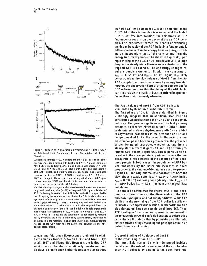
additional level of complexity is observed in the GroES spectral shifts and intrinsic quantum yield changes) do not contrib-release reaction. As shown in Figure 5A, when such ute significantly to the fluorescence changes observed, since con-ADP bullets are mixed with ATP and competitor GroES, trol experiments in which one of the energy transfer partners was
replaced with its unlabeled conjugate show only small signalapproximately 40% of the bound ES98-A is releasedchanges and the emission spectra of the ES98-A and EL315-D domuch more rapidly than during the steady-state experi-not change upon complex formation.ments (compare Figure 5A with Figure 4B). The data in
Figure 5A can be fit with a two-step (double exponential)model with rate constants of kfast 5 2.3 s21 and kslow 5 A second experiment (Figures 5B and 5C) was carried
out to confirm the existence of a fast release step and0.025 s21. The slower component in Figure 5A essentiallymatches the slow step measured in the steady-state to control for the possibility that an ADP bullet formed
by ATP hydrolysis in situ might be different in kineticexperiments above for the dissociation of GroES fromthe cis-ADP complex. behavior from one formed directly in ADP. It is possible
GroEL-GroES Cycling331
than free GFP (Weissman et al., 1996). Therefore, as theGroES lid of the cis complex is released and the foldedGFP is set free into solution, the anisotropy of GFPfluorescence reports on the decay of the cis-ADP com-plex. This experiment carries the benefit of examiningthe decay behavior of the ADP bullet in a fundamentallydifferent manner than the energy transfer assay, provid-ing an independent test of the conclusions from theenergy transfer experiment. As shown in Figure 5C, uponrapid mixing of the EL398 ADP bullets with ATP, a largedrop in the steady-state fluorescence anisotropy of thetrapped GFP is observed. The anisotropy changes re-quire a double exponential fit with rate constants ofkslow 5 0.051 s21 and kfast 5 0.5 s21. Again, kslow likelycorresponds to the slow release of GroES from the cis-ADP complex, as measured above by energy transfer.Further, the observation here of a faster component forGFP release confirms that the decay of the ADP bulletcan occur via a step that is at least an order of magnitudefaster than that previously observed.
The Fast Release of GroES from ADP Bullets IsStimulated by Denatured Substrate ProteinThe fast phase of GroES release identified in Figure5 strongly suggests that an additional step must beconsidered when describing the ADP bullet disassemblypathway. The greater significance of the fast pathwaybecomes clear when either denatured Rubisco (dRub)or denatured malate dehydrogenase (dMDH) is addedto asymmetric complexes in the presence of ATP andcompetitor GroES. As illustrated in Figure 6, the fastdissociation phase becomes prominent in the presenceof the denatured substrate, whether starting from asteady-state mixture (Figures 6A and 6E) or from pre-
Figure 5. Release of ES98-A from a Preformed ADP Bullet Revealsformed ADP bullets (Figure 6C). This is particularly no-an Additional Fast Component to the Dissociation of the cisticeable in the steady-state experiment, where the fastChamberdecay rate is not detected in the absence of the dena-(A) Release kinetics of ADP bullets monitored as loss of acceptortured protein. In both cases, the population of ADP bul-fluorescence upon mixing with GroES and ATP. A 2 mM sample of
ADP bullets made from EL315-D and ES98-A was mixed (1:1) with lets that decay by the faster rate increases in directGroES and ATP (20 mM GroES plus 5 mM ATP). The disassembly proportion to the amount of denatured substrate presentof the ADP bullet can be fit by a double exponential model with rate (Figures 6B and 6D), but the rate constants of both theconstants of kslow 5 0.025 6 0.0002 s21 and kfast 5 2.3 6 0.1 s21.
slow phase (steady state: kslow 5 0.034 s21; ADP bullet:(B) The change in fluorescence anisotropy (r) of folded GFP uponkslow 5 0.034 s21) and fast phase (steady state: kfast 5 1.3release from an EL398 cis chamber into solution can also be useds21; ADP bullet: kfast 5 1.9 s21) remain unchanged (datato measure the decay of the ADP bullet.
(C) Plot showing changes in the steady-state fluorescence anisot- not shown).ropy and total intensity (v12h) of trapped GFP upon addition of It should be noted that the effects of ATP and dena-ATP. Following formation of an ATP bullet with GFP trapped inside tured substrate protein on the disassembly of the ADPthe cis space, the sample was incubated for 3 hr to allow the slow
bullet are synergistic and not simply additive. While ATPhydrolysis of ATP to produce a population of ADP bullets. The ADPbinding to the trans ring of the ADP bullet is sufficientbullets (approximately 2 mM) containing trapped and folded GFPto initiate cis complex dissociation, neither ADP nor ADPwere then mixed (1:1) with 5 mM ATP in the stopped flow. The
change in anisotropy of the GFP can be fit by a double exponential plus denatured Rubisco can do so (Figure 6C). Thus,model with rate constants of kslow 5 0.051 6 0.0005 s21 and kfast 5 ATP binding in trans is an indispensable component of0.50 6 0.0001 s21. Because the total fluorescence intensity remains the release trigger, while unfolded substrate polypeptidenearly constant, the drop in anisotropy can be largely attributed to
can enhance this step either by populating an alternate,an increase in the rotational motion of the GFP monomers, reflectingfaster pathway or by catalyzing the passage of the ADPrelease of the GFP from the cis cavity into solution as the ADPbullet through a slow step.bullets disassemble.
to trap and fold green fluorescent protein (GFP) within Ordered Binding of Rubisco and GroESto the trans Ring of an ADP Bulleta cis complex formed between EL398 and GroES (Rye
et al., 1997 and Figure 5B). However, the folded GFP The most likely manner by which denatured Rubiscocould affect the rate of dissociation of the cis chamberwithin the cis chamber is rotationally constrained and
displays a significantly higher fluorescence anisotropy of an ADP bullet is by binding to the open trans ring.
Cell332
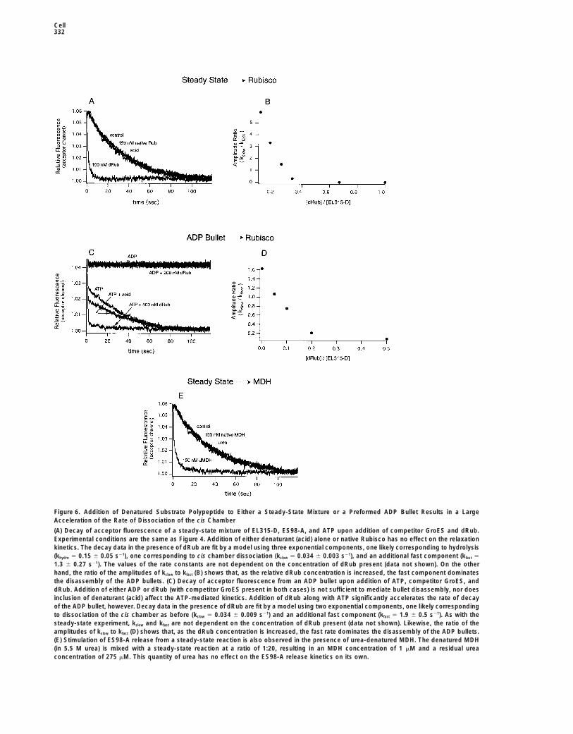
Figure 6. Addition of Denatured Substrate Polypeptide to Either a Steady-State Mixture or a Preformed ADP Bullet Results in a LargeAcceleration of the Rate of Dissociation of the cis Chamber
(A) Decay of acceptor fluorescence of a steady-state mixture of EL315-D, ES98-A, and ATP upon addition of competitor GroES and dRub.Experimental conditions are the same as Figure 4. Addition of either denaturant (acid) alone or native Rubisco has no effect on the relaxationkinetics. The decay data in the presence of dRub are fit by a model using three exponential components, one likely corresponding to hydrolysis(khydro 5 0.15 6 0.05 s21), one corresponding to cis chamber dissociation (kslow 5 0.034 6 0.003 s21), and an additional fast component (kfast 5
1.3 6 0.27 s21). The values of the rate constants are not dependent on the concentration of dRub present (data not shown). On the otherhand, the ratio of the amplitudes of kslow to kfast (B) shows that, as the relative dRub concentration is increased, the fast component dominatesthe disassembly of the ADP bullets. (C) Decay of acceptor fluorescence from an ADP bullet upon addition of ATP, competitor GroES, anddRub. Addition of either ADP or dRub (with competitor GroES present in both cases) is not sufficient to mediate bullet disassembly, nor doesinclusion of denaturant (acid) affect the ATP-mediated kinetics. Addition of dRub along with ATP significantly accelerates the rate of decayof the ADP bullet, however. Decay data in the presence of dRub are fit by a model using two exponential components, one likely correspondingto dissociation of the cis chamber as before (kslow 5 0.034 6 0.009 s21) and an additional fast component (kfast 5 1.9 6 0.5 s21). As with thesteady-state experiment, kslow and kfast are not dependent on the concentration of dRub present (data not shown). Likewise, the ratio of theamplitudes of kslow to kfast (D) shows that, as the dRub concentration is increased, the fast rate dominates the disassembly of the ADP bullets.(E) Stimulation of ES98-A release from a steady-state reaction is also observed in the presence of urea-denatured MDH. The denatured MDH(in 5.5 M urea) is mixed with a steady-state reaction at a ratio of 1:20, resulting in an MDH concentration of 1 mM and a residual ureaconcentration of 275 mM. This quantity of urea has no effect on the ES98-A release kinetics on its own.
GroEL-GroES Cycling333
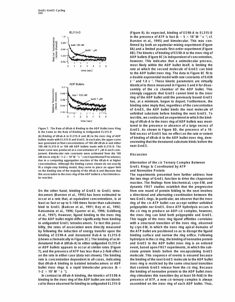
(Figure 8). As expected, binding of ES98-A to EL315-Din the presence of ATP is fast (k 5 5 3 107 M21 s21; cf.Burston et al., 1995) and bimolecular. This was con-firmed by both an equimolar mixing experiment (Figure8A) and a limited pseudo first-order experiment (Figure8B). The kinetics of binding of ES98-A to the trans ring ofADP bullets (Figure 8C) is independent of concentration,however. This indicates that a unimolecular process,most likely within the ADP bullet itself, is limiting therate at which the second molecule of GroES can bindto the ADP bullet trans ring. The data in Figure 8C fit toa double exponential model with rate constants of 0.038s21 and 1.0 s21. These kinetic parameters are virtuallyidentical to those measured in Figures 5 and 6 for disas-sembly of the cis chamber of the ADP bullet. Thisstrongly suggests that GroES cannot bind to the transring of the ADP bullet until the previously bound GroEShas, at a minimum, begun to depart. Furthermore, thebinding rates imply that, regardless of the concentrationof GroES, the ADP bullet binds the next molecule ofunfolded substrate before binding the next GroES. Totest this, we conducted an experiment in which the bind-ing of dRub-A to the trans ring of ADP bullets was moni-tored in the presence or absence of a large excess of
Figure 7. The Rate of dRub-A Binding to the ADP Bullet trans Ring GroES. As shown in Figure 8D, the presence of a 10-Is the Same as the Rate of Binding to Unliganded EL315-D
fold excess of GroES has no effect on the rate or extent(A) Binding of dRub-A to EL315-D and (B) to the trans ring of ADP of binding of dRub-A to the ADP bullet trans ring, dem-bullets made with EL315-D and GroES. In each plot, the upper curve
onstrating that the denatured substrate binds before thewas generated at final concentrations of 100 nM dRub-A and eithernext GroES.100 nM EL315-D or 100 nM ADP bullets made with EL315-D. The
lower curve was produced at a concentration of 1 mM in each com-ponent. Bimolecular rate constants were estimated from the 100 DiscussionnM traces only (k ≈ 1–2 3 107 M21 s21; see Experimental Procedures),due to a competing aggregation reaction of the dRub-A at higher
Alternation of the cis Ternary Complex Betweenconcentrations. Although the binding curves shown do not exactlyGroEL Rings Is Coordinated by ATPfit a single-step binding model, they serve to place an upper limit
on the binding rate of the majority of the dRub-A and illustrate that and Nonnative Proteinthis association to the trans ring of the ADP bullet is a fast bimolecu- The experiments presented here further address howlar reaction. the two rings of GroEL function to drive the chaperonin
reaction. The findings from biochemical, cryo-EM, anddynamic FRET studies establish that the progressionfrom one round of protein folding to the next involvesOn the other hand, binding of GroES to GroEL tetra-
decamers (Burston et al., 1995) has been estimated to a directional and alternating coordination between thetwo GroEL rings. In particular, we observe that the transoccur at a rate that, at equivalent concentrations, is at
least as fast or up to 5–100 times faster than substrates ring of the cis-ATP bullet can accept neither unfoldedpolypeptide nor GroES. Once ATP hydrolysis occurs inbind to GroEL (Badcoe et al., 1991; Roy et al., 1992;
Katsumata et al., 1996; Sparrer et al., 1996; Goldberg the cis ring to produce an ADP-cis complex, however,the trans ring can bind both polypeptide and GroES.et al., 1997). However, ligand binding to the trans ring
of the ADP bullet might differ significantly from binding This toggle of the trans ring ligand affinities correlateswith a structural transition of the trans ring, observedto unliganded GroEL tetradecamers. To test this possi-
bility, the rates of association were directly measured by cryo-EM, in which the trans ring apical domains ofthe ATP bullet are positioned so as to disrupt the ligandby following the induction of energy transfer upon the
binding of ES98-A and denatured Rub-A to EL315-D binding surface and narrow the ring orifice. Followinghydrolysis in the cis ring, the binding of nonnative protein(shown schematically in Figure 3C). The binding of acid-
denatured Rub-A (dRub-A) to either unliganded EL315-D and GroES to the ADP bullet trans ring is an orderedevent, based upon FRET experiments, in which the sub-or ADP bullets appears to occur at similar rates (Figure
7), and the presence of ATP has less than a 2-fold effect strate protein binds before the encapsulating GroESmolecule. This sequence of events is ensured becauseon the rate in either case (data not shown). The binding
rate is concentration dependent in all cases, indicating the binding of the next GroES molecule to the ADP bullettrans ring is restricted by the same structural transitionthat dRub-A binding to isolated tetradecamers or ADP
bullet trans rings is a rapid bimolecular process (k ≈ that controls GroES release from the cis ring. Becausethe binding of nonnative protein to the ADP bullet trans1–2 3 107 M21 s21).
In contrast to dRub-A binding, the kinetics of ES98-A ring stimulates this transition (by at least 50-fold) in thepresence of ATP, a new cis-ternary complex is alwaysbinding to the trans ring of the ADP bullet are not identi-
cal to those observed for binding to unliganded EL315-D assembled on the trans ring of each ADP bullet. Thus,

Cell334
Figure 8. Unfolded Substrate Polypeptide Binds before GroES to the trans Ring of an ADP Bullet and Stimulates Steady-State ATP Hydrolysis
(A) Binding of ES98-A to EL315-D at equimolar concentrations. In the upper trace, ES98-A and EL315-D were mixed at final concentrationsin the stopped flow of 50 nM each, and in the lower trace at 1 mM; ATP in each case was 2 mM. The bimolecular rate constants derived fromthese traces are 5.7 3 107 and 2.9 3 107 M21 s21 for the 50 nM and 1 mM traces, respectively.(B) A limited pseudo first-order experiment was conducted in which the ES98-A and ATP concentrations were maintained at 100 nM and 2mM, respectively, and the concentration of EL315-D in the binding reaction was varied. A plot of the pseudo first-order rate constants (kobs)versus the ratio of the components is shown. The bimolecular rate constant extracted from this analysis is 5 3 107 M21 s21.(C) Binding of ES98-A to the trans ring of ADP bullets (in 2 mM ATP) is independent of concentration. The binding data fit to a doubleexponential model with rate constants of 0.038 6 0.0006 s21 and 1 6 0.02 s21.(D) The binding of 100 nM dRub-A to the trans ring of 100 nM ADP bullets (plus 2 mM ATP) is the same in the absence and presence of 1mM GroES.(E) The steady-state ATP hydrolysis rate of GroEL, in the presence of GroES, is stimulated 2.3-fold by denatured MDH. A steady-state hydrolysisreaction was initiated by adding 2 mM ATP to a mixture of GroEL (125 nM) and GroES (500 nM). Either urea-denatured MDH (dMDH) or ureaalone was added at 5.5 min (1 mM final monomer concentration, approximately 80 mM residual urea after addition). Free phosphate wasassessed as described (Fenton et al., 1994).
given that GroES and polypeptide leave one ring prior steps per cycle, one on each ring. Recent work, how-ever, has shown that ATP binding alone on the transto, or simultaneously with, arrival of GroES to form a cis
complex on the opposite ring, it appears that folding- ring is sufficient to initiate discharge of the cis-GroEL-GroES complex and complete the reaction (Rye et al.,active states alternate between rings. Such behavior of
the GroEL-GroES system as a “two-stroke” machine 1997). This finding simplifies the states necessary todescribe a full turn of the cycle (Figure 9I; also see Kadhas been previously proposed (Lorimer, 1997; see also
Sparrer and Buchner, 1997; Kad et al., 1998). et al., 1998). Hydrolysis of ATP in the cis complex toform an ADP bullet occurs with a rate constant of 0.12These findings have been incorporated into two mod-
els of the GroEL reaction cycle (Figure 9): one in the s21 (Burston et al., 1995). Previous experiments with apyrene-labeled GroEL strongly suggested that the slow-absence of polypeptide substrate (I) and one in its pres-
ence (II). The nucleotide cycle of the GroEL-GroES sys- est step in the overall GroEL-GroES cycle in the absenceof nonnative polypeptide was the release of GroES fromtem was initially formulated to involve two hydrolytic

GroEL-GroES Cycling335
Figure 9. Proposed Models for Cycling of theGroEL-GroES System in the Presence andAbsence of Substrate
The GroEL-GroES system can be describedwith two reaction cycles, one in the absence(cycle I) and one in the presence (cycle II) ofdenatured substrate polypeptide (see Dis-cussion). Association of ligands with the ATPbullet trans ring is blocked by structural tran-sitions induced in the trans ring apical do-mains by the presence of ATP in the cis-ter-nary complex (indicated here as a narrowingof the trans ring). Association of the secondGroES molecule with the trans ring occurs ata rate similar to the rate at which the previousGroES is released and thus appears to be aconcerted event (indicated by the brackets).The switching of folding-active states be-tween rings is efficiently coupled to polypep-tide binding through a structural transition ofthe cis-ADP complex (“activated” cis ring). Asa result, the rate-limiting step of the reactioncycle in the presence of nonnative polypep-tide becomes cis chamber hydrolysis (desig-nated by the open arrow). The blue oval shownhere represents proteins that are committedto reaching the native state, while the irregu-lar blue symbol inside the cis ring representsproteins that are in the process of folding.The same irregular symbol is used followingcis complex disassembly to represent pro-teins that have not reached a committed stateand must be rebound for another round ofchaperonin-mediated folding.
the cis ring, occurring with a rate constant of 0.042 s21 bullet from which GroES dissociation is rapid. Althougha second GroES molecule might bind prior to this disso-(Burston et al., 1995). The direct examination of this step
by FRET (Figure 4) confirms this observation. Moreover, ciation, indicated by the brackets in the model, there isno kinetic requirement in our data for such involvement.the FRET assay has revealed that there must be an addi-
tional fast step (kfast ≈ 2 s21) in the release pathway of The GroEL cycle in the presence of substrate polypep-tide (Figure 9II), likely to be the physiologic state, is alsoGroES from the cis-ADP bullet. The simplest incorpora-
tion of this fast step places it immediately following the simplified by requiring only one round of ATP hydrolysisper cycle. In addition, the presence of unfolded sub-slowest step in the cycle and immediately preceding
the formation of a new ATP bullet (Figure 9I). Given these strate significantly accelerates the rate at which the ciscomplex dissociates. The rate constant measured forfindings, it is attractive to hypothesize that the slow,
rate-limiting step is a structural transition of the ADP GroES release in the presence of nonnative protein (1–2s21) is very similar to that of the fast step described inbullet, indicated schematically in Figure 9I. Passage of
the ADP bullet through this slow transition leads to the Figure 9I. This strongly suggests that it corresponds tothe same reaction (i.e., the dissociation of GroES frompopulation of an activated intermediate form of the ADP

Cell336
an activated ADP bullet). In this case, it appears that seems likely that some of the energy of polypeptidebinding to the trans ring is employed to accelerate thethe unfolded substrate catalyzes the structural re-
arrangement of the ADP bullet, which is slow in the structural change that is necessary for cis complex dis-sociation. This coupling helps drive the timely disruptionabsence of unfolded protein, so that it is no longer rate
limiting in the overall cycle. This directly couples dissoci- of the previous cis complex and increases the efficiencyof the GroEL machine. Because ATP binding to one ringation of one cis folding-active complex with the forma-
tion of the next. Furthermore, because the binding of simultaneously sets up a folding chamber on that ringwhile discharging the opposite ring, it is apparent thatunfolded substrate and GroES to the trans ring is or-
dered and sequential, with polypeptide binding first (Fig- turnover of one ringful of seven ATPs amounts to onecycle of folding. This is at least twice as efficient as aures 6 and 7), the efficient formation of the next cis
folding chamber is ensured. Once again, the kinetics of pathway in which GroEL must completely clear its ringsof ligands before initiating the next cycle. This efficiency,the cycle do not suggest the binding of a second GroES
before the first departs, but the transient formation of coupled with the increase in overall rate of the cyclein the presence of nonnative substrate, suggests thatan intermediate with a GroES molecule on each end of
the GroEL cylinder cannot be excluded, indicated by GroEL could be responsible for folding a greater fractionof cellular proteins than previously predicted (Lorimer,the bracketed species.1996). It also offers a mechanism by which the effective-ness of GroEL-mediated folding would be increasedEffect of Interring Allostery on Polypeptide Releasewhen the amount of unfolded protein increases, for ex-Following discharge of the cis-ADP complex, the va-ample, during heat shock.cated ring becomes the trans ring of a new ATP bullet
complex. As the biochemical and structural studies de-scribed above indicate, this ring has no significant af- Experimental Proceduresfinity for nonnative protein. An incompletely folded
Proteinssubstrate protein inside a decaying cis complex is,The G315C (EL315C) variant of GroEL places a single cysteine ontherefore, unlikely to be either retained or immediatelythe exterior of the apical domain of each GroEL subunit, whoserecaptured by the apical domains of such a ring. Indeed,endogenous cysteines at positions 138, 458, and 519 have been
not until the slowest step in the reaction cycle (cis hydro- replaced with alanine. The ES98C version of GroES adds a singlelysis) has been completed can substrate again bind to cysteine at the COOH terminus of each GroES subunit (Murai et al.,this ring. Thus, the allosteric changes transmitted to the 1996). The EL398 mutant of GroEL (D398A) was constructed as
previously described (Rye et al., 1997). Wild-type and mutant ver-opposite ring by formation of a cis-ATP complex ensuresions of GroEL and GroES, as well as Rubisco from Rhodospirillumthat the ring from the previous round of folding is com-rubrum, were expressed in E. coli and purified as previously de-pletely cleared of substrate protein. Substrate proteinsscribed (Weissman et al., 1995; Rye et al., 1997). Purified pig heart
are then ejected into solution and subjected to kinetic mitochondrial malate dehydrogenase (MDH) was obtained frompartitioning with each turn of the chaperonin cycle (Todd Boehringer-Mannheim (Indianapolis, IN).et al., 1994; Weissman et al., 1994; Smith and Fisher,1995; Taguchi and Yoshida, 1995; Ranson et al., 1997). Fluorescent Labeling of EL315C, ES98C, and Rubisco
EL315C was labeled with IAEDANS (Molecular Probes, Inc.) to pro-duce EL315-D (where “D” designates the use of the EDANS fluoro-Consequences of Polypeptide Bindingphore as an energy transfer donor). ES98C and Rubisco were labeledand Efficiency of the Folding Cyclewith fluorescein-5-maleimide (Molecular Probes, Inc.) to produceThe models presented above make a very specific pre- ES98-A and Rub-A, respectively (where “A” designates fluorescein
diction. In the presence of denatured polypeptide, the as an energy transfer acceptor). Labeling reactions were conductedrate-limiting step of the cycle becomes cis hydrolysis essentially as previously described (Rye et al., 1997). See below for
details regarding Supplemental Experimental Procedures.(Figure 9II), rather than the slow transition precedingFor Rubisco, a labeling ratio of one fluorescein to one Rubiscodissociation of the cis complex (Figure 9I). Thus the
monomer was obtained. For EL315-D, an average of 3–4 dye mole-steady-state ATPase rate of the GroEL-GroES systemcules per tetradecamer was achieved, and for ES98-A, approxi-should be stimulated by 2- to 3-fold in the presence mately 2 dye molecules were added per heptamer. Both labeled
of saturating amounts of denatured substrate protein. and unlabeled EL315C and ES98C were stored in 50 mM Tris (pHStimulation of the ATPase activity of the GroEL-GroES 7.4), 100 mM KCl, 0.5 mM EDTA, 4 mM DTT under argon and re-
mained stable for several months. Upon addition of either wild-typesystem has indeed been observed in the presence ofGroES or ES98-A, EL315-D refolded Rubisco in a GroES- and ATP-denatured lactate dehydrogenase (Staniforth et al.,dependent manner that was identical (in both refolding yield and1994). Here also, as shown in Figure 8E, the steady-rate) to a wild-type GroEL-GroES reaction. EL315-D did, however,state ATPase rate of GroEL-GroES is stimulated by 2.3- show a 30%–40% reduction in its steady-state ATPase rate. This
fold in the presence of saturating quantities of denatured was not due to the mutation at residue 315 or conjugation withMDH. This result provides specific confirmation of the IAEDANS but was the result of changing the endogenous cysteine
residues to alanine. Rub-A exhibited wild-type enzymatic activityconclusions drawn from the FRET experiments andand refolding.demonstrates a significant level of allosteric interaction
between the chaperonin system and its substrates.Gel Filtration of Bullet ComplexesWhereas the cryo-EM studies presented above pro-ATP and ADP bullets were formed with EL398 essentially as pre-vide some indication of the allosteric signal transmittedviously described (Rye et al., 1997). See below for details regardingto the trans ring by ATP hydrolysis in the cis ring, theSupplemental Experimental Procedures. All mixing was done in 50
structural changes involved in committing GroES to re- mM HEPES (pH 7.6), 5 mM KOAc, 10 mM Mg(OAc)2, and 2 mM DTT.lease, a step stimulated by polypeptide binding to the Following addition of either denatured Rub-A or ES98-A and ATP
to the bullet mixtures, the samples were injected onto a Tosohaastrans ring of the ADP bullet, are unknown. However, it
GroEL-GroES Cycling337
G4000SWXL HPLC gel filtration column in the same buffer and the folding and the hydrolysis of the trapped ATP. This sample was thenmixed (1:1) in the stopped flow with 5 mM ATP.elution profile monitored by an in-line fluorescence detector.
Data AnalysisCryo-Electron Microscopy, Three-Dimensional Reconstruction, Data records from the various stopped-flow experiments were firstand Atomic Structure Fitting smoothed to remove excess noise and then background corrected.Images of GroEL-GroES complexes, vitrified 6 s after addition of (See below for details regarding Supplemental Experimental Proce-2.5 mM ATP to a mixture of 1 mM each of GroEL and GroES, were dures.) Relative fluorescence was then calculated by dividing thecollected as previously described (Roseman et al., 1996) and digi- data record for the donor plus acceptor by a data record obtainedtized at 6.7 A/pixel on a Leafscan 45 linear CCD scanner (Ilford Ltd, under identical conditions using either the donor only (for donorCheshire, UK). A data set of 2550 side views contained a mixture monitored experiments) or acceptor only (for acceptor monitoredof four different structures, identified and separated by a combina- experiments). Decay experiments were fit to either single exponen-tion of multivariate statistical analysis (van Heel et al., 1996) and tials or sums of exponentials using nonlinear least-squares fittingangular refinement (Frank et al., 1996; Roseman et al., 1996). The with IGOR Pro (WaveMetrics Inc). Binding data for the pseudo first-structures were designated as apo, bullet, and football, correspond- order binding of ES98-A to EL315-D were fit to single exponentials.ing to GroEL complexes without GroES, and with one or two GroES Binding data obtained for experiments conducted at equivalent con-molecules, respectively. Two different bullet structures were pres- centrations of reactants were fit to the following simplified bindingent, and these were clearly identifiable as cis-ADP and cis-ATP equation (Gutfreund, 1995):bullets by their resemblance to preformed cis-ADP bullets and tobullet complexes formed in ATP under steady-state conditions, re- 1
D(t)2
1D(0)
5 ktspectively (Roseman et al., 1996).The positions and orientations of the trans apical domains were
determined by fitting the apical domain atomic coordinates (trans where D(t) is the concentration of unbound EL315-D at time t, D(0)ring from Xu et al., 1997) into the trans rings of the cis-ATP and cis- is the concentration of EL315-D at the beginning of the experiment,ADP bullet complexes, using O (Jones et al., 1991) and rigid body and k is the bimolecular rate constant for association.refinement in X-PLOR (Brunger, 1992). The fitted structures con-formed very well to the density envelope, with the exception of one Supplemental Experimental Proceduresexposed loop in the ATP bullet (residues 206–214). (See below for For more information on experimental methods, see http://www.details regarding Supplemental Experimental Procedures.) cell.com/cgi/content/full/97/3/325/DC1.
AcknowledgmentsStopped-Flow Fluorescence Energy Transfer and AnisotropyThe stopped-flow apparatus has been described (Rye et al., 1997).
We thank Erhard Hohenester, David Houldershaw, Ian Tickle, andFor fluorescence energy transfer experiments, the excitation wave-Richard Westlake for help with computing and members of thelength was 336 nm. Both detection channels were fitted with long-Horwich lab and Steve Burston for helpful discussions. This workpass barrier filters (KV370; Schott). The donor channel was config-was supported by The Wellcome Trust, Biotechnology and Biologi-ured with a blue separation filter (SWP filter, Reynard Corporation),cal Sciences Research Council, National Institutes of Health, How-and the acceptor channel was configured with a green separationard Hughes Medical Institute, and The Nelson Fund.filter (Reynard). All stopped-flow experiments were conducted at
258C in 50 mM HEPES (pH 7.6), 5 mM KOAc, 10 mM Mg(OAc)2, andReceived February 11, 1999; revised April 1, 1999.2 mM DTT .
For steady-state dissociation experiments, samples of EL315-Dand ES98-A were first mixed together with 5 mM ATP and then Referencesmixed in the stopped flow with either excess unlabeled GroES orexcess unlabeled GroEL. For ADP bullet dissociation experiments, Badcoe, I.G., Smith, C.J., Wood, S., Halsall, D.J., Holbrook, J.J.,ADP bullets were formed by first mixing 25 mM EL315-D with 25 mM Lund, P., and Clarke, A.R. (1991). Binding of a chaperonin to theES98-A and 1 mM ADP. This mixture was incubated at 238C in the folding intermediates of lactate dehydrogenase. Biochemistry 30,dark for 1.5–2 hr. Immediately prior to use, the ADP bullet mixture 9195–9200.was diluted with buffer and loaded into the stopped flow, then mixed Brunger, A.T. (1992). X-PLOR v. 3.1 Manual (New Haven, CT: Yalewith excess unlabeled GroES and ATP. For control experiments in University).which ES98-A was replaced with unlabeled GroES in the initial
Buckle, A.M., Zahn, R., and Fersht, A.R. (1997). A structural modelsteady-state mix, excess unlabeled GroEL was used to sequesterfor GroEL-polypeptide recognition. Proc. Natl. Acad. Sci. USA 94,the GroES in the stopped-flow experiments. For control experiments3571–3575.in which EL315-D was replaced with unlabeled GroEL, unlabeledBurston, S.G., Ranson, N.A., and Clarke, A.R. (1995). The originsGroES was used as the competitor.and consequences of asymmetry in the chaperonin reaction cycle.For experiments involving denatured Rubisco or denatured MDH,J. Mol. Biol. 249, 138–152.an additional syringe was employed to hold either acid-denatured
Rubisco in 25 mM glycine-phosphate (pH 2.0) or urea-denatured Chen, S., Roseman, A.M., Hunter, A.S., Wood, S.P., Burston, S.G.,MDH in 5.5 M fresh urea. Mixing was accomplished through the Ranson, N.A., Clarke, A.R., and Saibil, H.R. (1994). Location of asimultaneous push of three syringes, giving the final concentrations folding protein and shape changes in GroEL-GroES complexes im-of components noted in the figure legends. The final dilution of acid- aged by cryo-electron microscopy. Nature 371, 261–264.denatured Rubisco in each case was approximately 10-fold, which Corrales, F.J., and Fersht, A.R. (1996). Kinetic significance ofwas sufficient to neutralize the acid. The final dilution of the urea- GroEL14·(GroES7)2 complexes in molecular chaperone activity. Fold.denatured MDH was approximately 20-fold. Des. 1, 265–273.
For experiments involving GFP fluorescence anisotropy, theFenton, W.A., Kashi, Y., Furtak, K., and Horwich, A.L. (1994). Resi-stopped flow was reconfigured with a pair of crossed polarizers asdues in chaperonin GroEL required for polypeptide binding anddescribed previously (Rye et al., 1997). Both detection channelsrelease. Nature 371, 614–619.were fitted with long-pass filters (GG495, Schott), and the excitationFrank, J., Radermacher, M., Penczek, P., Zhu, J., Li, Y., Ladjadj, M.,wavelength was 400 nm. EL398 ATP bullets containing trapped GFPand Leith, A. (1996). SPIDER and WEB: processing and visualizationwere formed essentially as previously described (Rye et al., 1997).of images in 3D electron microscopy and related fields. J. Struct.See below for details regarding Supplemental Experimental Proce-Biol. 116, 190–199.dures. The purified ATP bullets (z2 mM), approximately one-half of
which contained a trapped GFP molecule, were then incubated in Goldberg, M.S., Zhang, J., Sondek, S., Matthews, C.R., Fox, R.O.,and Horwich, A.L. (1997). Native-like structure of a protein-foldingthe dark at 238C for 1–2 hr in order to permit completion of GFP
Cell338
intermediate bound to the chaperonin GroEL. Proc. Natl. Acad. Sci. Torok, Z., Vigh, L., and Goloubinoff, P. (1996). Fluorescence detec-tion of symmetric GroEL14(GroES7)2 heterooligomers involved in pro-USA 94, 1080–1085.tein release during the chaperonin cycle. J. Biol. Chem. 271, 16180–Gutfreund, H. (1995). Kinetics for the Life Sciences (Cambridge,16186.U.K.: Cambridge University Press).Van der Meer, B.W., Cocker, G.I., and Chen, S.Y.S. (1994). Reso-Inbar, E., and Horovitz, A. (1997). GroES promotes the T to R transi-nance Energy Transfer: Theory and Data (New York, NY: VCH Pub-tion of the GroEL ring distal to GroES in the GroEL-GroES complex.lishers, Inc.).Biochemistry 36, 12276–12281.van Heel, M., Harauz, G., and Orlova, E.V. (1996). A new generationItzhaki, L.S., Otzen, D.E., and Fersht, A.R. (1995). Nature and conse-of the IMAGIC image processing system. J. Struct. Biol. 116, 17–24.quences of GroEL-protein interactions. Biochemistry 34, 14581–Weissman, J.S., Kashi, Y., Fenton, W.A., and Horwich, A.L. (1994).14587.GroEL-mediated protein folding proceeds by multiple rounds ofJones, T.A., Zou, J.-Y., Cowan, S.W., and Kjeldgaard, M. (1991).binding and release of nonnative forms. Cell 78, 693–702.
Improved methods for building models in electron density mapsWeissman, J.S., Hohl, C.M., Kovalenko, O., Kashi, Y., Chen, S., Braig,and the location of errors in these models. Acta Crystallogr. A47,K., Saibil, H.R., Fenton, W.A., and Horwich, A.L. (1995). Mechanism110–119.of GroEL action: productive release of polypeptide from a seques-
Kad, N.M., Ranson, N.A., Cliff, M.J., and Clarke, A.R. (1998). Asym-tered position under GroES. Cell 83, 577–587.
metry, commitment and inhibition in the GroE ATPase cycle imposeWeissman, J.S., Rye, H.S., Fenton, W.A., Beechem, J.M., and Hor-alternating functions on the two GroEL rings. J. Mol. Biol. 278,wich, A.L. (1996). Characterization of the active intermediate of a267–278.GroEL-GroES-mediated protein folding reaction. Cell 84, 481–490.
Katsumata, K., Okazaki, A., and Kuwajima, K. (1996). Effect of GroELXu, Z., Horwich, A.L., and Sigler, P.B. (1997). The crystal structure ofon the re-folding kinetics of alpha-lactalbumin. J. Mol. Biol. 258,the asymmetric GroEL-GroES-(ADP)7 chaperonin complex. Nature827–838.388, 741–750.
Lin, Z., Schwartz, F.P., and Eisenstein, E. (1995). The hydrophobicnature of GroEL-substrate binding. J. Biol. Chem. 270, 1011–1014.
Llorca, O., Marco, S., Carrascosa, J.L., and Valpuesta, J.M. (1994).The formation of symmetrical GroEL-GroES complexes in the pres-ence of ATP. FEBS Lett. 345, 181–186.
Lorimer, G.H. (1996). A quantitative assessment of the role of thechaperonin proteins in protein folding in vivo. FASEB J. 10, 5–9.
Lorimer, G. (1997). Protein folding. Folding with a two-stroke motor.Nature 388, 720–721.
Murai, N., Makino, Y., and Yoshida, M. (1996). GroEL locked in aclosed conformation by an interdomain cross-link can bind ATPand polypeptide but cannot process further reaction steps. J. Biol.Chem. 271, 28229–28234.
Ranson, N.A., Burston, S.G., and Clarke, A.R. (1997). Binding, encap-sulation and ejection: substrate dynamics during a chaperonin-assisted folding reaction. J. Mol. Biol. 266, 656–664.
Roseman, A.M., Chen, S., White, H., Braig, K., and Saibil, H.R. (1996).The chaperonin ATPase cycle: mechanism of allosteric switchingand movements of substrate-binding domains in GroEL. Cell 87,241–251.
Roy, H., Kupferschmid, M., and Bell, J.A. (1992). Theory of chaper-onin action: inertial model for enhancement of prokaryotic Rubiscoassembly. Protein Sci. 1, 925–934.
Rye, H.S., Burston, S.G., Fenton, W.A., Beechem, J.M., Xu, Z., Sigler,P.B., and Horwich, A.L. (1997). Distinct actions of cis and trans ATPwithin the double ring of the chaperonin GroEL. Nature 388, 792–798.
Sigler, P.B., Xu, Z., Rye, H.S., Burston, S.G., Fenton, W.A., andHorwich, A.L. (1998). Structure and function in GroEL-mediated pro-tein folding. Annu. Rev. Biochem. 67, 581–608.
Smith, K.E., and Fisher, M.T. (1995). Interactions between the GroEchaperonins and rhodanese. Multiple intermediates and release andrebinding. J. Biol. Chem. 270, 21517–21523.
Sparrer, H., and Buchner, J. (1997). How GroES regulates bindingof nonnative protein to GroEL. J. Biol. Chem. 272, 14080–14086.
Sparrer, H., Lilie, H., and Buchner, J. (1996). Dynamics of the GroEL-protein complex: effects of nucleotides and folding mutants. J. Mol.Biol. 258, 74–87.
Staniforth, R.A., Burston, S.G., Atkinson, T., and Clarke, A.R. (1994).Affinity of chaperonin-60 for a protein substrate and its modulationby nucleotides and chaperonin-10. Biochem. J. 300, 651–658.
Taguchi, H., and Yoshida, M. (1995). Chaperonin releases the sub-strate protein in a form with tendency to aggregate and ability torebind to chaperonin. FEBS Lett. 359, 195–198.
Todd, M.J., Viitanen, P.V., and Lorimer, G.H. (1994). Dynamics ofthe chaperonin ATPase cycle: implications for facilitated proteinfolding. Science 265, 659–666.