Cell, Vol. 58, 955-968, September 8, 1989, Copyright 0 ... · Evolution of engrailed Expression 957...
14
Cell, Vol. 58, 955-968, September 8, 1989, Copyright 0 1989 by Cell Press Expression of engrailed Proteins in Arthropods, Annelids, and Chordates Nipam H. Patel: Enrique Martin-Blanco,t Kevin G. Coleman,? Stephen J. Poole,t Michael C. Ellis,’ Thomas B. Kornberg,t and Corey S. Goodman* * Howard Hughes Medical Institute Department of Biochemistry University of California Berkeley, California 94720 tDepartment of Biochemistry and Biophysics University of California San Francisco, California 94143 Summary engrailed is a homeobox gene that has an important role in Drosophila segmentation. Genes homologous to engrailed have been identified in several other or- ganisms. Here we describe a monoclonal antibody that recognizes a conserved epitope in the homeodo- main of engrailed proteins of a number of different ar- thropods, annelids, and chordates; we use this anti- body to isolate the grasshopper engrailed gene. In Drosophila embryos, the antibody reveals engrailed protein in the posterior portion of each segment dur- ing segmentation, and in a segmentally reiterated sub- set of neuronal cells during neurogenesis. Other arthropods, including grasshopper and two crusta- ceans, have similar patterns of engrailed expression. However, these patterns of expression are not shared by the annelids or chordates we examined. Our results provide the most comprehensive view that has been obtained of how expression patterns of a regulatory gene vary during evolution. On the basis of these patterns, we suggest that engrailed is a gene whose ancestral function was in neurogenesis and whose function was co-opted during the evolution of seg- mentation in the arthropods, but not in the annelids and chordates. Introduction Early pattern formation in Drosophila organizes the blastoderm into a series of repeated units and uniquely specifies each of these units. It is controlled by a hierarchy of maternal-effect, segmentation, and homeotic genes (reviewed by Ingham, 1988). One of these genes is en- grailed. In Drosophila, engrailed is essential during several de- velopmental phases, and it has characteristics common to bbth segmentation and homeotic genes. For instance, en- grailed mutant embryos do not segment normally (Korn- ‘berg, 1981; Niisslein-Volhard and Wieschaus, 1980). Other aspects of the engrailedmutant phenotype are suggestive of homeotic function (Garcia-Bellido, 1975; Lawrence and Morata, 1976), of a role in organizing the preblastoderm (Karr et al., 1985), and, in imaginal posterior compart- ments, of a role in maintaining compartment and segment borders (Morata and Lawrence, 1975; Komberg, 1981; Lawrence and Struhl, 1982). One further aspect of en- grailed function is its expression during neurogenesis in a segmentally reiterated subset of neuroblasts and neu- rons (DiNardo et al., 1985; Brower, 1986). Thus, engrailed, like other segmentation genes (Doe et al., 1988a, 1988b), appears to play multiple roles during development: during organization and growth of epidermal derivatives and dur- ing neurogenesis. During segmentation, the Drosophila engrailed protein is produced in the cells of the posterior region of each body segment (DiNardo et al., 1985; Karr et al., 1989). Consistent with its putative regulatory role, the engrailed protein contains a homeodomain (Fjase et al., 1985; Poole et al., 1985), localizes to nuclei (DiNardo et al., 1985; Karr et al., 1989), and has the capacity to bind tot DNA in vitro (Desplan et al., 1988). The Drosophila engrailed homeo- box sequence is distinctive, but it is not unique. The in- vetted gene, juxtaposed to engrailed on the Drosophila chromosome and expressed in an almost identical pattern during segmentation, has a homeobox sequence that is strikingly similar (52/61 amino acids) to that of engrailed (Coleman et al., 1987). In addition, invected has regions closely related to engrailed upstream (X3/26 identical amino acids) and downstream (26/30 amino acids) of its homeobox, and in a region near the amino terminus (8/12 amino acids). Thus Drosophila has a second engrailed gene, invected, the function of which is not known. A number of engrailed genes have been identified in other organisms. For example, honeybee (U. Walldorf and W. Gehring, personal communication), mouse (En-7 and En-2, Joyner et al., 1985; Joyner and %lartin, 1987), chicken (Darnell et al., 1986), zebrafish @j&e et al., 1988), and human (Poole et al., 1989; Logan ettali., 1989) contain two engrailed genes, and single engrail& genes have been found in sea urchin (Dolecki and Humphreys, 1988), leech (Weisblat et al., 1988), ,grasshopp&r (this report), and nematode (A. Kamb and T B. Kornberg, unpublished data). The homebdomain sequences of t&se genes are similar to Drosophila engrailed and, where sequence analysis extends sufficiently, to region? ihmedi&ly up- stream and downstream of the homeodcimain as well. For the m&se En-l gend, most of the codihdsequehce has been obtained, and in addition to the consbrved extended homeodomain regidn (69199 of the resjdlues are’ con- served), it contains the region near the Bmino terminus conserved’ in DrdSophila engrailed and h&ted (Frohman and Martin, personal communidation).* These two con- serve? regions,, t’he small amino-termi& bhe arid the ex- tend&d hdmeodomain, are the only seqaecces cons’erved among thdskthree genes. ConservatioQ df the hhhieobox regio’ns ‘of the &tier vertebrate and inve/rt$brate mehgrailed genes is comp&ble.“Since the sequence bf theengiaf/ed- type lhomeodamains is ‘distinctive and I uelike any of the other homeodomain sequences known (Schtt et al.., 1989), and since it is in every known case suriodnded by a con-
Transcript of Cell, Vol. 58, 955-968, September 8, 1989, Copyright 0 ... · Evolution of engrailed Expression 957...

Cell, Vol. 58, 955-968, September 8, 1989, Copyright 0 1989 by Cell Press
Expression of engrailed Proteins in Arthropods, Annelids, and Chordates
Nipam H. Patel: Enrique Martin-Blanco,t Kevin G. Coleman,? Stephen J. Poole,t Michael C. Ellis,’ Thomas B. Kornberg,t and Corey S. Goodman* * Howard Hughes Medical Institute Department of Biochemistry University of California Berkeley, California 94720 tDepartment of Biochemistry and Biophysics University of California San Francisco, California 94143
Summary
engrailed is a homeobox gene that has an important role in Drosophila segmentation. Genes homologous to engrailed have been identified in several other or- ganisms. Here we describe a monoclonal antibody that recognizes a conserved epitope in the homeodo- main of engrailed proteins of a number of different ar- thropods, annelids, and chordates; we use this anti- body to isolate the grasshopper engrailed gene. In Drosophila embryos, the antibody reveals engrailed protein in the posterior portion of each segment dur- ing segmentation, and in a segmentally reiterated sub- set of neuronal cells during neurogenesis. Other arthropods, including grasshopper and two crusta- ceans, have similar patterns of engrailed expression. However, these patterns of expression are not shared by the annelids or chordates we examined. Our results provide the most comprehensive view that has been obtained of how expression patterns of a regulatory gene vary during evolution. On the basis of these patterns, we suggest that engrailed is a gene whose ancestral function was in neurogenesis and whose function was co-opted during the evolution of seg- mentation in the arthropods, but not in the annelids and chordates.
Introduction
Early pattern formation in Drosophila organizes the blastoderm into a series of repeated units and uniquely specifies each of these units. It is controlled by a hierarchy of maternal-effect, segmentation, and homeotic genes (reviewed by Ingham, 1988). One of these genes is en- grailed.
In Drosophila, engrailed is essential during several de- velopmental phases, and it has characteristics common to bbth segmentation and homeotic genes. For instance, en- grailed mutant embryos do not segment normally (Korn- ‘berg, 1981; Niisslein-Volhard and Wieschaus, 1980). Other aspects of the engrailedmutant phenotype are suggestive of homeotic function (Garcia-Bellido, 1975; Lawrence and Morata, 1976), of a role in organizing the preblastoderm (Karr et al., 1985), and, in imaginal posterior compart-
ments, of a role in maintaining compartment and segment borders (Morata and Lawrence, 1975; Komberg, 1981; Lawrence and Struhl, 1982). One further aspect of en- grailed function is its expression during neurogenesis in
a segmentally reiterated subset of neuroblasts and neu- rons (DiNardo et al., 1985; Brower, 1986). Thus, engrailed, like other segmentation genes (Doe et al., 1988a, 1988b), appears to play multiple roles during development: during organization and growth of epidermal derivatives and dur- ing neurogenesis.
During segmentation, the Drosophila engrailed protein is produced in the cells of the posterior region of each body segment (DiNardo et al., 1985; Karr et al., 1989). Consistent with its putative regulatory role, the engrailed protein contains a homeodomain (Fjase et al., 1985; Poole et al., 1985), localizes to nuclei (DiNardo et al., 1985; Karr et al., 1989), and has the capacity to bind tot DNA in vitro (Desplan et al., 1988). The Drosophila engrailed homeo-
box sequence is distinctive, but it is not unique. The in- vetted gene, juxtaposed to engrailed on the Drosophila chromosome and expressed in an almost identical pattern during segmentation, has a homeobox sequence that is strikingly similar (52/61 amino acids) to that of engrailed (Coleman et al., 1987). In addition, invected has regions closely related to engrailed upstream (X3/26 identical amino acids) and downstream (26/30 amino acids) of its homeobox, and in a region near the amino terminus (8/12 amino acids). Thus Drosophila has a second engrailed gene, invected, the function of which is not known.
A number of engrailed genes have been identified in other organisms. For example, honeybee (U. Walldorf and W. Gehring, personal communication), mouse (En-7 and En-2, Joyner et al., 1985; Joyner and %lartin, 1987), chicken (Darnell et al., 1986), zebrafish @ j&e et al., 1988), and human (Poole et al., 1989; Logan ettali., 1989) contain two engrailed genes, and single engrail& genes have been found in sea urchin (Dolecki and Humphreys, 1988), leech (Weisblat et al., 1988), ,grasshopp&r (this report), and nematode (A. Kamb and T B. Kornberg, unpublished data). The homebdomain sequences of t&se genes are similar to Drosophila engrailed and, where sequence analysis extends sufficiently, to region? ihmedi&ly up- stream and downstream of the homeodcimain as well. For the m&se En-l gend, most of the codihdsequehce has been obtained, and in addition to the consbrved extended homeodomain regidn (69199 of the resjdlues are’ con- served), it contains the region near the Bmino terminus conserved’in DrdSophila engrailed and h&ted (Frohman and Martin, personal communidation).* These two con- serve? regions,, t’he small amino-termi& bhe arid the ex- tend&d hdmeodomain, are the only seqaecces cons’erved among thdskthree genes. ConservatioQ df the hhhieobox regio’ns ‘of the &tier vertebrate and inve/rt$brate mehgrailed genes is comp&ble.“Since the sequence bf theengiaf/ed- type lhomeodamains is ‘distinctive and I uelike any of the other homeodomain sequences known (Schtt et al.., 1989), and since it is in every known case suriodnded by a con-
Cell 956
Figure 1. Expression of engfeiled Proteins in Arthropods, Annelids, and Chordates Staining of Drosophila (A), grasshopper(B), crayfish (C), leech(D), zebrafish (E, G, H), and chick (F, I) embryos with the 4Dg MAb and HRP immunocy- tochemistry. In all three arthropods (A, 8, C), engrailed is expressed in the posterior region of each segment; in the other organisms examined (D-l), engfailed does not appear to play a role during metameric development (for example, arrowhead in F marks the developing somites in the chick

Evolution of engrailed Expression 957
served region, we consider this extended engrailed ho- meodomain region a signature of engrailed genes.
Proteins that share extensive sequence similarity have structural and functional identity in their conserved re- gions. Since the conserved portion of the engrailed pro- teins is in the putative DNA-binding domain, we assume that the biochemical function of these proteins as tran- scription regulators has been conserved. Given the strong correlation between patterns of engrailed expression and function in the Drosophila epidermis and the varied de- velopmental programs of the different animals in which engrailedgenes have been identified, it is of interest to de- termine if engrailed has a conserved pattern of expression indicating a common, and possibly ancestral, develop- mental function.
In the studies described here, a monoclonal antibody reagent (MAb 4D9) was found to recognize specifically en- &ailed proteins in a variety of species. Use of the 4D9 MAb has allowed us to show that engrailed proteins can be found in many organisms throughout the animal king- dom, including annelids, chordates, and arthropods (and other phyla not described here). In all arthropods exam- ined, engrailed is expressed during segmentation in the posterior portion of each segment, and subsequently in a subset of neuronal cells during neurogenesis. Among the members of the three phyla described here, expression of engrailed during neurogenesis is the only conserved fea- ture. We conclude that an ancestral function of engrailed may have been in neurogenesis, and its other functions in arthropods may represent a more recent addition, not shared by the annelids and chordates.
Results
Monoclonal Antibody 4D9 Lines of myeloma cells producing monoclonal antibodies directed against E. coli-derived engrailed and invected proteins were generated. Among these cell lines, two (4D9 and 4Fll) recognized epitopes conserved between the in- vetted and engrailed proteins. 4D9 and 4Fll antibodies bound to nuclei of Drosophila cell lines that express en- grailed or invected. These cell lines were derived from a Schneider 2 (S2) cell line that was transfected with plas- mids carrying either engrailed or invected cDNAs under lisp-70 promoter control (Gay et al., 1988). Both mono- clonals are effective histological reagents that localize en- grailed-invected proteins in Drosophila embryos (Figure IA), and the patterns they generate are consistent with previous studies (DiNardo et al., 1985; Karr et al., 1989). Cross-reaction of 4D9 or 4Fll with other antigens in em-
bryos is not detectable. The two MAbs also recognize both engrailed and invected proteins in Western blot assays (data not shown). Due to its ability to recognize engrailed in a wide variety of organisms (Figure l), MAb 409 was characterized further.
To identify the epitope recognized by the 4.D9 MAb, vari- ous portions of the engrailed protein were produced in E. coli, and these were used in Western blot assays. Whereas the complete engrailed protein reacts with MAb 4D9, the protein deleted of the homeodomain does not (Figure 2). The engrailed homeodomain, produced as afu- sion protein linked to the N-terminal portion of TRP-LE (see Experimental Procedures) is recognized by the MAb. Portions of the engrailed homeodomain we@ produced in the same way, and reaction of these constructs with 4D9 indicates that the epitope includes the 14 amino acids, 35-48, of the homeodomain (Figure 2). This {determination was confirmed with a synthetic peptide representing residues 38-50, which reacts directly with M,Ab 4D9 on dot blots and which blocks binding of MAb 4D9 to Drosophila embryos.
Eighty-one homeobox-containing genes have been iso- lated from avariety of organisms (Scott et al., 1989). Com- paring the sequence of the peptide recognized by 4D9 with the homologous portion of these other genes reveals that this region is highly variable and that the sequence of engrailed residues 36-50 is unique. Even among the known engrailed genes, some of the residues in this re- gion of the homeobox vary. Amino acid 36 {(serine in Dro- sophifaengrailed) is alanine in the murine, human, chicken, zebrafish, and sea urchin genes, Amino acid 37 (serine) is glycine in invected, glutamine in the murine, human, chicken, and zebrafish, and lysine in the sea urchin and leech genes. Amino acid 40 (glycine)‘is serine in the mu- rine and human, threonine in the sea urchiin, and aspara- gine in the leech gene. With the exception of invected, amino acid 44 (alanine) is se’rine in alf of the other genes. Residues 46-48 are invariant among the en&ailed-like genes, and are also highly conserved among other ho- meobox genes.
This comparative sequence information further limits the size of the MAb 4D9 epitope, since 4139 recognized some but not all of the engrailed genes,. For instance, when expressed in Drosophila 52 cells, a chicken en- grailed protein reacts with 4D9, but the mouse En-7 protein does not. This result suggests that residues 36,37, and 44 have little influence on antibody binding, since the murine and chicke‘n genes have identical amino aciids at these po- sitions. This result also implicates residue 40,as critical to antibody binding, and suggests that serin’e at this position
embryo, which do not express engreiled). In leech (D), engrailed is expressed in most if not all nuclei in the second suboesophageal ganglion (arrow in D), in a number of nuclei in the anterior region of the third suboesophageal ganglion, and in four nuclei in the fourth suboesophageal ganglion. In zebrafish (E, H) and chick (F, I), engrailed is expressed in the posterior mesencephalon and anterior metencephalon of the ‘developing neural tube (arrow in E). The arrow in (I) marks the invagination demarcating the mesencephalonlmetencephalon boundary. In zebrafish, a short time after somitogenesis begins, additional staining is observed in three to four nuclei in each somite (G; also see arrowhead in E). However, it is quite likely that engrailed is not involved in the process of segmentation in zebrafish, because the expression of engrailed in the somites occur:; after the delinea- tion of the somites. These nuclei are positioned in the most ventral and anterior parts of the somite and close to the spinal cord (arrowhead in G). Staining in these nuclei lags about four or five somites behind the position of the most newly formed somite. Calibration bar: (A) 85 nm; (6) 160 pm; (C) 60 pm; (D) 85 pm; (E) 200 pm; (F) 425 pm; (G) 35 pm; (H) 55 pm; (I) 115 pm.

Cell 958
A. engrailed Protein Homeodomain
c r II ;$p$
t N-terminal homology
111
hnAb 4D9 1 60
+
1 II ig’iiii @J-j _
6. Fusion Proteins
r_l
d 35 60
48 60 I
C. Protein Sequences 35 48
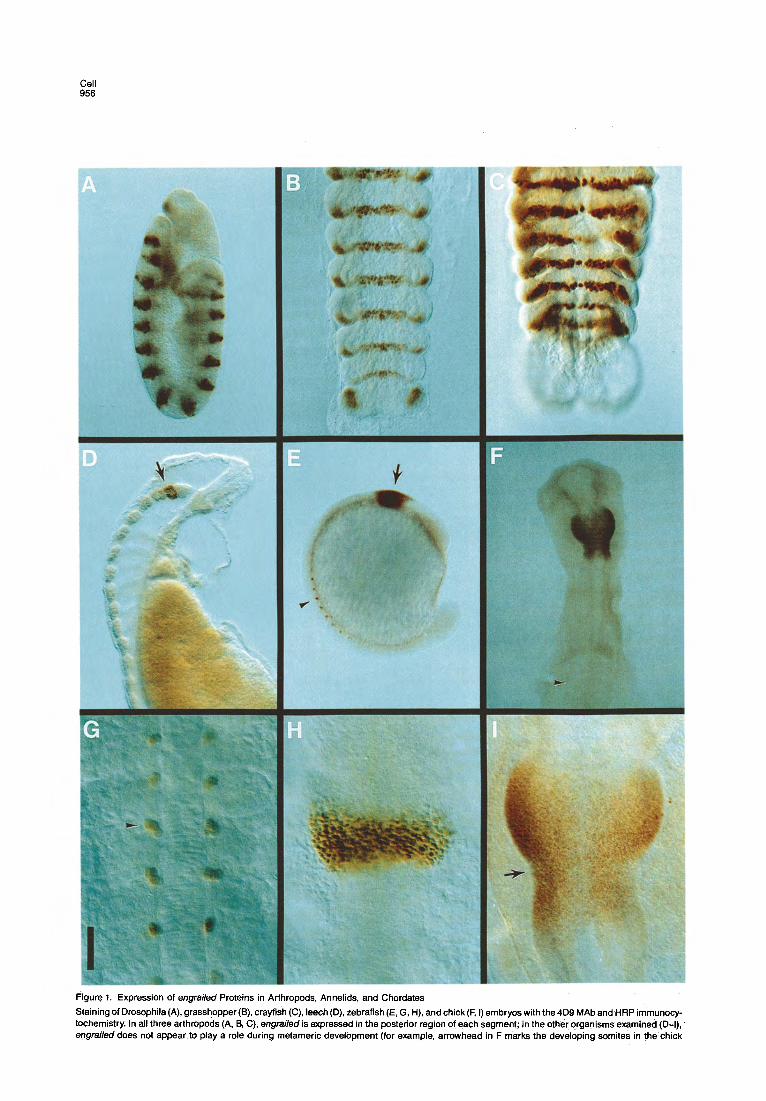
Drosophila en LSSF.l&LNF.AQIKI Drosophila inv --G----------- chicken +Q----S--- Leech --K--p--S---- Zebra Fish +Q----S--- Grasshopper -~----------
Mouse-1,2; Human -AQ-s---s---- sea urchin -~--FJ+S-S---
D. Peptides
prevents it. Indeed, a synthetic peptide representing res- idues 36-50, but in which serine substitutes for glycine at position 40, fails to block MAb 4D9 binding to Drosophila embryos. We conclude that the MAb 4D9 epitope is in the 11 residues 38-48 (Figure 2).
It is interesting to note that residues 38-48 of the homeodomain include a region that has sequence and structural homology (Laughon and Scott, 1984; Otting et al., 1988) to the DNA-binding portion of several bacterial proteins. These comparative studies suggest that the MAb 4D9 epitope is a turn region of a helix-turn-helix structure that directly interacts with DNA. Consistent with the hypothesis that this portion of the homeodomain mediates DNA binding, MAb 4D9 blocks association of the engrailed homeodomain peptide with DNA (E. Martin and T. B. Kornberg, unpublished data).
In summary, the evidence that the MAb 4D9 specifically recognizes engrailed protein is as follows. First, the MAb
Figure 2. Localization of the MAb 4D9 Epitope
The structure of the Drosophila engrailed pro- tein is portrayed with the homeobox and other regions of conservation indicated (A). MAb 409 binds on Western blots to the intact protein (+), but not to protein lacking the homeodomain (-). MAb 4D9 also binds to fusion proteins (B) and to synthetic peptides (D) containing resi- dues 36-48 of the homeodomain, and to en- grailed proteins in chicken (references denote source of sequence information for residues 36-48; Darnell et al., 1986), leech (Wedeen, Price, and Weisblat, personal communication), zebrafish (Fj&e et al., 1988), and grasshopper embryos (Pate1 et al., submitted) (C). MAb 4D9 does not bind to recombinant mouse (Joyner et al., 1985; Joyner and Martin, 1987) or human (Poole et al., 1989) engrailed protein, or to pro-
+ tein in sea urchin embryos (Dolecki and Hum- phreys, 1988), in which residue 40, a glycine, is instead serine or threonine. Nor does it bind to
+ a synthetic peptide (D), in which residue 40, a glycine, is instead serine.
+
+
4D9 staining pattern in Drosophila is consistent with previ- ous data: it is nuclear and includes only those cells known to express engrailed. Second, MAb 4D9 recognizes the protein product of the Drosophila engrailed and invected genes and a chicken engrailed gene when expressed in E. coli. Third, the MAb 4D9 epitope is in a variable region of the homeodomain that is unique to the known engrailed genes. We have obtained no evidence that it cross-reacts with homeodomain proteins other than those with se- quence homology to engrailed.
MAb 4D9 Recognizes the Grasshopper engrailed Protein We used MAb 4D9 to screen a grasshopper (Schistocerca americana) embryo hgtll cDNA library. Five antibody- positive plaques were isolated from a screen of 4.0 x lo5 recombinants. All five phages contain inserts that hybrid- ized at moderate str ingency to a Drosophila ‘engrailed
Evolution of engrailed Expression 959
homeobox probe. The largest cDNA was 2.6 kb and con- sisted of two EcoRl fragments of 1.1 and 1.5 kb. Hybridiza- tion experiments using a Drosophila engrailed homeobox probe indicated that an engrailed-type homeobox was present in the 1.1 kb fragment. To confirm the identity of the cDNA, we carried out tissue in situ hybridization analy- sis and sequenced the homeobox region of the cDNA.
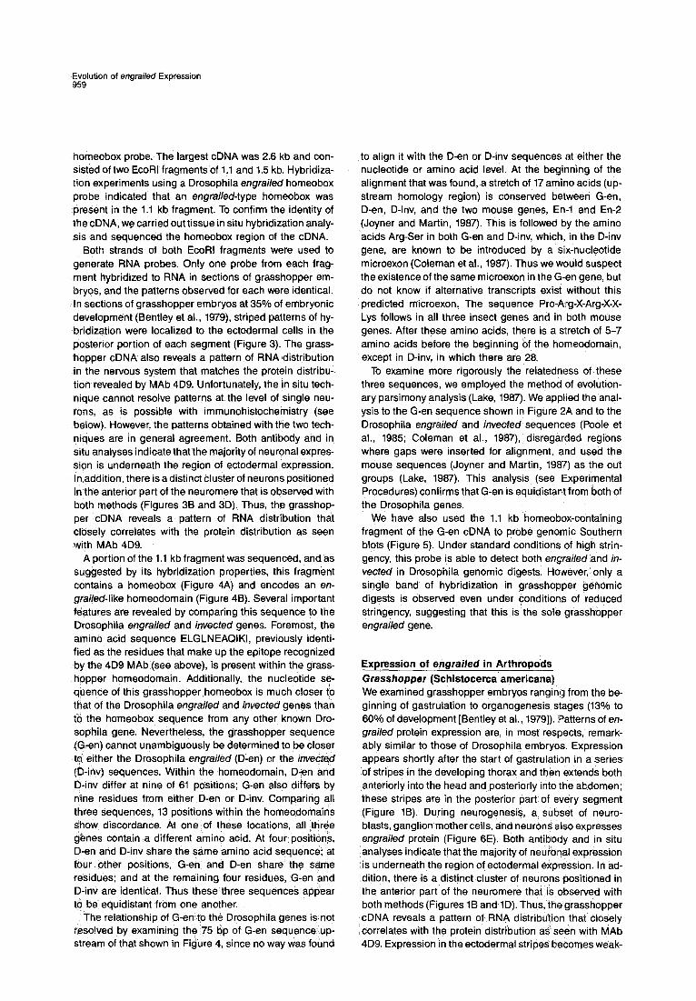
Both strands of both EcoRl fragments were used to generate RNA probes. Only one probe from each frag- ment hybridized to RNA in sections of grasshopper em- bryos, and the patterns observed for each were identical. In sections of grasshopper embryos at 35% of embryonic development (Bentley et al., 1979), striped patterns of hy- bridization were localized to the ectodermal cells in the posterior portion of each segment (Figure 3). The grass- hopper cDNA also reveals a pattern of RNA distribution in the nervous system that matches the protein distribu- tion revealed by MAb 4D9. Unfortunately, the in situ tech- nique cannot resolve patterns at the level of single neu- rons, as is possible with immunohistochemistry (see below). However, the patterns obtained with the two tech- niques are in general agreement. Both antibody and in situ analyses indicate that the majority of neuronal expres- sion is underneath the region of ectodermal expression. In addition, there is a distinct cluster of neurons positioned in the anterior part of the neuromere that is observed with both methods (Figures 36 and 3D). Thus, the grasshop- per cDNA reveals a pattern of RNA distribution thit closely correlates with the protein distribution as seen with MAb 4D9.
A portion of the 1.1 kb fragment was sequenced, and as suggested by its hybridization properties, this fragment contains a homeobox (Figure 4A) and encodes an en- grailed-like homeodomain (Figure 48). Several important features are revealed by comparing this sequence to the Drosophila engrailed and invected genes. Foremost, the ainino acid sequence ELGLNEAQIKI, previously identi- fied as the residues that make up the epitope recognized by the 4D9 MAb (see above), is present within the grass- hopper homeodomain. Additionally, the nucleotide se- quence of this grasshopper homeobox is much closer io that of the Drosophila engrailed and invected genes than to the homeobox sequence from any other known Dro- sbphila gene. Nevertheless, the grasshopper sequence (G-en) cannot unambiguously be determined to be closer to either the Drosophila engrailed (D-en) or the invedted (D-inv) sequences. Within the homeodomain, D-en aed D-inv differ at nine of 61 positions; G-en also differs by riine residues from either D-en or D-inv. Comparing all three sequences, 13 positions within the homeodomains s’how discordance. At one of these locations, all ,three genes contain a different amino acid. At four positions, D-en and D-inv share the same amino acid sequence; at four other positions, G-en and D-en share the same &sidues; and at the remaining four residues, G-en and D-inv are identical. Thus these ‘three sequences appear to be equidistant from one another.
The relationship of G-en to the Drosophila genes is not r,esolved by examining the 75 bp of G-en sequence up- stream of that shown in Figure 4, since no way was foutid
to align it with the D-en or D-inv sequences at either the nucleotide or amino acid level. At the beginning of the alignment that was found, a stretch of 17 amino acids (up- stream homology region) is conserved between G-en, D-en, D-inv, and the two mouse genes, En-l and En-2 (Joyner and Martin, 1987). This is followed by the amino acids Arg-Ser in both G-en and D-inv, which, in the D-inv gene, are known to be introduced by a six-nucleotide microexon (Coleman et al., 1987). Thus we wauld suspect the existence of the same microexon in the G-en gene, but do not know if alternative transcripts exist without this predicted microexon. The sequence Pro-Arg-X-Arg-X-X- Lys follows in all three insect genes and in both mouse genes. After these amino acids, there is a stretch of 5-7 amino acids before the beginning of the homeodomain, except in D-inv, in which there are 28.
To examine more rigorously the relatedness of these three sequences, we employed the method of evalution- ary parsimony analysis (Lake, 1987). We applied the anal- ysis to the G-en sequence shown in Figure 2A ancl to the Drosophila engrailed and invected sequences (Poole et al., 1985; Coleman et al., 1987), disregarded regions where gaps were inserted for alignment, and used the mouse sequences (Joyner and Martin, 1987) as the out groups (Lake, 1987). This analysis (see Experimental Procedures) confirms that G-en is equidistant from both of the Drosophila genes.
We have also used the 1.1 kb homeobox-containing fragment of the G-en cDNA to probe genomic Southern blots (Figure 5). Under standard conditions of high strin- gency, this probe is able to detect both engrailed and in- vetted in Drosophila genomic digests. However, only a single band of hybridization in grasshopper genomic digests is observed even under conditions of rsduced stringency, suggesting that this is the sole grasshopper engrailed gene.
Expression of engrailed in Arthropods Grasshopper (Schistocerca americana) We examined grasshopper embryos ranging from the be- ginning of gastrulation to organogenesis stages (13% to 60% of development [Bentley et al., 19791). Patterns of en- grailed protein expression are, in most respects, remark- ably similar to those of Drosophila embryoa. Expression appears shortly after the start of gastrulation in a series of stripes in the developing thorax and then extends both anteriorly into the head and posteriorly into the abdomen; these stripes are in the posterior part of every segment (Figure 1B). During neurogenesis, a subset of neuro- blasts, ganglion mother cells, and neurons also expresses engrailed protein (Figure 6E). Both antibody and in situ analyses indicate that the majority of neuronal expression is underneath the region of ectodermal expression. In ad- dition, there is a distinct cluster of neurons positioned in the anterior part of the neuromere that is (observed with both methods (Figures 1B and 1D). Thus, the grasshopper cDNA reveals a pattern of RNA distribution that closely correlates with the protein distribution as seen with MAb 4D9. Expression in the ectodermal striped becomes weak-

Cell 960

Evolution of engrailed Expression 961
Figure 4. Partial Nucleotide and Amino Acid Sequence of the Grasshopper engrailed Gene
The nucleotide sequence of the grasshopper engrailed gene (G-en) is shown in (A), and the deduced amino acid sequence is shown in (B). Dashes in the G-en nucleotide sequence in- troduce a gap to allow for afignment to the amino acid sequence shown ini (6). The ho- meobox is underlined. The deduced amino acid sequence of G-en and its alignment to Drosophila engrailed (D-en) and invected (D-inv) (Poole et al., 1965; Coleman et al., 1967) is shown in (6). The alignment is based in part on the published alignment of En-l and En-2 to engrailed and iwected (Joyner and Martin, 1967). Sets of three asterisks denote positions of amino acid identity between the G-en se- quence and the Drosophila errgraikd or in- vecredsequences. The dashes in the Drosoph- ila engrailed sequence coincide with the amino acids introduced by the microexons of G-en and invected. Dashes in the G-en sequence allow for alignment of both the upstream ho- mology region and the homeodomain. The
brackets in the invected sequence denote 22 amino acids that are not shown. The homeodomain is underlined in all three sequences, The three homeodomains differ by nine residues when analyzed in any pairwise combination. Comparing the three sequences at once, there a,re 13 residues that show mismatch. One of these (amino acid 37 of the homeodomain) is different in all three genes, Four (positions 22, 34, 36, and 60) are the same in D-en and D-inv, but different in G-en. Another four (positions 1, 11, 19, and 61) are the same in G-en and D-inv, but different in D-en. At the remaining four positions (2, 12, 30, and 57) G-en and D-en are the same, but D-inv is different.
er after about 35% of development, but is still detectable at all stages examined.
The principal difference between the patterns of en- giailedexpression in Drosophila and in grasshopper is the order in which the segmental stripes appear. In Drosoph- ila, the stripes appear in an order characterized by a rapid anterior-to-posterior gradient in which even-numbered stripes appear slightly before odd-numbered stripes (Fig- ure 6A). In grasshopper, segments are added with a rostral-caudal polarity and the stripes of engrailed expres- sion follow, forming one at a time. In grasshopper, no tran- sient pair-rule patterns appear (compare Figures 6A and 68). (For further details, see Pate1 et al., 1989.) Crayfish and Lobster (Procambarus clarki and Hbmarus americanus) Crayfish embryos were examined from the time of early abdominal segmentation through the period of neurogen- esis (equivalent to 25% through 45% of grasshopper de- velopment). As in grasshopper, the abdominal stripes of ehgrailed protein are added one at a time (Figure 6C) and correspond to the posterior part of each segment (see Fig- ure 1C). During neurogenesis, engrailed expression is al- most indistinguishable from that in grasshopper (Figure 6P) and includes a similar set of neuroblasts and appar- ently homologous neurons.
Lobster embryos were examined at only one stage of
embryonic development (equivalent to about 75% of grasshopper development). These embryos revealed ec- todermal stripes in the posterior part of each segment and a pattern of neuronal expression that was nearly identical with the pattern observed in the oldest crayfish embryos.
Expression of engrailed in Annelids Leech (Helobdella triserialis) We first observe staining with the 4D9 MAb in leech em- bryos during the phase of teloblast divisions (Ibetween late stage 7 and early stage 8, Stent et al., 1982). At this stage, engrailed expression is confined to the progeny of one of the teloblast lineages. Not all of the progeny of this partic- ular teloblast are engrailed-positive, but rather only a small group of progeny in a continuous band appear transiently to express engrailed protein. At late stage 8, by which time the two germinal bands have come together, expression is in two rows of bilaterally symmetric cells; thlese rows are positioned at the dorsal edge of the germinal plate. There is one 4D9-positive cell per hemisegment, and there are about 12 segments containing these stained cells. At this stage there is also a series of large, flat nuclei positioned around the yolk sac that also stain with the 4D9 MAb.
Nervous system expression of engrailed in the leech be- gins at early stage 9 and appears first in the subset of nuclei in the second suboesophageal ganglion. By late
Figure 3. Tissue In Situ Hybridization Analysis of the Grasshopper engrailed Gene
Double exposure bright-field and dark-field photographs of frontal (A, B, C) and parasagittal (D) sections of 35% grasshopper embryos. Anterior is up in’all panels. Sections were hybridized with an RNA probe from the 1.5 kb non-homeobox-containing EcoRl fragment of the G-en cDNA. Position of the second thoracic leg is indicated in (6) and (C). Hybridization (revealed by accumulation of silver grains that appear red in these photographs) is clearly locafized to the posterior region of each segment in the abdomen (A) and extends along the posterior margin of each leg (C). Hybridization is also seen in the posterior portion of each neuromere (B, D), and a cluster of neurons in the anterior region also contains the transcript (arrows in B and D). These patterns closely match those seen with the 4D9 MAb. Scale bar: (A, B) 135 nm; (C) 110 pm; (D) 40 pm.

Cell 962
21.7,
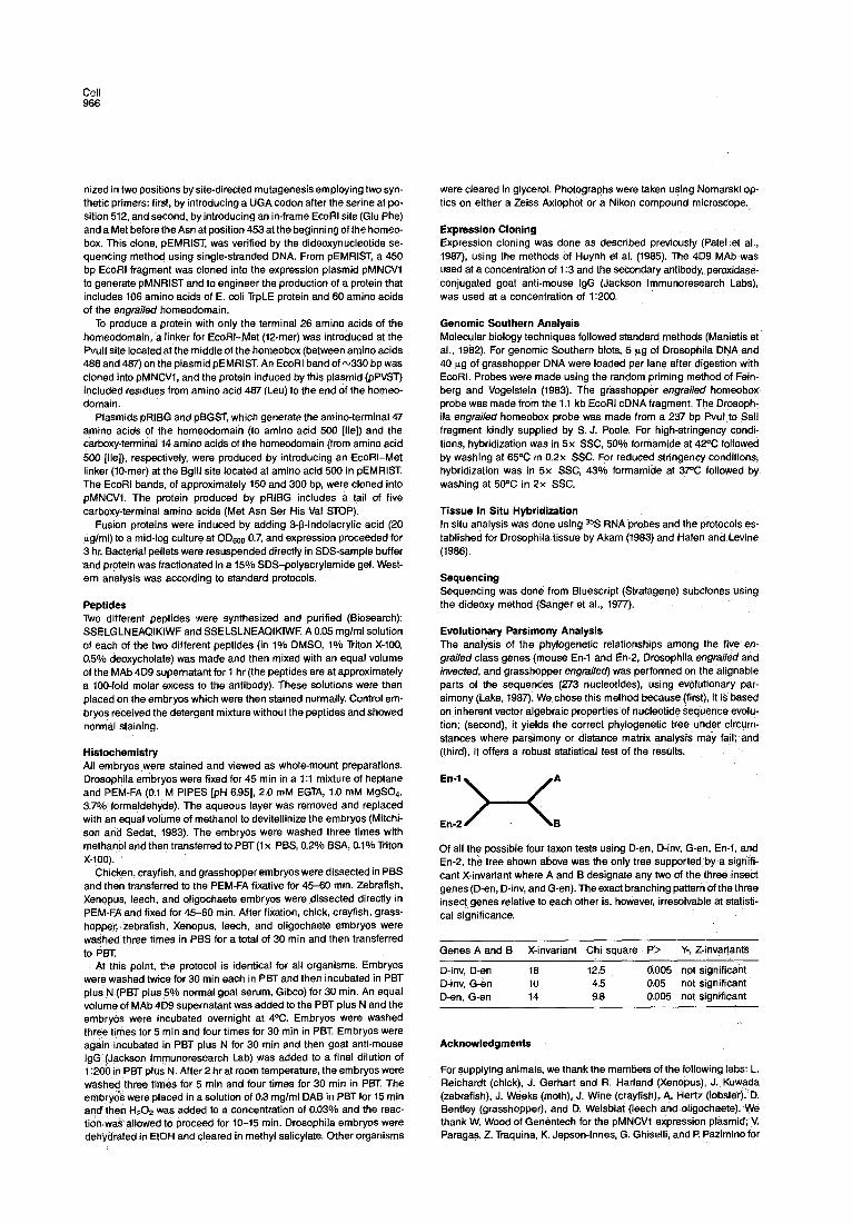
Figure 5. Grasshopper Has Only One errgrailed Gene
Genomic southern blots of Drosophila (lanes 1 and 2) and grasshopper (lanes 3, 4, and 5) DNA digested with EcoRl (lanes 1, 2, 4, and 5) or Hindlll (lane 3) and hybridized with a Drosophila engrailed homeobox probe (lane 1) or a grasshopper engrailed homeobox probe (lanes 2-5) at high (lanes l-4) or reduced (lane 5) stringency. In lane 1, a Drosoph- ila engrai/ed probe reveals EcoRl bands at 4.0 and 1.1 kb that cor- respond to the invected and engrailed genes, respectively. The grass- hopper engrailed homeobox probe hybridizes to these same two Drosophila genes even under high-stringency conditions (lane 2). When grasshopper genomic DNA is probed with the grasshopper en- grailed homeobox probe under high-stringency conditions, hybridiza- tion is seen to a single 5.9 kb band in a Hindlll digest (lane 3) a single 4.8 kb band in an EcoRl digest (lane 4) and a single 5.5 kb band in a Sal1 digest (not shown). No additional bands are seen in any of the digests under reduced-stringency conditions. lane 5 shows one such example: a single 4.8 kb band is present in a grasshopper EcoRl genomic digest hybridized with the grasshopper engrailed homeobox probe at reduced stringency. These conditions (see Experimental Procedures) are sufficiently low to allow one Antennapedia class homeobox to detect other Antennapedia class homeoboxes (McGinnis et al., 1984). Arrowheads mark positions of various DNA size standards (given in kb).
stage 9, most, if not all, nuclei in the second suboesopha- geal ganglion stain with MAb 4D9 and, in addition, there are also a number of positive nuclei in the anterior region of the third suboesophageal ganglion and four stained nuclei in the fourth suboesophageal ganglion (Figure 1D). There are also strongly stained nuclei extending ventrally from the second suboesophageal ganglion to the ecto- derm and occasionally a 4D9-positive nucleus in the ec- todermal layer. At this stage, staining at the dorsal edges of the germinal plate forms a series of arcs of weakly posi- tive cells extending out dorsally. Oligochaeta (Eisenia foetida) We examined oligochaete embryos during the period of
neurogenesis; this period corresponds to stage 9 of leech development. In this annelid, however, the pattern of MAb 4D9 staining is quite different from that observed in the leech. Expression of engrailed occurs in a segmentally reiterated pattern of neuronal nuclei in the developing central nervous system (data not shown). This is in marked contrast to the regionalized pattern observed in the central nervous system of leech embryos at the same stage.
Expression of engrailed in Chordates Zebrafish (Brachydanio rerio) Zebrafish embryos were examined from a period before gastrulation until formation of the most posterior somites. As in the other chordates (see below), expression was first observed when the neural tube forms (Figure 1E) in the posterior mesencephalon/anterior metencephalon (Fig- ure 1H). As development proceeds, a deep indentation separates these two regions of the central nervous sys- tem, and expression clearly extends across this morpho- logical boundary.
A short time after somitogenesis begins, additional staining was observed in three to four nuclei in each so- mite (Figure 1G). These nuclei are positioned in the most ventral and anterior parts of the somite and close to the spinal cord. No expression in the somites is present prior to or during the time that the somites become delineated. Rather, as the wave of somitogenesis proceeds posteri- orly, staining lags about four to five somites behind the most newly formed somite. It appears that these stained nuclei correspond to a particular set of muscle cells (C. Kimmel, personal communication). A second wave of ad- ditional somite staining follows this original somite expres- sion, and lags behind the first by about 10 somites. These additional lo-15 nuclei are stained less intensely than the original 3-4 nuclei and are loosely packed around the de- veloping spinal cord. These nucletare probably a subset of the sclerotome. The nervous system staining in the posterior mesencephalon/anterior metencephalon is main- tained through the oldest embryos examined. Frog (Xeno,pus laevis) Xenopus embryos were examined from the early gastrula period (stage 10; Nieuwkoop and Faber, 1956) to near ‘the end of somitogenesis (stage 30). At~stage 20, immediately following fusion of the neural folds, MAb 409 staining was observed on the dorsal surface of a region of the neural tube (data not shown). As in zebrafish, this region corre- sponds to the posterior mesencephalonlanterior meten- cephal,on. As more nuclei begin to express engrailed pro- tein, ktaining also extends ventratly around the neural tube. k stage 2.5 we noted a,few positive nuclei far anterior in the reg,ion of .the prosencephalon. Staining in the posteiior mesencephalon/anterior metencephalon per- sisted tfirough the oldest stages examined, and no ex- pression was observed in the somites or spinal cord. Chick (Gallirs doinesticus) Chilc~ken’embryos were examined from stage 7 (one So- mite;, Hamburger and Hamilton, 1951) to stage 15 (25 SO- mites). ‘We did not observe any MAb 4D9 staining until ap- proximately stage 9. At this time, just prior to the fusion of

Evolution of engrailed Expression 963
Figure 6. Expression of engrailed Proteins during Arthropod Segmentation and Neurogenesis
Expression of engrailed proteins during segmentation in Drosophila (A), grasshopper (B), and crayfish (C), and during neurogenlssis in Drosophila (D), grasshopper(E), and crayfish (F), as revealed by the 4D9 MAb and HRP immunocytochemistry. (A) This cellular blastoderm has virtually com- pleted the formation of segmentally reiterated stripes, although the formation of odd-numbered stripes lags behind the even-numbered stripes (arrow- heads). (B, C) The grasshopper and crayfish embryos, however, generate engrailed stripes one at a time (arrowheads) as their germ bands extend caudally. In grasshopper and crayfish, the abdominal stripes form without apparent pair-rule patterning. (E, F) During the period of neurogenesis, ,a subset of neuroblasts, ganglion mother cells, and neurons expressed engrailed protein; this pattern appears quite similar in Drosophila (A), grass- hopper (B), and crayfish (C). Calibration bar: (A) 90 urn; (B) 200 pm; (C) 110 urn; (D) 40 urn; (E) 50 urn; (F) 40 urn,
the neural folds, engrailed expression appears in a scat- mesencephalon and anterior metencephalon (Figure 1F). tering of nuclei on the dorsal surface of a rostra1 region of MAb 4D9 staining begins dorsally, but moves ventrally at the neural tube. As development progresses, increasing about the time of cranial flexure. The borders of the MAb numbers of nuclei in this region stain, and by stage 11, it 4D9 staining are well defined and clearly extend to both is clear that the region of expression is the posterior sides of the mesencephalonlmetencephafon invagination

(Figure II). There is also a region of stained nuclei at the dorsal surface of the neural tube extending posteriorly through the metencephalon. The pattern of expression of engrailed proteins in the developing chick neural tube is therefore generally similar to both zebrafish and Xenopus although, in contrast to zebrafish, no staining was de- tected in the chick somites.
Discussion
Analysis of pattern formation in the Drosophila embryo has led to the discovery of a hierarchy of genes that con- trols the generation and specification of the body seg- ments. Two aspects of these studies are relevant here. First, the regions in which these segmentation genes are expressed in the embryo correlate in most cases with the areas in which their functions are required. Therefore, in Drosophila, it is clear that their patterns of expression are intimately related to their functions. Second, many of these genes encode proteins that have homeodomain se- quences, and similar homeobox genes have been iso- lated in vertebrates. The patterns of expression of some of these vertebrate proteins have been described, and the presence of a homeodomain suggests a function in tran- scriptional regulation. For many of these proteins, how- ever, their developmental roles and their relationships to the Drosophila homeodomain proteins are unclear. Given the extraordinary sequence conservation among these regulatory proteins, it is important to determine how their functions and roles are related, and to understand how they evolved. Fortunately, the expression and evolution of one of these segmentation genes, engrailed, can be read- ily studied.
The MAb 4D9 binds to a conserved region in the ho- meodomain of the engrailed protein and recognizes this epitope in animals from many phyla (Figure 1). These in- clude Drosophila, a more primitive insect (grasshopper), and two crustaceans (crayfish and lobster), annelids from two different groups (a leech and an oligochaete), and chordates from three different groups: teleost fish (zebra- fish), amphibians (Xenopus), and birds (chick). Chordates and arthropods are thought to have diverged over 600 mil- lion years ago, yet engrailed genes are present in both phyla and their protein products are recognized by MAb 4D9.
Evidence that MAb 4D9 recognizes engrailed proteins in these organisms is strong. Its epitope is unique to en- grailed homeodomains. It stains Drosophila embryos in a manner consistent with the known patterns of engrailed expression, indicating that it binds selectively to Drosoph- ila, engrailed proteins. Furthermore, it has been used to isolate a grasshopper engrailed gene and, moreover, its staining patterns in grasshopper and other arthropods are similar to Drosophila. The antibody recognizes the protein product of the Drosophila engrailed and invected genes and a chicken engrailed gene when expressed in E. coli. MAb 4D9 also stains chicken, zebrafish, and frog with pat- ter,ns that have significantly greater resolution than, but are consistent with, patterns of expression previously de- tedted by in situ hybridization of zebrafish (Njelstad and
Fjke, 1988), chicken (Gardner et al., 1988) and mouse (Davis et al., 1988; Davidson et al., 1988; Davis and Joyner, 1988). As noted in the Introduction, conservation of a sequence of almost 100 amino acid residues in and around the homeodomain of engrailedgenes is an indica- tion of the functional relatedness of their protein products. This extended sequence similarity justifies their common designation as engrailedgenes and suggests that they are true evolutionary homologs. MAb 409 is therefore a probe that can be used specifically to detect engrailed proteins in distantly related animals.
While the evolutionary conservation of the engrailed homeodomain indicates a conserved biochemical func- tion as a transcriptional regulator, the patterns of expres- sion of engrailed protein in different animals suggests that engrailed has several developmental roles, only some of which have been conserved. Within the arthropods we ex- amined the pattern is consistent: engrailed is expressed in the posterior portion of each metamere during segmen- tation and in a segmentally reiterated subset of cells dur- ing neurogenesis. Interestingly, no other organism exam- ined expresses engrailed in developing metameres. MI do share one feature, namely, expression during neurogene- sis. This has important implications for comparison of metameric development in different phylogenetic line- ages and for determination of how, during their evolu- tion, different developmental programs utilized regulatory genes such as engrailed.
In young leech embryos, transient patterns of expres- sion in the progeny of a particular teloblast lineage are fol- lowed later by expression in the developing nervous sys- tem. Neuronal expression is regional (within only a few anterior segmental ganglia) and is not segmentally reiter- ated. In contrast, engrailed is expressed in the developing nervous system of an oligochaete in a segmentally reiter- ates subset of neurons. Therefore, these annelids feature engrailed expression in the developing nervous system in quite different patterns.
The three chordates examined in this study (zebrafish, Xenopus, and chick) have similar patterns: engrailedis ex- pressed in a specific region of the developing neural tube (posterior midbrain and anterior hindbrain), but~not in de- veloping metameres. This pattern of expression is remi- niscent of the leech patterns, and recalls the regional expression of homeotic genes in the developing Drosoph- ila nervous system (reviewed by Doe and Scott, 1988).
In zebrafish, engrailedis expressed in a region of the de- veloping neural tube and, a short time after somitogene- sis begins, in three to four nuclei in each somite. However, it /s likely that engrailed is not involved in the process of segmentation in zebrafish, because the expression of tin- grailed in the somites occurs after the delineation of the somites. Thus, in addition to its role in neurogenesis, en- grailed may also be involved in specifying the fate of a seg- mentally reiterated subset of mesodermal cells. When co,mparing the patterns of expression of different animals, it is relevant that many organisms have two ;engraihd genes, and MAb 4D9 may not recognize the products of both genes. For instance, MAb 4D9-positive mesodermal cells seen in zebrafish were not seen in Xenopus or chick,

Evolution of engrailed Expression 965
and this may reflect recognition by MAb 4D9 for the prod- ucts of only one of the two engrailed genes in chick and Xenopus and both engrailed genes in zebrafish.
Clearly, different phyla have different patterns of en- grailed expression. If we can infer function from pattern of expression, then it is only in the arthropods that engrailed plays a major role during the process of segmentation. It is possible, of course, that the engrailed function was ini- tially involved in chordate metamerization, but that this role was lost during evolution. However, the most par- simonious hypothesis to account for these staining pat- terns is that engrailed was never involved in chordate metamerization, and that an ancestral function of en- grailedwas to specify cell fate within the nervous system. Some present groups of organisms appear to use en- grailed to specify a region of the developing nervous sys- tem (e.g., in the leech and in chordates), whereas other or- ganisms appear to use engrailed to specify a reiterated subset of neurons within the developing nervous system (e.g., in an oligochaete and in arthropods).
The differences between the patterns of engrailed ex- pression in the annelids and in arthropods lead to the con- clusion that arthropods are not closely related to annelids. This argues against the popular notion that metamerism in arthropods evolved from annelids (Anderson, 1973). Our data are consistent with the evolutionary analysis of the sequences of 18s rRNA (Field et al., 1988), which also suggested that arthropods and annelids are not closely related. Moreover, our data help to clarify further another evolutionary question: the relationship of crustaceans and insects. Previous work on the pattern of neuronal cells (Thomas et al., 1984), on embryology (Weygolt, 1979), and on comparative anatomy (Boudreaux, 1979) of crusta- cea?s and insects suggested that these two groups of ar- thropods share a relatively recent common ancestor. The similar pattern of engrailed expression in these two classes is consistent with this hypothesis. This is in con- trast to the arguments of others (Anderson, 1979, 1982; Manton, 1977; Schramm, 1986), who asserted that crusta- ceans and insects represent unrelated groups sharing convergence toward “arthropodizationl’
Our analysis of a grasshopper engrailed gene has in- teresting implications with respect to the evolution of the engiailed gene. Whereas Drosophila contains two en- grailed genes, engrailed and invected (Coleman et al., 1987), only a single gene was found in grasshopper, and analysis of its sequence indicates that it is equally~relate$ to the two Drosophila genes. This arrangement may have be+ .g‘enerated in either of two ways. First, the two Dro- sophila genes may have arisen by duplication some time in the insect lineage after the last ancestor common to both Drosophila and grasshopper. This hypothesis sug- gests that the two genes in mouse (En-l and En-2;‘Joyner and Martin, 1987), chicken (Darnell et al., 198&), zebrafish (Fjase @t al., 1988), and human (Poole et al., 1989;’ Logan et al., 1989) also arose as an independent duplic,&ion of an anc$stral engrailed gene early in the chordate lineage. This is’ supported by the observation that, thei:mouse g&ies are m”ore similar to each other than either’ is to the Drosophila engrailedor ihected genes. The same’conclu-
sion has been suggested by Dolecki and Humphreys (1986), after finding a lone engrailed gene in sea urchin. The second possibility is that the duplication occurred be- fore the split of the lines leading to Drosophila and grass- hopper, and then one of the genes was latlsr lost in the grasshopper lineage. The lone grasshopper gene may ap- pear to be equidistant from the two Drosoptnila genes be- cause of certain functional constraints on thle G-en gene product.
Sequence analysis has also been used Irecently as a way of addressing some of the unresolved questions of phylogenetic relationships, and thus of supplementing previous morphological, embryological, and paleontologi- cal data(e.g., Field et al., 1988). The phylogenetic patterns of expression of regulatory genes such as engrailed pres- ent another way of using molecular data to examine phylogenetic relationships. Previous studies have exam- ined slow changes in highly conserved, hiouse-keeping molecules like 18s rRNA (Field et al., 1986). In contrast, the engrailedgene is a regulatory gene whose patterns of expression are conserved within groups of closely related organisms, and yet vary dramatically between more dis- tantly related groups. The nature of these differences may reflect the evolutionary radiation of differenl phylogenetic lines.
In summary, our results suggest that engrailed is a gene that predates the divergence of arthropods, annelids, and chordates; had an ancestral function controlling cell fate during neurogenesis; and was co-opted for ,the process of segmentation during the evolution of the arthropods but not the annelids and chordates. Furthermore, the data support the hypothesis that metameric development evolved independently enough in these three different phyletic lines (annelids, arthropods, and chordates) that a regulatory gene that plays a crucial role during segmenta- tion in one phylum (engrailed in arthropods) does not ap- pear to play the same role in the bther twlo.
Experimental Procedures
Generation of Monoclonal Antibodies Full-length engrailed protein and the C-terminal two-thirds of the in- vecfed protein were generated in E. coli with the.T7 polymerase ex- pression system (Studier and Moffatt, 1986). BALWc mice were im- munized with either protein by intraperitoneal injections; each injection contained about 100 pg of either engrailedor invected protein. Primary injections (300 ~1) consisted of protein suspended in 150 ~1 PBS and 150 VI complete adjuvant. Mice were given three boosts of ‘100 pg pro- tein in incomplete adjuvant at approximately 2 week intervals. Three days before fusion, a final injection of 50 pg was administered. Spleen cells were fused with NS-1 myeloma cells (Kohler a.nd Milstein, 1975; Oi and Herzenberg, 1980). Hybridoma supernatants were screened on Schneider 2 cell lines that express either engrailed or invected protein under HSWO promoter control (Gay et al., 1988), and hybridoma lines of interest were isolated and recloned by single-cell cloning. The MAb 4D9 was derived from a mouse injected with invecteldprotein; the MAb 4Fll was derived from a mouse injected with engrwled protein. The 4D9-producing line has been deposited with the A,merican Type Cul- ture Collection and is available on request.
Fusion Proteins The homeoboxfusion protein clone (pMNRIST) was produced from the plasmid pE18 BH, which contains DNA from the BamHl site at nucleo- tide 1405 to the EcoRl site at nucleotide 2017 of tha clone C2.1 (Poole et al., 1985). The BamHI-EcoRI fragment was sequentially mutage-
Cell 966
nized in two positions by site-directed mutagenesis employing two syn- thetic primers: first, by introducing a UGA codon after the serine at po- sition 512, and second, by introducing an in-frame EcoRl site (Glu Phe) and a Met before the Asn at position 453 at the beginning of the homeo- box. This clone, pEMRIST, was verified by the dideoxynucleotide se- quencing method using single-stranded DNA. From pEMRIST, a 450 bp EcoRl fragment was cloned into the expression plasmid pMNCV1 to generate pMNRlST and to engineer the production of a protein that includes 106 amino acids of E. coli TrpLE protein and 60 amino acids of the engrailed homeodomain.
To produce a protein with only the terminal 26 amino acids of the homeodomain, a linker for EcoRI-Met (12-mer) was introduced at the Pvull site located at the middle of the homeobox (between amino acids 466 and 487) on the plasmid pEMRIST An EcoRl band of ~330 bp was cloned into pMNCV1, and the protein induced by this plasmid (pPVST) included residues from amino acid 487 (Leu) to the end of the homeo- domain.
Plasmids pRlBG and pBGST, which generate the amino-terminal 47 amino acids of the homeodomain (to amino acid 500 [lie]) and the carboxy-terminal 14 amino acids of the homeodomain (from amino acid 500 [lie]), respectively, were produced by introducing an EcoRI-Met linker (lo-mer) at the Bglll site located at amino acid 500 in pEMRIST The EcoRl bands, of approximately 150 and 300 bp, were cloned into pMNCV1. The protein produced by pRlEG includes a tail of five carboxy-terminal amino acids (Met Asn Ser His Val STOP).
Fusion proteins were induced by adding 3-6-lndolacrylic acid (20 uglml) to a mid-log culture at OD so0 0.7, and expression proceeded for 3 hr. Bacterial pellets were resuspended directly in SDS-sample buffer and protein was fractionated in a 15% SDS-polyacrylamide gel. West- ern analysis was according to standard protocols.
Peptides Two different peptides were synthesized and purified (Biosearch): SSELGLNEAQIKIWF and SSELSLNEAQIKIWF. A 0.05 mglml solution of each of the two different peptides (in 1% DMSO, 1% Briton X-100, 0.5% deoxycholate) was made and then mixed with an equal volume of the MAb 4D9 supernatant for 1 hr (the peptides are at approximately a lOO-fold molar excess to the antibody). These solutions were then placed on the embryos which were then stained normally. Control em- bryos received the detergent mixture without the peptides and showed normal staining.
Histochemistry All embryos were stained and viewed as whole-mount preparations. Drosophila embryos were fixed for 45 min in a 1 :I mixture of heptane and PEM-FA (0.1 M PIPES [pH 6.951, 2.0 mM EGTA, 1.0 mM MgS04, 3.7% formaldehyde). The aqueous layer was removed and replaced with an equal volume of methanol to devitellinize the embryos (Mitchi- son and Sedat, 1983). The embryos were washed three times with methanol and then transferred to PBT (lx PBS, 0.2% BSA, 0.1% Triton X-100).
Chicken, crayfish, and grasshopper embryos were dissected in PBS and then transferred to the PEM-FA fixative for 45-60 min. Zebrafish, Xenopus, leech, and oligochaete embryos were dissected directly in PEM-FA and fixed for 45-80 min. After fixation, chick, crayfish, grass- hopper, zebrafish, Xenopus, leech, and oligochaete embryos were washed three times in PBS for a total of 30 min and then transferred to PET
At this point, the protocol is identical for all organisms. Embryos were washed twice for 30 min each in PBT and then incubated in PBT plus N (PBT plus 5% normal goat serum, Gibco) for 30 min. An equal volume of MAb 4D9 supernatant was added to the PBT plus N and the embryos were incubated overnight at 4OC. Embryos were washed three times for 5 min and four times for 30 min in PBT. Embryos were again incubated in PBT plus N for 30 min and then goat anti-mouse IgG (Jackson lmmunoresearch Lab) was added to a final dilution of 1:200 in PBT plus N. After 2 hr at room temperature, the embryos were washed three times for 5 min and four times for 30 min in PBT The embryos were placed in a solution of 0.3 mglml DAB in PBT for 15 min and then H202 was added to a concentration of 0.03% and the reac- tion was allowed to proceed for IO-15 min. Drosophila embryos were dehydrated in EtOH and cleared in methyl salicylate. Other organisms
were cleared in glycerol. Photographs were taken using Nomarski op- tics on either a Zeiss Axiophot or a Nikon compound microscope.
Expreosion Cloning Expression cloning was done as described previously (Pate1 et al., 1987) using the methods of Huynh et al. (1985). The 4D9 MAb was used at a concentration of I:3 and the secondary antibody, peroxidase conjugated goat anti-mouse IgG (Jackson lmmunoresearch Labs), was used at a concentration of 1:200.
Genomic Southern Analysis Molecular biology techniques followed standard methods (Maniatis et al., 1982). For genomic Southern blots, 5 ug of Drosophila DNA and 40 ug of grasshopper DNA were loaded per lane after digestion with EcoRI. Probes were made using the random priming method of Fein- berg and Vogelstein (1963). The grasshopper engrailed homeobox probe was made from the 1.1 kb EcoRl cDNA fragment. The Drosoph- ila engrailed homeobox probe was made from a 237 bp Pvul to Sall fragment kindly supplied by S. J. Poole. For high-stringency condi- tions, hybridization was in 5x SSC, 50% formamide at 42°C followed by washing at 65OC in 0.2x SSC. For reduced stringency conditions, hybridization was in 5x SSC, 43% formamide at 3PC followed by washing at 50°C in 2x SSC.
Tissue In Situ Hybridization In situ analysis was done using 35S RNA probes and the protocols es- tablished for Drosophila tissue by Akam (1983) and Hafen and Levine (1986).
Sequencing Sequencing was done from Bluescript (Stratagene) subclones using the dideoxy method (Sanger et al., 1977).
Evolutionary Parsimony Analysis The analysis of the phylogenetic relationships among the five en- grailed class genes (mouse En-l and En-2, Drosophila engrailed and invected, and grasshopper engrailed) was performed on the alignable parts of the sequences (273 nucleotides), using evolutionary par- simony (Lake, 1987). We chose this method because (first), it is based on inherent vector algebraic properties of nucleotide sequence evolu- tion; (second), it yields the correct phylogenetic tree under circum- stances where parsimony or distance matrix analysis may fail; and (third), it offers a robust statistical test of the results.
En-l A
En-2 B
Of all the possible four taxon tests using D-en, D-inv, G-en, En-l, and En-2, the tree shown above was the only tree supported by a signifi- cant X-invariant where A and B designate any two of the three insect genes (D-en, D-inv, and G-en). The exact branching pattern of the three insect genes relative to each other is, however, irresolvable at statisti- cal significance.
Genes A and B X-invariant Chi square P> Y-, Z-invariants
D-inv, D-en 16 12.5 0.005 not significant D-inv, G-en 10 4.5 0.05 not significant D-en, G-en 14 9.6 0.005 not significant
Acknowledgments
For supplying animals, we thank the members of the following labs: L. Reichardt (chick), J. Gerhart and R. Harland (Xenopus), J. Kuwada (zebrafish), J. Weeks (moth), J. Wine (crayfish), A. Hertz (lobster), D. Bentley (grasshopper), and D. Weisblat (leech and oligochaete). We thank W. Wood of Genentech for the pMNCV1 expression plasmid; V Paragas, 2. Traquina, K. Jepson-lnnes, G. Ghiselli, and R Pazimino for

Evolution of engrailed Expression 967
technical assistance; A. Sidow for performing the evolutionary par- simony analysis; C. Wedeen, D. Price, and A. Hemmati Brivanlou for help with dissections and interpretations of staining patterns; S. Smiley, A. Kolodkin, C. Kimmel, A. Joyner, and C. Lehner for helpful discussions; S. Smiley and K. Sander for thoughtful suggestions on the manuscript; and P Njelstad, A. Fjose, C. Wedeen, D. Price, and D. Weisblat for communication of results prior to publication. Supported by postdoctoral fellowships from the C. S. I. C. (to E. M.-B.) and the Weingart Foundation (to K. C.), by a predoctoral fellowship from the National Science Foundation (to M. C. E.), and by grants from the Na- tional Institutes of Health (to T B. K. and C. S. G.) and the Howard Hughes Medical Institute (to C. S. G.).
The costs of publication of this article were defrayed in part by the payment of page charges. This article must therefore be hereby marked “advertisement” in accordance with 18 U.S.C. Section 1734 solely to indicate this fact.
Received March 10, 1989; revised June 27, 1989.
References
Akam, M. (1983). The localization of Ultrabithorax transcripts in Dro- sophila tissue sections. EMS0 J. 2, 2075-2084.
Anderson, D. T (1973). Embryology and Phylogeny in Annelids and Ar- thropods (Oxford: Pergamon Press).
Anderson, D. T. (1979). Embryos, fate maps, and the phylogeny of ar- thropods. In Arthropod Phylogeny (San Francisco: Van Nostrand Reinhold).
Anderson, D. T. (1982). Embryology. In Embryology, Morphology, and Genetics (New York: Academic Press).
Bentley, D., Keshishian, H., Shankland, M., and Toroian-Raymond, A. (1979). Quantitative staging of embryonic development of the grass- hopper Schistocerca nitens. J. Embryol. Exp. Morphol. 54, 47-74. Boudreaux, H. B. (1979). Arthropod Phylogeny: With Special Refer- ence to Insects (New York: Wiley Interscience).
Brower, D. (1986). engrailed gene expression in Drosophila imaginal discs. EMBO J. 5, 2649-2656.
Carroll, S. B., and Scott, M. P (1985). Localization of the fushi tarazu protein during Drosophila embryogenesis. Cell 43, 47-57.
Coleman,.K. G., Poole, S. J., Weir, M. P., Soeller, W. C., and Kornberg, T. B.‘(1987). The hvecied gene of Drosophila: sequence analysis and expression studies reveal a close kinship to the engrailedgene. Genes Dev. 7, 19-28.
Darnell, D. K., Kornberg, T, and Ordahl, C. P (1986). ChickEn: a chick genomic clone with homology to the Drosophila engrailed homeo box. J. Cell Biol. 703, 311a.
Davidson, D., Graham, E., Sime, C., and Hill, R. (1988). A gene with sequence similarity to Drosophila engrailed is expressed during the development of the neural tube and vertebrae in the mouse. Develop ment 704, 365-316.
Davis, C., and Joyner, A. L. (1988). Expression patterns of the homeobox-containing genes en-7 and en-2, and the proto-oncogene int-7 diverge during mouse development. Genes Dev. 2, 1736-1744. Davis, C., NpbelTopham, S. E., Rossant, J., and Joyner, A. L. (1988). Expression cf the homeobox-containing gene En-2 delineates a spe- cific region ‘of the developing mouse brain. Genes Dev. 2, 361-371.
Desplan, C., Theis, J., and O’Farrell, P H. (1988). The sequence speci- ficity of homeodomain-DNA interaction. Cell 54, 1081-1090.
DiNardo, S., Kuner, J. M., Theis, J., and O’Farrell, P H. (1985). Develop- metit of embryonic pattern in D. melanogaster as revealed by accumu- lation of the nuclear engrailed protein. Cell 43, 59-69.
Do?, C. Q., and Scott, M. P (1988). Segmentation and homeotic action in the developing nervous system of Drosophila. Trends Neurosci. 77, 101-106.
Doe, C. Q., Hiromi, Y., Gehring, W. J., and Goodman, C. S. (1988a). Ex- pression and function of the segmentation gene fushi tarazu during Drosophila neurogenesis. Science 239, 170-175.
Doe, C. Q., Smouse, D., and Goodman, C. S. (1988b). Control of neu-
ronal fate by the Drosophila segmentation gene even-skipped. Nature 333, 376-378. Dolecki, G. J., and Humphreys, T. (1988). An engrailed class homeo box gene in sea urchins. Gene 64, 21-31.
Feinberg, A. P., and Vogelstein, B. (1983). A technique for radiolabeling DNA restriction endonuclease fragments to high specific activity. Anal. Biochem. 132, 6-13.
Field, K. G., Olsen, G. J., Lane, D. J., Giovannoni, S. J., Ghiselin, M. T., Raff, E. C., Pace, N. FL, and Raff, R. A. (1988). Molecular phy- logeny of the animal kingdom. Science 239, 748-753.
Fjbse, A., McGinnis, W., and Gehring, W. (1985). Isolation of a homeobox-containing gene from the engrailed region of Drosophila and the spatial distribution of its transcript. Nature 373, 284-289.
Fjose, A., Eiken, H. G., Njelstad, P R., Molven, A., and Hordvik, I. (1988). A zebrafish engrailed-like homeobox sequence expressed dur- ing embryogenesis. FEBS Lett. 231, 355-360.
Garcia-Bellido, A. (1975). Genetic control of wing disc development in Drosophila. In Cell Patterning (Amsterdam: Elsevier), pp. 161-182.
Gardner, C. A., Darnell, D. K., Poole, S. J., Ordahl, C. P, and Barald, K. F. (1988). Expression of an engrailed-like gene during development of the early embryonic chick nervous system. J. Neurosci. Res. 27, 426-437.
Gay, N. J., Poole, S. J., and Kornberg, T 8. (1988). The Drosophila en- grailed protein is phosphorylated by a serine-specific kinase. Nucl. Acids Res. 76, 6637-6647.
Hafen, E., and Levine, M. (1986). The localization 01 RNAs in Drosoph- i/a tissue sections by in situ hybridization. In Drosophila: A Practical Approach, D. B. Roberts, ed. (Washington, DC: IRL Press), pp. 139-157.
Hamburger, V., and Hamilton, H. L. (1951). A series’of normal stages in the development of the chick embryo. J. Morphol. 88, 49-92.
Huynh, T. V., Young, R. A., and Davis, R. W. (1965). (Constructing and screening cDNA libraries in lambda gtl0 and lambda gtll. In DNA Cloning, Volume 1, D. M. Glover, ed. (Washington, DC: IRL Press), pp. 49-78.
Ingham, P W. (1988). The molecular genetics of embryonic pattern for- mation in Drosophila. Nature 335, 25-34.
Joyner, A., and Martin, G. (1987). En-l and En-2, two mouse genes with sequence homology to the Drosophila ertgrailed gene: expression dur- ing embryogenesis. Genes Dev. 7, 29-38.
Joyner, A. L., Kornberg, T, Coleman; K. G., Cox, D. R., and Martin, G. Ft. (1985). Expression during embryogenesis of a mouse gene with sequence homology to the Drosophila engrailed gene. Cell 43, 29-37.
Karr, T L., Ali, Z., Drees, B, and Kornberg, T (1985). The engrailed lo- cus of D. melanogaster provides an essential zygotic function in precel- lular embryos. Cell 43, 591-601.
Karr, T L., Weir, M. J., Ali, Z., and Kornberg, T. (198!9). Patterns of en- grailed protein in early Drosophila embryos. Development 705, 605- 612.
Kohler, G., and Milstein, 6. (1975). Continuous cuttures of fused cells secreting antibody of predefined specificity. Nature 256, 495-497. Kornberg, T (1981). engrailed: a gene controlling compartment and segment formation in Drosophila. Proc. Nat!. Acad. Sci. USA 78, 1095-1099.
Lake, J. (1987). A rate-independent technique for analysis of nucleic acid sequences: evolutionary parsimony. Molt Biot. Evol. 4, 167-191.
Laughon, A., and Scott, M. (1964). Sequence of a Drosophila segmen- tation gene: protein structure homology with DNA-binding proteins, Nature 370, 25-31.
Lawrence, P, and Morata, G. (1976). Compartments in the wing of Dro- sophila: a study of the engrai/ed gene. Dev. Bioi. 50, 321-.337.
Lawrence, P, and Struhl, G. (1982). Further studies on the engrailed phenotype of Drosophila. EMBO J. 7, 827-833.
Logan, C., Willard, H. F., Rommens, J. M., and Joyner, A. L. (1989). Chromosomal localization of the human homeo box-containing genes En-l and En-2. Genomics 4, 206-209.
Maniatis, T., Fritsch, E. F., and Sambrook, J. (1962). IMolecular Clon-

Cell 968
ing: A Laboratory Manual (Cold Spring Harbor, New York: Cold Spring Harbor Laboratory).
Manton, S. M. (1977). The Arthropoda: Habits, Functional Morphology, and Evolution (Oxford, Clarendon Press).
McGinnis, W., Garber, R. L., Wirz, J., Kuroiwa, A., and Gehring. W. J. (1984). A homologous protein-coding sequence in Drosophila ho- meotic genes and its conservation in other metazoans. Cell 37, 403- 406.
Mitchison, T J., and Sedat, J. (1983). Localization of antigenic deter- minants in whole Drosophila embryos. Dev. Biol. 99, 261-264.
Morata, G., and Lawrence, f? A. (1975). Control of compartment devel- opment by the engrailed gene in Drosophi/a, Nature 255, 814-617.
Nieuwkoop, P, and Faber, J. (1956). Normal Table of Xenopus kiwis (Daudin) (Amsterdam: North Holland Publishing).
Njelstad, P R., and Fjbse, A. (1988). In situ hybridization patterns of zebrafish homeobox genes homologous to Hox-2.1 and En-2 of mouse. Biochem. Biophys. Res. Commun. 757, 426-432.
Niisslein-Volhard, C., and Wieschaus, E. (1980). Mutations affecting segment number and polarity in Drosophila. Nature 287; 795-801.
Oi, V T., and Herzenberg, L. A. (1980). Selected Methods in Cellular Immunology (San Francisco: Freeman Press).
Otting, G., Qian, Y., Miiller, M., Affolter, M., Gehring, W., and Wfithrich, K. (1988). Secondary structure determination for the Antennapedia homeodomain by nuclear magnetic resonance and evidence for a helix-turn-helix motif. EMBO J. 7, 4305-4309.
Patel, N. H., Snow, P. M. and Goodman, C. S. (1987). Characterization and cloning of fasciclin Ill: a glycoprotein expressed on a subset of neurons and axon pathways in Drosophila. Cell 48, 975-988.
Patel, N. H., Kornberg, T. B., and Goodman, C. S. (1989). Expression of engrailed during segmentation in grasshopper and crayfish. Devel- opment, in press.
Poole, S. J., Kauvar, L., Drees, B., and Kornberg, T (1985). The en- grailed locus of Drosophila: structural analysis of an embryonic tran- script. Cell 40, 37-43.
Poole, S. J., Law, M. L., Kao, F., and Lau, Y. (1989). Isolation and chro- mosomal localization of the human En-2 gene. Genomics 4, in press.
Sanger, F, Nicklen, S., and Coulson, A. Ft. (1977). DNA sequencing with chain-terminating inhibitors. Proc. Natl. Acad. Sci. USA 74, 5463-5467
Schramm, F. A. (1986). Crustacae (Oxford: Oxford University Press).
Scott, M. P., Tamkun, J. W., and Hartzell, G. W., III (1989). The struc- ture and function of the homeodomain. BBA Rev. Cancer, in press.
Stent, G. S., Weisblat, D. A., Blair, S. S., and Zackson, S. L. (1982). Cell l ineage in the development of the leech nervous system. In Neuronal Development (New York: Plenum).
Studier, W. F., and Moffatt, B. A. (1986). Use of T-7 RNA polymerase to direct selective high-level expression of cloned genes. J. Mol. Biol. 789, 113-130.
Thomas, J. B., Bastiani, M. J., Bate, C. M., and Goodman, C. S. (1984). From grasshopper to Drosophila: a common plan for neuronal devel- opment. Natu’re 370, 203-207.
Weisblat, D. A., Price, D. J., and Wedeen, C. J. (1988). Segmentation in leech development. Development 704 (Suppl.), 161-168. Weygolt, P. (1979). Significance of later embryonic stages and head de- velopment in arthropod phylogeny. In Arthropod Phylogeny (San Fran- cisco: Van Nostrand Reinhold).