Cell tour
40
Tour of the Cell Key Terms to know about he cell organelles a. organism b. cell theory c. micrograph d. organelle e. plasma membrane f. nucleus g. cytoplasm h. cell wall i. prokaryotic cell j. Eukaryotic cell
-
Upload
hurricanemaine -
Category
Technology
-
view
818 -
download
0
description
Transcript of Cell tour
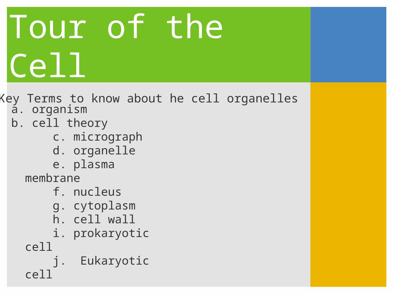
Tour of the Cell1. Key Terms to know about he cell organelles
a. organismb. cell theory
c. micrographd. organellee. plasma
membranef. nucleusg. cytoplasmh. cell walli. prokaryotic cellj. Eukaryotic cell
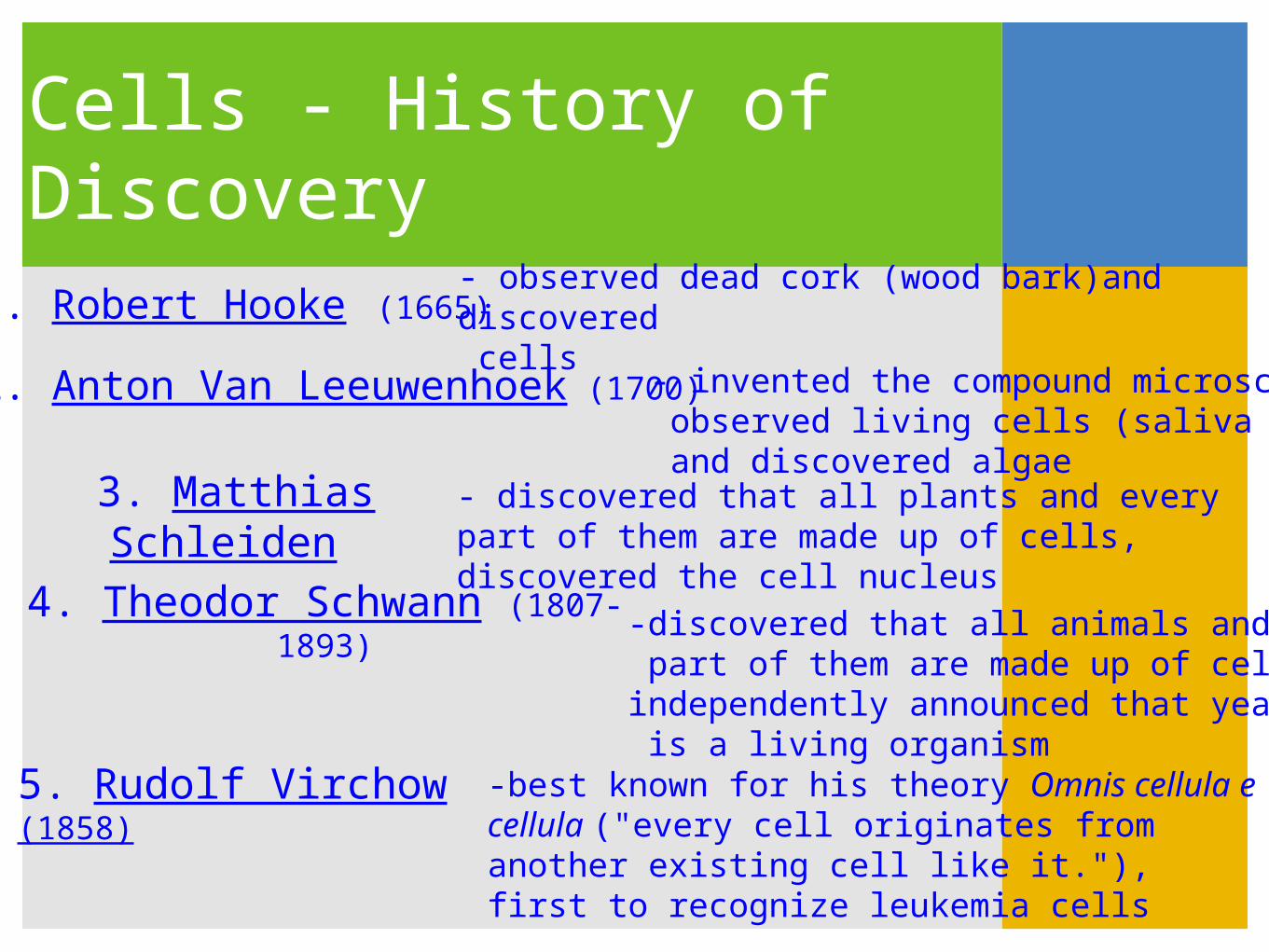
Cells - History of Discovery
1. Robert Hooke (1665)- observed dead cork (wood bark)and discovered cells
2. Anton Van Leeuwenhoek (1700)- invented the compound microscope, observed living cells (saliva & blood), and discovered algae
3. Matthias Schleiden
- discovered that all plants and every part of them are made up of cells, discovered the cell nucleus
4. Theodor Schwann (1807-1893)
-discovered that all animals and every part of them are made up of cells, independently announced that yeast is a living organism
5. Rudolf Virchow (1858)
-best known for his theory Omnis cellula e cellula ("every cell originates from another existing cell like it."), first to recognize leukemia cells

Cell Theory
Schleiden, Schwann, and Virchow are creditedfor the cell theory.
Cell Theory 1.) that all living things are composed of cells2.) cells are the basic unit of structure3.) cells are the basic unit of function in living things

Microscopes1.) Light- up to 1000x - living
cells
2.) Electron - up to 1,000,000x - dead cells
a.) SEM surface structureb.) TEM internal structure
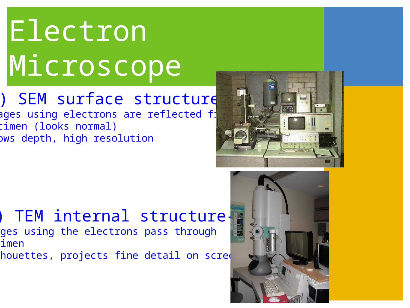
Electron Microscope
1.) SEM surface structure--images using electrons are reflected from specimen (looks normal)-shows depth, high resolution
2.) TEM internal structure--images using the electrons pass through specimen-silhouettes, projects fine detail on screen

Plant Cell
Cell Wall
Cell Membrane
Cytoplasm
Smooth ER (Endoplasmic reticulum)
Ribosomes
Rough ER (Ribosomes)
Mitochondria
Golgi AparatusGolgi Vesicles
Vacuole membrane
Large centralvacuole
Cholorplast
NucleusNucleolus
Raphide Crystal
Druse Crystal
Amyloplast (starch grain)

Animal Cell
Cell (plasma)Membrane
Pinocytotic vesicle
Cytoplasm
Smooth ER (Endoplasmic Reticulum)
Ribosomes
Rough ER (Ribosomes)
LysosomesMitochondrion
Golgi ApparatusGolgi Vesicles
Microtubules
Centrioles (2)Each composed of
9 microtubule triplets
Nucleus
Nucleolus

Another view

Plant vs. Animal Cell

Cell Wall*Only found in plant cells,
*The cell wall provides the cell with additional strength.
*Cell walls are thick walls built around the cell. These walls are made from cellulose.

Cell Membrane
*Found in both plant and animal cells, the cell membrane is the outside wall of a cell.
*In plant cells, it is a second wall, and is found just inside the main cell wall.
*The cell membranes found in animal cells contain a chemical called cholesterol. This chemical makes the membrane harder.
*Plant cells do not need cholesterol, because they have a cell wall, as a result, their cell membranes are softer.

Cytoplasm
* Found in both plant and animal cells
*Helps to hold the cell's organelles (small organs) in place.
*Gives the cell structure.
*Helps the cell move proteins, chromosomes and other materials including the cells organelles around the cell.

Nucleus - Cell’s Brain*A cell's nucleus, or brain, is responsible for directing the activities of the cell, in the same way that your brain directs the activities of your body.
*Nuclear Envelope - has pores, surrounds nucleus.
*In the nucleus you will see many small rod like objects called Chromosomes. They contain blueprints for how cells and organisms should be built.
*The chromosomes are made from smaller molecules called DNA, and RNA (information-rich molecules)
*Nucleolus contain parts that make up ribosomes.

Endoplasmic Reticulum
*Found in both animal and plant cells
*Clear tubes travel to all parts of the cell, known as the “Cellular Highway”.
*Carries materials where they need to go.
*The ER is also connected to the nuclear envelope.
*Rough ER: contains ribosomes
*Smooth ER: transports materials
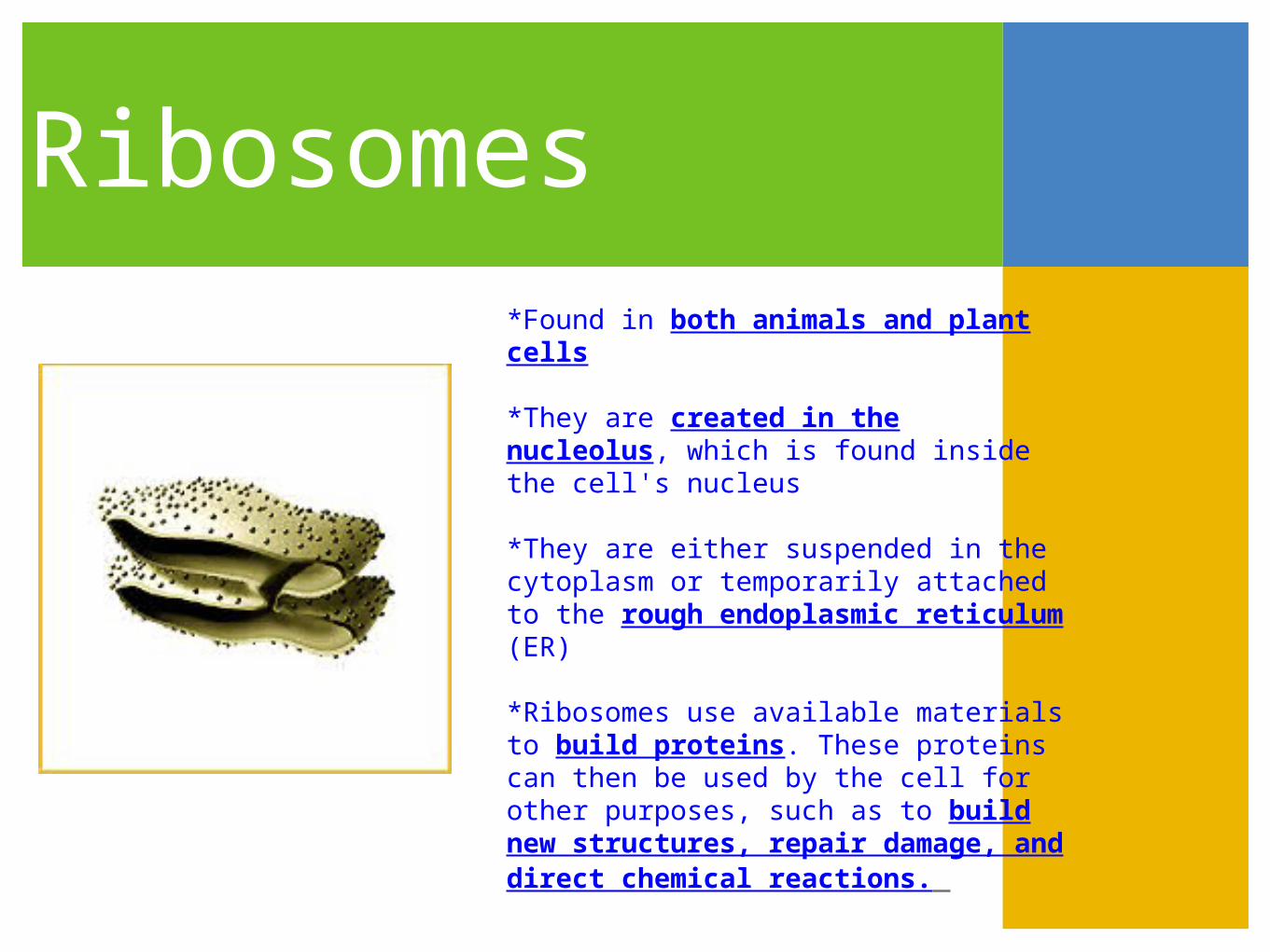
Ribosomes
*Found in both animals and plant cells
*They are created in the nucleolus, which is found inside the cell's nucleus
*They are either suspended in the cytoplasm or temporarily attached to the rough endoplasmic reticulum (ER)
*Ribosomes use available materials to build proteins. These proteins can then be used by the cell for other purposes, such as to build new structures, repair damage, and direct chemical reactions.

Moving Proteins
*Some proteins are made by ribosomes (the red structure) on the rough ER and packaged in vesicles.
*After further processing in other parts of the cell, these proteins will eventually move to other organelles or to the plasma membrane.

Golgi Apparatus (bodies)
*Found in both plant and animal cells
*Modifies, stores, and dispatch products *Takes the proteins which were created by the ribosomes, and makes them bigger and better
*When the golgi apparatus is done, it releases the new proteins into the cell, where they can be used to strengthen and build up the cell.
Lysosome
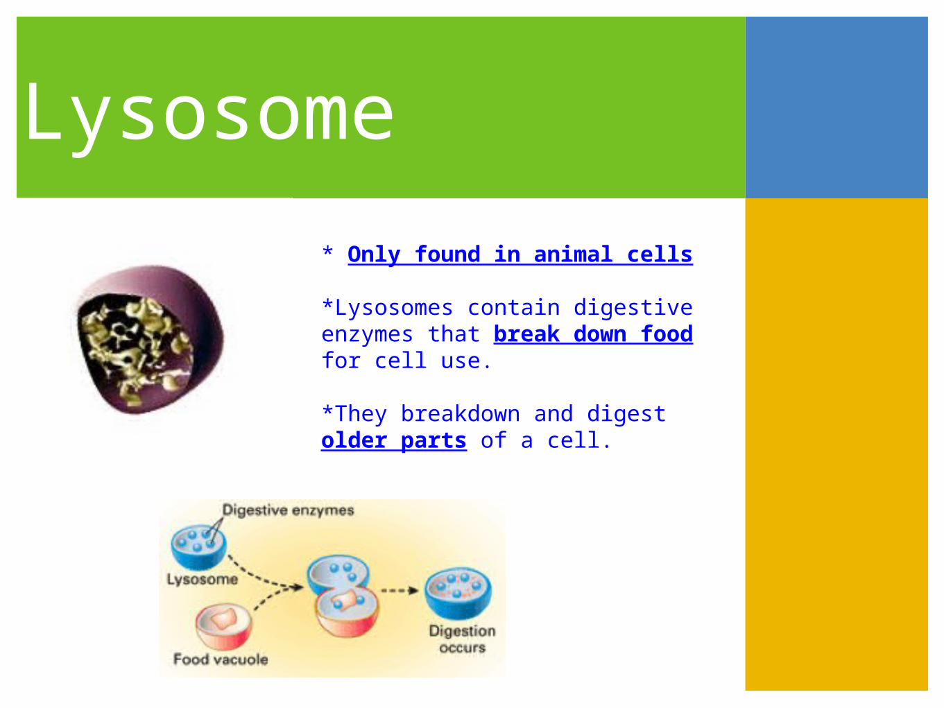
* Only found in animal cells
*Lysosomes contain digestive enzymes that break down food for cell use.
*They breakdown and digest older parts of a cell.

Membrane Pathways -how they work
*Products made in the ER move through membrane pathways in a cell.

Vacuoles*Large ones found in plant cells, small ones in animal cells
*This large membraned sac’s function is to storea. water & foodb. wastec. undigested nutrientsd. mineralse. proteinsf. pigments
*It helps in plant growth, and plays an important structural role for the plant.

Chloroplasts*Only found in plant cells
*Small pill shaped organelle that is like a miniature “solar collector”
*The discs are green because they are filled with a green pigment, or chemical called Chlorophyll that reacts with light
*Chlorophyll is used by a plant to capture light energy from the sun, which transforms into chemical energy through photosynthesis to create food.
Mitochondrion
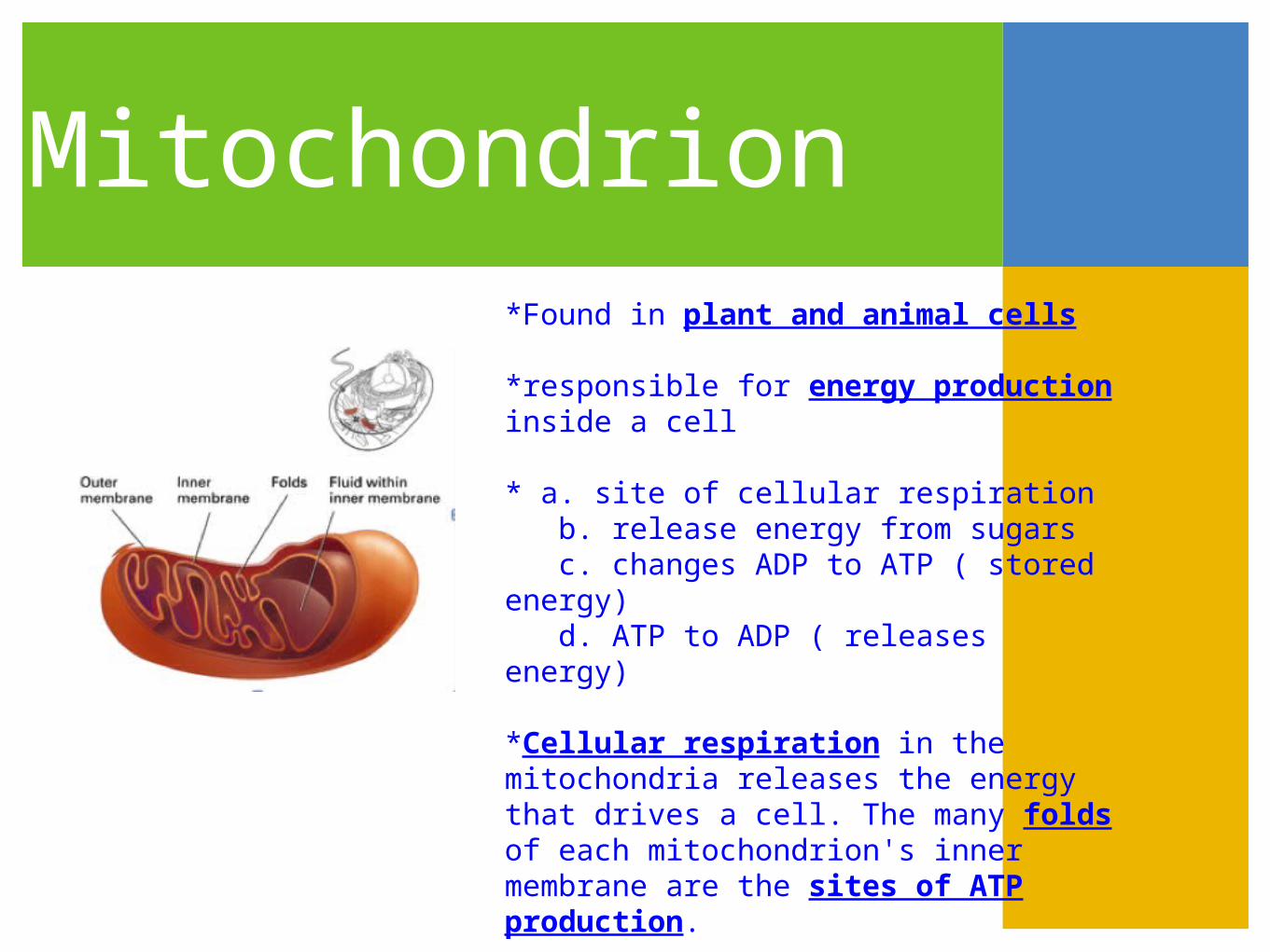
*Found in plant and animal cells
*responsible for energy production inside a cell
* a. site of cellular respiration b. release energy from sugars c. changes ADP to ATP ( stored energy) d. ATP to ADP ( releases energy)
*Cellular respiration in the mitochondria releases the energy that drives a cell. The many folds of each mitochondrion's inner membrane are the sites of ATP production.

Tour of the Cell
2. Key Terms to know about the Cell Membrane
1. Terms: a. phospholipid layers b. diffusion c. equilibrium d. selectively permeable membrane e. passive transport f. facilitated diffusion g. osmosis h. hypertonic i. hypotonic j. isotonic k. active transport l. vesicle m. exocytosis n. endocytosis 2. Structure of Cell Membrane

Transport- Diffusion
Dye molecules diffuse across a membrane. At equilibrium, the concentration of dye is the same throughout the container.

Passive Transport
Both diffusion and facilitated diffusion are forms of passive transport, as neither process requires the cell to expend energy. In facilitated diffusion, solute particles pass through a channel in a transport protein.

Osmosis
OSMOSIS IS DIFFUSION OF WATER FORM REGION OF LESSER CONCENTRATION OF SOLUTE TO GREATER CONCENTRATION OF SOLUTE UNTIL EQUILIBRIUM OCCURS
A selectively permeable membrane (the bag) separates two solutions of different sugar concentrations. Sugar molecules cannot pass through the membrane.

Active Transport
Like an enzyme, a transport protein recognizes a specific solute, molecule or ion. During active active transporttransport, the protein uses energy, usually moving the solute in a direction from lesser concentration to greater concentration.

Transport of large molecule
Active transport plays a part in maintaining the cell's chemical environment. Pinocytosis- Pino= drink (liquid) Phagocytosis- Phago= eat (solid)
Exocytosis (top left) expels molecules from the cell that are too large to pass through the plasma membrane.
Endocytosis (bottom left) brings large molecules into the cell and packages them in vesicles.

Why are cells so small? The cell theory never states that cell must be
small.But, there are two reasons given for their size:1. Efficiency- surface area is increased. Cells require nutrients and oxygen to get rid of waste and must move across the membrane to do so. If the cell were too big, these nutrients and wastes would have to cover large distances in order to get to the proper destination inside the cell.2. Specialization- having numerous small cells permits specialization and different cells have different functions.

Prokaryotic CellThese are the simplest of all cells.Most only have a cell wall and ribosomes. 1. DNA loop- naked in the cytoplasm, which contains all genetic info(processes)2. Ribosomes- freely floating in the cytoplasm for protein synthesis(antibiotics, like tetracycline, bind to ribosome and interfere with protein synthesis)3. Plasmids- contain small loop of extrachromosomal DNA

Prokaryotic Cell4. Cell Wall- gives shape and protection from unfavorable outside environment for cell membrane
Two types of cell walls that classify bacteria:Gram Positive- contain a thick peptidoglycan(protein-carbohydrate mix) layer and no outer membrane layerGram Negative- have a multilayered and complex wall made of an outer lipopolysaccharide and thin peptidoglycan inner layer
Antibiotics like penicillin inhibit cell wall development, which prevents reproduction of the prokaryotic cell. Enzymes in tears, mucus, and saliva dissolve the cell wall, rupturing the cell and killing the bacteria.

Prokaryotic Cell5. Capsule- a jelly-like coating that surround the cell wall; there are four functions of the capsule:1. prevents from drying out2. helps cells stick together on other surfaces3. helps slide on surfaces4. defense mechanism from being destroyed by host organisms’ cells
6. Flagella- can be one or many; provide locomotion by spinning like a propeller; they are structurally different from plant/animal flagella
7. Pili- short bristle-like appendages that have two functions:1. attach to surfaces2. assist in the transfer of DNA from one to another

Prokaryotic CellEubacteria Shapes:1. Coccus- spherical shape allows for less distortion in a dried out environment2. Bacillus- rod shape has more surface area to take up more nutrients from the environment3. Spirillium- spiral shape4. Spirochete- spiral shape with flagella5. Vibrio- 1/2 spiral
Spiral shapes are very motile, they move using a corkscrew type of movement.

Prokaryotic Cell
Movement is by way of something called chemotaxis. Chemotaxis is the movement of an organism towards or away from a chemical. Chemicals influence the organism to move toward them are called attractants (positive chemotaxis) or away from them are called repellents (negative chemotaxis).

Prokaryotic CellSurvival
When environmental conditions are unfavorable, bacteria will become inactive.Some species form endospores in which a thick wall forms around the genetic material and the rest of the cell disintegrates.Endospores are dormant and do not reproduce or show any signs of life, withstanding the harshest of environmental conditions.When conditions improve, endospores germinate and form an active cell again.

Prokaryotic CellReproduction1. Asexual fission- single loop of DNA is copied and the cell splits in half by pinching between the two DNA loops.2. Sexual conjugation- a bridge is formed between two cells using pili. Requires the F plasmid (F for fertility) and controls the formation of the F pilus. If the cell contains this plasmid, it is an F+ cell and can give an F- cell these genes, thus making it an F+ cell.R plasmids contain the genes for making a cell resistant to antibiotics and must integrate into main DNA of cell to make it resistant.

Reproduction cont.Transformation- living bacteria absorb the genetic material of a dead or naked genetic material in the environment
Transduction- transfer of DNA from a host to another cell by means of a virus. Viruses are non-living, pieces of DNA or RNA enclosed by a protein coat that can infect bacteria. Their DNA is small and contains information for making proteins involved in infection.

Metabolic DiversityHeterotroph- dependent on outside sources of organic moleculesPhotoheterotrophs- can use light to produce ATP but must obtain carbon from another sourceChemoheterotrophs- most bacteria assume this metabolism, there are three types:Saprobes- decomposers that absorb the nutrients from dead or decaying organic matterParasites- absorb nutrient from the body fluids of living hostsPhagotrophs- ingest food and digest it enzymatically within cell or multicellular bodies Autotrophs- synthesize organic molecule from inorganic substancesPhotosynthetic- harness light energy to drive organic compounds from CO2 and use an internal membrane system with light absorbing pigmentsChemosynthetic- use energy from specific inorganic substances to produce organic substances from CO2
Chemoautotrophs- need only CO2 as their carbon source and obtain energy from by oxidizing inorganic nutrients like H2S, NH4, Fe2O3; a unique group for prokaryotes
O2 RequirementsOxygen requirements also helps classify prokaryotic organisms:1. Obligate aerobes- must need and use oxygen for cellular respiration- cannot be without it2. Facultative anaerobes- will use oxygen if present, but can grow by fermentation without oxygen3. Obligate anaerobes- cannot use oxygen and are killed by the presence of it

Archaebacteria
Primitive forms of modern day bacteria that are thriving in different environment conditions
1. Methanogens- use hydrogen to reduce CO2 into methane; are obligate anaerobes that live in swamps, marshes and the guts of animals like cows, sheep, and camels; are used as important decomposers in sewage treatment plants2. Extreme Halophiles- like high salinity (salt) environments; this can color water pink because of their photosynthetic pigment bacteriorhodopsin3. Thermoacidophiles- need an environment that is hot (140-180 degrees fahrenheit) and acidic (pH of 2-4), they have no cell wall and can grow aerobically or anaerobically; examples are hot springs, water heaters, and coal piles