
Cell Staining - · PDF fileviable cell staining. ... (CFSE), and Fluorescein diacetate (FDA),...
13
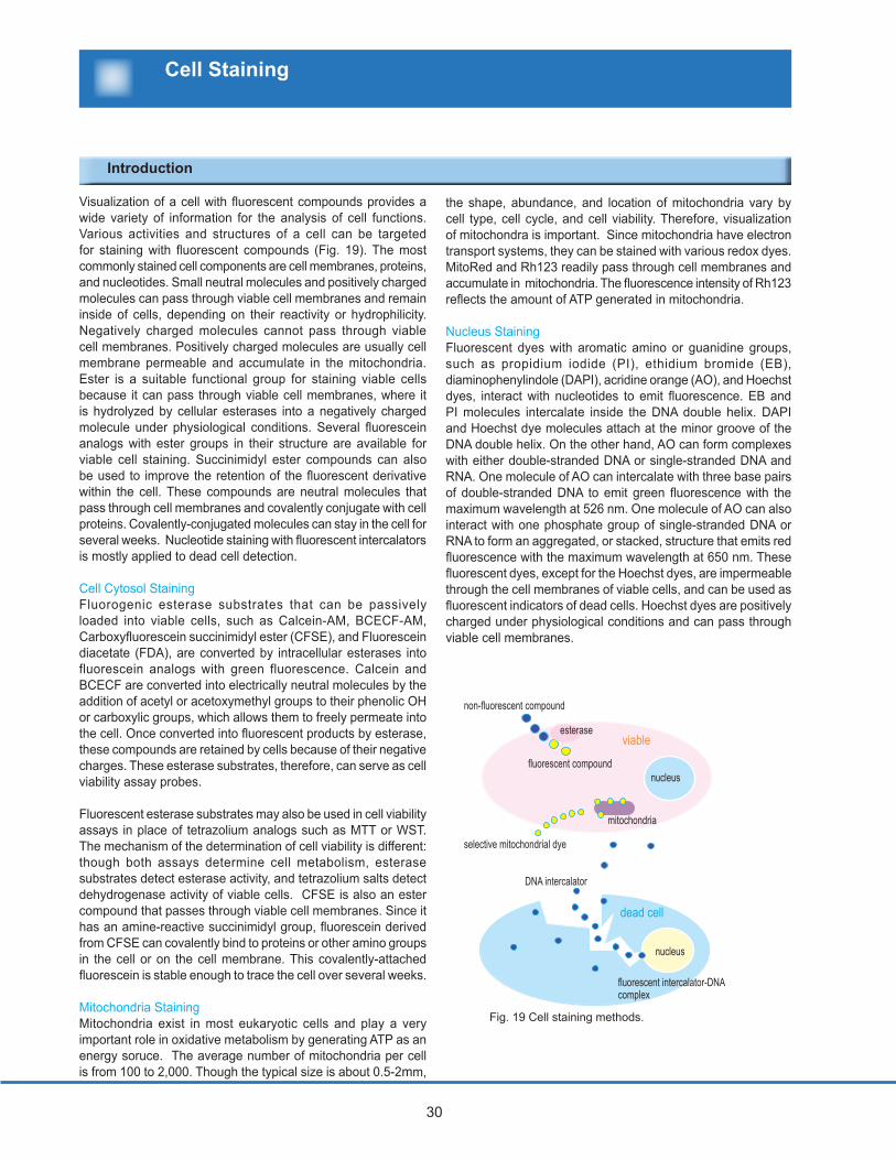
30 Visualization of a cell with fluorescent compounds provides a wide variety of information for the analysis of cell functions. Various activities and structures of a cell can be targeted for staining with fluorescent compounds (Fig. 19). The most commonly stained cell components are cell membranes, proteins, and nucleotides. Small neutral molecules and positively charged molecules can pass through viable cell membranes and remain inside of cells, depending on their reactivity or hydrophilicity. Negatively charged molecules cannot pass through viable cell membranes. Positively charged molecules are usually cell membrane permeable and accumulate in the mitochondria. Ester is a suitable functional group for staining viable cells because it can pass through viable cell membranes, where it is hydrolyzed by cellular esterases into a negatively charged molecule under physiological conditions. Several fluorescein analogs with ester groups in their structure are available for viable cell staining. Succinimidyl ester compounds can also be used to improve the retention of the fluorescent derivative within the cell. These compounds are neutral molecules that pass through cell membranes and covalently conjugate with cell proteins. Covalently-conjugated molecules can stay in the cell for several weeks. Nucleotide staining with fluorescent intercalators is mostly applied to dead cell detection. Cell Cytosol Staining Fluorogenic esterase substrates that can be passively loaded into viable cells, such as Calcein-AM, BCECF-AM, Carboxyfluorescein succinimidyl ester (CFSE), and Fluorescein diacetate (FDA), are converted by intracellular esterases into fluorescein analogs with green fluorescence. Calcein and BCECF are converted into electrically neutral molecules by the addition of acetyl or acetoxymethyl groups to their phenolic OH or carboxylic groups, which allows them to freely permeate into the cell. Once converted into fluorescent products by esterase, these compounds are retained by cells because of their negative charges. These esterase substrates, therefore, can serve as cell viability assay probes. Fluorescent esterase substrates may also be used in cell viability assays in place of tetrazolium analogs such as MTT or WST. The mechanism of the determination of cell viability is different: though both assays determine cell metabolism, esterase substrates detect esterase activity, and tetrazolium salts detect dehydrogenase activity of viable cells. CFSE is also an ester compound that passes through viable cell membranes. Since it has an amine-reactive succinimidyl group, fluorescein derived from CFSE can covalently bind to proteins or other amino groups in the cell or on the cell membrane. This covalently-attached fluorescein is stable enough to trace the cell over several weeks. Mitochondria Staining Mitochondria exist in most eukaryotic cells and play a very important role in oxidative metabolism by generating ATP as an energy soruce. The average number of mitochondria per cell is from 100 to 2,000. Though the typical size is about 0.5-2mm, the shape, abundance, and location of mitochondria vary by cell type, cell cycle, and cell viability. Therefore, visualization of mitochondra is important. Since mitochondria have electron transport systems, they can be stained with various redox dyes. MitoRed and Rh123 readily pass through cell membranes and accumulate in mitochondria. The fluorescence intensity of Rh123 reflects the amount of ATP generated in mitochondria. Nucleus Staining Fluorescent dyes with aromatic amino or guanidine groups, such as propidium iodide (PI), ethidium bromide (EB), diaminophenylindole (DAPI), acridine orange (AO), and Hoechst dyes, interact with nucleotides to emit fluorescence. EB and PI molecules intercalate inside the DNA double helix. DAPI and Hoechst dye molecules attach at the minor groove of the DNA double helix. On the other hand, AO can form complexes with either double-stranded DNA or single-stranded DNA and RNA. One molecule of AO can intercalate with three base pairs of double-stranded DNA to emit green fluorescence with the maximum wavelength at 526 nm. One molecule of AO can also interact with one phosphate group of single-stranded DNA or RNA to form an aggregated, or stacked, structure that emits red fluorescence with the maximum wavelength at 650 nm. These fluorescent dyes, except for the Hoechst dyes, are impermeable through the cell membranes of viable cells, and can be used as fluorescent indicators of dead cells. Hoechst dyes are positively charged under physiological conditions and can pass through viable cell membranes. viable dead cell non-fluorescent compound esterase mitochondria nucleus selective mitochondrial dye fluorescent intercalator-DNA complex DNA intercalator nucleus fluorescent compound Cell Staining Introduction Fig. 19 Cell staining methods.
Transcript of Cell Staining - · PDF fileviable cell staining. ... (CFSE), and Fluorescein diacetate (FDA),...
30
Visualization of a cell with fluorescent compounds provides a wide variety of information for the analysis of cell functions. Various activities and structures of a cell can be targeted for staining with fluorescent compounds (Fig. 19). The most commonly stained cell components are cell membranes, proteins, and nucleotides. Small neutral molecules and positively charged molecules can pass through viable cell membranes and remain inside of cells, depending on their reactivity or hydrophilicity. Negatively charged molecules cannot pass through viable cell membranes. Positively charged molecules are usually cell membrane permeable and accumulate in the mitochondria. Ester is a suitable functional group for staining viable cells because it can pass through viable cell membranes, where it is hydrolyzed by cellular esterases into a negatively charged molecule under physiological conditions. Several fluorescein analogs with ester groups in their structure are available for viable cell staining. Succinimidyl ester compounds can also be used to improve the retention of the fluorescent derivative within the cell. These compounds are neutral molecules that pass through cell membranes and covalently conjugate with cell proteins. Covalently-conjugated molecules can stay in the cell for several weeks. Nucleotide staining with fluorescent intercalators is mostly applied to dead cell detection.
Cell Cytosol StainingFluorogenic esterase substrates that can be passively loaded into viable cells, such as Calcein-AM, BCECF-AM, Carboxyfluorescein succinimidyl ester (CFSE), and Fluorescein diacetate (FDA), are converted by intracellular esterases into fluorescein analogs with green fluorescence. Calcein and BCECF are converted into electrically neutral molecules by the addition of acetyl or acetoxymethyl groups to their phenolic OH or carboxylic groups, which allows them to freely permeate into the cell. Once converted into fluorescent products by esterase, these compounds are retained by cells because of their negative charges. These esterase substrates, therefore, can serve as cell viability assay probes.
Fluorescent esterase substrates may also be used in cell viability assays in place of tetrazolium analogs such as MTT or WST. The mechanism of the determination of cell viability is different: though both assays determine cell metabolism, esterase substrates detect esterase activity, and tetrazolium salts detect dehydrogenase activity of viable cells. CFSE is also an ester compound that passes through viable cell membranes. Since it has an amine-reactive succinimidyl group, fluorescein derived from CFSE can covalently bind to proteins or other amino groups in the cell or on the cell membrane. This covalently-attached fluorescein is stable enough to trace the cell over several weeks.
Mitochondria StainingMitochondria exist in most eukaryotic cells and play a very important role in oxidative metabolism by generating ATP as an energy soruce. The average number of mitochondria per cell is from 100 to 2,000. Though the typical size is about 0.5-2mm,
the shape, abundance, and location of mitochondria vary by cell type, cell cycle, and cell viability. Therefore, visualization of mitochondra is important. Since mitochondria have electron transport systems, they can be stained with various redox dyes. MitoRed and Rh123 readily pass through cell membranes and accumulate in mitochondria. The fluorescence intensity of Rh123 reflects the amount of ATP generated in mitochondria.
Nucleus StainingFluorescent dyes with aromatic amino or guanidine groups, such as propidium iodide (PI), ethidium bromide (EB), diaminophenylindole (DAPI), acridine orange (AO), and Hoechst dyes, interact with nucleotides to emit fluorescence. EB and PI molecules intercalate inside the DNA double helix. DAPI and Hoechst dye molecules attach at the minor groove of the DNA double helix. On the other hand, AO can form complexes with either double-stranded DNA or single-stranded DNA and RNA. One molecule of AO can intercalate with three base pairs of double-stranded DNA to emit green fluorescence with the maximum wavelength at 526 nm. One molecule of AO can also interact with one phosphate group of single-stranded DNA or RNA to form an aggregated, or stacked, structure that emits red fluorescence with the maximum wavelength at 650 nm. These fluorescent dyes, except for the Hoechst dyes, are impermeable through the cell membranes of viable cells, and can be used as fluorescent indicators of dead cells. Hoechst dyes are positively charged under physiological conditions and can pass through viable cell membranes.
viable
dead cell
non-fluorescent compound
esterase
mitochondria
nucleus
selective mitochondrial dye
fluorescent intercalator-DNA complex
DNA intercalator
nucleus
fluorescent compound
Cell Staining
Introduction
Fig. 19 Cell staining methods.

31 301.987.2667 1.877.987.2667
Characteristics of Dye
Applications: Fluorescence Microscopy, Flowcytometry, Electrophoresis (Nucleic Acid Screening)
Cell Staining Reagents
Target Dye Excitation Emission Excitation filter Color Characteristic
BCECF-AM 490 nm 526 nm B excitation yellowish green
Calcein-AM 490 nm 515 nm B excitation yellowish green
CFSE 496 nm 516 nm B excitation yellowish green
CytoRed 535 nm 590 nm G excitation red
FDA 488 nm 530 nm B excitation yellowish green
DAPI 360 nm 460 nm W excitation blue
PI 530 nm 620 nm G excitation red
AO (dsDNA) 500 nm 526 nm B excitation red
(ssDNA & RNA) 420-460 nm 630-650 nm B excitation yellow
Hoechst33258 350 nm 461 nm W excitation blue
Hoechst33342 352 nm 461 nm W excitation blue
MitoRed 560 nm 580 nm G excitation red
Rh123 507 nm 529 nm B excitation yellowish green
Living cells
Dead cells
Mitochondria
Fluorescence is produced by hydrolysis inside the cell.
Fluorescence is produced by interacting with the nucleus of dead cells.
Fluorescence is produced by combining with single stranded and double stranded DNA.Fluorescence is produced by combining with the nucleus of living and dead cells.Fluorescence is produced by accumulation in the mito-chondria.
Nucleous
Product Information
-Cellstain- SeriesProduct name Product code Unit
BCECF-AM B262-10 1 mgCalcein-AM C326-10 1 mgCalcein-AM solution C396-10 1 mlCFSE C375-10 1 mgCytoRed solution C410-10 1 mlFDA F209-10 1 mgDAPI D212-10 1 mgDAPI solution D523-10 1 mlPI P346-10 1 mgPI solution P378-10 1 mlAO A386-10 1 mgAO solution A430-10 1 mlHoechst33258 solution H341-10 1 mlHoechst33342 solution H342-10 1 mlMitoRed R237-10 50 mg x 8 vialsRh123 R233-10 1 mg
32
Devices, Tools CO2 incubator Clean bench Fluorescence microscope Cytometer or cell counter Centrifuge Slide glass, cover glass, or chamber slide
Reagents
Required Materials
Cell Staining Reagents
The following is a general protocol for preparing assay solutions. In order to obtain the best results, optimization of staining conditions, such as changing the reagent concentration and staining time will be required.
Some reagents are stable in the solution. However, some reagents are not stable. Please follow the storage conditions for each reagent. Generally, the reagents offered in the solution form are fairly stable. If no microbalance is available to weigh small amounts of the reagent, add an appropriate amount of solvent described in the chart, aliquot, and store it in a freezer.
Product name Characteristic Storage Mol. Weight Unit size Stock Solution (DMSO) Staining solution-Cellstain- Calcein-AM white-yellowish solid avoid light, freeze 994.86 1 mg 0.5 - 1mmol/l
-Cellstain- Calcein-AM solution colorless liquid avoid light, freeze 994.86 1 ml 1mmol/l -Cellstain- CFSE white-yellowish solid avoid light, freeze 557.64 1 mg 0.5 - 1mmol/l -Cellstain- CytoRed solution orange-yellow liquid avoid light, freeze 313.31 1 ml 1mmol/l -Cellstain- FDA white crystal avoid light, freeze 416.38 1 mg 0.5 - 1mmol/l BCECF-AM Special packaging orange-brown solid avoid light, freeze 688.59 50 µg x 8 0.5 - 1mmol/l
Staining solutions are not stable for a storage. Discard the remaining staining solution after each use.
1-20 µmol/l (Storage solution diluted by PBS (-))
Living Cell Staining Dyes-Cellstain- Calcein-AM (product code: C326) -Cellstain- Calcein-AM solution(product code: C326) -Cellstain- CFSE (product code: C375) -Cellstain- CytoRed solution (product code:C410 ) -Cellstain- FDA (product code: F209) BCECF-AM (product code:B262)
DMSO, Sterilized Water, PBS(-)
Dead Cell Staining Dyes-Cellstain- DAPI (product code: D212) -Cellstain- DAPI solution (product code: D523) -Cellstain- PI (product code: P346) -Cellstain- PI solution (product code:P378)
Nucleus Staining Dyes-Cellstain- AO (product code: A386) -Cellstain-AO solution (product code: A430) -Cellstain- Hoechst 33258 (product code: H341) -Cellstain-Hoechst 33342 (product code: H342)
Mitochondria Staining Dyes-Cellstain- MitoRed (product code: R237) -Cellstain- Rh123 (product code: R233)
Dyes for Living Cell Staining
If the reagent is in a solid form, use DMSO to prepare a solution with a certain concentration. Since CFSE has a succinimidyl group, the stability of the prepared DMSO solution is poor. After the preparation of the DMSO solution, aliguot in an appropri-ate volume and store at -20 oC. The DMSO solution can be used for several months. The working solutions prepared by PBS(-) are not stable enough to store. Discard the remaining working solution after each use.
Other Reagents
Preparation of Staining Solution
33 301.987.2667 1.877.987.2667
Product name Characteristic Storage Mol. Weight Units Stock Solution (H20) Staining Solution
-Cellstain-DAPI yellow solid avoid light, freeze 350.25 1 mg 1 mg/ml -Cellstain-DAPI soln. light yellow liquid avoid light, refrigerate 350.25 1 ml 1 mg/ml* -Cellstain-PI red-brown solid avoid light, refrigerate 668.39 1 mg 1 mg/ml -Cellstain-PI soln. red liquid avoid light, freeze 668.39 1 ml 1 mg/ml
Since the dyes directly stain the nucleus, these dyes are considered mutagens. Gloves, safety goggles, and masks are necessary when handling. If it comes in contact with your skin, immediately wash with a copious amount of water.
Dyes for Dead Cell Staining
1-10 µg/ml (Storage solution diluted by PBS (-))
Dyes for Nucleus Staining
Product name Characteristic Storage Mol. Weight Units Stock Solution (H20) Staining Solution
-Cellstain-AO red-brown avoid light, refrigerate 301.81 1 mg 1 mg/ml -Cellstain-AO soln. orange-yellow liquid avoid light, freeze 301.81 1 ml 1 mg/ml -Cellstain-Hoechst 33258 soln. yellow liquid avoid light, refrigerate 533.88 1 ml 1 mg/ml-Cellstain-Hoechst 33342 soln. yellow liquid avoid light, refrigerate 561.93 1 ml 1 mg/m
Dyes for Mitochondria Staining
Only mitochondria in the living cells will be stained.
Product name Characteristic Storage Mol. Weight Units Stock Solution (H20) Staining Solution
-Cellstain-MitoRed purple brown solid avoid light, refrigerate 637.17 50 µgx8 1 mmol/l* 20 - 200 nmol/l-Cellstain-Rh123 brown powder avoid light, refrigerate 380.82 1 mg 1 mg/ml 20 - 100 nmol/l
When disposing remaining dye solution and solution containing staining dyes, follow handling guidelines and regula-tions, and entrust disposal to an industrial waste disposal company. If the amount of the dye solution is fairly small and disposal rules and regulations of your institute allow, use a paper towel to adsorb and mix it with plastic tubes used for the preparation of the staining dye solution to incinerate.
If the reagent is in a solid form, use sterilized water to prepare a solution and store according to the indicated condition. Prepare DAPI solution with PBS. Working solutions prepared using PBS(-) are not stable enough to store. Discard the remaining working solution after use.
If the reagent is in a solid form, use sterilized water to prepare a solution and store according to the indicated condition. Working solutions prepared using PBS(-) are not stable enough to store. Discard the remaining working solution after use.
Prepare the stock solution using DMSO.
* Use buffer to prepare a solution
1-10 µg/ml (Storage solution diluted by PBS (-))
Since the dyes directly stain the nucleus, these dyes are considered mutagens. Gloves, safety goggles, and masks are necessary when handling. if it comes in contact with your skin immediately wash with a copious amount of water.
When disposing remaining dye solution and solution containing staining dyes, follow handling guidelines and regula-tions and entrust disposal to an industrial waste disposal company. If the amount of the dye solution is fairly small and disposal rules and regulations of your institute allow, use a paper towel to adsorb and mix it with plastic tubes used for the preparation of the staining dye solution to incinerate.
Cell Staining Reagents
* Avoid storing in the solution.

34
Add 104 -105 cells to each well with cell culture medium and incubate overnight.
Remove cell culture medium and wash cells by PBS(-) buffer.
Remove PBS(-) buffer, add 0.2 ml “Staining Solution” to cells and incubate cells at 37oC for 15-30 min.
Remove “Staining Solution” and wash cells by PBS(-) buffer for 1-2 times.
Use a cytometer or a cell counter to mea-sure cell number.When using a glass bottom plate, the clear image of cells may be obtained.
Gently pipette to avoid damaging the cells.
Gently remove the medium and add PBS buffer without damaging the cells.When using a staining dye for staining living cells, the dye will be hydrolized and emit fluorescence if esterase in the media remains. This is one of the factor for a high background, so it is important to wash the cells several times.
Refer to the previous section “Prepara-tion of Assay Solution” and prepare the “Staining Solution”. In order to obtain the best fluorescent image, it is necessary to determine the optimal reagent concentra-tion and staining time.
Procedure Precautions & Tips
Staining Procedure for a Fluorescence Microscopy (for 24-well plate)
If you use suspention cells, it’s necessary to centrifuge the cell sus-pension at 500 xg for 3 min before removing the medium, PBS(-) buffer or “Staining Solution”.
Observe the fluorescent image under a fluores-cence microscope.
Cell Staining Reagents
35 301.987.2667 1.877.987.2667
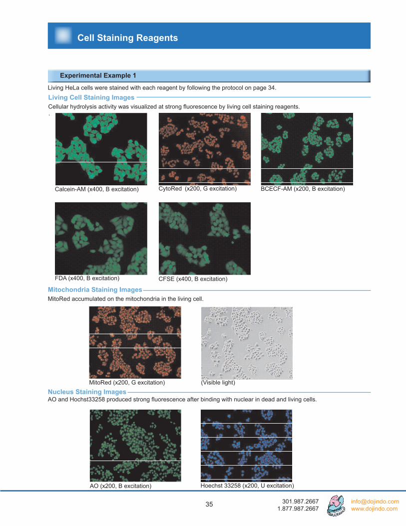
Living Cell Staining Images Cellular hydrolysis activity was visualized at strong fluorescence by living cell staining reagents.
Experimental Example 1
Calcein-AM (x400, B excitation)
AO (x200, B excitation)
Mitochondria Staining Images MitoRed accumulated on the mitochondria in the living cell.
CytoRed (x200, G excitation) BCECF-AM (x200, B excitation)
MitoRed (x200, G excitation)
.
Nucleus Staining Images AO and Hochst33258 produced strong fluorescence after binding with nuclear in dead and living cells.
Hoechst 33258 (x200, U excitation)
FDA (x400, B excitation) CFSE (x400, B excitation)
Living HeLa cells were stained with each reagent by following the protocol on page 34.
(Visible light)
Cell Staining Reagents
36
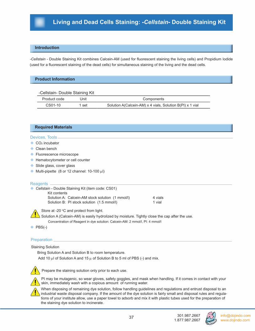
Fluorescent Staining of Fixed CellsNIH3T3 cells that were fixated with 3% glutaraldehyde were stained with the nuclic acid staining reagent Hoechst 33258. Then, actin filaments were stained with biotin-labeled phalloidin and anti-biotin antibody labeled with HiLyte FluorTM 555.*
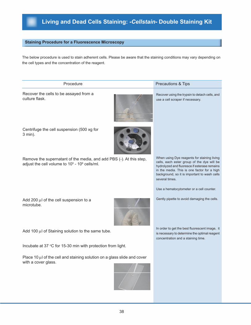
Flowcytometry Example HL60 cells were stained with Calcein-AM, a reagent used to stain the living cells. The cells were then measured using flowcytometry (excitation: 488 nm). The fluorescence of the stained living cells (blue line) increased dramatically compared to the unstained cells (white line).
Fluorescent Intensity
Cel
l Num
ber
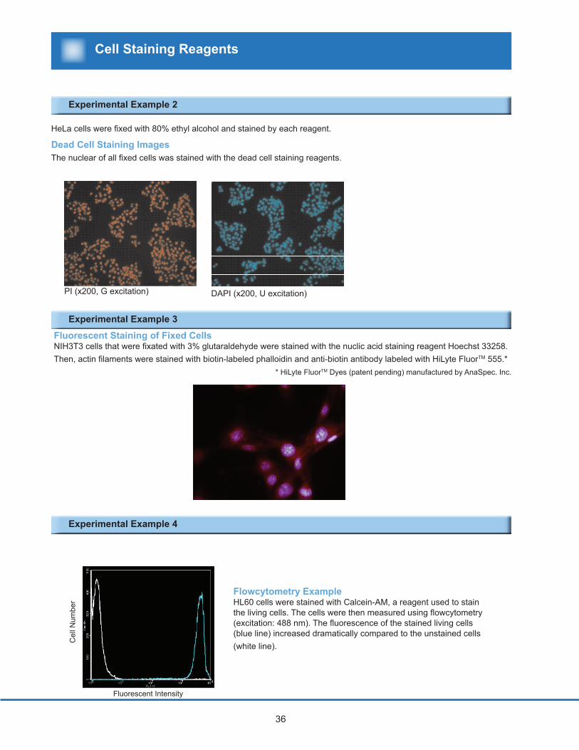
PI (x200, G excitation) DAPI (x200, U excitation)
* HiLyte FluorTM Dyes (patent pending) manufactured by AnaSpec. Inc.
Dead Cell Staining Images The nuclear of all fixed cells was stained with the dead cell staining reagents.
HeLa cells were fixed with 80% ethyl alcohol and stained by each reagent.
Experimental Example 2
Experimental Example 3
Experimental Example 4
Cell Staining Reagents
37 301.987.2667 1.877.987.2667
Preparation
Devices, Tools CO2 incubator Clean bench Fluorescence microscope Hematocytometer or cell counter Slide glass, cover glass Multi-pipette (8 or 12 channel: 10-100 µl)
Reagents Cellstain - Double Staining Kit (item code: CS01) Kit contents Solution A: Calcein-AM stock solution (1 mmol/l) 4 vials Solution B: PI stock solution (1.5 mmol/l) 1 vial
PBS(-)
Required Materials
Living and Dead Cells Staining: -Cellstain- Double Staining Kit
Introduction
-Cellstain - Double Staining Kit combines Calcein-AM (used for fluorescent staining the living cells) and Propidium Iodide (used for a fluorescent staining of the dead cells) for simultaneous staining of the living and the dead cells.
Store at -20 oC and protect from light.Solution A (Calcein-AM) is easily hydrolized by moisture. Tightly close the cap after the use.
Staining Solution Bring Solution A and Solution B to room temperature. Add 10 µl of Solution A and 15 µ of Solution B to 5 ml of PBS (-) and mix.
Prepare the staining solution only prior to each use.
PI may be mutagenic, so wear gloves, safety goggles, and mask when handling. If it comes in contact with your skin, immediately wash with a copious amount of running water.
Concentration of Reagent in dye solution: Calcein-AM: 2 mmol/l, PI: 4 mmol/l
When disposing of remaining dye solution, follow handling guidelines and regulations and entrust disposal to an industrial waste disposal company. If the amount of the dye solution is fairly small and disposal rules and regula-tions of your institute allow, use a paper towel to adsorb and mix it with plastic tubes used for the preparation of the staining dye solution to incinerate.
Product Information
-Cellstain- Double Staining KitProduct code Unit Components
CS01-10 1 set Solution A(Calcein-AM) x 4 vials, Solution B(PI) x 1 vial
38
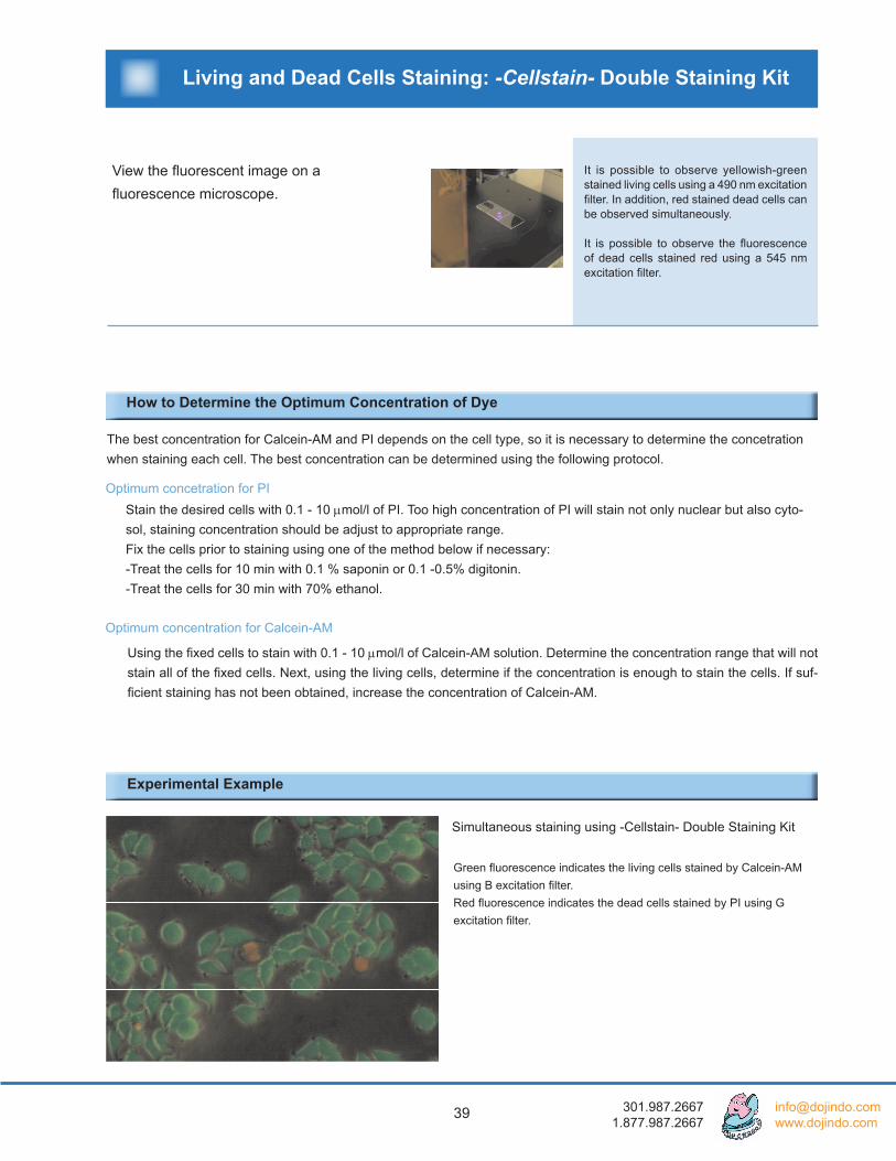
Recover the cells to be assayed from a culture flask.
Centrifuge the cell suspension (500 xg for 3 min).
Remove the supernatant of the media, and add PBS (-). At this step, adjust the cell volume to 105 - 106 cells/ml.
Add 200 µl of the cell suspension to a microtube.
Add 100 µl of Staining solution to the same tube.
Incubate at 37 oC for 15-30 min with protection from light.
Place 10 µl of the cell and staining solution on a glass slide and cover with a cover glass.
Recover using the trypsin to detach cells, and use a cell scraper if necessary.
When using Dye reagents for staining living cells, each ester group of the dye will be hydrolyzed and fluoresce if esterase remains in the media. This is one factor for a high background, so it is important to wash cells several times.
Use a hematocytometer or a cell counter.
Gently pipette to avoid damaging the cells.
In order to get the best fluorescent image, it is necessary to determine the optimal reagent concentration and a staining time.
Procedure Precautions & Tips
Staining Procedure for a Fluorescence Microscopy
The below procedure is used to stain adherent cells. Please be aware that the staining conditions may vary depending on the cell types and the concentration of the reagent.
Living and Dead Cells Staining: -Cellstain- Double Staining Kit
39 301.987.2667 1.877.987.2667
Simultaneous staining using -Cellstain- Double Staining Kit
Experimental Example
View the fluorescent image on a fluorescence microscope.
It is possible to observe yellowish-green stained living cells using a 490 nm excitation filter. In addition, red stained dead cells can be observed simultaneously.
It is possible to observe the fluorescence of dead cells stained red using a 545 nm excitation filter.
The best concentration for Calcein-AM and PI depends on the cell type, so it is necessary to determine the concetration when staining each cell. The best concentration can be determined using the following protocol.
Optimum concetration for PIStain the desired cells with 0.1 - 10 µmol/l of PI. Too high concentration of PI will stain not only nuclear but also cyto-sol, staining concentration should be adjust to appropriate range.Fix the cells prior to staining using one of the method below if necessary:-Treat the cells for 10 min with 0.1 % saponin or 0.1 -0.5% digitonin.-Treat the cells for 30 min with 70% ethanol.
Optimum concentration for Calcein-AM
Using the fixed cells to stain with 0.1 - 10 µmol/l of Calcein-AM solution. Determine the concentration range that will not stain all of the fixed cells. Next, using the living cells, determine if the concentration is enough to stain the cells. If suf-ficient staining has not been obtained, increase the concentration of Calcein-AM.
Green fluorescence indicates the living cells stained by Calcein-AM using B excitation filter.Red fluorescence indicates the dead cells stained by PI using G excitation filter.
How to Determine the Optimum Concentration of Dye
Living and Dead Cells Staining: -Cellstain- Double Staining Kit

40
Troubleshooting
The cells are not stained well.
The dye seems not to stay inside of the viable cell after staining.
The dye remains insoluble with the solvent.
High fluorescent background is observed.
The staining dye was hydrolized or decomposed due to the exceedingly long term storage or incorrect storage conditions.
The dye in the working solution was hydrolized or decomposed because the solution was not freshly prepared.
The dye or the working solution was decomposed by the exposure to light.
The concentration of the dye in the working solution is too low.
The viable cell expels the dye due to the cell function.
Not enough reagent was used for the cells.
Since a vacuum centrifuge was used to prepare the dye product, the dye is tightly packed on the bottom of the tube.
The dye was decomposed or hy-drolized
The wrong solvent was used to dis-solve.
Not enough washing and the dye still remained after the washing process.
Too much dye was used for the staining.
Check the purchase date of the reagent and the storage conditions. If the reagent has been stored for a year from the purchase date, do not use. The staining dye may not work properly.
Some of the reagent is unstable in a buffer solution. In particular, viable staining dye is fairly unstable in the buffer solution. Prepare a working solution only prior to each use.
Light may accelerate the oxidation process of the dyes. Keep the reagent under the proper storage conditions. Pro-tect the working solution from light during the experiment.
Increase the concentration of the dye in the working solu-tion. If there is no change, use Pluronic F-127 or another low toxic detergent to improve the dye uptake by the cell if it is allowed
The dye did not dissolve completely with the solvent. Make sure that the proper solvent was used and the proper con-centration was prepared.
Use the stained cell as quickly as possible for your experi-ments.
Probenecid, a transporter inhibitor, may be used to block the leakage of the dye from the cell.
Use a vortex mixer or ultra sonic bath to dissolve the dye with the solvent completely.
Check the purchase date of the reagent & storage condi-tions. If the reagent has been stored for over a year from the purchase date, do not use. The staining dye may be decomposed or hydrolized.
Simultaneuos Staining of living and dead cells Use the proper solvent to prepare a dye solution
Repeat washing with PBS(-) or an appropriate buffer to remove excess dye from the cells.
Reduce the concentration of the dye in the working solu-tion.
Problem Possible Cause Solution
Living and Dead Cells Staining: -Cellstain- Double Staining Kit
41 301.987.2667 1.877.987.2667
Q&A
Staining reagents for living cells
Q: What should the powder-type reagent be dissolved in?A: Please dissolve the reagent in DMSO for viable cell staining reagents. Since DMSO easily absorbs moisture, please use fresh DMSO.
Q: Among all the staining reagents used for living cells, which one remains the longest inside cells?A: CFSE remains relatively the longest inside the cells. It has been reported in a paper that the fluorescent dye was retained within cells for up to 8 weeks. Also, the fluorescence of Calcein-AM and BCECF-AM have been observed in cells for up to three days. Please refer to the following for more details:
ES.A.Weston, et.al., J.Immunol.Methods, 133, 87-97 (1990) EH.P.Zhong, et.al., Hum.Immunol., 37, 264-270 (1993)
Q: Which staining reagents used for living cells have the lowest cytotoxicity?A: Calcein-AM and BCECF-AM seem to have the lowest cytotoxicity, Please refer to the following for more details:
EL.S.D.Clerck, et.al., J.Immunol.Methods, 172, 115-124 (1994)
Q: What are the characteristics of staining dyes used for the living cells?A: Refer to the list below for characteristics of each product:
BCECF-AM : This was originally used to measure pH inside the cell, and is also used as a dye to stain living cells. Calcein-AM: This has the least effect on cell function. CFSE: After entering into a cell, it combines with the amino base of protein in the cell membrane on the cytoplasm side. As a result, it leaks out of the cell comparatively less than other dyes. CytoRed: A compound produced by Dojindo, it posses a higher fluorescence intensity than Calcein-AM. FDA: The oldest known dye. It leaks out of the cell relatively quickly.
Q: Are there any papers that report on the toxicity of the dyes?A: Refer to the below paper comparing the toxicity of Calcein-AM, BCECF-AM, CFDA, and CFSE.
L. S. D. Clerck, et al.,J.Immunol. Methods, 172,115 (1994)
Q: Which dye should be used to stain the bacteria?A: Since bacterial cells have a cell wall, most cell staining dyes cannot penetrate. For example, Calcein-AM and
BCECF-AM will pass through the cell membrane of animal cells, but will not pass through the bacteria cell wall. AO can be used to stain bacteria such as malaria parasites. PI, and DAPI can be used to stain dead bacteria cells. There is a report of using FDA to stain living bacteria. Refer to the paper below for more information:
Appl. Microbiol. Biotechnol., 38, 268 (1992)
Cell Staining
42
Nucleus staining reagents (dead cells)
Q: What are the differences between the nuclues staining reagents AO, Hoechst 33258, and Hoechst 33342 other than fluorescent wavelength?
A: The differences are listed below: AO : It is possible to distinguish between single stranded DNA and double stranded DNA using the difference in
fluorescence wavelength when intercalating with a double stranded DNA and when combining with the phosphoric acid of a single stranded DNA. AO passes through the membrane of living cells.
Hoechst 33258, Hoechst 33342 :Binds specifically with adenine - thyamine base pairs of DNA. They pass through the cell membrane, and stain the DNA of living cells. Hoechst 33342 has a higher membrane permeability. A better staining is possible when cells are fixed.
Q: What is the method of disposal after use?
A: PI is a possible carcinogen. When disposing remaining dye solution and solution containing staining dyes, follow handling guidelines and regulations and entrust disposal to an industrial waste disposal company. If the amount of the dye solution is fairly small and disposal rules and regulations of your institute allow, use a paper towel to adsorb and
mix it with plastic tubes used for the preparation of the staining dye solution to incinerate.
Nucleus staining reagents (living / dead cells)
Q: What is the difference among dyes used to stain the nucleus?
A: Some notable differences other than wavelength are listed below; PI: It does not have base specificity. It binds to all DNA and RNA., but the fluorescence intensity is higher when inter-calating and can be used widely among variety of cells. DAPI:This will bind with the minor groove of a double chain, and has a high affinity for adenine - thimine base pairs.
Mitochondria staining reagents
Q: Why do MitoRed and Rh123 stain the mitochondria?
A: Both MitoRed and Rh123 employ the chemical structure Rhodamine. Rhodamine has the characteristic of gathering to mitochondria after entering the cell, so it is used as a mitochondria staining dye. When too much dye is introduced into the cell, other areas are stained also, so it is necessary to determine the best concentration in advance.
Cell Staining


![Spectrofl uorometer - GrupoBios · Calibration Curve of Fluorescein Solutions Spectra of Fluorescein Solutions Spectrum of quinine sulfate solution Wavelength [nm] 300 400 500 600](https://static.fdocuments.in/doc/165x107/5fb614bb5457d74a9a1fd826/spectrofl-uorometer-grupobios-calibration-curve-of-fluorescein-solutions-spectra.jpg)