Cell-Mediated Immunity during Measles Infection · 'Abbreviations used in this paper: FCS, fetal...
7
Cell-Mediated Immunity during Natural Measles Infection H. C. WHITTLE, J. DOSSETOR, A. ODULOJU, A. D. M. BRYCESON, and B. M. GREENWOOD, Departments of Medicine and Pediatrics, Ahmadu Bello University Hospital, Zaria, Kaduna State, Nigeria A B S T R A C T Natural measles causes prolonged depression of cell-mediated immunity yet little is known as to how the infection influences lymphocyte function. Therefore, we studied the properties and function of lymphocytes during and after measles. The number and proportion of circulating thymus-derived lymphocytes was low during the acute stage of measles, and at this time 37% of these cells showed positive immunofluorescent staining for measles virus after stimulation with phytohemagglutinin. 7% of B cells were shown to contain virus but their numbers did not alter during the infection. Acute-phase lymphocytes, when stimulated, yielded infective virus and half were killed on incubation with autologous serum and com- plement. In acute measles the increase in [3H]- thymidine uptake of lymphocytes when stimulated with an optimal dose of PHA was normal in media with 10% fetal calf serum and low in media con- taining 10% autologous serum: the mean values were 56.8±34.1 and 23.7±25.9 cpm x 103 per 106 lympho- cytes, respectively. Stimulation of acute-phase lympho- cytes by Candida antigen was also low in media containing autologous serum averaging 1.2 x 10³ cpm per 106 lymphocytes. On recovery 4-6 wk later this rose significantly to 18.9±19.8. The mean migration index of leukocytes to heat-killed candida cells in acute measles was 0.84±SD 0.08, and this fell signifi- cantly to 0.75±SD 0.08 4 wk later. Thus, depletion of T cells, an inhibitor of lymphocyte proliferation in the serum and a possible defect in antigen process- ing, interacts to depress cell-mediated immunity in measles. INTRODUCTION Natural measles infection suppresses both cell- mediated and humoral immune responses (1), and this, Received for publication 22 April 1977 and in revised form 8 May 1978. coupled with malnutrition, leads to the death'of many children from secondary infection (2). Von Pirquet in 1908 (3), first noticed that the skin test to old tuber- culin became negative during measles, and since then many scientists have investigated this phenomenon. Smithwick and Berkovich (4) infected lymphocytes from tuberculous children with measles virus and showed a diminished blastogenic response of these cells to tuberculin. Osunkoya et al. (5, 6) studied lymphocyte transformation to phytohemagglutinin (PHA)l during natural measles infection: tritiated thymidine incorporation and blast-cell counts were normal despite the appearance of many giant cells infected with measles virus. However, Finkel and Dent (7) noted impairment of lymphocyte response to sub- optimal doses of PHA but were unable to show that this was because of serum inhibitory factors that are known to occur in some viral infections (8). Experi- ments by Zweiman and Miller (9-11) demonstrated that both live and killed measles virus could suppress the response of human lymphocytes to PHA and purified protein derivative (PPD). Sullivan et al. (12) extended this work and postulated that depression by live virus of lymphocyte stimulation by PHA re- sulted from direct viral inhibition of host cell DNA synthesis. Other hypotheses for the depression of cell-mediated immune responses following measles have been ad- vanced. Osunkoya et al. (6) suggested this was be- cause of a transient reduction in number of T lympho- cytes as a result of cytopathic destruction, and this idea has been supported by the work of Joseph et al. (13) who have shown immune lysis of measles-in- fected HeLa cells by antibody and complement. They also showed that in vitro both T and B lymphocytes 'Abbreviations used in this paper: FCS, fetal calf serum; HBSS, Hanks' balanced salt solution; PBS, phosphate- buffered saline; PHA, phytohemagglutinin; PWM, pokeweed mitogen; RPMI, Roswell Park Memorial Institute medium; SRBC, sheep erythrocytes. J. Clin. Invest. X) The American Society for Clinical Investigationt, Inc., 0021-9738/78/0901-678 $1.00 678
Transcript of Cell-Mediated Immunity during Measles Infection · 'Abbreviations used in this paper: FCS, fetal...

Cell-Mediated Immunity during Natural
Measles Infection
H. C. WHITTLE, J. DOSSETOR,A. ODULOJU, A. D. M. BRYCESON,andB. M. GREENWOOD,Departments of Medicine and Pediatrics, Ahmadu BelloUniversity Hospital, Zaria, Kaduna State, Nigeria
A B S T RA C T Natural measles causes prolongeddepression of cell-mediated immunity yet little isknown as to how the infection influences lymphocytefunction. Therefore, we studied the properties andfunction of lymphocytes during and after measles. Thenumber and proportion of circulating thymus-derivedlymphocytes was low during the acute stage of measles,and at this time 37% of these cells showed positiveimmunofluorescent staining for measles virus afterstimulation with phytohemagglutinin. 7% of B cellswere shown to contain virus but their numbers did notalter during the infection. Acute-phase lymphocytes,when stimulated, yielded infective virus and half werekilled on incubation with autologous serum and com-plement. In acute measles the increase in [3H]-thymidine uptake of lymphocytes when stimulatedwith an optimal dose of PHA was normal in mediawith 10% fetal calf serum and low in media con-taining 10% autologous serum: the mean values were56.8±34.1 and 23.7±25.9 cpm x 103 per 106 lympho-cytes, respectively. Stimulation of acute-phase lympho-cytes by Candida antigen was also low in mediacontaining autologous serum averaging 1.2 x 10³ cpmper 106 lymphocytes. On recovery 4-6 wk later thisrose significantly to 18.9±19.8. The mean migrationindex of leukocytes to heat-killed candida cells inacute measles was 0.84±SD 0.08, and this fell signifi-cantly to 0.75±SD 0.08 4 wk later. Thus, depletionof T cells, an inhibitor of lymphocyte proliferationin the serum and a possible defect in antigen process-ing, interacts to depress cell-mediated immunity inmeasles.
INTRODUCTION
Natural measles infection suppresses both cell-mediated and humoral immune responses (1), and this,
Received for publication 22 April 1977 and in revisedform 8 May 1978.
coupled with malnutrition, leads to the death'of manychildren from secondary infection (2). Von Pirquet in1908 (3), first noticed that the skin test to old tuber-culin became negative during measles, and since thenmany scientists have investigated this phenomenon.
Smithwick and Berkovich (4) infected lymphocytesfrom tuberculous children with measles virus andshowed a diminished blastogenic response of thesecells to tuberculin. Osunkoya et al. (5, 6) studiedlymphocyte transformation to phytohemagglutinin(PHA)l during natural measles infection: tritiatedthymidine incorporation and blast-cell counts werenormal despite the appearance of many giant cellsinfected with measles virus. However, Finkel and Dent(7) noted impairment of lymphocyte response to sub-optimal doses of PHAbut were unable to show thatthis was because of serum inhibitory factors that areknown to occur in some viral infections (8). Experi-ments by Zweiman and Miller (9-11) demonstratedthat both live and killed measles virus could suppressthe response of human lymphocytes to PHA andpurified protein derivative (PPD). Sullivan et al. (12)extended this work and postulated that depressionby live virus of lymphocyte stimulation by PHA re-sulted from direct viral inhibition of host cell DNAsynthesis.
Other hypotheses for the depression of cell-mediatedimmune responses following measles have been ad-vanced. Osunkoya et al. (6) suggested this was be-cause of a transient reduction in number of T lympho-cytes as a result of cytopathic destruction, and thisidea has been supported by the work of Joseph et al.(13) who have shown immune lysis of measles-in-fected HeLa cells by antibody and complement. Theyalso showed that in vitro both T and B lymphocytes
'Abbreviations used in this paper: FCS, fetal calf serum;HBSS, Hanks' balanced salt solution; PBS, phosphate-buffered saline; PHA, phytohemagglutinin; PWM,pokeweedmitogen; RPMI, Roswell Park Memorial Institute medium;SRBC, sheep erythrocytes.
J. Clin. Invest. X) The American Society for Clinical Investigationt, Inc., 0021-9738/78/0901-678 $1.00678

and monocytes can be infected by measles virus (14).Finkel and Dent (7) suggested that phagocytic cellsresponsible for antigen processing are primarily af-fected by the virus, and Kantor raised the pos-sibility that the virus might stimulate some lympho-cytes to release a suppressor of cell-mediated im-munity (15).
In an attempt to gain further knowledge and toadvance treatment of the natural infection in the en-vironment of tropical Africa we have tested some ofthese theories by investigating patients during and af-ter measles. Virus was identified in different types oflymphocytes, T and B cells were counted, and PHAfunction was studied. Leukocyte migration andlymphocyte stimulation with Canidida antigen wasexamined and, in addition, pooled measles sera whichmight contain inhibitory factors have been tested onlymphocytes when stimulated with PHAand Candidaantigen.
NIETHODS
Children with acute measles diagnosed by the presence ofKoplik's spots and(or) a typical rash, were studied as out-patients at the Ahmadu Bello University Hospital, Zaria,Kaduna State, Nigeria. Only well-nourishedl children (weightfor age greater than 80% of Harvard standard) (16) with a rashless than 4 days old were selected. The meani age of thepatienits was 20 mo (range 9-60( mo).
10( ml of blood were taken by venipuncture; this procedurewas repeatedl 4-6 wk later when the child had completelyrecovered. At this time an intradermal skinl test with Candlidaantigeni was made and the result was read 48 h later.
Controls were childreni of simiiilar age who had completelyrecovered fromii an attack of Plasmodooiuin falciparumi malaria4-6 wk earlier and who were having blood takeni in thecourse of another study. The study was approved by theethical commilittee of the Faculty of NMedicine, Ahinadu BelloUniversity.
Separationt of lymlphocytes. 10 ml of blood were collectedin 100 IU of preservative-free heparin (Boots Pure Drug Co.,Nottinghamii, England) and the total an(d differential leuko-cyte count was (letermnined. Blood was diluted 1:2 withHank's balanced salt solution (HBSS, Oxoid, Lonidon, Eng-land) and layered onto Ficoll-sodium metrizoate (Lympho-prep, Nyegaard, Oslo, Norway) at a ratio of 3:1 in 100 x 16-mmplastic tissue culture tubes (Sterilin, Teddingtoni, Eng-land). The tubes were centrifuged for 30 mnii at 400 g at20°C, the supernate remnoved, the cells collected with apasteur pipette, and the suspension diluted more than 5times with HBSS before centrifugation at 400 g for 10 minat 10°C. After two washes in HBSSthe tests were performed.
Idenitificationt of I 'and B lytiphoci tes. 0.25 ml of a 0.5%suspension of washed sheep erythrocytes (SRBC) was adcledlto 1 x 10" lymphocytes in 0.25 ml HBSSconitaininlg 0.15 ml offetal calf serum (FCS) which has been previously absorbedlwith sheep erythrocytes. After incubationi at 37C for 5 minthe miiixture was spuIn at 200 g for 5 mnm and theni left over-niight at 4°C. Cells were theni resuspend(lecl by flickinig thetube, anid the niumiiber of lymphocytes bindilng 3 or imioreerythrocytes were counited by phase-conitrast microscopy. Blymphocytes were counited by dlarkfieldl UVmicroscopy (ZeissIlluminator, Carl Zeiss, Inc., New York, exciter filter BG 3,barrier filter 440) after staininig with fluorescein isothiocvanate-
labeled sheep antihuman immunoglobulin (WellcomeReagents, WVellcome Research Laboratories, Beckenham,England) at a concentration of 1:4. This conjugate wascentrifuged at 30,000 g filtered with a microsyringe filterholder and a 0.22-,um filter (Millipore Corp., Bedford, Mass.),and stored in small aliquots which were only used on a fewoccasions.
Separationi of T lytmtphocytes. Nylon fiber (FenwalLaboratories, Morton Grove, Ill.) was washed in 0.2 NHCL then washed thoroughly with distilled water and driedat 37°C. The washed nylon was loosely packed to the 0.5-ml mark in 1-inl plastic syringes (Argyle, Feltham, Middle-sex, England) washed through with Roswell Park MemorialInstitute medium (RPMI 1640, Flow, Irvine, Scotland) whichwas buffered with 20 mM/liter Hepes and 20 mM/litersodium bicarbonate and contained penicillin 100 IU/ml andstreptomycin 100 ,ug/ml. Before adding lymphocytes thecolumn was washed with RPMI/10% FCS prewarmed to37°C. Lymphocytes, about 10 x 10" in 0.2 ml RPMI/10% FCSwere added to the column which was kept at 37°C for 30min before eluting with RPMI at 37°C. Eluted lympho-cytes were washed twice with RPMI and adjusted to 106lymphocytes/ml in RPMI/10% FCS. Contamiiination with Blymphocytes averaged less than 2%.
Separationt of B lyImphocytes. Lymphocytes at a concen-tration of 5 x 10⁶ ml in RPMI were dispensed in 0.5-mlaliquots in 12 x 75-mm plastic tubes (Falcon Plastics, Div.of BioQuest, Oxnard, Calif.). 0.25 ml of a 2% suspension ofSRBC and 0.4 ml of heat-inactivated FCS (56°C for 0.5 h)previously absorbed with SRBC was added to each tube.The tube was mixed thoroughly, incubated for 5 min at 370C,spun at 200 g for 7 min at 4°C, and incubated for a further3-4 h in crushed ice. The pellets were then gently resus-pended by flicking the tube, pooled, layered onto Lympho-prep, and spun at 400 g for 20 min at 20°C. B lymphocyteswere removed from the interface, washed thrice in RPMI,and suspended at 1 x 10" lymphocytes/ml in RPMI/10% FCS.Contaminationi with T lymphocytes averaged less than 2%.
IdenttificationI at d isolationt of measles virusfroml lyItmpho-cytes. Blood was defibrinated with glass beads; the erythro-cytes were sedimented with an equal volume of 3% Dextran(Pharmacia, Inc., Uppsala, Sweden) in phosphate-bufferedsaline (PBS), pH 7.2, and the supernate was removed. Thesedimented leukocytes were washed twice with medium andcultured at a concentration of 1 x 10⁶ lymphocyte per ml inRPMI 1640, prepared as above, 10% decomplemenited FCS,and 5 ,ug per ml of purified PHA (Wellcome Reagents).The cells were dispenised in 0.2-ml amounts in flat bottommicrotiter plates (Cooke Laboratory Products, Div. DynatechLaboratories, Inc., Alexandria, Va.), and after incubation at37°C for 3 days in 5% CO2 in air they were washedwith PBS and stained by direct immunofluorescence, with ahigh titer fluorescein or rhodamine isothiocyanate-labeled,measles immunie human serum at a concentrationi of 1:8. Thisserum had been absorbed against pig liver powder and thenspuIn at 30,000 g for 30 min. The successful absorption ofantibody with measles virus showed that this fluoresceinatedantiserum was specific for measles. It also did not stainnormal PHA-stimulated lymphocytes which were used as acontrol. To determinie if B cells were infected with virus,stimiiulated cells were first stained with fluoresceinated anti-humiian immiiiiunoglobulin (see identification of B lympho-cytes), washedl, and theni counterstained with the rhodaminlleisothiocyaniate-labeled measles immilunie serum. The remiiaini-inig cells anid the supernate were frozen at -20(C for upto 1 imio. After thawinig, 1 ml was inoculated onto a layerof Vero cells which has been growni in 25-cml2 tissue cul-ture flasks (Corniing Glass Works, Corninlg, N. Y.) coni-
Cell-AIedia ted In inii n itl (di urinig Natu ral M.easles Inifection 679

taining Eagle's minimum essential medium (Autopow, Flow),10% FCS, penicillin 100 U/ml, streptomycin, 100 Ag/ml andamphotericin B 2.5 ,ug/ml. After 1 h, 10 ml of medium wasadded and after incubation at 37°C for 14-21 days thevirus was identified by its cytopathic effects and by im-munofluorescence. Purified T and B lymphocytes werecultured as above in U microtiter plates (Linbro ScientificCo., New Haven, Conn.) with either 5 ,ug/ml PHA or a 1:100dilution of Pokeweed mitogen (PWM, Grand Island Biologi-cal Co., Grand Island, N. Y.) before identification of measlesvirus by immunofluorescence and culture.
Cytotoxicity studies. Lymphocytes separated on Lympho-prep were grown for 3 days with or without PHA in Uwell microtiter plates in RPMI 1640/10% FCS. After 3 daysthe cells were pooled, centrifuged at 250 g for 5 min at 10°C,the supernate was removed, and the cells were washedwith 10 ml RPMI/10% FCS. The lymphocytes were thensuspended at 1 x 10⁶/ml in RPMI/10% FCS and dispensedin 0.2-ml amounts in a V well microtiter plate, centri-fuged, and the supernate was removed with a pasteurpipette. 0.1 ml of serum was added to the button of cellsand the suspension was incubated for 90 min at 37°C beforeadding 0.5% trypan blue and assessing viability. The num-ber of cells excluding trypan blue was counted at x320magnification by phase-contrast microscopy and expressed asa percentage of the total counted. The sera used for theseexperiments with the exception of the FCS, was preparedby clotting blood in glass bottles at room temperature for 2 h.It was then separated, and stored for no longer than 3 days at-25°C. Specimens were coded and all viabilities were read bythe same observer.
Lymitphocyte culture with PHA. Lymphocytes separatedon Lymphoprep were cultured at 5 x 10⁵/ml in RPMI 1640 inmicrotiter U plates containing either 10% decomplementedFCS or 10% autologous serum which was not decomple-mented. After incubation in 5% CO2 at 37°C for =54 h,with the appropriate dilution of PHA 0.8 ,uc of [3H]-thymidine (Radiochemical Center, Amersham, England) wasadded to each well. After another 18 h the cells were pooled,washed in 10 ml PBS and harvested on 2.5-cm glass fiberdisks (GFC, Whatman, Inc., Clifton, N. J. and Reeve Angel,London) with a sampling manifold (Millipore Corp.). Eachdisk was first rinsed with 10% trichloracetic acid followedby 95% ethanol. After drying at 100'C for 0.5 h in hot air,the disks were placed in 3 ml scintillation fluid (NE 233,Nuclear Enterprises Ltd., Reading, England), with 10%Hyamine (Nuclear Enterprises Ltd.), kept overnight at 40C inthe dark and counted for 100 s under the same conditionsin a hand-operated 8 scintillation counter (NE 5503,Nuclear Enterprises Ltd.). Results were expressed in countsper minute per 106 lymphocytes, and the increase in countswas calculated by subtracting the counts for the culture notcontaining PHA from that of the PHA cultures. The dose ofthe PHA that gave optimal stimulation in this system was5 jug/ml.
Lymitphocyte culture with Catndida antigetn. Lymphocyteswere separated by Dextran sedimentation and thereafterthe procedure was the same as for the PHA culturesexcept that Candida antigen at a final concentration of 20,ug/ml was added to each well and the cells were incubatedfor 6 days before harvesting.
Candida antigen was prepared from the same strain as thatused in the leukocyte migration test (see below) by dis-rupting whole cells in a Dynomill disintegrator for 30 min.This antigen gave optimal lymphocyte stimulation at a finalconcentration of 20 /.g/ml.
Skin tests with Candlida antigen. 0.1 ,ug of the disruptedCancdida antigen in 0.1 ml of PBS was injected intra-
dermally into the volar surface of the forearm. After 48 hthe mean diameter of the indurated area was measured witha ruler. Positive responses varied from 3 to 15 mm indiameter.
Study of serum factors. Pools of five or six sera werestored in aliquots at -20°C for up to 3 mo before testing.
Leukocyte migratiotn test. A standard method was used(17). Leukocytes were separated froirm heparinized blood bysedimentation in 100 x 16-mm tissue culture tubes (Sterilin)for 0.5-1 h. After washing in Eagle's minimum essentialmedium they were suspended at a concentration of 1 x 10⁸/mlin this medium containing 10% FCS and used to fill 12.5-glcapillaries (half of a 25-,ul micro capillary, DrummondScientific Co., Broomall, Pa.). The tubes were plugged withparaffin wax (BDH Chemicals, Poole, England) spun at 800 gfor 5 min, cut with a diamond pen, and the cell buttonswere placed in plastic migration chambers (Sterilin) con-taining medium and 10% FCS with or without antigen,sealed with coverslips and incubated at 37°C for 18 h. Alloperations were carried out at room temperature before thefinal incubation. The area of cell migration was projected by aprism attachment to a standard microscope, drawn on paper,and measured by weighing the cut-out area. Results have beenexpressed as a migration index which is the ratio of theaverage area of migration in the presence of antigen to theaverage area of migrationi in the absence of antigen. Each ofthese averages were calculated from four observations.
The antigen used was heat-killed Cancdida albicatns cells(70°C for 0.5 h, kindly supplied by Dr. D. W. R. McKenzie,Mycological Reference Laboratory, London School ofHygiene and Tropical Medicine) at a concentration of 1 x 10⁷cells per mnl.
Statistics. Student's t test or Student's paired t test, or theMann-Whitney U tests were used to test the significance ofthe difference between means. A P value of <0.05 was re-garded as significant.
Tests were done in mixed batches containing samples frompatients with acute measles, patients who had recoveredfrom measles, and from controls.
RESULTS
T atnd B lymphocytes. Table I shows the numberof lymphocytes anid the percentage of T, B and nulllymphocytes in 15 children with acute measles, in10 children 4-6 wk after measles and in 19 con-trol children. Both the children with measles and those
TABLE INumber of Lymtiphocytes antd Meant Percenttage of
Lymphocyte Subpopulations during Measles
No. lymn-Group phocytes T Cells B Cells Null cells
1(911iter % % %
Acutemeasles(15) 3.8+ 1.5 38.7+±13.8 32.7+8.4 26.7±16.5
4 wk later(10) 5.4+2.5 42.2±7.1 29.9+6.0 27.9+9.5
Controls(19) 4.1+1.6 53.3+10.0 32.3+9.0 14.4+10.3
All values are +SD.
680 Whittle, Dossetor, Oduloju, Brycesont, antd Greetnwood
who had recovered had a significantly lower per-centage of T lymphocytes in their blood than the con-trols (P < 0.01 in both cases). They also had a higherpercentage of null cells (P < 0.01 in both cases).
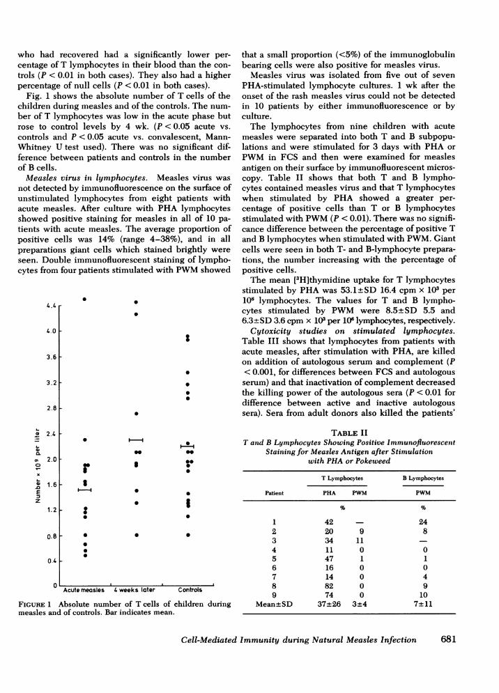
Fig. 1 shows the absolute number of T cells of thechildren during measles and of the controls. The num-ber of T lymphocytes was low in the acute phase butrose to control levels by 4 wk. (P <0.05 acute vs.controls and P < 0.05 acute vs. convalescent, Mann-Whitney U test used). There was no significant dif-ference between patients and controls in the numberof B cells.
Measles virus in lymphocytes. Measles virus wasnot detected by immunofluorescence on the surface ofunstimulated lymphocytes from eight patients withacute measles. After culture with PHA lymphocytesshowed positive staining for measles in all of 10 pa-tients with acute measles. The average proportion ofpositive cells was 14% (range 4-38%), and in allpreparations giant cells which stained brightly wereseen. Double immunofluorescent staining of lympho-cytes from four patients stimulated with PWMshowed
4.4
4.0
3.6
3.2 -
2.8 F
a}
L-
'.0o
Ez
2.4 t
2.0
1.6 F
1.21-
0.8 F
0.4 F
0
0 0
.
0
.
r
00
0
000
II
0
S
00*0000
0
0
0
0
Acute measles 4 weeks later Controls
FIGURE 1 Absolute number of T cells of children duringmeasles and of controls. Bar indicates mean.
that a small proportion (<5%) of the immunoglobulinbearing cells were also positive for measles virus.
Measles virus was isolated from five out of sevenPHA-stimulated lymphocyte cultures. 1 wk after theonset of the rash measles virus could not be detectedin 10 patients by either immunofluorescence or byculture.
The lymphocytes from nine children with acutemeasles were separated into both T and B subpopu-lations and were stimulated for 3 days with PHA orPWMin FCS and then were examined for measlesantigen on their surface by immunofluorescent micros-copy. Table II shows that both T and B lympho-cytes contained measles virus and that T lymphocyteswhen stimulated by PHA showed a greater per-centage of positive cells than T or B lymphocytesstimulated with PWM(P < 0.01). There was no signifi-cance difference between the percentage of positive Tand B lymphocytes when stimulated with PWM.Giantcells were seen in both T- and B-lymphocyte prepara-tions, the number increasing with the percentage ofpositive cells.
The mean [3H]thymidine uptake for T lymphocytesstimulated by PHAwas 53.1±+SD 16.4 cpm x 103 per106 lymphocytes. The values for T and B lympho-cytes stimulated by PWMwere 8.5± SD 5.5 and6.3±+SD 3.6 cpm x 103 per 106 lymphocytes, respectively.
Cytoxicity studies on stimulated lymphocytes.Table III shows that lymphocytes from patients withacute measles, after stimulation with PHA, are killedon addition of autologous serum and complement (P< 0.001, for differences between FCS and autologousserum) and that inactivation of complement decreasedthe killing power of the autologous sera (P < 0.01 fordifference between active and inactive autologoussera). Sera from adult donors also killed the patients'
TABLE IIT and B Lymphocytes Showing Positive Immunofluorescetit
Staining for Measles Antigen after Stimulationwith PHAor Pokeweed
T Lymphocytes B Lymphocytes
Patient PHA PWM PWM
1 42 242 20 9 83 34 114 11 0 05 47 1 16 16 0 07 14 0 48 82 0 99 74 0 10
Mean+SD 37+26 3±4 7+11
Cell-Mediated Immunity during Natural Measles Infection 681

TABLE IIIViable Lymphocytes after Stimulation with PHAand Incubation in Various Sera
Autologous Autologous Adult MeaslesDonor FCS serum + C serum - C serum + C serum + C
Acute measles (9) 60.7+8.2 29.4+8.5 45.4± 10.4 46.7±8.4Adult (8) 58.3±7.1 59.7±8.1 53.8±7.2
All percentages are ±SD.
stimulated lymphocytes (P < 0.001 for difference fromFCS), but the effect was not so great as when thepatients' own serum was used. Stimulated lymphocytesfrom healthy adult volunteers were not affected bytheir own serum but showed a slight and insignificantreduction in viability when incubated in acute-phasemeasles sera.
Unstimulated lymphocytes from the same patientswith measles were not killed on incubation with theirown sera and complement. The viability counts forthese cells were 67.3%+SD 11.8 in FCS and 66.3%+SD 8.9 in autologous serum.
PHAstimulation. The results of culturing lympho-cytes of patients and controls with a suboptimal doseof PHA(0.1 ,g/ml) or an optimal dose of PHA(5 ,ug/ml)are shown in Table IV. In the acute stage of measlesthe increase in [3H]thymidine uptake was significantlyhigher in medium containing FCS than in autologousserum at both concentrations of PHA, (P <0.05 for0.1 ,ug, P < 0.025 for 5 ,ug/ml PHA). Control childrenhad a significantly higher increase in [3H]thymidineuptake than children with acute measles in both typesof media at the low concentration of PHA (P < 0.05),but at optimal concentration only the increase in 10%autologous serum was significantly higher (P < 0.025).
[3H]Thymidine uptake in unstimulated cultures didnot differ significantly between groups or sera. Itaveraged 0.8+SD 0.5 x 10³ cpm per 106 lymphocytes.There was also no significant difference in viabilities
TABLE IVEffect of 10% Fetal Calf or Autologous Serum* on PHA
Response of Lymphocytest during Measles
0.1 jag PHA/ml 5 iLg PHA/ml
Autologous AutologousFCS serum FCS serum
Acute measles(10) 3.1±3.8 0.1±0.3 56.8±34.1 23.7±25.9
4 wk later (10) 9.9±17.0 0.5±0.9 67.8±49.9 52.0±61.2Controls (12) 17.5±20.8 12.2±19.2 37.7±19.3 56.5±33.5
* Obtained at the time of culture.$Expressed as mean increase in [3H]thymidine uptake in cpm x 103per 106 lymphocytes.
of stimulated or unstimulated lymphocytes in the vari-ous cultures. This averaged 66.0+12.8%.
In another set of experiments lymphocytes from 10children with acute measles were cultured with anoptimal dose of PHA in media containing either 15%pooled-acute, 15% pooled-recovered serum, or 15%FCS. The increase in [3H]thymidine uptake was sig-nificantly lower (P < 0.001) in acute-phase serum: thisaveraged 40.7+ SD31.6 x 103 cpm per 106 lymphocytesand 80.3+SD 51.0 and 95.8±SD 54.2 in recoveredand FCS, respectively.
Lymphocyte stimulation by Candida antigen. Themean uptake of [3H]thymidine in 10% autologousserum in unstimulated and Candida containing cul-tures in children with measles is shown in Table V.In the acute stage uptake was low in both unstimu-lated and antigen cultures and on recovery it rosesignificantly in both types of culture (P < 0.02 for bothcases). The rise on recovery was not obviouslyrelated to the presence of a positive skin test whichwas noted in 7 of the 10 patients at this time. The viabil-ities of lymphocytes (mean 67± SD 6.5%) did notdiffer significantly between any of the cultures.
In a separate set of experiments lymphocytes fromnine children with acute measles were cultured withCandida antigen in media containing either 20%pooled-acute or 20% pooled-convalescent serum.There was no significant difference in the increasein [3H]-thymidine uptake of stimulated over controlcultures in these media. This was 2.7+SD 3.9 and2.4±SD 4.6 x 10³ cpm per 106 lymphocytes for acuteand recovered serum, respectively.
TABLE VResponse of Lymphocytes* in 10O%Autologous SerumI to
Candida Antigen during Measles
Stage of disease Unstimulated Candida
Acute measles (10) 0.5+0.4 1.2±+1.44 wk later (10) 4.0+4.3 18.9±+19.8
* Expressed as mean [3H]thymidine uptake in cpm x 103per 106 lymphocytes.t Obtained at the time of culture.
682 Whittle, Dossetor, Oduloju, Bryceson, and Greenwood

Leukocyte migration to Candida antigen. Themean migration index for 17 healthy adult volunteerswho had a positive skin test to Candida antigenwas 0.69±SD 0.11 which was significantly lower thanthe mean of 0.87±SD 0.06 in 6 adults who had anegative skin test (P <0.01). This inhibition wasreversible by puromycin at a concentration of 10 jig/ml.
Fig. 2 shows the migration indices to heat-killedcandida cells in 12 patients during and 4-6 wk aftermeasles. The mean index for the children in theacute stage of measles was 0.84±SD 0.08 and onrecovery this fell to 0.75±SD 0.08 (P <0.02). Thosewith positive skin tests on recovery tended to havelower migration indices.
DISCUSSION
Our work shows that natural measles lowers the num-ber of T lymphocytes thus offering an explanationfor the transient lymphopenia reported in this infec-tion (18). A high proportion of T cells containedmeasles virus, and, when stimulated, lymphocyteswere lysed by autologous antibody and complement.We think this is one of the mechanisms by which theT cells are depleted. In contrast, few B cells wereshown to be infected and their numbers did not altersignificantly during the infection. It is also of interestthat the proportion of T cells remained low for over4 wk after the acute infection, which may explain
1-0 r
0.9
0.8
0.7x
~0a06- 05aL-
xn /.L
F
L
0.3 .
0.2 [
0.1
Acu te 4 weeksmeasles af ter
FIGURE 2 Migration indices to Candida antigen in childrenduring measles. Bar indicates mean. Open circle signifies apositive skin test to Candida antigen.
why cutaneous-delayed hypersensitivity reactions arediminished for several weeks after the acute infection(19). The proportion of T cells that contained virusvaried according to the degree of mitogenic stimu-lation, for when weakly stimulated by PWMno morepositive cells were seen than when B lymphocyteswere stimulated by this mitogen. However, if inmeasles T cells are more powerfully stimulated thanB cells, more T lymphocytes would express virus andso be more susceptible to lysis by antibody.
The proliferative response of lymphocytes to PHAwas also lowered during the acute phase of measles.This was shown to be the result of an inhib-itory factor in acute phase serum, for the lympho-cytes responded normally to an optimum dose of PHAin convalescent serum or FCS. This conclusion con-tradicts experimental evidence (10, 12) which suggeststhat the decreased [3H]thymidine uptake is the resultof direct viral inhibition of host-cell DNA synthesis,an effect which is only achieved with an unnaturallylarge dose of virus. Viability counts showed this in-hibition was not a result of killing of lymphocytes.We think it is separate from the cytotoxic effect ofundiluted acute-phase sera on measles-infected lym-phocytes which is critically dependent on the amountof complement, over 90% of lytic activity being lostwhen complement is diluted to 1:4 (13). In our lym-phocyte cultures sera were diluted 1:5 or 1:10 so comple-ment-dependent lysis in this system would have beenminimal or absent.
A third major finding was that the response oflymphocytes to Candida antigen was markedly de-pressed. The explanation for this, which is more com-plicated, lies in favor of a defect in antigen handling.We have some evidence of monocyte dysfunction inthat the response to a suboptimal dose of PHA wasdepressed (20). Also, the proliferative response ofacute-phase lymphocytes to antigen was lowered inboth acute and recovered serum which suggest thecells were at fault. Still further evidence of defectivelymphocyte or monocyte function was given by theabnormally high leukocyte migration indices in theacute stage of the illness. This indicates that leuko-cyte migration inhibitory factor, a soluble lymphocytefactor (21), was not produced at this stage. Impair-ments in polymorph function such as deficient randommigration and chemotaxis have been found in the acutestage of measles (22), but this is an unlikely ex-planation for our results as we have found leuko-cyte migration tests are still abnormal 2 wk after theonset of the rash (23), a time when polymorphfunction has returned to normal (22). Perhaps the in-fected macrophages are unable to process antigen orperhaps antigen receptors on the surface of lympho-cytes are disturbed. This possibility is raised as altera-tions in the cell membrane have been shown in cells
Cell-Mediated Immunity during Natural Measles Infection 6
1_-
U-4r
683

infected with Newcastle disease virus (24). In additionthese defects were reinforced by the suppressive effectof acute-phase serum on lymphocyte proliferation.This was apparent even in the unstimulated cultureswhich showed a low [3H]uptake in media containingthis serum.
These three major abnormalities, T-cell depletion, apowerful inhibitor in the serum, and defective antigenhandling help explain why cell-mediated immune re-sponses are profoundly depressed during measles. Thisis important in Nigeria for measles affects the youngwho are often malnourished, and in such children thedisease is severe and prolonged (23). As a result in-tercurrent infections such as tuberculosis, candidiasis(23), and herpes simplex,2 which are normally con-trolled by cell-mediated immunity, became rampantafter measles and contribute to the alarmingly highmortality of the disease (2).
ACKNOWLEDGMENTWewish to thank Jernina Werblinska for her excellent tech-nical help and J. K. Mayaki, Ibrahim Dabo, and Adama Ab-dullahi for their work in the clinic and field.
This study was supported by a grant from the MedicalResearch Council, England.
REFERENCES1. Whittle, H. C., A. Bradley-Moore, A. Fleming, and B. M.
Greenwood. 1973. Effects of measles on the immune re-sponse of Nigerian children. Arch. Dis. Child. 48:753-756.
2. Morely, D. 1969. Severe measles in the tropics. I.Br. Med. J. 1: 297-300.
3. Von Pirquet, C. 1908. Das verhalten der kutanen tuber-culin-reacktion wahrend der masem. Dtsch. Med. Woch-enischr. 34: 1297-1300.
4. Smithwick, E. M., and S. Berkovick. 1966. In vitrosuppression of the lymphocyte response to tuberculin bylive measles virus. Proc. Soc. Exp. Biol. Med. 123:276-278.
5. Oshunkoya, B. O., A. R. Cooke, 0. Ayeni, and T. A.Adejumo. 1974. Studies on leucocyte cultures in measles1. Lymphocyte transformation and giant cell formation inleucocyte cultures from clinical cases of measles. Arch.Virusforsh. 44: 313-322.
6. Oshunkoya, B. O., G. L. Adeleye, T. A. Adejumo, andL. S. Salimonu. 1974. Studies in leucocyte cultures inmeasles. Arch. Gesamte Virusforsch. 44: 323-329.
2 Unpublished observations.
7. Finkel, A., and P. B. Dent. 1973. Abnormalities inlymphocyte proliferation in classical and atypicalmeasles infection. Cell Immunol. 6: 41-48.
8. Newble, D. I., K. T. Holmes, A. G. Wangel, and I. J.Forbes. 1975. Immune reactions in acute viral hepatitis.Clini. Exp. Immunol. 20: 17-28.
9. Zweiman, B. 1971. In vitro effects of measles virus onproliferating human lymphocytes. J. Immunol. 106:1154-1158.
10. Zweiman, B. 1971. Effect of viable and nonviable measlesvirus on proliferating human lymphocytes. Int. Arch. Al-lergy. Appl. Immunol. 43: 600-607.
11. Zweiman, B., and M. F. Miller. 1974. Effects of non-viablemeasles virus on proliferating human lymphocytes. 2.Characteristics of the suppressive reaction. Int. Arch. Al-lergy. 46: 822-833.
12. Sullivan, J. L., D. W. Barry, P. Albrecht, and S. J. Lucas.1975. Inhibition of lymphocyte stimulation of measlesvirus.J. Immunol. 114: 1458-1461.
13. Joseph, B. S., N. R. Cooper, and M. B. A. Oldstone. 1975.Immunologic injury of cultured cells infected withmeasles virus. 1. Role of IgG antibody and the alterna-tive complement pathway. J. Exp. Med. 141: 761-774.
14. Joseph, B. S., P. W. Lampert, and M. B. A. Oldstone.1975. Replication and persistence of measles virus indefined subpopulations of human leucocytes.J. Virol. 16:1638-1649.
15. Kantor, F. S. 1975. Infection, anergy and cell-mediatedimmunity N. Engl. J. Med. 292: 629-634.
16. Jellife, D. B., 1966. The assessment of the nutritionalstatus of the community. Wld. Hlth Org. Geneva. 222.
17. Maini, R. N., L. M. Roffe, I. I. Magrath, and D. C.Dumonde. 1973. Standardization of the leucocyte migra-tion test. Itmt. Arch. Allergy Appl. Immutnol. 45: 308-324.
18. Benjamin, B., and S. M. Ward. 1932. Leucocyte responseto measles. Amer. J. Dis. Child. 44: 921-963.
19. Starr, S., and S. Berkovich. 1964. Effects of measles,gamma-globulin-modified measles and vaccine measleson the Tuberculin test. N. Engl. J. Med. 270: 386-391.
20. Oppenheim, J. J., B. G. Leventhal, and E. M. Hersh. 1968.The transformation of column-purified lymphocytes withnon-specific and specific antigenic stimuli. J. Immunol.101: 262-270.
21. Rocklin, R. E. 1974. Products of activated lymphocytes:leucocyte inhibitory factor (LIF) distinct from migrationinhibitory factor; (MIF).J. Immunol. 112: 1461-1466.
22. Anderson, R., A. R. Rabson, R. Sher, and H. J. Koornhof.1976. Defective Neutrophil motility in children withmeasles. J. Paediatrics. 89: 27-32.
23. Dossetor, J., H. C. Whittle, and B. M. Greenwood. 1977.Persistent measles infection in malnourished childrenBr. Med. J. 1: 1633- 1635.
24. Poste, G., and P. Reeve, 1974. Increased mobility andredistribution of concanavalin A receptors on cells in-fected with Newcastle Disease Virus. Nature (Lotnd.).247: 469-471.
684 Whittle, Dossetor, Oduloju, Bryceson, and Greetnwood