Cell Host & Microbe Resource - Victor Nizetnizetlab.ucsd.edu/Publications/TLR-Cofactors.pdf · Cell...
13
Cell Host & Microbe Resource Cofactors Required for TLR7- and TLR9-Dependent Innate Immune Responses Chih-yuan Chiang, 1,8 Alex Engel, 2,8 Amanda M. Opaluch, 1,8 Irene Ramos, 3 Ana M. Maestre, 3 Ismael Secundino, 4 Paul D. De Jesus, 1 Quy T. Nguyen, 1 Genevieve Welch, 6 Ghislain M.C. Bonamy, 6,7 Loren J. Miraglia, 6 Anthony P. Orth, 6 Victor Nizet, 4,5 Ana Fernandez-Sesma, 3 Yingyao Zhou, 6 Gregory M. Barton, 2 and Sumit K. Chanda 1, * 1 Infectious and Inflammatory Disease Center, Sanford-Burnham Medical Research Institute, La Jolla, CA 92037, USA 2 Division of Immunology and Pathogenesis, Department of Molecular and Cell Biology, University of California, Berkeley, Berkeley, CA 94720-3200, USA 3 Department of Microbiology and The Global Health and Emerging Pathogens Institute, Mount Sinai School of Medicine, New York, NY 10029, USA 4 Department of Pediatrics 5 Skaggs School of Pharmacy and Pharmaceutical Sciences University of California, San Diego, La Jolla, CA 92093, USA 6 The Genomics Institute of the Novartis Research Foundation, San Diego, CA 92121, USA 7 Hudson-Alpha Institute for Biotechnology, Huntsville, AL 35801, USA 8 These authors contributed equally to this work *Correspondence: [email protected] DOI 10.1016/j.chom.2012.02.002 SUMMARY Pathogens commonly utilize endocytic pathways to gain cellular access. The endosomal pattern re- cognition receptors TLR7 and TLR9 detect path- ogen-encoded nucleic acids to initiate MyD88- dependent proinflammatory responses to microbial infection. Using genome-wide RNAi screening and integrative systems-based analysis, we identify 190 cofactors required for TLR7- and TLR9-directed sig- naling responses. A set of cofactors were cross- profiled for their activities downstream of several immunoreceptors and then functionally mapped based on the known architecture of NF-kB signaling pathways. Protein complexes and pathways in- volved in ubiquitin-protein ligase activities, sphingo- lipid metabolism, chromatin modifications, and ancient stress responses were found to modulate innate recognition of endosomal nucleic acids. Addi- tionally, hepatocyte growth factor-regulated tyrosine kinase substrate (HRS) was characterized as neces- sary for ubiquitin-dependent TLR9 targeting to the endolysosome. Proteins and pathways identified here should prove useful in delineating strategies to manipulate innate responses for treatment of auto- immune disorders and microbial infection. INTRODUCTION Vertebrates use interconnected branches of the immune system to dictate responses to commensal and pathogenic microbes and to maintain host health. In one strategy of immune sensing, invariant receptors, such as toll-like receptors (TLRs), are employed to recognize conserved molecules associated with microorganisms. Activation of these receptors allows cells to integrate contextual cues and signal for tissue repair, inflamma- tion, and protective immunity (Kumar et al., 2011). Nucleic acid species are one class of microbial ligand sensed by multiple families of innate immune receptors. The recognition of nucleic acids within endosomes is medi- ated by TLR3, TLR7/8, and TLR9, which sense double-stranded RNA (dsRNA), single-stranded RNA (ssRNA), and DNA, respec- tively (Blasius and Beutler, 2010). Many ssRNA, dsRNA, and dsDNA viruses have been found to activate endosomal TLRs, including human immunodeficiency virus (HIV), influenza virus, Sendai virus (SeV), and Newcastle disease virus (NDV) (Kawai and Akira, 2006; Melchjorsen et al., 2005; Paun et al., 2008). Furthermore, both bacterial and parasitic genomes can activate TLR7 and TLR9 (Arpaia et al., 2011; Takeuchi and Akira, 2010; Zinkernagel et al., 2011). To sense pathogen infection, TLR7/9 must traffic from their site of synthesis in the endoplasmic reticulum (ER) to the endo- lysosomal network. Determinants for intracellular localization reside in both the transmembrane and cytosolic domains of these TLRs (Barton et al., 2006; Mouchess et al., 2011). The multipass transmembrane protein UNC93B1 is specifically required for endosomal TLR trafficking (Kim et al., 2008). During trafficking, TLR9 traverses the Golgi en route to acidified endoly- sosomes where it is proteolytically processed by cathepsins and asparagine endopeptidase to yield a functional N-terminally truncated receptor (Ewald et al., 2008; Park et al., 2008; Sepulveda et al., 2009). However, it remains unclear how sorting from the Golgi to the endolysosomal compartment is achieved. Intriguingly, the AP-3 complex, as well as protein-sorting com- plexes required for the formation of lysosome-related organelles, has been implicated in a late TLR9-trafficking event (Blasius et al., 2010; Sasai et al., 2010). Unlike TLR3, both TLR7 and TLR9 signal through the adaptor MyD88 to produce either inflammatory cytokines or type I inter- ferons (IFNs), depending on the cell type (Blasius and Beutler, 2010). In the proinflammatory arm of the signaling pathway, the 306 Cell Host & Microbe 11, 306–318, March 15, 2012 ª2012 Elsevier Inc.
Transcript of Cell Host & Microbe Resource - Victor Nizetnizetlab.ucsd.edu/Publications/TLR-Cofactors.pdf · Cell...

Cell Host & Microbe
Resource
Cofactors Required for TLR7- and TLR9-DependentInnate Immune ResponsesChih-yuan Chiang,1,8 Alex Engel,2,8 Amanda M. Opaluch,1,8 Irene Ramos,3 Ana M. Maestre,3 Ismael Secundino,4
Paul D. De Jesus,1 Quy T. Nguyen,1 Genevieve Welch,6 Ghislain M.C. Bonamy,6,7 Loren J. Miraglia,6 Anthony P. Orth,6
Victor Nizet,4,5 Ana Fernandez-Sesma,3 Yingyao Zhou,6 Gregory M. Barton,2 and Sumit K. Chanda1,*1Infectious and Inflammatory Disease Center, Sanford-Burnham Medical Research Institute, La Jolla, CA 92037, USA2Division of Immunology and Pathogenesis, Department of Molecular and Cell Biology, University of California, Berkeley, Berkeley,
CA 94720-3200, USA3Department of Microbiology and The Global Health and Emerging Pathogens Institute, Mount Sinai School of Medicine, New York,
NY 10029, USA4Department of Pediatrics5Skaggs School of Pharmacy and Pharmaceutical Sciences
University of California, San Diego, La Jolla, CA 92093, USA6The Genomics Institute of the Novartis Research Foundation, San Diego, CA 92121, USA7Hudson-Alpha Institute for Biotechnology, Huntsville, AL 35801, USA8These authors contributed equally to this work
*Correspondence: [email protected] 10.1016/j.chom.2012.02.002
SUMMARY
Pathogens commonly utilize endocytic pathwaysto gain cellular access. The endosomal pattern re-cognition receptors TLR7 and TLR9 detect path-ogen-encoded nucleic acids to initiate MyD88-dependent proinflammatory responses to microbialinfection. Using genome-wide RNAi screening andintegrative systems-based analysis, we identify 190cofactors required for TLR7- and TLR9-directed sig-naling responses. A set of cofactors were cross-profiled for their activities downstream of severalimmunoreceptors and then functionally mappedbased on the known architecture of NF-kB signalingpathways. Protein complexes and pathways in-volved in ubiquitin-protein ligase activities, sphingo-lipid metabolism, chromatin modifications, andancient stress responses were found to modulateinnate recognition of endosomal nucleic acids. Addi-tionally, hepatocyte growth factor-regulated tyrosinekinase substrate (HRS) was characterized as neces-sary for ubiquitin-dependent TLR9 targeting to theendolysosome. Proteins and pathways identifiedhere should prove useful in delineating strategies tomanipulate innate responses for treatment of auto-immune disorders and microbial infection.
INTRODUCTION
Vertebrates use interconnected branches of the immune system
to dictate responses to commensal and pathogenic microbes
and to maintain host health. In one strategy of immune sensing,
invariant receptors, such as toll-like receptors (TLRs), are
employed to recognize conserved molecules associated with
306 Cell Host & Microbe 11, 306–318, March 15, 2012 ª2012 Elsevie
microorganisms. Activation of these receptors allows cells to
integrate contextual cues and signal for tissue repair, inflamma-
tion, and protective immunity (Kumar et al., 2011). Nucleic acid
species are one class of microbial ligand sensed by multiple
families of innate immune receptors.
The recognition of nucleic acids within endosomes is medi-
ated by TLR3, TLR7/8, and TLR9, which sense double-stranded
RNA (dsRNA), single-stranded RNA (ssRNA), and DNA, respec-
tively (Blasius and Beutler, 2010). Many ssRNA, dsRNA, and
dsDNA viruses have been found to activate endosomal TLRs,
including human immunodeficiency virus (HIV), influenza virus,
Sendai virus (SeV), and Newcastle disease virus (NDV) (Kawai
and Akira, 2006; Melchjorsen et al., 2005; Paun et al., 2008).
Furthermore, both bacterial and parasitic genomes can activate
TLR7 and TLR9 (Arpaia et al., 2011; Takeuchi and Akira, 2010;
Zinkernagel et al., 2011).
To sense pathogen infection, TLR7/9 must traffic from their
site of synthesis in the endoplasmic reticulum (ER) to the endo-
lysosomal network. Determinants for intracellular localization
reside in both the transmembrane and cytosolic domains of
these TLRs (Barton et al., 2006; Mouchess et al., 2011). The
multipass transmembrane protein UNC93B1 is specifically
required for endosomal TLR trafficking (Kim et al., 2008). During
trafficking, TLR9 traverses the Golgi en route to acidified endoly-
sosomes where it is proteolytically processed by cathepsins and
asparagine endopeptidase to yield a functional N-terminally
truncated receptor (Ewald et al., 2008; Park et al., 2008;
Sepulveda et al., 2009). However, it remains unclear how sorting
from the Golgi to the endolysosomal compartment is achieved.
Intriguingly, the AP-3 complex, as well as protein-sorting com-
plexes required for the formation of lysosome-related organelles,
has been implicated in a late TLR9-trafficking event (Blasius
et al., 2010; Sasai et al., 2010).
Unlike TLR3, both TLR7 and TLR9 signal through the adaptor
MyD88 to produce either inflammatory cytokines or type I inter-
ferons (IFNs), depending on the cell type (Blasius and Beutler,
2010). In the proinflammatory arm of the signaling pathway, the
r Inc.

Cell Host & Microbe
Cofactors Required for TLR7/9 Signaling
kinases IRAK1, IRAK2, and IRAK4 activate the E3 ubiquitin ligase
TRAF6. Together with Ubc13 and Uev1A, TRAF6 catalyzes the
K63-linked ubiquitination of substrates, including TRAF6 itself,
as well as NEMO and TAK1. Ubiquitination of NEMO induces
the formation of the IKK signalosome, where activated TAK1
phosphorylates IKK-b, leading to IkBa phosphorylation and
degradation (Akira et al., 2006). This allows NF-kB to translocate
to the nucleus and, with the transcription factor AP-1, activate
proinflammatory cytokine production.
Ligand availability, receptor expression and localization, and
downstreamsignalingnetworksare tightly regulated toeffectively
enable host defense while simultaneously limiting autoimmunity.
Tomorecomprehensivelyunderstand themolecular components
that govern this process, we conducted a genome-wide RNAi
analysis of the cellular requirements for endosomally initiated
MyD88-dependent innate immune signaling. Integration of these
results with orthogonal systems-based data sets yielded func-
tional insight toward the regulatory hierarchies that preserve the
critical balance between productive innate immune responses
topathogenchallengeandhyperresponsiveness toautoantigens.
RESULTS
Genome-wide RNAi Analysis of MyD88-DependentEndocytic SignalingToestablishasystems-level understandingof thecompendiumof
genes that regulate innate immune responses to endosomal
nucleic acids, we performed genome-wide RNAi screens to iden-
tify cofactors required for TLR7/9 signaling. To elucidate regula-
tors of these pathways in a cell type that is tractable for high-
throughput genetics, we selected HEK293T cells. Although these
are not hematopoietic cells, they are able to recapitulate tran-
scriptional responses to TLR ligands after constitution with
appropriate receptors (Chuang and Ulevitch, 2004). To monitor
activities of these innate pathways, HEK293T cells stably ex-
pressing either TLR7 or TLR9 were engineered to harbor an
NF-kB luciferase reporter element (HEK293T/TLR7/NF-kB lucif-
erase or HEK293T/TLR9/NF-kB luciferase reporter cell lines).
Reporter cell lines were transfected with an arrayed genome-
wide siRNA library targeting �20,000 human genes (Konig
et al., 2009). Subsequently, cells were stimulated with a synthetic
TLR7 ligand,R848, or a TLR9 ligand, theCpG-containing oligonu-
cleotide ODN2006-G5, and NF-kB luciferase activity was moni-
tored (Figure 1A). We observed significant separation between
positiveandnegativecontrol siRNAs (p<10�100), anda large frac-
tion of canonical TLR signaling components, including MyD88,
IRAK1, TAK1, and IKK-a, were identified from the primary screen
(Figure S1A and Table S1 available online, and data not shown).
Integrative Evidence-Based Analysis of Genome-wideRNAi Data SetsNext, we used an integrative approach for candidate cofactor
selection that considered additional lines of functional, biochem-
ical, or transcriptional data (collectively called evidence-based
analysis; see Figure 1A, Figure S1B, and the Supplemental
Experimental Procedures). This approach is based on the
assumption that a factor is more likely to be a physiologically
relevant regulator of TLR signaling if multiple orthogonal lines
of evidence can support experimentally derived RNAi activities.
Cell Ho
Thus, in addition to weighing individual well and multiple well
RNAi activities for a gene (siRNA activity and RSA activity,
respectively), we also considered if a candidate factor has a
high level of reported protein interactions with other canonical
or candidate innate immune factors (protein network), is ex-
pressed in relevant cell types, or is induced by TLR stimulation
(gene expression) (Figure S1C) (Konig et al., 2007; Zhou et al.,
2005). We combined these multiple lines of evidence into
a weighted decision matrix and utilized a genetic algorithm to
prioritize those factors that were the most likely to function as
bona fide regulators of TLR signaling (Figures S1B and S1C,
Table S2, and Supplemental Experimental Procedures).
This integrative evidence-based approach identified 546
genes as strongly supported candidate TLR7/9 cofactors. The
activities of 190 cofactors were reconfirmed, employing cri-
teria wherein each gene must be targeted by at least two
sequence-independent siRNAs which attenuate R848- or CpG-
mediated TLR signaling activation, while not significantly
affecting cellular viability. These criteria significantly reduce,
but do not eliminate, the possibility of RNAi-mediated off-
target effects. Importantly, 80% of the known components of
the TLR7/9 signaling pathways were identified in the primary
screen and reconfirmed using these approaches (Table S1 and
Table S2).
Gene ontology analyses of the 190 reconfirmed innate
signaling cofactors revealed, as expected, enrichment of path-
ways and functions associated with innate immunity, including
TLR and IL-1R signaling, innate responses to RNA viruses,
NF-kB activation, and inflammation (Table S3). Furthermore,
a collection of cofactors that are involved in chromatin modifica-
tion, the sphingomyelin and ceramide synthesis pathways, and
the unfolded protein response (UPR) were also enriched in this
analysis (Figure S1D and Table S3). Significantly, 42 candidate
cofactors could be genetically linked to multiple immune-related
diseases through genome-wide association studies (GWAS) and
expression analyses (Table 1 and Table S4).
To determine if the factors confirmed in the screen were ex-
pressed in cell types relevant to TLR7 and TLR9 surveillance
activities, we extracted mRNA expression profiles from the
GNF Tissue Atlas (Su et al., 2002). The expression of 67% of
the confirmed cofactors could be detected in cells and tissues
of myeloid and lymphoid origin, and more than 50% were
predominantly expressed in hematopoietic cell types (p < 3 3
10�6), including plasmacytoid dendritic cells (pDCs) (Figure S1E).
Furthermore, 30 of these genes were found to have highly signif-
icant expression correlation coefficients (r2 = 0.69) with TLR7
and MyD88 (Figure 1B). Taken together, gene expression
profiles of these identified molecules are largely consistent
with their potential roles as TLR7/9 innate signaling cofactors.
We also evaluated the density of protein interactions between
the 190 confirmed factors and known TLR signaling pathway
members (see the Supplemental Experimental Procedures).
This analysis identified a highly significant protein interaction
network (p < 0.001) containing 105 total proteins and 85 con-
firmed gene products (Figure S1F). By expanding this network
to include 546 primary screen hits, we generated an additional
protein interaction network including 191 proteins and 109
confirmed factors (Figure 1C and Figure S1G). This interaction
map reflects the probable biochemical hierarchies of innate
st & Microbe 11, 306–318, March 15, 2012 ª2012 Elsevier Inc. 307
A
C
siRNA Activity Level
Proposed Mechanism
Inactive Active
TLR7/9 Specific (new)
TLR7/9 Specific (known)
MyD88-dependent Signaling (new)
MyD88-dependent Signaling (known)
General NF- B Activity (new)
General NF- B Activity (known)
D
BBloodOthers
Known Modulators
Novel Modulators
Relative Tissue Expression
highlow
TL
R7
TL
R9
TN
FR
Me
ch
an
ism
TL
R5
IL-1
R
DKC1 MMP2 RXRB CD40 RING1 PHPT1 PKN2 JAK1 NTRK1 SP3 NME2P1 TMPRSS11D USP33 BRD2 USP24 THUMPD2 UQCRC1 GNG8 VARS HIST1H3I HRS EPC1 FBXW11 WRN KRT2 LAMA3 SNRPD3 PDK2 DVL3 UNC93B1 CDK9 PIGF FBXL7 C4B C10orf125 ADAM15 CYP2F1 ANKRD31 HERPUD1 SMURF1 PPFIA4 SPTAN1 SCARF2 MYD88 IRAK1 MED12 KIR2DS5 EIF3A PSAP GTF2A2 MAP2K5 MUC17 ASB10 HIST1H2BO PRMT2 DTX3 SSTR5 DVL1 CELSR1 PROKR2 KIFC2 KDM5C KCNJ11 SCFD1 CDK18 SNRPA1 QARS CTSF PSMA8 MEX3A CCR7 CTSL1 GHSR LIF CARD11 TM4SF18 LRSAM1 ARSE APOB FLG CEACAM16 SPEN POLR2C CHUK RELA
p<0.001
Reconfirmation &
Toxicity Analysis
Genome-wide
RNAi Analysis
Integration of RNAi data
with Protein Interaction,
Gene Expression, Pathway,
and Gene Ontology Analyses
At least 2 Active
siRNAs/Gene with
Minimal Toxicity
HEK293T/TLR7/NF- B Luciferase HEK293T/TLR9/NF- B Luciferase
6 siRNAs/Gene
Targeting 20,000
Human Genes
Evidence-based
Hit Selection546
Genes
190
Genes
R848CpG
DNA
Pro
po
se
d
MyD88
UBE2V1
UBE2N
TRAF6 ECSIT
TLR7
UNC93B1
IRAK4
TLR9
IRAK1
TAB3TAB1 TAK1
TAB2
IKBKB
NFKB1RELA
IKBKG
NFKBIA
CHUK
TLR7 MYD88 NFKB1 UNC93B1
IKBKG
TAB2
IRAK1
NFKBIA UBE2V1
CHUK
Blood Others
RELA
Figure 1. Genome-wide Integrative RNAi Analysis of TLR7- and TLR9-Mediated Innate Immune Responses
(A) HEK293T/TLR7/NF-kB or HEK293T/TLR9/NF-kB reporter cell lines were reverse transfected with an arrayed genome-wide siRNA library and stimulated with
cognate ligands, and activation of the NF-kB pathway was monitored (yellow shaded area). Integration of these data with orthogonal systems-level data sets
drove the selection of 546 genes (tan shaded area). One hundred ninety genes were subsequently confirmed to be required components of the TLR7 and/or TLR9
signaling response (blue shaded area).
(B) Tissue expression of 30 confirmed factors that have the strongest correlations with expression profiles of both TLR7 and MyD88. Tissues and cell types are
depicted on the x axis (Blood, hematopoietic origin; Others, nonhematopoietic origin). Genes identified through the RNAi analyses are reflected on the y axis.
Previously known or unknown pathway members are indicated by black or blue bars, respectively. A continuum of blue (low expression) to red (high expression)
depicts relative expression levels.
Cell Host & Microbe
Cofactors Required for TLR7/9 Signaling
308 Cell Host & Microbe 11, 306–318, March 15, 2012 ª2012 Elsevier Inc.

Table 1. Confirmed Factors Involved in Autoimmune Diseases, Inflammatory Diseases, and Viral Infection
Disease Analysis TLR7/9 Signaling Cofactors P Value
Systemic sclerosis GWAS RXRB, BRD2, RING1 0.000084
Systemic lupus erythematosus GWAS PHRF1 3.00E-10
Expression profiling EIF2AK2, PSMA6, UNC93B1, CTSL1, CASP3,
TMUB2, MYD88, USP33, C6orf125, TLR7, PKN2
0.0333
Crohn’s disease GWAS FUT2, LOC400456, CAPN10, CELSR1, GNAS,
KRT2, LDLRAD1, LINGO2MAP2K5, MAST2, MKL2,
MUC17, SMURF1, SP3, TRPC4AP, USP33, WRN
<9.7E-4
Multiple sclerosis GWAS CD40, BRD2 <9.712E-5
Expression profiling BCKDK, TBL3, POLR2I, PFKL, UNC93B1, ECSIT, RING1,
MTMR14, COMT, KDM5C, HRS, RELA, MAP3K14, IKBKG,
STK11, DVL1, POLR2C
0.0131
Inflammatory bowel disease Expression profiling ADAM15, KDM5C, MLL5, UNC13D, FBXW11, PIH1D1,
BRD2, MMP2, WRN, PFKL, ACTN4, TEX264, MAP2K5,
ANKRD31, CD40
0.000021
Rheumatoid arthritis GWAS CD40, ADRA1B, BRD2, APOB, C6orf125, CAPN10,
GNAS, IL20RA, LAMA3, LINGO2, LRSAM1, MAST2,
POLR2C, RING1
<9.7E-4
Systematic juvenile idiopathic
arthritis
Expression profiling NR4A2, ANPEP, UQCRC1, NFKBIA, KIFC2, BCKDK,
COMT, PRDX5, VPS16, POLR2C, BRD2, FTL, PTPN6,
MED30, GNAS
0.0232
Psoriasis Expression profiling KRT2, MYD88, CTSL1, TUBA3C, UBE2N, NFKB1,
IRAK1, FLG, VCP, EIF3F, BRD2, GTF2A2, PSMA6,
PSMA5, SPEN
0.0461
Staphylococcus aureus
infection
Expression profiling CCR7, NR4A2, LIF, EIF2AK2, CD40, NDP, RELA,
NFKB1, CSF1, NFKBIA, UNC93B1, CTSL1, MYD88,
PNRC1, DVL3
0.0067
Dengue virus infection Expression profiling C6orf125, VARS, CTSL1, HSP90B1, C1orf112, TBL3,
RHBDD3, VCP, GHSR, MMP2, CAD, PHPT1, UQCRC1,
POLR2I, SOX9
6.2E-10
Type I diabetes GWAS HIST1H3I, RXRB, VARS, BRD2, RING1, HIST1H2BO, C4B,
MAP3K7, UBASH3A, C6orf125, LINGO2, VARS
<0.0125
References are contained in Table S4. Explanation of the column headings in table are as follows: Disease, name of the medical condition; Analysis,
type of study conducted; TLR7/9 Signaling Cofactors, name of the cofactors that are associated with the disease; P Value, p value associated with that
overlap.
Cell Host & Microbe
Cofactors Required for TLR7/9 Signaling
immune regulatory proteins within the context of the canonical
TLR7/9 signaling pathway. By identifying dense local interac-
tions, our analysis also suggested that the identified regulatory
proteins participate in higher-order biochemical interactions
and protein complexes (Table S5 and Figures S1H–S1N).
Functional Classification of TLR7/9 CofactorsTo functionally characterize the regulatory role of 80 confirmed
cofactors, we profiled their ability to modulate the NF-kB
signaling pathway in response to ligand stimulation through
TNF-a receptor (TNFR), IL-1b receptor (IL-1R), and TLR5. Based
upon the known signaling architecture of these pathways, we
were able to infer the likely impact upon individual steps of the
TLR pathway for each gene (Figure 1D). RNAi against 29 genes,
as well as UNC93B1, specifically impaired TLR7- and TLR9-
(C) The network relationship between identified innate regulators and the canonica
expression patterns are distinguished by differential edge color, node color, and
(D) Confirmed TLR7/9 signaling cofactors were crossprofiled for their activities u
dependent reporter. Data used to generate the heat map depicted in (D) are repr
Also see Figure S1.
Cell Ho
mediated NF-kB activation, suggesting this category of genes
may be involved in trafficking, processing, signaling, or tran-
scriptional events associated specifically with TLR7/9 (Figure 1D;
‘‘TLR7/9 Specific’’). RNAi against 13 genes, in addition toMyD88
and IRAK1, impaired TLR5-, TLR7-, TLR9-, and IL-1R-mediated
NF-kB activation, indicating that these genes are preferentially
required for MyD88-dependent signaling responses (Figure 1D;
‘‘MyD88-dependent Signaling’’). Finally, RNAi against 38 genes,
as well as IKK-a and p65, impaired all interrogated receptor
signaling pathways, implicating these genes in the global regula-
tion of NF-kB-dependent responses (Figure 1D; ‘‘General NF-kB
Activity’’).
Next, we manually curated the subcellular localizations and
known functions of these 80 mediators of TLR7/9 signaling.
This information was integrated with ligand profiling data
l TLR7/9 signaling components is depicted. (Interaction type, protein type, and
node shape, respectively, as indicated in Figure S1G.)
pon TLR5-, TLR7-, TLR9-, TNFR-, and IL-1R-mediated induction of a NF-kB-
esentative of duplicate assays, with three wells per gene tested in each assay.
st & Microbe 11, 306–318, March 15, 2012 ª2012 Elsevier Inc. 309
Chromatinmodification
Acetylation Methylation Citrullination
Ribonuclearprotein
complex
HIST1H
2BO
PHF17
KDM5C
LysosomeEndosome
Kinase/Phosphatase
Metabolism
UB/DUB
TLR7/9
TLR7/9
UNC93B1
HRS
PDK2
TLR7/9
PPFIA4VARS
QARS
USP33USP24SCFD1FUT2
PRMT2
MAP2K5
FBXW9
FBXL7
SNRPA1DKC1
SNRPD3
GNG8BRD2
Transcription/Initiation/Elongation
EPC1
RING1
WRN
ARSE
HERPUD1
VC
P
CYP2F1M
ED12
SPEN
POLR
2C
POLR
2I
GHSR
KIR2DS5
KCNJ11
SSTR5
PROKR2
TM4SF18
UQCRC1
BCKDK
HDAC5
HECTD1
LRSAM1
ASB10
FB
XW
5
TRPC4AP
EIF3FEIF2AK2
NR4A2
Translation
ANPEP
KIR3DL1
GNAS
STK11
MAP3K
14
PTPN6
SpliceosomeRBM5
SOX9MED30
MED19
GTF2A2
HNF1A
SMURF1
PSMA8
DTX3
PSMA5
Proteasome
PFKL
DVL3
KRT2
TUBA3D
SPTAN1
FLG
ACTN4
Cytoskeleton
FBXW11
JAK1PHPT1
Trans-
mem
brane
Wnt
DVL1
MyD88
TRAF6
IκBα
p65
p65
IKKα/β/γ
DUPD1
Cell
Adhesion
LAMA3SCARF2
CELSR1
MUC17
ADAM15
CD40
TAK1
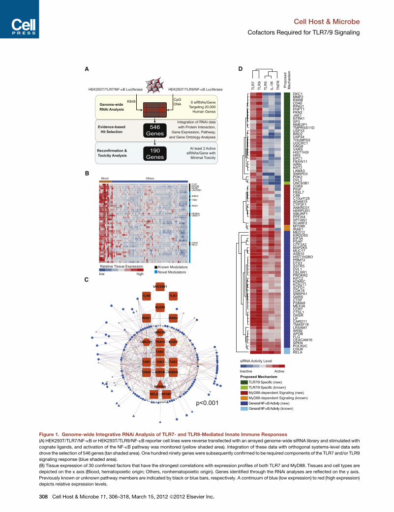
Figure 2. Comprehensive Model of Functional and Regulatory Roles of Confirmed TLR7/9 Cofactors
Subcellular localization and functional data for identified innate regulators were curated using multiple databases (Table S6). Innate cofactors were mapped to
their likely regulatory roles in TLR7 and TLR9 signaling (solid circles). SVM-based gene regulatory activity predictions are also depicted (open circles) (see
Table S6). TLR7/9-specific factors are represented by green nodes, MyD88-dependent factors are indicated by red nodes, and regulators of general NF-kB
activation are shown as blue nodes. Lines between proteins predict biochemical interactions (see Figure 1D).
Cell Host & Microbe
Cofactors Required for TLR7/9 Signaling
(Figure 1D) and protein interaction data (Figures S1F and S1G) to
construct a comprehensive model that reflects the likely func-
tional and regulatory intersections between the innate signaling
molecules identified here and the canonical TLR7/9 pathway
(Figure 2 and Table S6; also see the Supplemental Experimental
Procedures).
Molecular and Biochemical Analyses of ConfirmedCofactorsSubsequently, to verify that depletion of the confirmed innate
cofactors impacted the transcriptional response to TLR ligands,
we profiled the role of these factors in the induction of NF-kB-
dependent target genes. Silencing of >80% of confirmed factors
in HEK293T cells reduced IL-8 target gene induction (Figure S2A
and Table S7). Furthermore, of the 91 genes found to most
potently regulate TLR7/9 responses, inhibition of 78 genes
(85.7%) decreased R848-induced ICAM-1 mRNA expression in
THP-1 monocytic cells (Figure 3A, Figures S2A and S2B, and
Table S7). Importantly, these data demonstrate that the cofac-
tors identified by our analysis impact innate immune signaling
in a myeloid-lineage cell line.
Next, we measured the effects of siRNA-mediated depletion
of confirmed factors upon R848-induced p65 nuclear transloca-
tion. An evaluation of both target gene data and p65 localization
data allowed us to distinguish between innate cofactors with
310 Cell Host & Microbe 11, 306–318, March 15, 2012 ª2012 Elsevie
nuclear versus cytoplasmic activities (Figures S2A and S2C,
and Table S7). For example, siRNAs targeting several mitochon-
drial-, ER-, or cytoplasmic-localized proteins impaired not only
IL-8 mRNA expression but also p65 nuclear translocation, sug-
gesting these genes regulate upstream TLR signaling events.
In contrast, siRNAs targeting a group of nuclear localized pro-
teins inhibited IL-8 mRNA expression while p65 nuclear trans-
location remained intact, providing evidence to support a role
for these genes in transcriptional or epigenetic control of the
inflammatory response (Figure 2).
Finally, we tested a subset of confirmed TLR7/9 cofactors for
their ability to regulate the endosomal response to SeV infection.
Since SeV robustly induces a RIG-I-mediated immune response
(Melchjorsen et al., 2005), we evaluated the cofactors in a RIG-I-
deficient background. Silencing of TLR7, UNC93B1, or MyD88
significantly reduced induction of NF-kB by SeV, indicating
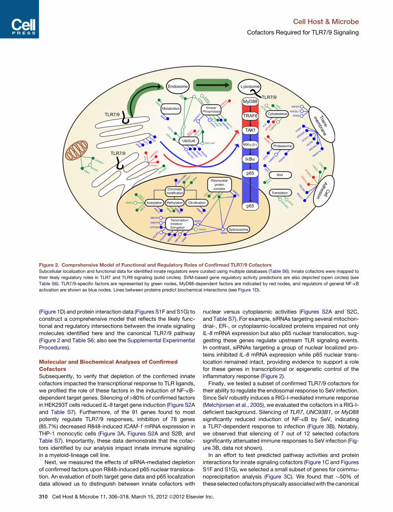
a TLR7-dependent response to infection (Figure 3B). Notably,
we observed that silencing of 7 out of 12 selected cofactors
significantly attenuated immune responses to SeV infection (Fig-
ure 3B, data not shown).
In an effort to test predicted pathway activities and protein
interactions for innate signaling cofactors (Figure 1C and Figures
S1F and S1G), we selected a small subset of genes for coimmu-
noprecipitation analysis (Figure 3C). We found that �50% of
theseselectedcofactors physically associatedwith the canonical
r Inc.

Cell Host & Microbe
Cofactors Required for TLR7/9 Signaling
TLR pathway member predicted by our protein-protein interac-
tion analysis (Figure 3C and Figure 4A; data not shown). For
example, protein network analyses suggested that pyruvate
dehydrogenase kinase, isozyme 2 (PDK2) physically interacts
with TRAF6, Uev1A, Ubc13, and TAK1 through an intermediate
protein (Figure 1C and Figure S1G); coimmunoprecipitation
confirmed that PDK2 does physically associate with both
TRAF6 and TAK1, although we were unable to detect a physical
interaction between PDK2 and Uev1A or Ubc13 (Figure 3C). In
addition, PDK2 depletion inhibited ligand-induced expression of
IL-8, p65 nuclear translocation, and altered IkBa degradation
kinetics (Figures 3D–3F, Figure S2D); this suggests that PDK2
functions upstream of IKK activation, and is consistent with the
predicted PDK2 activity derived from ligand profiling studies (Fig-
ure 1D). Additional coimmunoprecipitation experiments revealed
biochemical interactions between the ubiquitin protein ligase,
FBXL7 (F box and leucine-rich repeat protein 7), and TLR7/9
pathwaymembers IRAK1, TRAF6, andTAK1,which are all known
be modified by ubiquitin following receptor activation (Conze
et al., 2008; Takeuchi and Akira, 2010) (Figure 3C). Interestingly,
we observed that TLR pathway members interacted with ex-
pected (�54 kDa) and higher molecular weight forms of FBXL7,
suggesting that this F box protein may be subject to posttransla-
tional modification. Since depletion of FBXL7 also impaired IkBa
degradation kinetics, p65 nuclear translocation, and IL-8 upregu-
lation (Figures 3D–3F, and Figure S2E), our data suggest that
FBXL7 may be a MyD88-dependent regulator of ubiquitination
events that occur prior to the activation of the IKK signalosome
complex.
Finally, coimmunoprecipitation experiments using ectopically
expressed tagged TLR9 and HRS revealed that TLR9 interacted
with HRS, and the affinity of HRS for TLR9 was stronger than
its affinity for cell-surface-expressed TLR2 (Figure 4A). Taken
together with data demonstrating that HRS was uniquely
required for endosomal signaling (Figure 1D and Figure 2),
this finding was of particular interest, since HRS has been
shown to mediate plasma membrane-associated receptor
trafficking toward the lysosome (Saksena et al., 2007; Wegner
et al., 2011).
A Role for HRS in Endosomal TLR TraffickingTo pursue the hypothesis that HRS might play a role in targeting
TLR7/9 to the endolysosomal compartment, we verified deple-
tion of HRS protein and further assessed the impact of HRS on
NF-kB signaling (Figure S3A). In HRS-depleted cells, NF-kB
promoter activation was significantly reduced after stimulation
with R848 or CpG, but remained intact after stimulation with
TNF-a (Figure 4B). Furthermore, R848-induced secretion of
IL-8 was abrogated with HRS knockdown in HEK293T cells (Fig-
ure 4C), and HRS silencing in THP-1 cells reduced target gene
induction in response to TLR7 ligand, but not TNF-a, or the
TLR2 agonist Pam3CSK4 (Figure S3B). HRS knockdown in
HEK293T cells also resulted in the blockage of p65 nuclear trans-
location in response to R848, as well as the impairment of ligand-
mediated IkBa degradation (Figures 4D and 4E). Importantly,
silencing of HRS in primary macrophage-derived dendritic cells
(MDDCs) reduced TLR-dependent induction of TNF-a and
IFN-a following infection with NDV (Figure 4F), underscoring
the critical role of HRS in the endosomal innate response to virus
Cell Ho
infection. Together, these results indicate that HRS specifically
regulates TLR7/9-initiated signaling events that occur prior to
TAK1 activation, and are consistent with a role for HRS in endo-
somal receptor trafficking.
When delivered to acidified endosomal compartments, TLR9
(150 kDa) is proteolytically processed, generating an �80 kDa
cleaved species that is a hallmark of proper TLR9 trafficking
(Ewald et al., 2008; Park et al., 2008). Furthermore, in the
absence of the trafficking chaperone UNC93B1, no cleaved
TLR9 species is observed (Figure 5A). In order to evaluate if
HRSwas required for this endosomal trafficking event, we exam-
ined whether TLR9 was properly cleaved following HRS knock-
down. Silencing of HRS in a RAW264.7 cell line resulted in
a 70% reduction of HRS protein levels, which corresponded to
an �60% decrease in the abundance of cleaved TLR9 (Fig-
ure 5A). Thus, these data support an essential role for HRS in
TLR9 receptor trafficking.
It has been previously shown that HRS recognizes ubiquiti-
nated cargo (Saksena et al., 2007). Because HRS physically
interacts with TLR9 and is required for endolysosomal localiza-
tion of the receptor, we investigated the role of TLR9 ubiq-
uitination in receptor trafficking. When TLR9 was immunopre-
cipitated from immortalized macrophages or RAW264.7 cells
expressing wild-type (WT) TLR9-HA, a high molecular weight
ubiquitinated species was detected (Figure 5B and data not
shown). Mutation of the three lysine residues in the cytosolic
linker and TIR (toll IL-1 receptor) domain of murine TLR9
(K878, K932, K963, called ‘‘TLR9 KallR mutant’’) resulted in
a highly significant reduction in the amount of ubiquitin staining
in the TLR9 immunoprecipitate, suggesting that TLR9 is directly
ubiquitinated (Figure 5B). Since HRS binds ubiquitinated cargo
at the surface of early endosomes, we evaluated whether TLR9
ubiquitination might occur at this location (Saksena et al.,
2007). In MEF cell lines, ectopic expression of UNC93B1 is
required for the exit of TLR9 from the ER (Ewald et al., 2008).
Consistent with ubiquitination of TLR9 in a post-ER com-
partment, coexpression of UNC93B1 was required for ubiq-
uitination of TLR9 (Figure 5C). This UNC93B1-dependent
ubiquitination also required TLR9 cytoplasmic lysines. Impor-
tantly, we find that while WT TLR9 strongly interacted with
HRS, the TLR9 KallR mutant exhibited a very low affinity for
HRS, suggesting that HRS specifically binds the ubiquitinated
receptor (Figure 5D).
To determine the functional relevance of TLR9 ubiquitination
for signaling, we stimulated TLR9�/� immortalizedmacrophages
expressing single, double, and triple lysine-substituted con-
structs and measured the production of TNF-a. Whereas WT
TLR9 and TLR9-K878,963R were able to robustly produce
TNF-a in response to CpG-B oligonucleotides, signaling by
TLR9-K932R was strongly diminished, and the TLR9 KallR
mutant was completely defective for signaling (Figure 5E).
Though TLR9 K932 is strongly conserved across all TLR family
members (Figure S4A), this lysine is not universally required for
TLR signaling, as an analogous substitution in TLR2 (K698R)
did not alter responsiveness to Pam3CSK4 (Figure S4B). How-
ever, substitution of two corresponding lysines in TLR7 (K952
and K953) reduced R848-mediated signaling, possibly reflecting
an important and unique role for ubiquitination of these lysines in
endosomal TLR function (Figure S4C).
st & Microbe 11, 306–318, March 15, 2012 ª2012 Elsevier Inc. 311
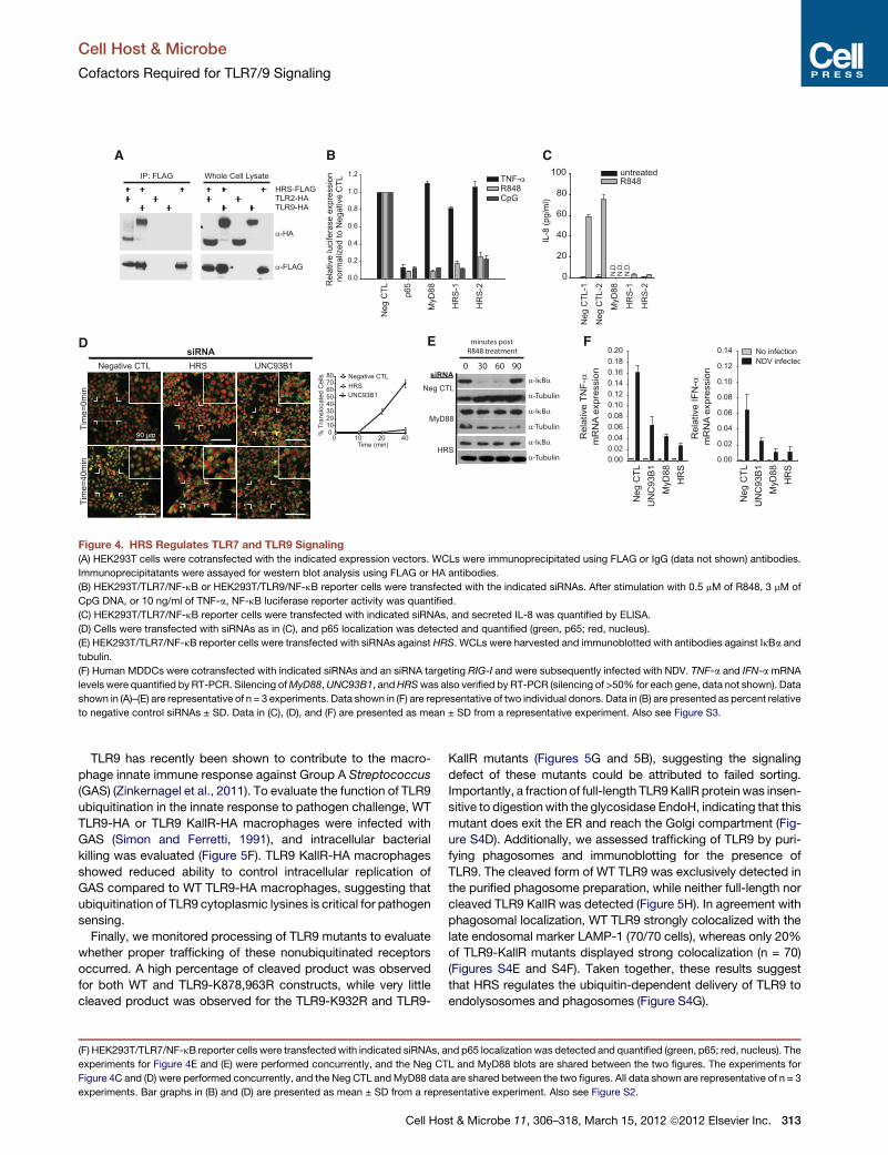
Figure 3. Characterization of Selected Confirmed Genes onto the TLR Signaling Pathway(A) THP-1 cells were transfected with indicated siRNAs and stimulated with 5 mMof R848. Five hours poststimulation, mRNA levels of ICAM-1were quantified by
RT-PCR. The red bar reflects a threshold at which 50% of the ICAM-1 mRNA expression was inhibited compared to the negative control (Neg CTL) siRNA.
(B) HEK293T/TLR7/NF-kB reporter cells were transfected with the indicated siRNAs together with an siRNA targeting RIG-I and infected with Sendai virus, and
luciferase reporter activity was quantified.
(C) HEK293T cells were cotransfected with indicated expression vectors. Whole-cell lysates (WCLs) were immunoprecipitated using FLAG antibody.
Immunoprecipitatants were assayed by western blot analysis using FLAG or Myc antibodies.
(D) HEK293T/TLR7/NF-kB reporter cells were transfected with indicated siRNAs and stimulated with 3 mM R848 for 12 hr, and secreted IL-8 was quantified by
ELISA.
(E) HEK293T/TLR7/NF-kB reporter cells were transfected with indicated siRNAs. Cells were stimulated with 10 mMof R848, and WCLs were immunoblotted with
antibodies against IkBa and tubulin.
Cell Host & Microbe
Cofactors Required for TLR7/9 Signaling
312 Cell Host & Microbe 11, 306–318, March 15, 2012 ª2012 Elsevier Inc.
A B C
D E F
Figure 4. HRS Regulates TLR7 and TLR9 Signaling(A) HEK293T cells were cotransfected with the indicated expression vectors. WCLs were immunoprecipitated using FLAG or IgG (data not shown) antibodies.
Immunoprecipitatants were assayed for western blot analysis using FLAG or HA antibodies.
(B) HEK293T/TLR7/NF-kB or HEK293T/TLR9/NF-kB reporter cells were transfected with the indicated siRNAs. After stimulation with 0.5 mM of R848, 3 mM of
CpG DNA, or 10 ng/ml of TNF-a, NF-kB luciferase reporter activity was quantified.
(C) HEK293T/TLR7/NF-kB reporter cells were transfected with indicated siRNAs, and secreted IL-8 was quantified by ELISA.
(D) Cells were transfected with siRNAs as in (C), and p65 localization was detected and quantified (green, p65; red, nucleus).
(E) HEK293T/TLR7/NF-kB reporter cells were transfected with siRNAs against HRS. WCLs were harvested and immunoblotted with antibodies against IkBa and
tubulin.
(F) Human MDDCs were cotransfected with indicated siRNAs and an siRNA targeting RIG-I and were subsequently infected with NDV. TNF-a and IFN-amRNA
levels were quantified by RT-PCR. Silencing ofMyD88,UNC93B1, andHRSwas also verified by RT-PCR (silencing of >50% for each gene, data not shown). Data
shown in (A)–(E) are representative of n = 3 experiments. Data shown in (F) are representative of two individual donors. Data in (B) are presented as percent relative
to negative control siRNAs ± SD. Data in (C), (D), and (F) are presented as mean ± SD from a representative experiment. Also see Figure S3.
Cell Host & Microbe
Cofactors Required for TLR7/9 Signaling
TLR9 has recently been shown to contribute to the macro-
phage innate immune response against Group A Streptococcus
(GAS) (Zinkernagel et al., 2011). To evaluate the function of TLR9
ubiquitination in the innate response to pathogen challenge, WT
TLR9-HA or TLR9 KallR-HA macrophages were infected with
GAS (Simon and Ferretti, 1991), and intracellular bacterial
killing was evaluated (Figure 5F). TLR9 KallR-HA macrophages
showed reduced ability to control intracellular replication of
GAS compared to WT TLR9-HA macrophages, suggesting that
ubiquitination of TLR9 cytoplasmic lysines is critical for pathogen
sensing.
Finally, we monitored processing of TLR9 mutants to evaluate
whether proper trafficking of these nonubiquitinated receptors
occurred. A high percentage of cleaved product was observed
for both WT and TLR9-K878,963R constructs, while very little
cleaved product was observed for the TLR9-K932R and TLR9-
(F) HEK293T/TLR7/NF-kB reporter cells were transfected with indicated siRNAs, a
experiments for Figure 4E and (E) were performed concurrently, and the Neg CT
Figure 4C and (D) were performed concurrently, and the Neg CTL andMyD88 data
experiments. Bar graphs in (B) and (D) are presented as mean ± SD from a repre
Cell Ho
KallR mutants (Figures 5G and 5B), suggesting the signaling
defect of these mutants could be attributed to failed sorting.
Importantly, a fraction of full-length TLR9KallR proteinwas insen-
sitive to digestionwith the glycosidase EndoH, indicating that this
mutant does exit the ER and reach the Golgi compartment (Fig-
ure S4D). Additionally, we assessed trafficking of TLR9 by puri-
fying phagosomes and immunoblotting for the presence of
TLR9. The cleaved form of WT TLR9 was exclusively detected in
the purified phagosome preparation, while neither full-length nor
cleaved TLR9 KallR was detected (Figure 5H). In agreement with
phagosomal localization, WT TLR9 strongly colocalized with the
late endosomal marker LAMP-1 (70/70 cells), whereas only 20%
of TLR9-KallR mutants displayed strong colocalization (n = 70)
(Figures S4E and S4F). Taken together, these results suggest
that HRS regulates the ubiquitin-dependent delivery of TLR9 to
endolysosomes and phagosomes (Figure S4G).
nd p65 localization was detected and quantified (green, p65; red, nucleus). The
L and MyD88 blots are shared between the two figures. The experiments for
are shared between the two figures. All data shown are representative of n = 3
sentative experiment. Also see Figure S2.
st & Microbe 11, 306–318, March 15, 2012 ª2012 Elsevier Inc. 313

A
E F G
B C D
H
Figure 5. TLR9 Ubiquitination, Signaling, and Cleavage Are Dependent on TLR9 Cytoplasmic Lysines
(A) RAW264.7-TLR9-HA cells were transducedwith the indicated shRNAs.WCLswere harvested and immunoblotted with antibodies against HA, HRS, or tubulin.
The ratio between cleaved TLR9 and tubulin was quantified by densitometry.
(B) WCLs from TLR9�/� immortalized macrophages expressing either WT-TLR9-HA or KallR-TLR9-HA were subjected to immunoprecipitation using HA anti-
bodies and assayed by western blot analysis with the indicated antibodies.
(C) UNC93B1 was coexpressed with WT-TLR9-HA and KallR-TLR9-HA in TLR9�/� MEF cell lines. WCLs were immunoprecipitated with HA antibodies and
assayed by western blot analysis with the indicated antibodies.
(D) HEK293T cells were cotransfected with the indicated plasmids, and WCLs were subjected to immunoprecipitation using anti-HA beads or IgG antibodies
(data not shown). Proteins were detected by western blot analysis using antibodies against HA, FLAG, or ubiquitin.
(E) Cell lines described in (B) were stimulated with the indicated TLR ligands, and intracellular TNF-awas quantified. Data shown are the percent of total live cells
present in the TNF+ gate for a representative experiment.
(F) Cell lines described in (B) were infected with GAS, and intracellular bacteria content was quantified.
(G) Western blot analysis of WCLs from (E).
(H) Purified phagosomes/endolysosomes were assayed by western blotting with an anti-HA antibody. Data shown in (A)–(E) and (G) and (H) are representative of
n = 3 experiments. Data shown in (F) are representative of n = 2 experiments, each done in triplicate. Bar graphs in (E) and (F) are presented as mean ± SD from
a representative experiment. Also see Figure S4.
Cell Host & Microbe
Cofactors Required for TLR7/9 Signaling
DISCUSSION
Here, we describe a genome-wide analysis of the cofactors that
are required for MyD88-dependent endosomal innate signaling.
By integrating orthogonal systems-level approaches, including
RNAi data, cellular protein-protein interaction network analyses,
and gene expression profiling data, we were able to identify and
confirm 190 cofactors that mediate TLR7/9 signaling responses.
Based on the activity of the confirmed factors in response to
TLR5, TLR7, TLR9, TNFR, and IL-1R stimulation, we categorized
a subset of genes as three different types of NF-kB regulators:
TLR7/9 specific, MyD88-dependent signaling, and general
NF-kB activity. Using these functional predictions, we were
able to further classify an additional 38 candidate innate regula-
tory factors using a support vector machine (SVM) algorithm
314 Cell Host & Microbe 11, 306–318, March 15, 2012 ª2012 Elsevie
(Figure 2 and Table S6; also see the Supplemental Experimental
Procedures). Finally, these data were consolidated to construct
a comprehensive predictive model that defines the likely regula-
tory hierarchies of the identified innate regulatory genes during
the innate response to pathogen challenge.
Transcriptional Control of the Inflammatory Responsethrough Chromatin RemodelingHistones are subject to posttranslational modifications that
govern gene expression through the remodeling of global chro-
matin environments (Kouzarides, 2007). Interestingly, we found
that several chromatin modifiers were involved in the regulation
of general proinflammatory responses. For example, PRMT2,
HDAC5, and PHF17 affected NF-kB activation in response to
all ligands tested. This suggests that these proteins facilitate
r Inc.
Cell Host & Microbe
Cofactors Required for TLR7/9 Signaling
the maintenance of chromatin conformations that enable rapid
transcriptional responses to proinflammatory stimuli.
However, we also identified chromatin-modifying factors,
such as BRD2, EPC1, GNG8, and WRN, that have activities
specific for only TLR7- or TLR9-mediated NF-kB transcriptional
activation. Consistent with a role in directing epigenetic modifi-
cations in response to endosomal TLR activation, we find that
in cells depleted of the TLR7/9-specific chromatin-modifying
factors identified here, ligand-induced upregulation of IL-8
mRNA is impaired, while p65 nuclear translocation is unim-
peded; this suggests that the chromosomal architecture of
NF-kB target genes may be regulated in a ligand-dependent
manner, and that upstream pathway regulators may be able to
encode stimulus-specific information that is transmitted to the
downstream transcription machinery. Importantly, such activi-
ties may enable overlapping, but unique, transcriptional re-
sponses driven by a variety of proinflammatory stimuli. Taken
together, these results may provide insight into the mechanisms
bywhich different pattern recognition receptors are able to utilize
shared pathway architectures to mediate distinct innate immune
transcriptional responses.
Sphingolipid Metabolism Contributes to TLR-MediatedProinflammatory SignalingSphingolipid synthesis and metabolism are critical for the
formation of cellular membranes, lipid rafts, and the generation
of second lipid messenger ceramide; importantly, these path-
ways have previously been linked to induction of immune
responses (Cinque et al., 2003). Here, we have found a significant
enrichment of genes involved in both ceramide and sphingomye-
lin synthesis (p < 2 3 10�4) (Table S3; HNF1A, PROKR2, PSAP,
CXCR1, FLG, and APOB) to be required for TLR-, IL-1R-, and
TNFR-mediated NF-kB activation, further underscoring the
connection between lipid metabolism and innate immune sig-
naling. Ceramide and sphingomyelin have been shown to be
agonists for MAPK, AP-1, or NF-kB activation in the absence
of other environmental stimuli (Cinque et al., 2003), and our
results indicate that the endogenous sphingolipid metabolism
is an important component of TLR-mediated activation of innate
target genes. Consistent with our observations, treatment with
D609 (an inhibitor of PC-PLC, which regulates acid sphingomye-
linase) has been shown to protect mice from septic shock, sug-
gesting that lipid metabolism is intricately linked to the efficient
production of proinflammatory cytokines by TLRs (Machleidt
et al., 1996). Importantly, our results indicate that this metabolic
pathway is an integral component of the general NF-kB signaling
response to pathogen challenge, since all ceramidemetabolism-
associated genes identified by RNAi analysis affected NF-kB
activation in response to all stimuli tested.
Involvement of Organelles and Ancient StressResponses in TLR7/9 SignalingOrganelles support general cell homeostasis through special-
ized functions but can also serve as platforms for signal initiation
or as points of integration. Indeed, organelle-bound proteins are
associated with pathways governing ancient stress responses,
including the glycolytic pathway, the UPR, and innate immunity.
For example, innate responses mediated by the cytosolic PRR,
RIG-I, are dependent on the mitochondrial membrane protein,
Cell Ho
MAVS (Seth et al., 2005; Yoneyama et al., 2004). The present
study has identified genes that are associated with the mito-
chondria, suggesting the existence of a parallel innate signaling
circuit that routes through this organelle. Additionally, we have
also found a number of genes that participate in ER-associated
UPR, as well as factors that localize to theGolgi (see Figure 2 and
Table S3).
In times of cellular stress or danger, limited ambient oxygen
results in a switch to mitochondrial energy production by
anaerobic glycolysis, a process that is primarily governed by
posttranslational regulation of hypoxia-inducible factor-1 alpha
(HIF-1a) (Carmeliet et al., 1998; Maxwell et al., 1999). Impor-
tantly, several studies have provided compelling evidence to
support an intricate relationship between this hypoxic response
and innate immunity (Tannahill and O’Neill, 2011). Here, we find
that the glycolytic enzyme PDK2 and a component of the elec-
tron transport chain, UQCRC1, are mitochondrial proteins that
regulate TLR7/9-specific innate regulatory responses. Interest-
ingly, PDK2 also regulates the glycolytic production of pyruvate
and triggers upregulation of the transcription factor HIF-1a,
providing a potential link between innate immune and glycolytic
regulation of HIF-1a transcription (Sun et al., 2009). We also
demonstrate that PDK2 can physically associate with IRAK1,
TRAF6, and TAK1, suggesting that endosomal MyD88-depen-
dent signaling events are reliant upon an intermediate mitochon-
drial complex containing PDK2 and other membrane-spanning
proteins. Taken together, these data highlight the expanding
role for cellular organelles as platforms for host defense and
innate signaling responses.
TLR7/9 Cofactors in SLE and Immune DiseasesImportantly, 138 genes identified in our RNAi screen have been
previously associated with genetic or gene expression variations
(by GWAS or microarray, respectively) that underlie autoimmune
or infection-related phenotypes. One autoimmune disorder,
systemic lupus erythematosus (SLE), is characterized by the
production of autoantibodies directed at nuclear antigens;
genetic and experimental evidence suggests that deregulated
innate immune responses can trigger this autoimmune disease
(Waldner, 2009). Intriguingly, our analysis identified a number
of molecular components associated with SLE disease onset
or pathology as regulators of TLR signaling, including nuclear
autoantigens (histones H2A, H2B, and SNRPD3 [Sm-D]), a
regulator of antigen presentation (CD40), and a mediator of
complement response (C4b) (Cook and Botto, 2006; Zieve and
Khusial, 2003). These findings support a hypothesis that SLE
autoantigens may also act as ‘‘endogenous-adjuvants.’’
In addition, this analysis identified PHRF1 (also known as
KIAA1542) as an essential regulator of the transcriptional re-
sponse to TLR7/9 activation. A genome-wide association scan
identified a single nucleotide polymorphism in an intronic region
of PHRF1 (rs4963128) that was strongly linked to patients with
SLE (Harley et al., 2008). This polymorphism is 23 kb telomeric
to IRF7, and the two loci are in strong linkage disequilibrium. As
a consequence, it has been suggested that deregulation of
IRF7may result in the production of specific autoantibodies (Sal-
loumet al., 2010). However, a functional link between PHRF1 and
TLR7 signaling responses supports the possibility that variations
in PHRF1 activity may play a causal role in the onset of SLE.
st & Microbe 11, 306–318, March 15, 2012 ª2012 Elsevier Inc. 315
Cell Host & Microbe
Cofactors Required for TLR7/9 Signaling
HRS and TLR7/9 TraffickingHRS is a proximal component of the endosomal sorting complex
required for transport (ESCRT) machinery; we find that HRS is
required for proper trafficking of TLR7/9 to endolysosomal
networks, supporting a positive role for an upstream component
of the ESCRTmachinery in endosomal receptor sorting. Interest-
ingly, Drosophila HRS was also recently shown to be necessary
for robust signaling of the ancestral Toll pathway (Huang et al.,
2010).
In classical models of ESCRT-mediated protein trafficking,
activated receptors are downregulated from the plasma mem-
brane, packaged into MVBs, and ultimately degraded as MVBs
fuse with lysosomes (Hanson et al., 2009). Such negative regula-
tion has been observed for TLRs: for example, TLR4 is ubiquiti-
nated, interacts with HRS, and is degraded following LPS
sensing (Husebye et al., 2006). As our results show a weak inter-
action between HRS and TLR2, it is possible that additional
plasma membrane-associated TLRs, including TLR2, are regu-
lated in a similar manner. In contrast to this model, we show
that TLR9 is positively regulated via a physical interaction with
HRS, and that this association leads to delivery of the receptor
to the endolysosome, enabling signaling responses, instead of
favoring protein degradation. We show these events are also
dependent on the ubiquitination of TLR9 in a post-ER compart-
ment; this modification of the receptor permits a highly specific
interaction with HRS that is critical for TLR9 activation, since
mutation of only three lysine residues impairs receptor interac-
tion with HRS, as well as trafficking, processing, and signaling.
Importantly, previous studies have shown that the impairment
of HRS function does not disrupt endosome formation or acidifi-
cation (Bache et al., 2003; Vaccari et al., 2010). Together with our
observation that both mutations of the TLR9 cytoplasmic lysines
and depletion of HRS result in similar defects in receptor traf-
ficking, these data support a unique role for HRS in the ubiqui-
tin-dependent targeting of TLR9 to the endosome.
We further hypothesize that the targeting of TLR7/9 to endoly-
sosomes by HRS is dependent on a noncanonical ESCRT
pathway, because TLR7/9 would not be able to signal from
intralumenal vesicles within MVBs and would be targeted for
lysosomal degradation. This is consistent with evidence also
suggesting that, in some cases, HRS or ESCRT complexes
may govern sorting steps that do not result in receptor degrada-
tion. For example, HRS directs recycling of the b2-adrenergic
receptor to the plasma membrane (Hanyaloglu et al., 2005),
and ESCRT-I promotes sorting of Tyrp1 from early endosomes
to melanosomes (Truschel et al., 2009). Taken together, our
results support a role for HRS in ubiquitin-dependent, nondegra-
dative TLR7/9 receptor trafficking; further studies will be
required to elucidate the potential noncanonical role of the
ESCRT pathway in the processing and lysosomal trafficking
of TLR9.
Based upon the integration of RNAi, proteomic, and gene
expression data sets, this study provides a comprehensive
systems-level analysis of the MyD88-dependent signaling re-
sponse to endosomal nucleic acids. Secondary genetic and
biochemical analyses enabled the modeling of an extended
signaling network hierarchy that mediates the transcriptional
response to TLR7/9 activation, and defined roles for proteins
involved in ubiquitination and proteasomal degradation, chro-
316 Cell Host & Microbe 11, 306–318, March 15, 2012 ª2012 Elsevie
matin remodeling, ancient stress responses, and receptor traf-
ficking in the regulation of innate immune signaling responses.
Recently, a study describing a systematic survey of TLR-respon-
sive genes reported 280 putative innate immune signaling com-
ponents (Chevrier et al., 2011). We observed limited overlap
between the factors identified by Chevrier et al. and the TLR7/
9 cofactors described here (data not shown). This discrepancy
may be attributable to the differences in underlying experimental
approaches, but additionally suggests that genetic screens have
not reached saturation and that additional TLR regulatory com-
ponents remain to be discovered. In total, our study provides
global insight toward the molecular circuits that govern the
cellular response to microbial infection, and identifies thera-
peutic targets for the treatment of autoimmunity and microbial
infection.
EXPERIMENTAL PROCEDURES
Reagents
Latex beads were purchased from Polysciences. Human CpG oligonucleo-
tides, murine CpG oligonucleotides (TCCATGACGTTCCTGACGTT)with phos-
phorothioate linkages, and all primers used for RT-PCR were from IDT. R848
and flagellin were purchased from Invivogen. Human TNF-a, human IL-1b,
and antibodies against HA, MYC, ubiquitin, IkBa, tubulin, and b-actin were
from Cell Signaling Technology; p65 antibody was from Santa Cruz Biotech-
nology; goat anti-mouse Alexa 488 secondary antibody was from Invitrogen;
anti-FLAG antibody was from Sigma; anti-HRS antibody was from Abcam.
Cell Lines, Plasmids, and Tissue Culture
TheHEK293T/TLR7/NF-kBandHEK293T/TLR9/NF-kB reporter cell lines were
generated by transfecting HEK293T cells with TLR7 or TLR9 expression
vectors, along with a 53 NF-kB luciferase reporter construct. HEK293T/
TLR7/NF-kB and the HEK293T/TLR9/NF-kB cell lines were cultured in
DMEM supplemented with 10% FBS (Thermo Scientific), L-glutamine, and
penicillin/streptomycin, with the addition of 5 mg/ml of Blasticidin and
2 mg/ml of puromycin. RAW264.7 cells purchased from ATCC were cultured
in RPMI supplemented with 10% FBS, L-glutamine, and penicillin/strepto-
mycin. RAW264.7-TLR9-HA stable line was generated as described
(Ewald et al., 2008). TLR9�/� immortalized macrophages expressing either
WT-TLR9-HA or a KallR-TLR9-HA were generated by retroviral transduction
using the MSCV2.2 vector. RAW264.7-TLR9-HA cells were transduced
with LMP microRNA-adapted retroviral vectors (Thermo Scientific) targeting
luciferase, UNC93B1, or HRS to generate stable knockdown cell lines. The
WT-TLR9-HA and KallR-TLR9-HA expression vectors used in coimmunopre-
cipitation experiments were obtained by subcloning WT-TLR9-HA from
MSCV2.2 retroviral vector into pCDNA3.1 vector.
Immunoprecipitation and Immunoblot Analysis
HEK293T cells were transiently transfected with expression vectors using
Lipofectamine 2000 (Invitrogen). Forty-eight hours posttransfection, the cells
were lysed (50 mM Tris-HCl [pH 7.4], 250 mM NaCl, 1 mM EDTA, 1%
TRITON-100, supplemented with a complete protease inhibitor cocktail and
phosphatase inhibitor cocktail [Sigma]). FLAG-HRS was immunoprecipitated
using Dynabeads coated with protein G according to manufacturer’s protocol
(Invitrogen). For immunoblotting with ubiquitin antibody, RIPA buffer (25 mM
Tris-HCl [pH 7.6], 150 mM NaCl, 1% NP-40, 1% sodium deoxycholate,
0.1% SDS) was supplemented with 40 mM N-ethylmaleimide.
p65 Translocation Assay
HEK293T/TLR7/NF-kB reporter cells were transfected with siRNAs. Three
days posttransfection, the cells were stimulated with 1 mM of R848 for
40 min. The cells were fixed with 1% paraformaldehyde and then permeabi-
lized and blocked using 0.3% TRITON-100 and 10% goat serum. Cells were
then incubated overnight with a mouse anti-p65 antibody at 1:400 dilution.
After three washes, cells were incubated for 1 hr with a goat anti-mouse Alexa
r Inc.

Cell Host & Microbe
Cofactors Required for TLR7/9 Signaling
488 secondary antibody. Finally, cells were washed three more times and
stained with Draq5.
ELISA
HEK293T/TLR7/NF-kB reporter cells were transfected with siRNAs. Three
days posttransfection, transfectants were stimulated with 3 mM of R848 for
12 hr. Human IL-8 immunoassay (eBioscience, catalog number 88-8086)
was performed according to the manufacturer’s instructions.
Sendai Virus Infection
HEK293/TLR7/NF-kB reporter cells were reverse transfected with siRNAs.
Seventy-two hours posttransfection, the media was removed and 25 ml of
RPMI containing the appropriate amount of SeV was added (9 HA units).
SeVwas grown in 9-day-old embryonated chicken eggs (Spafas Charles River)
and was titrated by hemagglutination assay (HA) using chicken red blood cells
(Lampire Biological Laboratories). After infection, the plates were incubated for
45min at 37�C. Then, 75 ml of RPMI with 10% FBSwas added, cells were incu-
bated at 37�C for an additional 10 hr, and the luciferase assay was performed.
More detail regarding the procedures outlined here, as well as additional
experimental procedures, can be found in the Supplemental Experimental
Procedures.
SUPPLEMENTAL INFORMATION
Supplemental Information includes Supplemental Experimental Procedures,
Supplemental References, four figures, and seven tables and can be found
with this article online at doi:10.1016/j.chom.2012.02.002.
ACKNOWLEDGMENTS
This work was supported in part by the Defense Threat Reduction Agency
Joint Science and Technology Office (DTRA-JSTO) contract HDTRA1-07-9-
0001 from the Department of Defense Chemical and Biological Defense
program through the Defense Threat Reduction Agency (DTRA). The work is
also supported by an Alliance for Lupus Research Grant, San Diego Center
for Systems Biology (grant P50GM085764), National Institutes of Health
(NIH) F31 grant AG032171 (to A.M.O.), the Irvington Institute Fellowship
Program of the Cancer Research Institute (to A.E.), NIH/National Institute of
Allergy and Infectious Diseases (NIAID) grant P01 AI090935 (HIV immune
networks team, HINT), and NIH grant AI77780. The authors thank the Small
Molecule Immune Potentiators (SMIP) team at Novartis for helpful discussions.
Received: August 23, 2011
Revised: December 4, 2011
Accepted: February 6, 2012
Published: March 14, 2012
REFERENCES
Akira, S., Uematsu, S., and Takeuchi, O. (2006). Pathogen recognition and
innate immunity. Cell 124, 783–801.
Arpaia, N., Godec, J., Lau, L., Sivick, K.E., McLaughlin, L.M., Jones, M.B.,
Dracheva, T., Peterson, S.N., Monack, D.M., and Barton, G.M. (2011). TLR
signaling is required for Salmonella typhimurium virulence. Cell 144, 675–688.
Bache, K.G., Brech, A., Mehlum, A., and Stenmark, H. (2003). Hrs regulates
multivesicular body formation via ESCRT recruitment to endosomes. J. Cell
Biol. 162, 435–442.
Barton, G.M., Kagan, J.C., and Medzhitov, R. (2006). Intracellular localization
of Toll-like receptor 9 prevents recognition of self DNA but facilitates access to
viral DNA. Nat. Immunol. 7, 49–56.
Blasius, A.L., and Beutler, B. (2010). Intracellular toll-like receptors. Immunity
32, 305–315.
Blasius, A.L., Arnold, C.N., Georgel, P., Rutschmann, S., Xia, Y., Lin, P.,
Ross, C., Li, X., Smart, N.G., and Beutler, B. (2010). Slc15a4, AP-3, and
Hermansky-Pudlak syndrome proteins are required for Toll-like receptor
signaling in plasmacytoid dendritic cells. Proc. Natl. Acad. Sci. USA 107,
19973–19978.
Cell Ho
Carmeliet, P., Dor, Y., Herbert, J.M., Fukumura, D., Brusselmans, K.,
Dewerchin, M., Neeman, M., Bono, F., Abramovitch, R., Maxwell, P., et al.
(1998). Role of HIF-1alpha in hypoxia-mediated apoptosis, cell proliferation
and tumour angiogenesis. Nature 394, 485–490.
Chevrier, N., Mertins, P., Artyomov, M.N., Shalek, A.K., Iannacone, M.,
Ciaccio, M.F., Gat-Viks, I., Tonti, E., DeGrace, M.M., Clauser, K.R., et al.
(2011). Systematic discovery of TLR signaling components delineates viral-
sensing circuits. Cell 147, 853–867.
Chuang, T.H., and Ulevitch, R.J. (2004). Triad3A, an E3 ubiquitin-protein ligase
regulating Toll-like receptors. Nat. Immunol. 5, 495–502.
Cinque, B., Di Marzio, L., Centi, C., Di Rocco, C., Riccardi, C., and Grazia
Cifone, M. (2003). Sphingolipids and the immune system. Pharmacol. Res.
47, 421–437.
Conze, D.B., Wu, C.J., Thomas, J.A., Landstrom, A., and Ashwell, J.D. (2008).
Lys63-linked polyubiquitination of IRAK-1 is required for interleukin-1
receptor- and toll-like receptor-mediated NF-kappaB activation. Mol. Cell.
Biol. 28, 3538–3547.
Cook, H.T., and Botto, M. (2006). Mechanisms of Disease: the complement
system and the pathogenesis of systemic lupus erythematosus. Nat. Clin.
Pract. Rheumatol. 2, 330–337.
Ewald, S.E., Lee, B.L., Lau, L., Wickliffe, K.E., Shi, G.P., Chapman, H.A., and
Barton, G.M. (2008). The ectodomain of Toll-like receptor 9 is cleaved to
generate a functional receptor. Nature 456, 658–662.
Hanson, P.I., Shim, S., and Merrill, S.A. (2009). Cell biology of the ESCRT
machinery. Curr. Opin. Cell Biol. 21, 568–574.
Hanyaloglu, A.C., McCullagh, E., and von Zastrow, M. (2005). Essential role of
Hrs in a recycling mechanism mediating functional resensitization of cell
signaling. EMBO J. 24, 2265–2283.
Harley, J.B., Alarcon-Riquelme, M.E., Criswell, L.A., Jacob, C.O., Kimberly,
R.P., Moser, K.L., Tsao, B.P., Vyse, T.J., Langefeld, C.D., Nath, S.K., et al.
(2008). Genome-wide association scan in women with systemic lupus erythe-
matosus identifies susceptibility variants in ITGAM, PXK, KIAA1542 and other
loci. Nat. Genet. 40, 204–210.
Huang, H.R., Chen, Z.J., Kunes, S., Chang, G.D., and Maniatis, T. (2010).
Endocytic pathway is required for Drosophila Toll innate immune signaling.
Proc. Natl. Acad. Sci. USA 107, 8322–8327.
Husebye, H., Halaas, O., Stenmark, H., Tunheim, G., Sandanger, O., Bogen,
B., Brech, A., Latz, E., and Espevik, T. (2006). Endocytic pathways regulate
Toll-like receptor 4 signaling and link innate and adaptive immunity. EMBO
J. 25, 683–692.
Kawai, T., and Akira, S. (2006). TLR signaling. Cell Death Differ. 13, 816–825.
Kim, Y.M., Brinkmann, M.M., Paquet, M.E., and Ploegh, H.L. (2008). UNC93B1
delivers nucleotide-sensing toll-like receptors to endolysosomes. Nature 452,
234–238.
Konig, R., Chiang, C.Y., Tu, B.P., Yan, S.F., DeJesus, P.D., Romero, A.,
Bergauer, T., Orth, A., Krueger, U., Zhou, Y., et al. (2007). A probability-based
approach for the analysis of large-scale RNAi screens. Nat. Methods 4,
847–849.
Konig, R., Stertz, S., Zhou, Y., Inoue, A., Hoffmann, H.H., Bhattacharyya, S.,
Alamares, J.G., Tscherne, D.M., Ortigoza, M.B., Liang, Y., et al. (2009).
Human host factors required for influenza virus replication. Nature 463,
813–817.
Kouzarides, T. (2007). Chromatin modifications and their function. Cell 128,
693–705.
Kumar, H., Kawai, T., and Akira, S. (2011). Pathogen recognition by the innate
immune system. Int. Rev. Immunol. 30, 16–34.
Machleidt, T., Kramer, B., Adam, D., Neumann, B., Schutze, S., Wiegmann, K.,
and Kronke, M. (1996). Function of the p55 tumor necrosis factor receptor
‘‘death domain’’ mediated by phosphatidylcholine-specific phospholipase C.
J. Exp. Med. 184, 725–733.
Maxwell, P.H., Wiesener, M.S., Chang, G.W., Clifford, S.C., Vaux, E.C.,
Cockman, M.E., Wykoff, C.C., Pugh, C.W., Maher, E.R., and Ratcliffe, P.J.
(1999). The tumour suppressor protein VHL targets hypoxia-inducible factors
for oxygen-dependent proteolysis. Nature 399, 271–275.
st & Microbe 11, 306–318, March 15, 2012 ª2012 Elsevier Inc. 317
Cell Host & Microbe
Cofactors Required for TLR7/9 Signaling
Melchjorsen, J., Jensen, S.B., Malmgaard, L., Rasmussen, S.B., Weber, F.,
Bowie, A.G., Matikainen, S., and Paludan, S.R. (2005). Activation of innate
defense against a paramyxovirus is mediated by RIG-I and TLR7 and TLR8
in a cell-type-specific manner. J. Virol. 79, 12944–12951.
Mouchess, M.L., Arpaia, N., Souza, G., Barbalat, R., Ewald, S.E., Lau, L., and
Barton, G.M. (2011). Transmembrane mutations in Toll-like receptor 9 bypass
the requirement for ectodomain proteolysis and induce fatal inflammation.
Immunity 35, 721–732.
Park, B., Brinkmann,M.M., Spooner, E., Lee, C.C., Kim, Y.M., and Ploegh, H.L.
(2008). Proteolytic cleavage in an endolysosomal compartment is required for
activation of Toll-like receptor 9. Nat. Immunol. 9, 1407–1414.
Paun, A., Reinert, J.T., Jiang, Z., Medin, C., Balkhi, M.Y., Fitzgerald, K.A., and
Pitha, P.M. (2008). Functional characterization of murine interferon regulatory
factor 5 (IRF-5) and its role in the innate antiviral response. J. Biol. Chem. 283,
14295–14308.
Saksena, S., Sun, J., Chu, T., and Emr, S.D. (2007). ESCRTing proteins in the
endocytic pathway. Trends Biochem. Sci. 32, 561–573.
Salloum, R., Franek, B.S., Kariuki, S.N., Rhee, L., Mikolaitis, R.A., Jolly, M.,
Utset, T.O., and Niewold, T.B. (2010). Genetic variation at the IRF7/PHRF1
locus is associated with autoantibody profile and serum interferon-alpha
activity in lupus patients. Arthritis Rheum. 62, 553–561.
Sasai, M., Linehan, M.M., and Iwasaki, A. (2010). Bifurcation of Toll-like
receptor 9 signaling by adaptor protein 3. Science 329, 1530–1534.
Sepulveda, F.E., Maschalidi, S., Colisson, R., Heslop, L., Ghirelli, C., Sakka, E.,
Lennon-Dumenil, A.M., Amigorena, S., Cabanie, L., and Manoury, B. (2009).
Critical role for asparagine endopeptidase in endocytic Toll-like receptor
signaling in dendritic cells. Immunity 31, 737–748.
Seth, R.B., Sun, L., Ea, C.K., and Chen, Z.J. (2005). Identification and charac-
terization of MAVS, a mitochondrial antiviral signaling protein that activates
NF-kappaB and IRF 3. Cell 122, 669–682.
Simon, D., and Ferretti, J.J. (1991). Electrotransformation of Streptococcus
pyogenes with plasmid and linear DNA. FEMS Microbiol. Lett. 66, 219–224.
Su, A.I., Cooke, M.P., Ching, K.A., Hakak, Y., Walker, J.R., Wiltshire, T., Orth,
A.P., Vega, R.G., Sapinoso, L.M., Moqrich, A., et al. (2002). Large-scale anal-
ysis of the human and mouse transcriptomes. Proc. Natl. Acad. Sci. USA 99,
4465–4470.
318 Cell Host & Microbe 11, 306–318, March 15, 2012 ª2012 Elsevie
Sun, W., Zhou, S., Chang, S.S., McFate, T., Verma, A., and Califano, J.A.
(2009). Mitochondrial mutations contribute to HIF1alpha accumulation via
increased reactive oxygen species and up-regulated pyruvate dehydrogen-
ease kinase 2 in head and neck squamous cell carcinoma. Clin. Cancer Res.
15, 476–484.
Takeuchi, O., and Akira, S. (2010). Pattern recognition receptors and inflam-
mation. Cell 140, 805–820.
Tannahill, G.M., and O’Neill, L.A. (2011). The emerging role of metabolic regu-
lation in the functioning of Toll-like receptors and the NOD-like receptor Nlrp3.
FEBS Lett. 585, 1568–1572.
Truschel, S.T., Simoes, S., Setty, S.R., Harper, D.C., Tenza, D., Thomas, P.C.,
Herman, K.E., Sackett, S.D., Cowan, D.C., Theos, A.C., et al. (2009). ESCRT-I
function is required for Tyrp1 transport from early endosomes to the melano-
some limiting membrane. Traffic 10, 1318–1336.
Vaccari, T., Duchi, S., Cortese, K., Tacchetti, C., and Bilder, D. (2010). The
vacuolar ATPase is required for physiological as well as pathological activation
of the Notch receptor. Development 137, 1825–1832.
Waldner, H. (2009). The role of innate immune responses in autoimmune
disease development. Autoimmun. Rev. 8, 400–404.
Wegner, C.S., Rodahl, L.M., and Stenmark, H. (2011). ESCRT proteins and cell
signalling. Traffic 12, 1291–1297.
Yoneyama, M., Kikuchi, M., Natsukawa, T., Shinobu, N., Imaizumi, T.,
Miyagishi, M., Taira, K., Akira, S., and Fujita, T. (2004). The RNA helicase
RIG-I has an essential function in double-stranded RNA-induced innate anti-
viral responses. Nat. Immunol. 5, 730–737.
Zhou, Y., Young, J.A., Santrosyan, A., Chen, K., Yan, S.F., and Winzeler, E.A.
(2005). In silico gene function prediction using ontology-based pattern identi-
fication. Bioinformatics 21, 1237–1245.
Zieve, G.W., and Khusial, P.R. (2003). The anti-Sm immune response in auto-
immunity and cell biology. Autoimmun. Rev. 2, 235–240.
Zinkernagel, A.S., Hruz, P., Uchiyama, S., von Kockritz-Blickwede, M.,
Schuepbach, R.A., Hayashi, T., Carson, D.A., and Nizet, V. (2011).
Importance of Toll-like receptor 9 in host defense against M1T1 group A
Streptococcus infections. J. Innate. Immun. 4, 213–218.
r Inc.