Cell density during differentiation can alter the phenotype of
5
SHORT REPORT Open Access Cell density during differentiation can alter the phenotype of bone marrow-derived macrophages Chan Mi Lee 1,2 and Jim Hu 1,2* Abstract Background: Bone marrow-derived macrophages (BMDMs) are widely used primary cells for studying macrophage function. However, despite numerous protocols that are currently available, lack of a notable consensus on generating BMDMs may obscure the reliability in comparing findings from different studies or laboratories. Findings: In this study, we addressed the effect of cell density on the resulting macrophage population. With reference to previously published methods, bone marrow cells from wild type C57BL/6 mice were plated at either 4 × 10 5 cells or 5 × 10 6 cells per 10 cm and cultured in 20% L-cell conditioned media for 7 days, after which they were analyzed for cell surface markers, production of proinflammatory cytokines, and responsiveness to polarizing signals. Reproducibly, cells plated at lower density gave a pure population of CD11b + F4/80 + macrophages (97.28 ± 0.52%) with majority being Ly-6C - Ly-6G - and c-Fms + , while those plated at higher density produced less CD11b + F4/80 + cells and a considerably higher proportion of CD11b + F4/80 + CD11c + (68.72 ± 2.52%) and Ly-6C - Ly-6G + (71.10 ± 0.90%) cells. BMDMs derived from higher plating density also secreted less proinflammatory cytokines such as IL-6, IL-12 and TNF-α and were less phagocytic, and had a different pattern of expression for M1- and M2-related genes upon LPS or IL-4 stimulation. Conclusions: Overall, our findings indicate that altering cell density during BMDM differentiation can give rise to distinct macrophage populations that could vary the outcome of a functional study. Keywords: Bone marrow-derived macrophages, Plating density, Macrophage phenotype Findings Bone marrow-derived macrophages (BMDMs) are commonly used but methods are not conformed Macrophages are phagocytic cells which play a crucial role in the first line of defense against pathogens and environ- mental toxins, and provide a link between innate and adaptive immune response. They express a wide range of Toll-like receptors (TLRs) and pattern-recognition recep- tors (PRRs) to detect endogenous danger signals and dis- play an incredible plasticity, where their functions can be significantly and specifically altered by surrounding cyto- kines [1]. As a model system to study the function of this highly diversified and complex cell type, bone marrow- derived macrophages (BMDM) differentiated in vitro from bone marrow myeloid progenitors have enabled gene function studies in macrophages in general, as well as pro- viding an abundant source of primary cells that are other- wise difficult to obtain. However, despite decades of application in a wide variety of experimental settings, there has not been a uniformly agreed BMDM culture method that is available to the scientific community. This notable lack of consensus on generating BMDMs may ob- scure the reliability in comparing findings from different studies or laboratories, as publications often do not pro- vide detailed description of the procedures involved in the generation of primary cells. Culturing of bone marrow cells under different densities during in vitro differentiation To generate BMDMs, the myeloid progenitor cells isolated from the bone marrow are cultured in the presence of macrophage colony-stimulating factor (M-CSF), which signals through its receptor M-CSFR (= c-Fms) to mediate monocytic lineage proliferation and differentiation [2,3]. M-CSF is now commercially available as a recombinant * Correspondence: [email protected] 1 Physiology and Experimental Medicine, SickKids, 555 University Avenue, M5G 1X8, Toronto, Ontario, Canada 2 Laboratory Medicine & Pathobiology, University of Toronto, 1 King’s College Circle, M5S 1A8, Toronto, Ontario, Canada Cell & Bioscience © 2013 Lee and Hu; licensee BioMed Central Ltd. This is an Open Access article distributed under the terms of the Creative Commons Attribution License (http://creativecommons.org/licenses/by/2.0), which permits unrestricted use, distribution, and reproduction in any medium, provided the original work is properly cited. Lee and Hu Cell & Bioscience 2013, 3:30 http://www.cellandbioscience.com/content/3/1/30
Transcript of Cell density during differentiation can alter the phenotype of

Cell & BioscienceLee and Hu Cell & Bioscience 2013, 3:30http://www.cellandbioscience.com/content/3/1/30
SHORT REPORT Open Access
Cell density during differentiation can alter thephenotype of bone marrow-derived macrophagesChan Mi Lee1,2 and Jim Hu1,2*
Abstract
Background: Bone marrow-derived macrophages (BMDMs) are widely used primary cells for studying macrophagefunction. However, despite numerous protocols that are currently available, lack of a notable consensus ongenerating BMDMs may obscure the reliability in comparing findings from different studies or laboratories.
Findings: In this study, we addressed the effect of cell density on the resulting macrophage population. Withreference to previously published methods, bone marrow cells from wild type C57BL/6 mice were plated at either4 × 105 cells or 5 × 10⁶ cells per 10 cm and cultured in 20% L-cell conditioned media for 7 days, after which they wereanalyzed for cell surface markers, production of proinflammatory cytokines, and responsiveness to polarizing signals.Reproducibly, cells plated at lower density gave a pure population of CD11b+F4/80+ macrophages (97.28 ± 0.52%) withmajority being Ly-6C-Ly-6G- and c-Fms+, while those plated at higher density produced less CD11b+F4/80+ cells and aconsiderably higher proportion of CD11b+F4/80+CD11c+ (68.72 ± 2.52%) and Ly-6C-Ly-6G+ (71.10 ± 0.90%) cells. BMDMsderived from higher plating density also secreted less proinflammatory cytokines such as IL-6, IL-12 and TNF-α andwere less phagocytic, and had a different pattern of expression for M1- and M2-related genes upon LPS or IL-4stimulation.
Conclusions: Overall, our findings indicate that altering cell density during BMDM differentiation can give rise todistinct macrophage populations that could vary the outcome of a functional study.
Keywords: Bone marrow-derived macrophages, Plating density, Macrophage phenotype
FindingsBone marrow-derived macrophages (BMDMs) arecommonly used but methods are not conformedMacrophages are phagocytic cells which play a crucial rolein the first line of defense against pathogens and environ-mental toxins, and provide a link between innate andadaptive immune response. They express a wide range ofToll-like receptors (TLRs) and pattern-recognition recep-tors (PRRs) to detect endogenous danger signals and dis-play an incredible plasticity, where their functions can besignificantly and specifically altered by surrounding cyto-kines [1]. As a model system to study the function of thishighly diversified and complex cell type, bone marrow-derived macrophages (BMDM) differentiated in vitro frombone marrow myeloid progenitors have enabled gene
* Correspondence: [email protected] and Experimental Medicine, SickKids, 555 University Avenue,M5G 1X8, Toronto, Ontario, Canada2Laboratory Medicine & Pathobiology, University of Toronto, 1 King’s CollegeCircle, M5S 1A8, Toronto, Ontario, Canada
© 2013 Lee and Hu; licensee BioMed Central LCommons Attribution License (http://creativecreproduction in any medium, provided the or
function studies in macrophages in general, as well as pro-viding an abundant source of primary cells that are other-wise difficult to obtain. However, despite decades ofapplication in a wide variety of experimental settings,there has not been a uniformly agreed BMDM culturemethod that is available to the scientific community. Thisnotable lack of consensus on generating BMDMs may ob-scure the reliability in comparing findings from differentstudies or laboratories, as publications often do not pro-vide detailed description of the procedures involved in thegeneration of primary cells.
Culturing of bone marrow cells under different densitiesduring in vitro differentiationTo generate BMDMs, the myeloid progenitor cells isolatedfrom the bone marrow are cultured in the presence ofmacrophage colony-stimulating factor (M-CSF), whichsignals through its receptor M-CSFR (= c-Fms) to mediatemonocytic lineage proliferation and differentiation [2,3].M-CSF is now commercially available as a recombinant
td. This is an Open Access article distributed under the terms of the Creativeommons.org/licenses/by/2.0), which permits unrestricted use, distribution, andiginal work is properly cited.

Lee and Hu Cell & Bioscience 2013, 3:30 Page 2 of 5http://www.cellandbioscience.com/content/3/1/30
protein, or, more classically, from L-929 fibroblast cell lineconditioned media. To test the reliability of utilizing differ-ent BMDM culture methods, we analyzed the effect of celldensity during BMDM differentiation on the resultingmacrophage population, as different plating density evi-dently varied between established protocols. With refer-ence to previously published methods (such as [4-7]),bone marrow (BM) was flushed from femur and tibia ofwild-type 9–12 week-old C57BL/6 mice, dispersed into
CD11b
F4/
80(b)
FSC
SS
C
(a)4 x 10⁵ 5 x 10⁶ 4 x 10⁵
4 x 10⁵ 5 x 10⁶
CD11b
c-F
ms
97.280.52%
86.702.95%
57.582.13%
(c) (d)
Figure 1 Differentiation of bone marrow cells at different densities rehigher density produces (a) smaller and rounder macrophages, with top pphase contrast images; (b) less CD11b+F4/80+ cells but more CD11b+F4/80For flow cytometric analysis, 2 × 10⁵ BMDM cells detached from culture disprimary antibodies, using unstained cells and isotype antibodies as negativexclusion. Data was acquired by BD LSRII flow cytometer and analyzed witexperiments of three independent samples each, and statistical analysis wa*** P < 0.0005; ** P < 0.005; * P < 0.05.
single cell suspension after red blood cell (RBC) lysis andwere plated at either 4 × 10⁵ cells or 5 × 10⁶ cells per10 cm non-tissue culture-treated petri dishes in 10 mL of20% L-929-conditioned media (20% L-cell conditionedmedia, 10% FBS, 2 mM L-glutaMAX, 10 IU/mL penicillinand 10 μg/mL streptomycin in 1× RPMI). On Day 3, add-itional 5 mLs of the growth media were added per petridish and cells were harvested for assays on Day 7. The twocell populations were then analyzed for their expression of
CD
11c
CD11b
5 x 10⁶ 4 x 10⁵ 5 x 10⁶
89.181.14%
68.722.52%
9.4221.64%
28.112.47%
71.100.90%
Ly-6G (CD11b+)
Ly-6
C (
CD
11b+
)
4 x 10⁵ 5 x 10⁶
sults in phenotypically distinct BMDM populations. Plating cells atanel showing the forward-side scatter plot bottom panel showing the+CD11c+; (c) less CD11b + c-Fms+; and (d) more CD11b+Ly-6G+ cells.hes by 5 mM EDTA 1× PBS were stained with appropriate conjugatede controls and Fixable Violet™ staining (Invitrogen) for dead cellh Flowjo software. Results represent the mean (±SE) of threes done by student t test with Welch’s correction where appropriate.

Lee and Hu Cell & Bioscience 2013, 3:30 Page 3 of 5http://www.cellandbioscience.com/content/3/1/30
cell surface markers, secretion of inflammatory cytokines,phagocytosis, and expression of macrophage polarizationgenes. All animal work was performed in compliance withThe Toronto Centre for Phenogenomics (TCP) AnimalCare Committee (ACC)’s guidelines and approved AnimalUse Protocol (AUP#0062).
Macrophages grown under different cell densities expressdistinct cell surface markersFlow cytometric analysis of monocytic/macrophagematuration markers of BMDMs generated under low(4 × 10⁵) or high (5 × 10⁶) plating concentrations re-vealed that the same bone marrow cells grown underdifferent densities can give rise to phenotypically dis-tinct populations (Figure 1). Reproducibly, cells platedat lower density gave a pure population of CD11b+F4/
(a)
(b)
Figure 2 Functional comparison of BMDMs differentiated under diffemeasured from culture supernatant using ELISA kits from Peprotech followand stimulated with 100 ng/mL LPS (Escherichia coli serotype 0128:B12, Sig1x105 cells on a 96-well plate in triplicates and incubating with fluorescein-signals were blocked with trypan blue. Fluorescence was detected on theexcitation and 520 nm emission wavelengths. (c) Presence of nitric oxide (measured colometrically using Greiss reagent (Molecular Probes) followingthree biological samples each. Statistical analysis was done by one-way AN
80+ macrophages (97.28 ± 0.52%) (Figure 1b) with ma-jority being Ly-6C-Ly-6G- (60.46 ± 2.88%) and c-Fms+
(86.70 ± 2.95%) (Figure 1c, d) which denotes a maturephenotype [8], while those plated at higher density pro-duced less CD11b+F4/80+ (89.18 ± 1.14%) cells and aconsiderably higher proportion of CD11c+ (68.72 ± 2.52%)and Ly-6C-Ly-6G+ (71.10 ± 0.90%) cells (Figure 1b,d).
Different macrophage differentiation conditions can leadto functional disparityTo examine whether the observed phenotypic differencehad a functional consequence, BMDMs grown under dif-ferent densities were compared for their ability to se-crete cytokines and phagocytose foreign materials.Noticeably, BMDMs derived from higher plating densitysecreted less proinflammatory cytokines such as TNF-α,
(c)
rent plating densities. (a) TNF-α, IL-6, IL-12, KC and MIP-1α wereing the manufacturer’s instructions. Cells were plated on 6-well platesma) for 18 hours. (b) Phagocytosis assay was performed by platingtagged K-12 bioparticle (Invitrogen) for two hours and nonspecificMolecular Devices Spectra Max 190 microplate reader at 480 nmNO) in the culture supernatant of the LPS-stimulated BMDM wasthe manufacturer’s instructions. Results represent three experiments ofOVA with Bonferroni post-test. *** P < 0.0005; ** P < 0.005; * P < 0.05.
Lee and Hu Cell & Bioscience 2013, 3:30 Page 4 of 5http://www.cellandbioscience.com/content/3/1/30
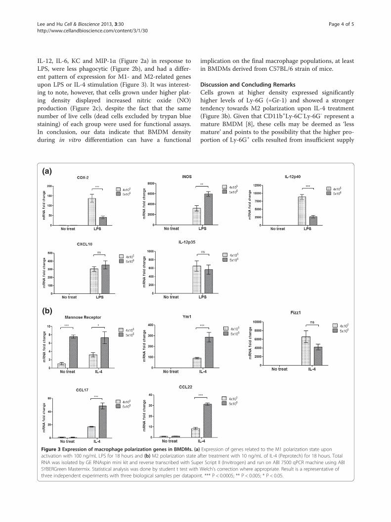
IL-12, IL-6, KC and MIP-1α (Figure 2a) in response toLPS, were less phagocytic (Figure 2b), and had a differ-ent pattern of expression for M1- and M2-related genesupon LPS or IL-4 stimulation (Figure 3). It was interest-ing to note, however, that cells grown under higher plat-ing density displayed increased nitric oxide (NO)production (Figure 2c), despite the fact that the samenumber of live cells (dead cells excluded by trypan bluestaining) of each group were used for functional assays.In conclusion, our data indicate that BMDM densityduring in vitro differentiation can have a functional
(a)
(b)
Figure 3 Expression of macrophage polarization genes in BMDMs. (a)activation with 100 ng/mL LPS for 18 hours and (b) M2 polarization state aRNA was isolated by GE RNAspin mini kit and reverse transcribed with SupSYBERGreen Mastermix. Statistical analysis was done by student t test withthree independent experiments with three biological samples per datapoin
implication on the final macrophage populations, at leastin BMDMs derived from C57BL/6 strain of mice.
Discussion and Concluding RemarksCells grown at higher density expressed significantlyhigher levels of Ly-6G (=Gr-1) and showed a strongertendency towards M2 polarization upon IL-4 treatment(Figure 3b). Given that CD11b+Ly-6C-Ly-6G- represent amature BMDM [8], these cells may be deemed as ‘lessmature’ and points to the possibility that the higher pro-portion of Ly-6G+ cells resulted from insufficient supply
Expression of genes related to the M1 polarization state uponfter treatment with 10 ng/mL of IL-4 (Peprotech) for 18 hours. Totaler Script II (Invitrogen) and run on ABI 7500 qPCR machine using ABIWelch’s correction where appropriate. Result is a representative oft. *** P < 0.0005; ** P < 0.005; * P < 0.05.

Lee and Hu Cell & Bioscience 2013, 3:30 Page 5 of 5http://www.cellandbioscience.com/content/3/1/30
of M-CSF in the growth medium. However, the M-CSF:BM cell ratio required for full BMDM maturation iscurrently unknown, and published studies typically use10-30% L929-conditioned media or 20–100 ng/mL re-combinant M-CSF over a period of 5–8 days. The possibil-ity that M-CSF fell short during differentiation of cellscultured at 5 × 10⁶ density may be tested by growing afixed number of BM cells under different known con-centrations of recombinant M-CSF. Morphologically,BMDMs matured under higher density tended to besmaller and rounder (Figure 1a), despite the fact that theoverall final cell density on Day 7 was similar between thetwo groups of macrophages. It is thus questionablewhether bone marrow stromal cells that eventuallyundergo apoptosis during the in vitro monocytic differen-tiation might have contributed to this effect. One way totest the effect of stromal cells in BMDM maturationwould be to co-culture bone marrow stromal cell line suchas M2-10B4 with the myeloid progenitor cells at differentratios and characterizing the final BMDM population. Inregards to the relationship between the maturation statesof a monocytic cell to their polarization propensity, anearlier study had indicated that immature macrophagessecrete more IL-12 [9]. However, a maturation status of amacrophage might be more complex than just definedby cell surface markers or morphology. Myeloid-derivedsuppressor cells (MDSCs), which are known to beCD11b+Gr-1+ in mice for example, have been demon-strated to undertake an immunosuppressive role in can-cer, but monocytic MDSCs produce high levels of NO[10], which is reflective of characteristics displayed bythe CD11b+Ly-6C-Ly-6G+ BMDMs in this study. There-fore, this calls for better understanding of monocyticmaturation steps and their corresponding function andthe environmental factors that might influence thecourse of maturation, as well as for a commonlyestablished protocol for BMDM generation to ensureconsistency between studies.
AbbreviationsBMDM: Bone marrow-derived macrophages; BM: Bone marrow;RBC: Red blood cell; M-CSF: Macrophage colony-stimulating factor;M-CSFR: Macrophage colony-stimulating factor receptor;LPS: Lipopolysaccharide; MDSC: Myeloid-derived suppressor cell.
Competing interestsBoth authors claim no competing interests in regards to this project.
Authors’ contributionCML conceived and executed the experimental procedures and drafted themanuscript. JH supervised the project. Both authors read and approved thefinal manuscript.
AcknowledgementsThe authors gratefully acknowledge the financial support of CanadianInstitute of Health Research Grants to JH for this study. Chan Mi Lee wassupported by Ontario Graduate Scholarship (OGS) and SickKids RestracompStudentship Award.
Received: 31 May 2013 Accepted: 10 July 2013Published: 29 July 2013
References1. Murray PJ, Wynn TA: Protective and pathogenic functions of macrophage
subsets. Nat Rev Immunol 2011, 11(11):723–737.2. Klappacher G, Lunyak VV, Sykes DB, Sawka-Verhelle D, Sage J, Brard G,
Ngo SD, Gangadharan D, Jacks T, Kamps MP, Rose DW, Rosenfeld MG,Glass CK: An induced Ets repressor complex regulates growth arrestduring terminal macrophage differentiation. Cell 2002, 109:169–180.
3. Stanley ER, Berg KL, Einstein DB, Lee PSW, Pixley FJ, Wang Y, Yeung Y:Biology and action of colony-stimulating factor-1. Mol Reprod Dev 1997,46(1):4–10.
4. Manzanero S: Generation of mouse bone marrow-derived macrophages.In Methods in Molecular Biology. 844th edition. Edited by Anonymous. ;2012:177–181.
5. Weischenfeldt J, Porse B: Bone marrow-derived macrophages (BMM):Isolation and applications. Cold Spring Harb Protoc 2008, 3(12):1–6.
6. Zanoni I, Ostuni R, Granucci F: Generation of mouse bone marrow-derivedmacrophages (BM-MFs). Protoc Exch 2009. doi:10.1038/nprot.2009.136.
7. Zhang X, Goncalves R, Mosser DM: The isolation and characterization ofmurine macrophages. Curr Protoc Immunol 2008, 14(SUPPL 83):14.1.1–14.1.14.
8. Francke A, Herold J, Weinert S, Strasser RH, Braun-Dullaeus RC: Generationof mature murine monocytes from heterogeneous bone marrow anddescription of their properties. J Histochem Cytochem 2011, 59(9):813–825.
9. Oliveira MAP, Lima GMAC, Shio MT, Leenen PJM, Abrahamsohn IA: Immaturemacrophages derived from mouse bone marrow produce large amounts ofIL-12p40 after LPS stimulation. J Leukoc Biol 2003, 74(5):857–867.
10. Gabrilovich DI, Nagaraj S: Myeloid-derived suppressor cells as regulatorsof the immune system. Nat Rev Immunol 2009, 9(3):162–174.
doi:10.1186/2045-3701-3-30Cite this article as: Lee and Hu: Cell density during differentiation canalter the phenotype of bone marrow-derived macrophages. Cell &Bioscience 2013 3:30.
Submit your next manuscript to BioMed Centraland take full advantage of:
• Convenient online submission
• Thorough peer review
• No space constraints or color figure charges
• Immediate publication on acceptance
• Inclusion in PubMed, CAS, Scopus and Google Scholar
• Research which is freely available for redistribution
Submit your manuscript at www.biomedcentral.com/submit