Caustic Injuries - SUNY Downstate Medical Center injuries and should be done within first 12 ......
50
Caustic Injuries Caustic Injuries Kings County Medical Center Eliana A. Soto MD
Transcript of Caustic Injuries - SUNY Downstate Medical Center injuries and should be done within first 12 ......
Caustic InjuriesCaustic Injuries
Kings County Medical CenterEliana A. Soto MD
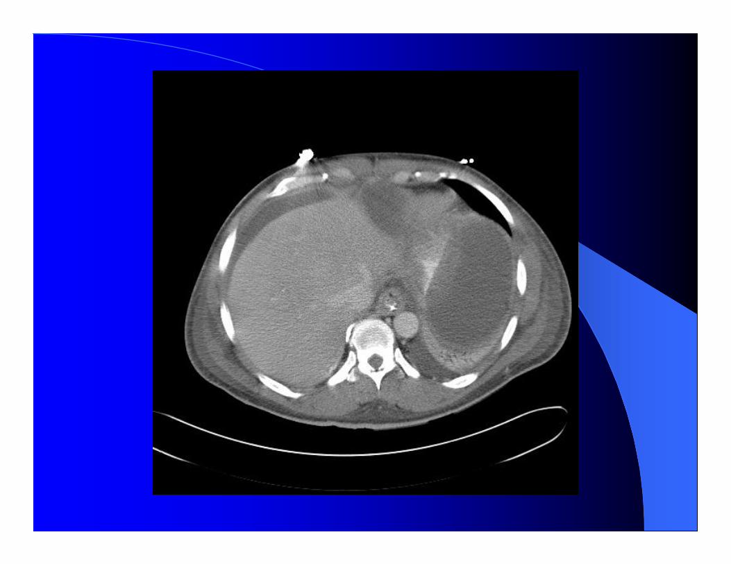
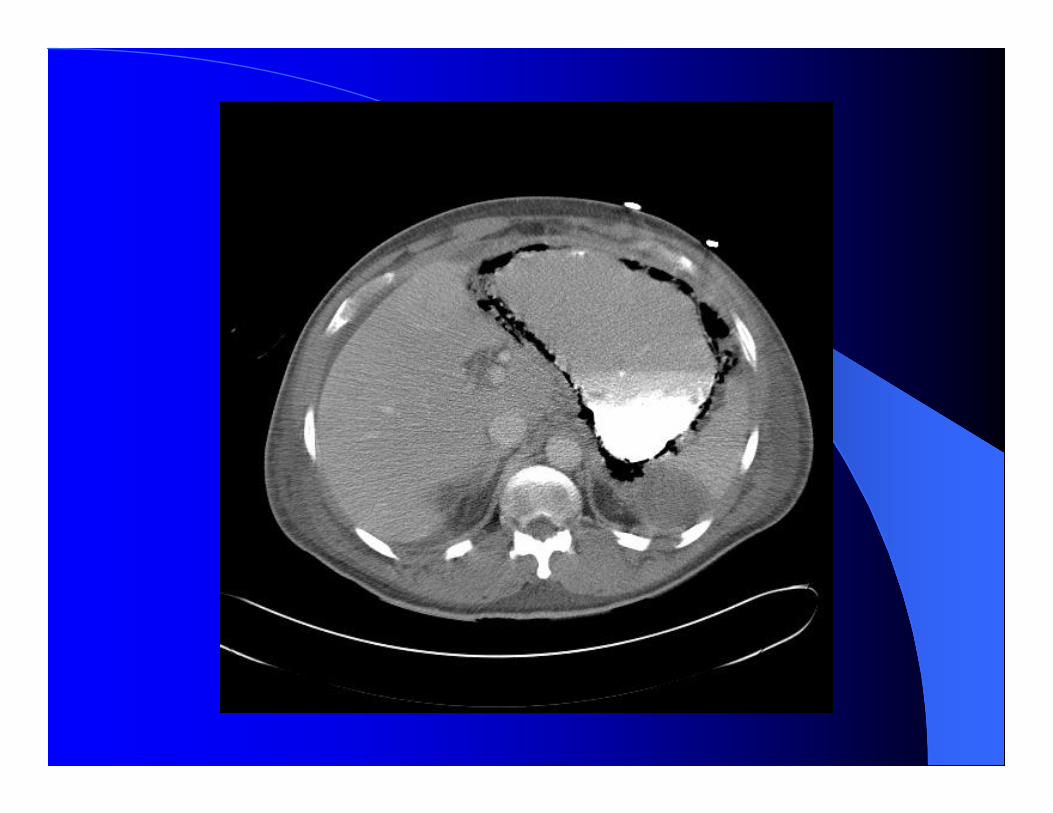
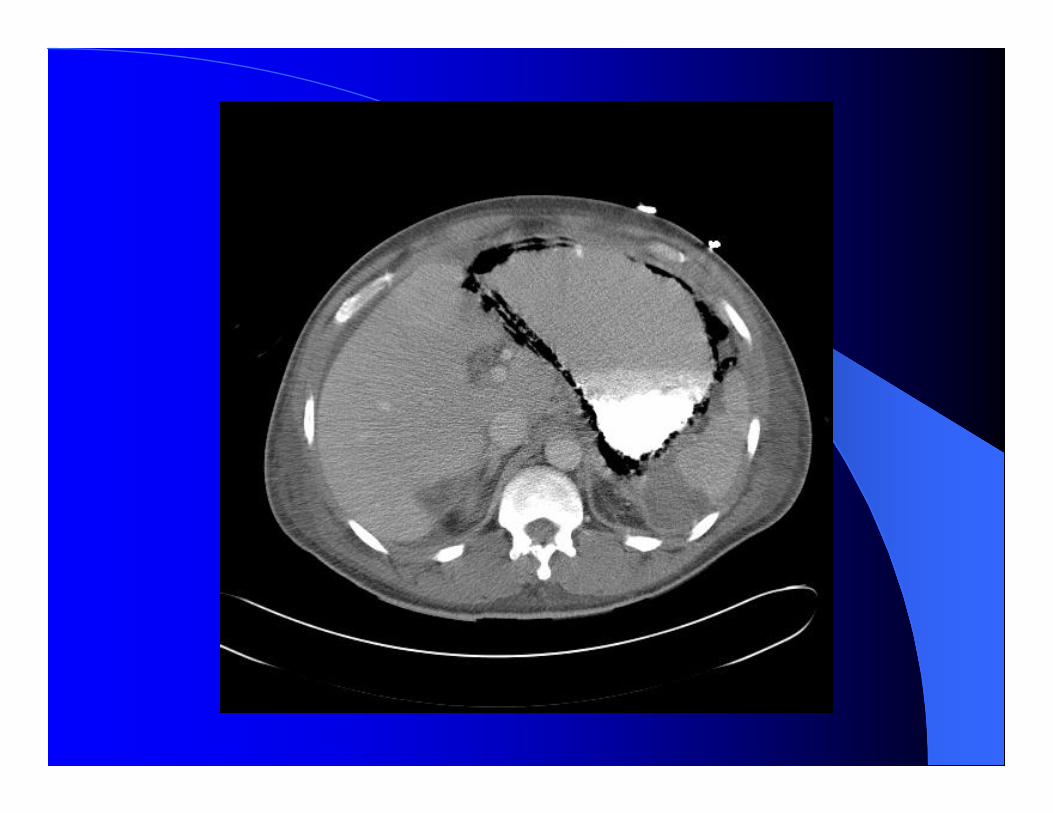

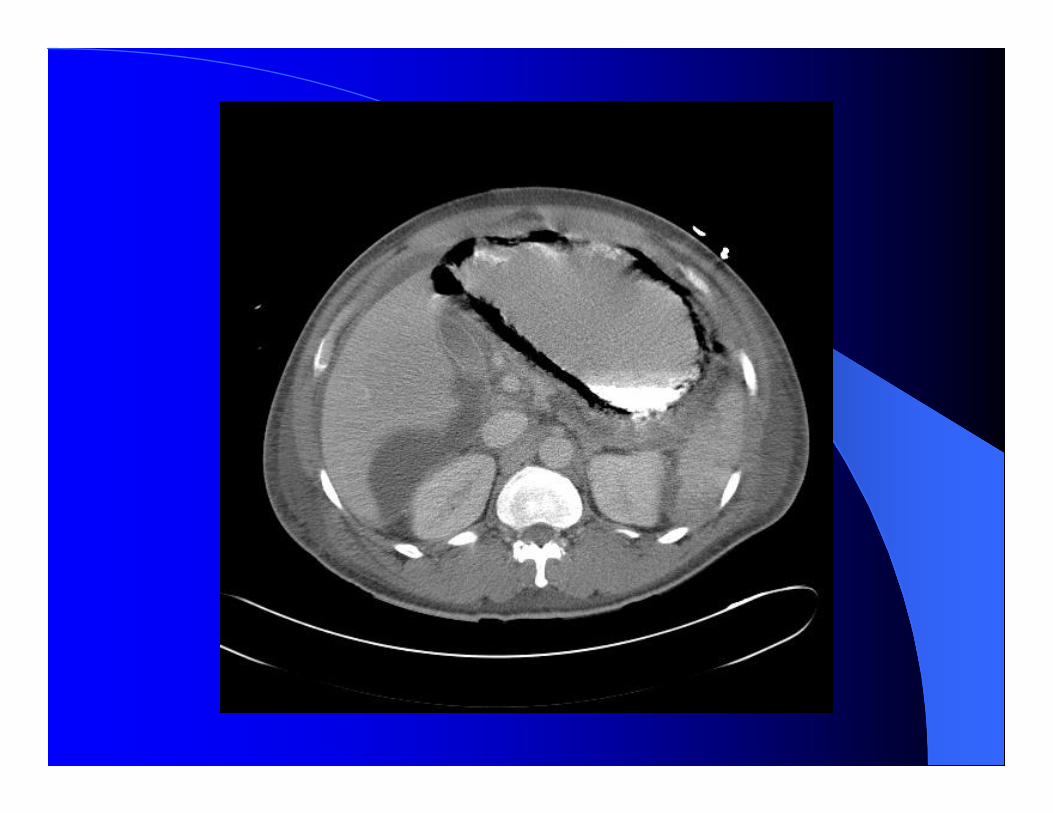
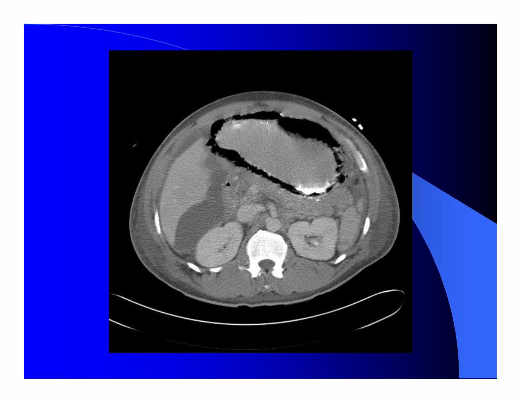


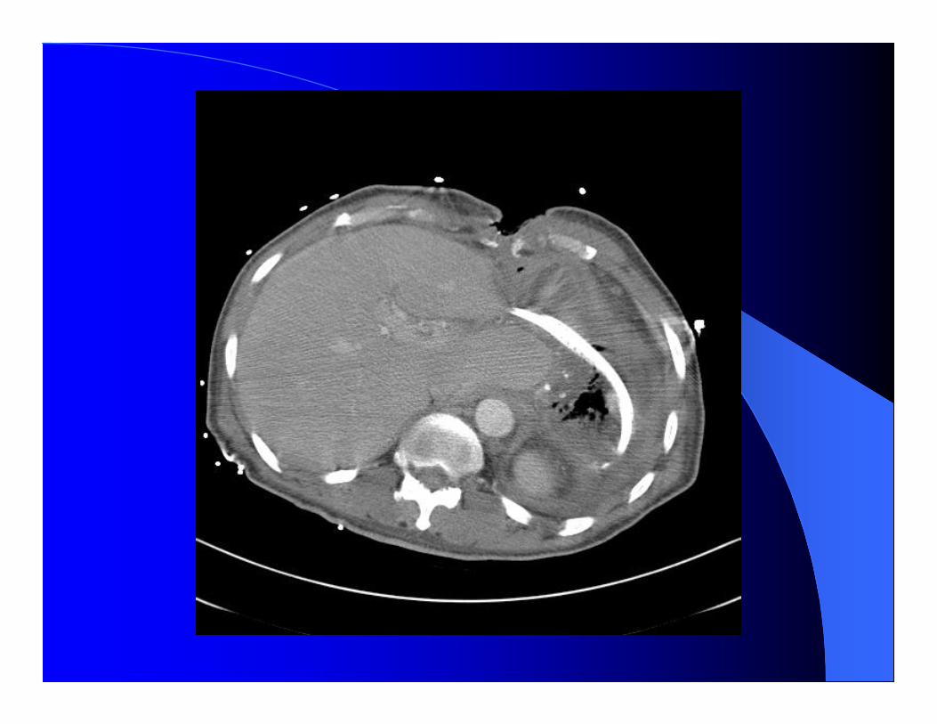
OR-necrotic stomach with viable GE junction-perforated transverse colon-splenectomy-cholecystectomy-patch proximal jejunum ? Viability-left chest tube
Caustic IngestionCaustic Ingestion
Before 1960, crystalline lye caused significant oropharyngeal burns to mucous membranes because of the adherent nature of the crystals.The immediate pain elicited from the oropharyngeal burns induced protective reflexes and rapid expulsion of the remaining crystals.Hence, deep burns only occurred in infants without protective reflexes.
The introduction of concentrated liquid oven cleaners in the 1960s resulted in more marked distal injuries involving the esophagus and stomach.Lobbying by the medical profession has reduced the lye concentration commercially available from 30% to 5% sodium hydroxide.

Chemical burns of the esophagus result from either strong acids or strong alkalis.Extent of injury to the esophagus depends on the degree of exposure to the agent: the concentration, duration and quantity.

Alkalis-liquid alkalis are odorless and tasteless and are easily swallowed without triggering protective reflexes.Sodium hydroxide commonly used as a drain cleaner.

Alkalis are more viscous and therefore have a longer transit time and penetrate deeper into tissue.Significant injury occurs at a pH exceeding 11.Alkalis produce liquefaction necrosis with enhanced tissue penetration.

Acids-bitter taste and painful cause rapid expectoration and minimal exposure to esophagus.Hydrochloric acid found in toilet bowl cleaners and battery.
Acids produce coagulation necrosis producing eschar and preventing deeper penetration into tissue.

Area of greatest contact is at areas of natural closing of the esophagus where corrosives can pool and cause circumferential burn with extensive delayed stricture formation.Alkalis and acids cause intense reflex pylorospasm with pooling of the agent in the antrum and sparing of the fundus.

Significant tissue destruction usually noted within 24 hours.3 phases of injuryPhase I-erythema, edema and inflammation within seconds.Phase II-vascular thrombosis results in further necrosis and sloughing of mucosa within 2-4 days and is period of greatest wall weakness.
Phase III-fibroblast proliferation with scar formation and stricturing over weeks to months.

Clinical features of caustic ingestion include oropharyngeal pain, chest pain, dysphagia with salivation and constant drooling.Hoarseness and stridor indicate laryngeal and epiglottal edema.Stridor suggests laryngeal injury and requires orotracheal intubation.
Up to 37% of severe esophageal lesions occur without any stigmata of oropharyngeal burns.Severe injuries with peritoneal signs, cervical emphysema, and/or retrosternal back pain indicate esophageal perforation with impending mediastinitis.Usually see change in mental status, pleural effusion and serum ph<7.2.

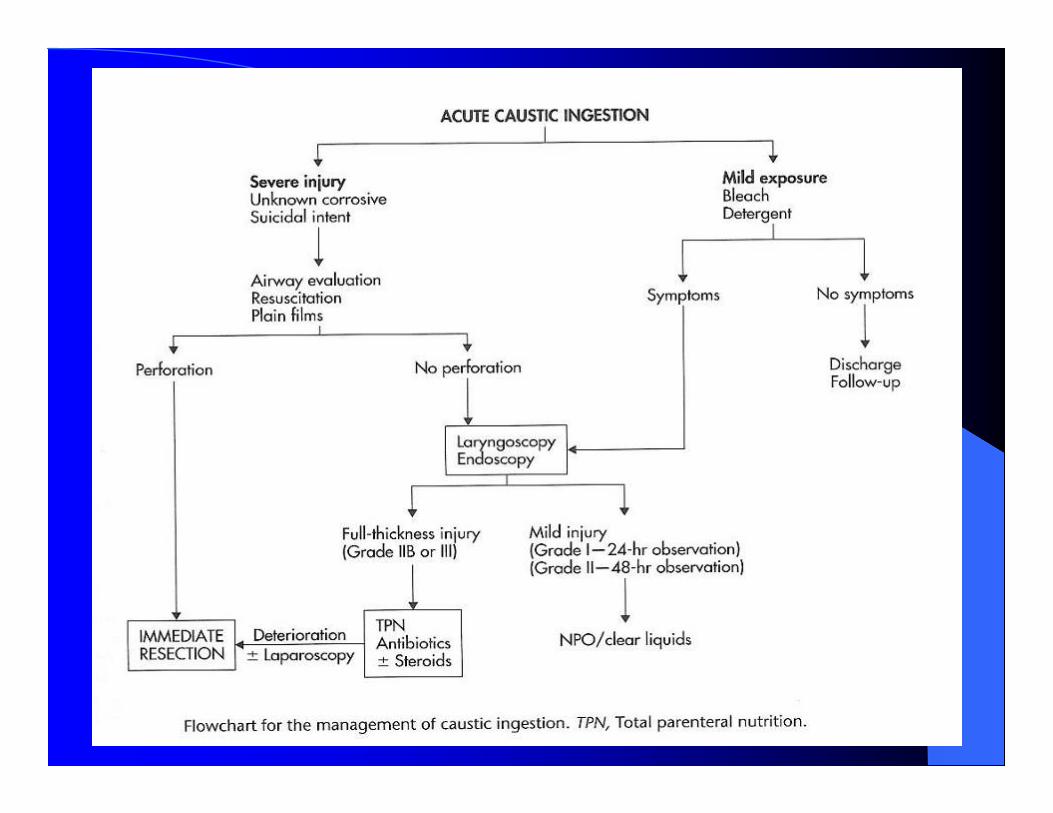
Initial management-airway assessment and fluid resuscitation.With concentrated acid injuries ingestion of large volumes of water can help neutralize acid only if given within first few minutes of exposure.Ingestion of water or milk can elicit vomiting with reexposure to corrosive agent.

Attempts to neutralize acids or alkalis can cause exothermic reaction and progression of tissue damage.Activated charcoal interferes with endoscopy and complicates management.
CXR and abdominal x-rays to r/o pneumoperitoneum, pneumomediastinum and pleural effusions which need emergent surgery.Early endoscopy is gold standard for evaluation of caustic injuries and should be done within first 12 to 24 hours.Most perforations occur around 2nd to 3rd day after injury.

Indications for endoscopy-all patients with stridor, all patients with intended ingestions, and any symptomatic children.Endoscopy is not necessary for asymptomatic children or patients already meeting criteria for surgery.Lamireau et al and Gupta et al in 2001 both published retrospective studies on children showing that all patients who were asymptomatic had normal findings on endoscopy. Zargar et al in 1989 published an endoscopic grading system for esophageal burns and modified


Grades I and IIA do not evoke strictures.All grade III and 70-100% of grade IIB injuries have variable progression to strictures.Differentiation between 2nd and 3rd degree injury may be difficult but 3rd degree injury is often indicated by presence of gray slough, thrombosed submucosal vessels and extensive black eschar.
Extent of tissue injury dictates management.Webb et al. in 1970 showed that severity of endoscopic grade of injury correlates with incidence of subsequent strictures.Endoscopic grades of injury exceeding grade IIA are associated with complications of esophageal perforation, infection and stricture.Laparoscopy can be helpful in documenting full-thickness injury.

Goal is to limit severity of injury because esophageal replacement is not as good as maintaining a scarred, damaged but functional esophagus.Use of corticosteroids for the prevention of strictures is controversial.Use of steroids began in 1960s with case series by Vascomi, Yarrington and Cannon that showed decrease in the incidence of strictures when systemic steroids and antibiotics were used.
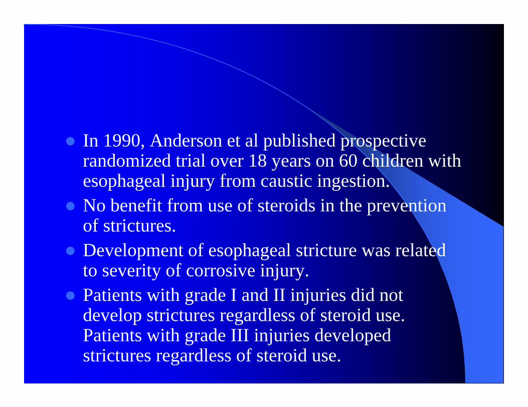
In 1990, Anderson et al published prospective randomized trial over 18 years on 60 children with esophageal injury from caustic ingestion.No benefit from use of steroids in the prevention of strictures.Development of esophageal stricture was related to severity of corrosive injury.Patients with grade I and II injuries did not develop strictures regardless of steroid use. Patients with grade III injuries developed strictures regardless of steroid use.

Grade IIB injuries-need more studies to evaluate effect of steroids.Antibiotics to cover oropharyngeal flora in grade IIB or III injuries because tissue disruption provides a path for infection.Fever and impending perforation may be masked by prophylactic antibiotics.Use of antibiotics should be used in patients receiving steroids.

Gastric acid suppression with H2 blockers is empirically recommended to decrease acid reflux from esophageal dysmotility.
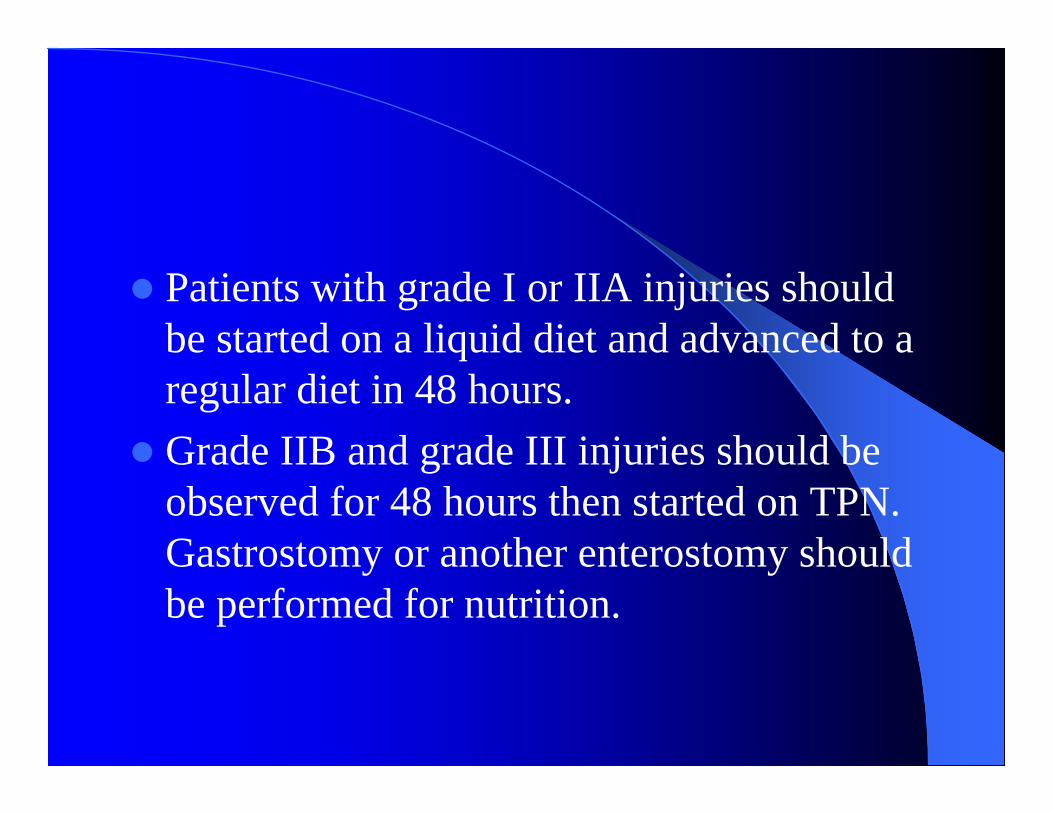
Patients with grade I or IIA injuries should be started on a liquid diet and advanced to a regular diet in 48 hours.Grade IIB and grade III injuries should be observed for 48 hours then started on TPN. Gastrostomy or another enterostomy should be performed for nutrition.

When to operate?When to operate?
Overall mortality rate of caustic ingestion injuries has been reduced from 20% to 3% over last 20 years as a result of improved and timely surgery with better antibiotics, anesthesia and nutrition.It is controversial whether laparoscopy or laparotomy should be performed for all patients with grade IIB and III burns for direct inspection of serosal surfaces.

In 1986 Estrera published a 6 year review of 62 patients with corrosive ingestion.In the first 2 years, patients received conservative treatment including esophagoscopy, steroids, antibiotics and dilation.5 of 9 patients with 2nd degree burns developed strictures requiring dilation. All 4 patients with full-thickness necrosis died.
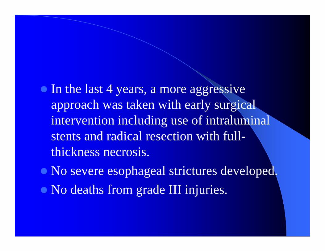
In the last 4 years, a more aggressive approach was taken with early surgical intervention including use of intraluminal stents and radical resection with full-thickness necrosis.No severe esophageal strictures developed.No deaths from grade III injuries.
Emergent operative intervention best approached via abdomen for complete visualization of stomach and contiguous structures.Esophageal resection can be completed by a transhiatal approach with a cervical esophagostomy.
Full-thickness injury of stomach may warrant total gastrectomy A cervical esophagostomy is created for a salivary fistula and a feeding jejunostomy is optimally placed for feeding.After resection, reconstruction is delayed for several months until full recovery of the patient with antibiotics and nutrition.
To reconstruct the esophagus may use colon, stomach or jejunum.Colon interposition using right or transverse colon is most widely used reconstruction.Microvascularized free jejunal graft based on superior thyroid artery can be used for residual defects.

Development of strictures-dilation usually begun with a documented stricture at 4 weeks post injury.Early bougienage, prolonged esophageal rest and lathyrogens have not been proven to be beneficial.Refractory strictures require esophagectomy and an esophageal bypass with an esophagogastric anastomosis or a colon interposition.

Intralesional corticosteroid injection for the prevention of strictures has been reported with variable success.Small case series in 1980s and 1990s showed success with intraluminal stenting to prevent stricture formation. Silastic stents or nasogastric tubes were used for a period usually of 21 days and left in place until esophagus fully healed.

Risk of developing carcinoma in a strictured residual esophagus is 100-1000x higher risk than general population.Interval between injury and development of cancer averages >40 years.Location of cancer is usually at site of stricture and type is usually squamous cell.Long-term annual follow-up of patients with grade IIB and III injuries is recommended.