Catastrophic Unbalanced Genome Rearrangements Cause ... · Catastrophic Unbalanced Genome...
16
Catastrophic Unbalanced Genome Rearrangements Cause Somatic Loss of Berry Color in Grapevine 1 Pablo Carbonell-Bejerano, a,2,3 Carolina Royo, a,2 Rafael Torres-Pérez, a,2 Jérôme Grimplet, a Lucie Fernandez, b José Manuel Franco-Zorrilla, c Diego Lijavetzky, d Elisa Baroja, a Juana Martínez, a Enrique García-Escudero, a Javier Ibáñez, a and José Miguel Martínez-Zapater a a Instituto de Ciencias de la Vid y del Vino, Consejo Superior de Investigaciones Científicas-Universidad de La Rioja-Gobierno de La Rioja, 26007 Logroño, Spain b Unité Mixte de Recherche Biologie du Fruit et Pathologie, Institut National de la Recherche Agronomique, F-33140 Villenave d’Ornon, France c Centro Nacional de Biotecnología, CNB-Consejo Superior de Investigaciones Científicas, 28049 Madrid, Spain d Instituto de Biología Agrícola de Mendoza, Consejo Nacional de Investigaciones Científicas y Técnicas- UNCuyo-FCA, M5528AHB Chacras de Coria, Argentina ORCID IDs: 0000-0002-7266-9665 (P.C.-B.); 0000-0001-5067-882X (C.R.); 0000-0002-3265-4012 (J.G.); 0000-0003-0006-658X (L.F.); 0000-0001-6769-7349 (J.M.F.-Z.); 0000-0003-4207-3067 (D.L.); 0000-0002-6286-5638 (J.I.); 0000-0001-7217-4454 (J.M.M.-Z.). Grape (Vitis vinifera) color somatic variants that can be used to develop new grapevine cultivars occasionally appear associated with deletion events of uncertain origin. To understand the mutational mechanisms generating somatic structural variation in grapevine, we compared the Tempranillo Blanco (TB) white berry somatic variant with its black berry ancestor, Tempranillo Tinto. Whole-genome sequencing uncovered a catastrophic genome rearrangement in TB that caused the hemizygous deletion of 313 genes, including the loss of the functional copy for the MYB transcription factors required for anthocyanin pigmentation in the berry skin. Loss of heterozygosity and decreased copy number delimited interspersed monosomic and disomic regions in the right arm of linkage groups 2 and 5. At least 11 validated clustered breakpoints involving intrachromosomal and interchromosomal translocations between three linkage groups flanked the deleted fragments, which, according to segregation analyses, are phased in a single copy of each of the affected chromosomes. These hallmarks, along with the lack of homology between breakpoint joins and the randomness of the order and orientation of the rearranged fragments, are all consistent with a chromothripsis-like pattern generated after chromosome breakage and illegitimate rejoining. This unbalanced genome reshuffling has additional consequences in reproductive development. In TB, lack of sexual transmission of rearranged chromosomes associates with low gamete viability, which compromises fruit set and decreases fruit production. Our findings show that catastrophic genome rearrangements arise spontaneously and stabilize during plant somatic growth. These dramatic rearrangements generate new interesting phenotypes that can be selected for the improvement of vegetatively propagated plant species. Somatic variation is a major source of diversity in multicellular organisms and is generated by unique mutation events affecting single dividing cells. Somatic mutations can spread by cell division to generate mutant sectors or cell lines derived from the original mutant cell. Somatic mutations range from single- nucleotide variation (SNV) and insertions-deletions (INDELs) to complex genome structural variation (SV) originating from chromosomal rearrangements (Li, 2016). Recently, the rapidly growing number of medical genomic studies has unveiled highly complex forms of SV that are collectively known as chromoa- nagenesis (Collins et al., 2017). Among them, the most commonly reported case is chromothripsis (for chro- mosome shattering), a cellular catastrophe initially described in human cancer that affects a few chro- mosomes (one to four) or chromosome arms and re- sults in multiple clustered rearrangements (five or more breakpoints) and deletions after chromosome breakage and illegitimate rejoining (Stephens et al., 2011; Korbel and Campbell, 2013; Collins et al., 2017). Chromothripsis has been reported in mammals, but it has never been related to natural somatic variation in the plant kingdom. 1 This work was partially supported by the project GrapeReSeq from the Spanish Ministry of Education and Sciences (EUI2008- 03752), by the European Union Seventh Framework Programme (KBBE Contract 311775 Innovine), and by the Spanish Ministry of Economy (MINECO) (BIO2014-59324-R). This work also benefited from the networking activities within the funded European Cooper- ation in Science and Technology (COST) Action FA1106 QualityFruit. 2 These authors contributed equally to the article. 3 Address correspondence to [email protected]. The author responsible for distribution of materials integral to the findings presented in this article in accordance with the policy de- scribed in the Instructions for Authors (www.plantphysiol.org) is: Pablo Carbonell-Bejerano ([email protected]). J.M.M.-Z. conceived the research; C.R., L.F., P.C.-B., D.L., and J.M.F.-Z. performed experiments; R.T.-P., P.C.-B., C.R., J.G., J.M.F.-Z., and J.I. ana- lyzed data; E.B., J.M., and E.G.-E. provided plant material; P.C.-B. and C.R. wrote the article with input and comments from all the other authors. www.plantphysiol.org/cgi/doi/10.1104/pp.17.00715 786 Plant Physiology Ò , October 2017, Vol. 175, pp. 786–801, www.plantphysiol.org Ó 2017 American Society of Plant Biologists. All Rights Reserved. www.plantphysiol.org on May 19, 2020 - Published by Downloaded from Copyright © 2017 American Society of Plant Biologists. All rights reserved.
Transcript of Catastrophic Unbalanced Genome Rearrangements Cause ... · Catastrophic Unbalanced Genome...
Catastrophic Unbalanced Genome Rearrangements CauseSomatic Loss of Berry Color in Grapevine1
Pablo Carbonell-Bejerano,a,2,3 Carolina Royo,a,2 Rafael Torres-Pérez,a,2 Jérôme Grimplet,a Lucie Fernandez,b
José Manuel Franco-Zorrilla,c Diego Lijavetzky,d Elisa Baroja,a Juana Martínez,a Enrique García-Escudero,a
Javier Ibáñez,a and José Miguel Martínez-Zapatera
aInstituto de Ciencias de la Vid y del Vino, Consejo Superior de Investigaciones Científicas-Universidad de LaRioja-Gobierno de La Rioja, 26007 Logroño, SpainbUnité Mixte de Recherche Biologie du Fruit et Pathologie, Institut National de la Recherche Agronomique,F-33140 Villenave d’Ornon, FrancecCentro Nacional de Biotecnología, CNB-Consejo Superior de Investigaciones Científicas, 28049 Madrid, SpaindInstituto de Biología Agrícola de Mendoza, Consejo Nacional de Investigaciones Científicas y Técnicas-UNCuyo-FCA, M5528AHB Chacras de Coria, Argentina
ORCID IDs: 0000-0002-7266-9665 (P.C.-B.); 0000-0001-5067-882X (C.R.); 0000-0002-3265-4012 (J.G.); 0000-0003-0006-658X (L.F.);0000-0001-6769-7349 (J.M.F.-Z.); 0000-0003-4207-3067 (D.L.); 0000-0002-6286-5638 (J.I.); 0000-0001-7217-4454 (J.M.M.-Z.).
Grape (Vitis vinifera) color somatic variants that can be used to develop new grapevine cultivars occasionally appear associatedwith deletion events of uncertain origin. To understand the mutational mechanisms generating somatic structural variation ingrapevine, we compared the Tempranillo Blanco (TB) white berry somatic variant with its black berry ancestor, Tempranillo Tinto.Whole-genome sequencing uncovered a catastrophic genome rearrangement in TB that caused the hemizygous deletion of313 genes, including the loss of the functional copy for the MYB transcription factors required for anthocyanin pigmentation inthe berry skin. Loss of heterozygosity and decreased copy number delimited interspersed monosomic and disomic regions in theright arm of linkage groups 2 and 5. At least 11 validated clustered breakpoints involving intrachromosomal and interchromosomaltranslocations between three linkage groups flanked the deleted fragments, which, according to segregation analyses, are phased ina single copy of each of the affected chromosomes. These hallmarks, along with the lack of homology between breakpoint joins andthe randomness of the order and orientation of the rearranged fragments, are all consistent with a chromothripsis-like patterngenerated after chromosome breakage and illegitimate rejoining. This unbalanced genome reshuffling has additional consequencesin reproductive development. In TB, lack of sexual transmission of rearranged chromosomes associates with low gamete viability,which compromises fruit set and decreases fruit production. Our findings show that catastrophic genome rearrangements arisespontaneously and stabilize during plant somatic growth. These dramatic rearrangements generate new interesting phenotypesthat can be selected for the improvement of vegetatively propagated plant species.
Somatic variation is a major source of diversity inmulticellular organisms and is generated by uniquemutation events affecting single dividing cells. Somatic
mutations can spread by cell division to generatemutant sectors or cell lines derived from the originalmutant cell. Somatic mutations range from single-nucleotide variation (SNV) and insertions-deletions(INDELs) to complex genome structural variation(SV) originating from chromosomal rearrangements(Li, 2016). Recently, the rapidly growing number ofmedical genomic studies has unveiled highly complexforms of SV that are collectively known as chromoa-nagenesis (Collins et al., 2017). Among them, the mostcommonly reported case is chromothripsis (for chro-mosome shattering), a cellular catastrophe initiallydescribed in human cancer that affects a few chro-mosomes (one to four) or chromosome arms and re-sults in multiple clustered rearrangements (five ormore breakpoints) and deletions after chromosomebreakage and illegitimate rejoining (Stephens et al.,2011; Korbel and Campbell, 2013; Collins et al., 2017).Chromothripsis has been reported in mammals, butit has never been related to natural somatic variationin the plant kingdom.
1 This work was partially supported by the project GrapeReSeqfrom the Spanish Ministry of Education and Sciences (EUI2008-03752), by the European Union Seventh Framework Programme(KBBE Contract 311775 Innovine), and by the Spanish Ministry ofEconomy (MINECO) (BIO2014-59324-R). This work also benefitedfrom the networking activities within the funded European Cooper-ation in Science and Technology (COST) Action FA1106 QualityFruit.
2 These authors contributed equally to the article.3 Address correspondence to [email protected] author responsible for distribution of materials integral to the
findings presented in this article in accordance with the policy de-scribed in the Instructions for Authors (www.plantphysiol.org) is:Pablo Carbonell-Bejerano ([email protected]).
J.M.M.-Z. conceived the research; C.R., L.F., P.C.-B., D.L., and J.M.F.-Z.performed experiments; R.T.-P., P.C.-B., C.R., J.G., J.M.F.-Z., and J.I. ana-lyzed data; E.B., J.M., and E.G.-E. provided plant material; P.C.-B. andC.R.wrote the articlewith input and comments from all the other authors.
www.plantphysiol.org/cgi/doi/10.1104/pp.17.00715
786 Plant Physiology�, October 2017, Vol. 175, pp. 786–801, www.plantphysiol.org � 2017 American Society of Plant Biologists. All Rights Reserved. www.plantphysiol.orgon May 19, 2020 - Published by Downloaded from
Copyright © 2017 American Society of Plant Biologists. All rights reserved.
Given their capacity for asexual propagation, plantsmay be better systems than animals in which to studysomatic variation and its effects on somatic cells(Whitham and Slobodchikoff, 1981). Plant species lack aseparated germ line, and somatic genetic variationrepresents an important source of phenotypic variationthat may be adaptive and transmitted to the next sexualgeneration when present in the meristem cell layergiving rise to gametes (D’Amato, 1997). Somatic vari-ation is especially relevant in plants such as long-livingtrees or vegetatively propagated species in which singlegenotypes are perpetuated for very long periods andcan colonize vast extensions, which increase the likeli-hood of somatic mutation emergence and accumula-tion. This case is applicable to woody crops such asgrapevine (Vitis vinifera), from which cultivar geno-types are asexually propagated by cuttings and per-petuated from the original sexual seedling that, inmanycases, germinated several centuries ago (This et al.,2006). Somatic mutations generating new interestingphenotypes that are stabilized in grapevine plants aspericlinal chimeras or that extend to all cell layers havebeen selected as new clones of wine grape cultivars oras new derivative cultivars (This et al., 2006; Pelsy et al.,2010; Torregrosa et al., 2011). Recently, genetic andgenomic approaches identified SNV and new trans-posable element (TE) insertions responsible for emer-gent phenotypes in several grapevine somatic variants(Boss and Thomas, 2002; Fernandez et al., 2010, 2013;Battilana et al., 2011; Emanuelli et al., 2014).Given their conspicuousness, berry color somatic
variants have occasionally been selected and studied ingrapevine (Kobayashi et al., 2004; Walker et al., 2006;Yakushiji et al., 2006; Furiya et al., 2009). Grape colorresults from the accumulation of anthocyanins gener-ally in the berry skin (Boss et al., 1996), which is ge-netically controlled by a major locus on linkage group(LG) 2 (Doligez et al., 2006). This locus colocalizes witha cluster of tandemly repeated VviMybA genes (Wonget al., 2016). FunctionalVviMybA1 andVviMybA2 genesare essential for grape pigmentation, whereas thepresence of a Gret1 retrotransposon insertion in thepromoter of VviMybA1 along with a small INDELcausing a frame shift in VviMybA2 are responsible for anull allele of the grape color locus that is frequently inhomozygosis in white berry cultivars (Kobayashi et al.,2004; Lijavetzky et al., 2006; This et al., 2007; Walkeret al., 2007). Black berry cultivars heterozygous for thenull allele occasionally display grape color variantswith either red/gray or white berries depending onwhether only the L2 or both L1 and L2 meristem celllayers, respectively, carry mutations at the color locus(Walker et al., 2006; Furiya et al., 2009; Vezzulli et al.,2012; Migliaro et al., 2014; Pelsy et al., 2015). Initialcharacterization of red/gray and white berry somaticvariants of Cabernet Sauvignon and Pinot Noir usingSouthern blots showed that the absence of anthocya-nins in the berry was related to deletion of the func-tional allele of the color locus (Walker et al., 2006;Yakushiji et al., 2006). Recently, the use of loss of
heterozygosity (LOH) analyses along LG 2 suggestedthat the size of these deletions can be quite variable indifferent berry color somatic variants (Vezzulli et al.,2012; Migliaro et al., 2014; Pelsy et al., 2015). Unfortu-nately, no information is available yet on the break-points delimiting these deletions, which is required tounderstand their mutational origin.
Tempranillo Blanco (TB) is a white berry somaticvariant that originally appeared as a bud sport mutantof Tempranillo Tinto (TT) (Martinez et al., 2006). Likemany black berry grapevine cultivars, TT is heterozy-gous for the color locus functional allele, and TB hasbeen proposed to appear by deletion of this allele(Ibáñez et al., 2012; Migliaro et al., 2014). To understandthe origin of the TB variant, we carried out SV analysisafter whole-genome sequencing (WGS) of TT and TBlines. SV breakpoints were confirmed by DNA se-quencing of specific amplicons, and self-cross progenywere used for haplotype phasing and transmissionanalysis. Globally, the results show the relevance ofcomplex SV as a driver of clonal variation in plants.
RESULTS
Hemizygous Deletions around the Color Locus Associatewith Anthocyanin Biosynthesis Blockage in TB
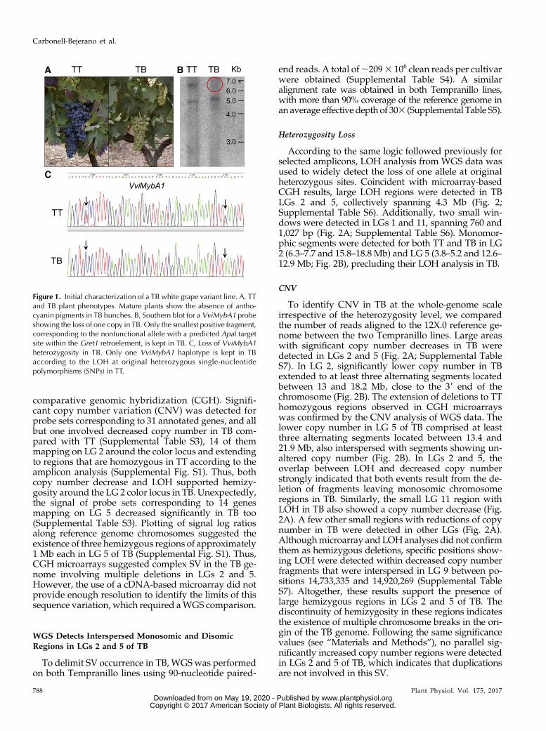
TB is a grapevine cultivar generated by vegetativepropagation of the white-berried mutant phenotypethat appeared as a spontaneous bud sport in a TT plant(Martinez et al., 2006; Fig. 1A). In TB, the presence of ahemizygous deletion eliminating the functional allele ofthe VviMybA1 gene on the color locus at LG 2 wasconfirmed using DNA-blot hybridization of DNAextracted from both TT and TB plants (Fig. 1B). Thesequence of a specific amplicon corresponding to partof the VviMybA1 gene showed LOH in TB, furtherconfirming the presence of a hemizygous deletion af-fecting at least this gene in TB (Fig. 1C). Given the highheterozygosity of grapevine cultivars (Laucou et al.,2011), the extent of the deletion in TB was first assessedusing primer pairs (Supplemental Table S1) intendedfor LOH analysis of amplified fragments along theputatively affected region in LG 2. This analysisshowed that putative hemizygous deletions extendedoutside of theMybA gene cluster in LG 2 (SupplementalTable S2). Unfortunately, the efficiency of this methodto identify the loss of one allele is limited to originallyheterozygous genomic regions, and, coincident withother studies (Migliaro et al., 2014), many testedamplicons only provided monomorphic sequences inthe TT original cultivar, with no polymorphic sitedetected after position chr2:15,861,181 (SupplementalTable S2) in the PN40024 12X.0 reference genome as-sembly (https://urgi.versailles.inra.fr/Species/Vitis/Data-Sequences/Genome-sequences).
To globally predict the extension of hemizygousregions in TB irrespective of the heterozygosity levelin TT, the GrapeGen GeneChip cDNA-based micro-array (Lijavetzky et al., 2012) was initially used for
Plant Physiol. Vol. 175, 2017 787
Chromothripsis-Mediated Loss of Grape Color
www.plantphysiol.orgon May 19, 2020 - Published by Downloaded from Copyright © 2017 American Society of Plant Biologists. All rights reserved.
comparative genomic hybridization (CGH). Signifi-cant copy number variation (CNV) was detected forprobe sets corresponding to 31 annotated genes, and allbut one involved decreased copy number in TB com-pared with TT (Supplemental Table S3), 14 of themmapping on LG 2 around the color locus and extendingto regions that are homozygous in TT according to theamplicon analysis (Supplemental Fig. S1). Thus, bothcopy number decrease and LOH supported hemizy-gosity around the LG 2 color locus in TB. Unexpectedly,the signal of probe sets corresponding to 14 genesmapping on LG 5 decreased significantly in TB too(Supplemental Table S3). Plotting of signal log ratiosalong reference genome chromosomes suggested theexistence of three hemizygous regions of approximately1 Mb each in LG 5 of TB (Supplemental Fig. S1). Thus,CGH microarrays suggested complex SV in the TB ge-nome involving multiple deletions in LGs 2 and 5.However, the use of a cDNA-based microarray did notprovide enough resolution to identify the limits of thissequence variation, which required aWGS comparison.
WGS Detects Interspersed Monosomic and DisomicRegions in LGs 2 and 5 of TB
To delimit SV occurrence in TB, WGSwas performedon both Tempranillo lines using 90-nucleotide paired-
end reads. A total of;2093 106 clean reads per cultivarwere obtained (Supplemental Table S4). A similaralignment rate was obtained in both Tempranillo lines,with more than 90% coverage of the reference genome inan average effective depth of 303 (Supplemental Table S5).
Heterozygosity Loss
According to the same logic followed previously forselected amplicons, LOH analysis from WGS data wasused to widely detect the loss of one allele at originalheterozygous sites. Coincident with microarray-basedCGH results, large LOH regions were detected in TBLGs 2 and 5, collectively spanning 4.3 Mb (Fig. 2;Supplemental Table S6). Additionally, two small win-dows were detected in LGs 1 and 11, spanning 760 and1,027 bp (Fig. 2A; Supplemental Table S6). Monomor-phic segments were detected for both TT and TB in LG2 (6.3–7.7 and 15.8–18.8 Mb) and LG 5 (3.8–5.2 and 12.6–12.9 Mb; Fig. 2B), precluding their LOH analysis in TB.
CNV
To identify CNV in TB at the whole-genome scaleirrespective of the heterozygosity level, we comparedthe number of reads aligned to the 12X.0 reference ge-nome between the two Tempranillo lines. Large areaswith significant copy number decreases in TB weredetected in LGs 2 and 5 (Fig. 2A; Supplemental TableS7). In LG 2, significantly lower copy number in TBextended to at least three alternating segments locatedbetween 13 and 18.2 Mb, close to the 39 end of thechromosome (Fig. 2B). The extension of deletions to TThomozygous regions observed in CGH microarrayswas confirmed by the CNV analysis of WGS data. Thelower copy number in LG 5 of TB comprised at leastthree alternating segments located between 13.4 and21.9 Mb, also interspersed with segments showing un-altered copy number (Fig. 2B). In LGs 2 and 5, theoverlap between LOH and decreased copy numberstrongly indicated that both events result from the de-letion of fragments leaving monosomic chromosomeregions in TB. Similarly, the small LG 11 region withLOH in TB also showed a copy number decrease (Fig.2A). A few other small regions with reductions of copynumber in TB were detected in other LGs (Fig. 2A).Althoughmicroarray and LOHanalyses did not confirmthem as hemizygous deletions, specific positions show-ing LOH were detected within decreased copy numberfragments that were interspersed in LG 9 between po-sitions 14,733,335 and 14,920,269 (Supplemental TableS7). Altogether, these results support the presence oflarge hemizygous regions in LGs 2 and 5 of TB. Thediscontinuity of hemizygosity in these regions indicatesthe existence of multiple chromosome breaks in the ori-gin of the TB genome. Following the same significancevalues (see “Materials and Methods”), no parallel sig-nificantly increased copy number regions were detectedin LGs 2 and 5 of TB, which indicates that duplicationsare not involved in this SV.
Figure 1. Initial characterization of a TB white grape variant line. A, TTand TB plant phenotypes. Mature plants show the absence of antho-cyanin pigments in TB bunches. B, Southern blot for a VviMybA1 probeshowing the loss of one copy in TB. Only the smallest positive fragment,corresponding to the nonfunctional allele with a predicted ApaI targetsite within the Gret1 retroelement, is kept in TB. C, Loss of VviMybA1heterozygosity in TB. Only one VviMybA1 haplotype is kept in TBaccording to the LOH at original heterozygous single-nucleotidepolymorphisms (SNPs) in TT.
788 Plant Physiol. Vol. 175, 2017
Carbonell-Bejerano et al.
www.plantphysiol.orgon May 19, 2020 - Published by Downloaded from Copyright © 2017 American Society of Plant Biologists. All rights reserved.
SV Junction Sites Reveal Multiple Interchromosomal andIntrachromosomal Translocations in the Genome of TB
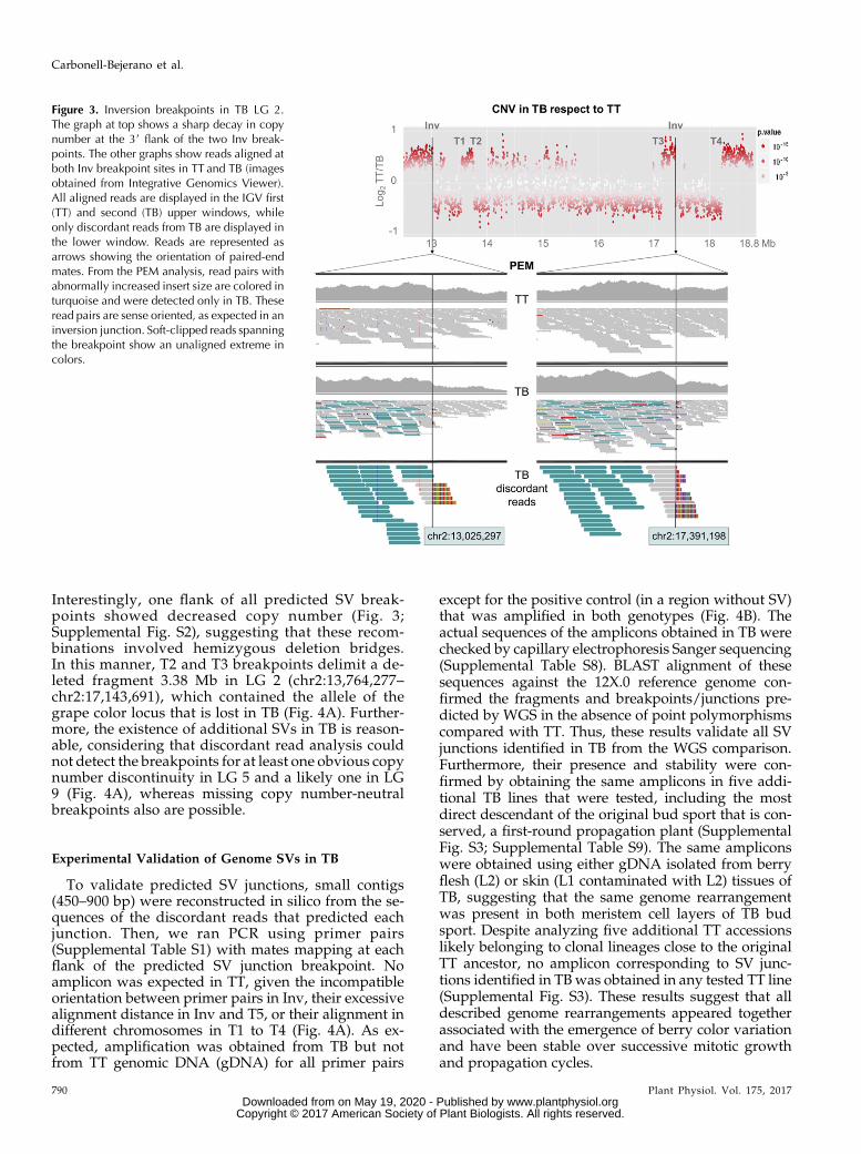
WGS reads were explored to delimit the breakpointsof fragments deleted in TB and to elucidate the nature ofthe SV. Discordant pair-endmapping (PEM) is useful todetect SV breakpoints from short-read sequencing data(Korbel et al., 2007; Marroni et al., 2014). Thus, TB-specific discordant PEM and soft-clipped reads withrespect to the grapevine reference genome revealed sixSV junctions in the TB genome not present in TT. Col-lectively, they comprised six breakpoints in LG 2, five inLG 5, and one in LG 9 (Table I).An inversion junction in LG 2 (Inv) was predicted by
the identification in TB but not in TT of read pairsmapping on LG 2 with mates separated one from an-other by more than 4.36 Mb (Fig. 3). Mates at both po-sitions displayed the same alignment orientation,
which is compatible with an inversion (Korbel et al.,2007; Rausch et al., 2012). TB-specific soft-clipped readswere detected at both flanks of the junction (Fig. 3).BLASTN identified that the unaligned extreme ofthese soft-clipped reads matches to the 59 extremeof the mate inversion flank, unveiling the putativeinversion junction breakpoint at nucleotide levelresolution (between positions chr2:13,025,297 andchr2:17,391,598; Table I). Similarly, discordant readsidentified four SV junctions with flanks in differentchromosomes of the reference genome (T1–T4; TableI). These rearrangements were interpreted as inter-chromosomal translocations and involved LGs 2 and5 in all cases except T1, involving LGs 2 and 9. For theT5 event, discordant alignment identified an SVjunction joining LG 5 fragments originally separatedby ;5 Mb, which is compatible with an intra-chromosomal translocation (Supplemental Fig. S2).
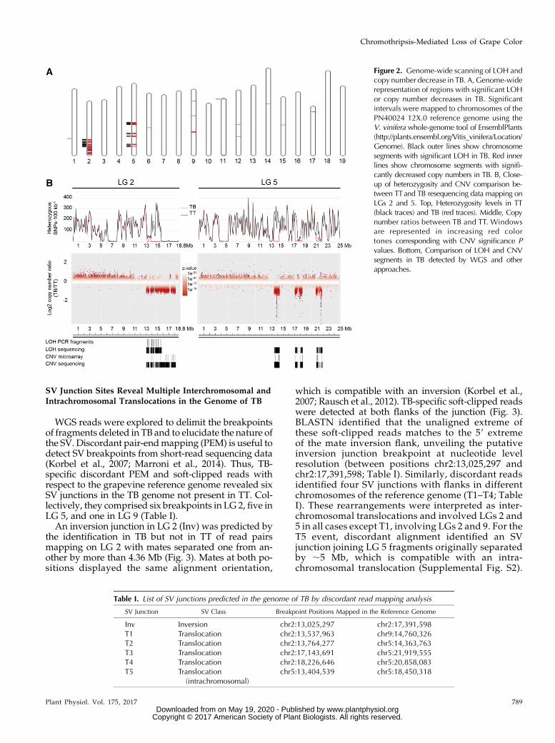
Figure 2. Genome-wide scanning of LOH andcopy number decrease in TB. A, Genome-widerepresentation of regions with significant LOHor copy number decreases in TB. Significantintervals were mapped to chromosomes of thePN40024 12X.0 reference genome using theV. vinifera whole-genome tool of EnsemblPlants(http://plants.ensembl.org/Vitis_vinifera/Location/Genome). Black outer lines show chromosomesegments with significant LOH in TB. Red innerlines show chromosome segments with signifi-cantly decreased copy numbers in TB. B, Close-up of heterozygosity and CNV comparison be-tween TTand TB resequencing data mapping onLGs 2 and 5. Top, Heterozygosity levels in TT(black traces) and TB (red traces). Middle, Copynumber ratios between TB and TT. Windowsare represented in increasing red colortones corresponding with CNV significance Pvalues. Bottom, Comparison of LOH and CNVsegments in TB detected by WGS and otherapproaches.
Table I. List of SV junctions predicted in the genome of TB by discordant read mapping analysis
SV Junction SV Class Breakpoint Positions Mapped in the Reference Genome
Inv Inversion chr2:13,025,297 chr2:17,391,598T1 Translocation chr2:13,537,963 chr9:14,760,326T2 Translocation chr2:13,764,277 chr5:14,363,763T3 Translocation chr2:17,143,691 chr5:21,919,555T4 Translocation chr2:18,226,646 chr5:20,858,083T5 Translocation
(intrachromosomal)chr5:13,404,539 chr5:18,450,318
Plant Physiol. Vol. 175, 2017 789
Chromothripsis-Mediated Loss of Grape Color
www.plantphysiol.orgon May 19, 2020 - Published by Downloaded from Copyright © 2017 American Society of Plant Biologists. All rights reserved.
Interestingly, one flank of all predicted SV break-points showed decreased copy number (Fig. 3;Supplemental Fig. S2), suggesting that these recom-binations involved hemizygous deletion bridges.In this manner, T2 and T3 breakpoints delimit a de-leted fragment 3.38 Mb in LG 2 (chr2:13,764,277–chr2:17,143,691), which contained the allele of thegrape color locus that is lost in TB (Fig. 4A). Further-more, the existence of additional SVs in TB is reason-able, considering that discordant read analysis couldnot detect the breakpoints for at least one obvious copynumber discontinuity in LG 5 and a likely one in LG9 (Fig. 4A), whereas missing copy number-neutralbreakpoints also are possible.
Experimental Validation of Genome SVs in TB
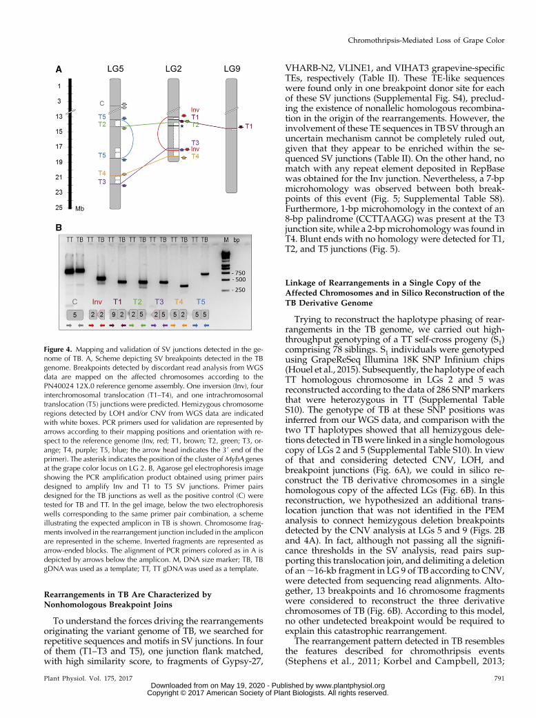
To validate predicted SV junctions, small contigs(450–900 bp) were reconstructed in silico from the se-quences of the discordant reads that predicted eachjunction. Then, we ran PCR using primer pairs(Supplemental Table S1) with mates mapping at eachflank of the predicted SV junction breakpoint. Noamplicon was expected in TT, given the incompatibleorientation between primer pairs in Inv, their excessivealignment distance in Inv and T5, or their alignment indifferent chromosomes in T1 to T4 (Fig. 4A). As ex-pected, amplification was obtained from TB but notfrom TT genomic DNA (gDNA) for all primer pairs
except for the positive control (in a region without SV)that was amplified in both genotypes (Fig. 4B). Theactual sequences of the amplicons obtained in TB werechecked by capillary electrophoresis Sanger sequencing(Supplemental Table S8). BLAST alignment of thesesequences against the 12X.0 reference genome con-firmed the fragments and breakpoints/junctions pre-dicted by WGS in the absence of point polymorphismscompared with TT. Thus, these results validate all SVjunctions identified in TB from the WGS comparison.Furthermore, their presence and stability were con-firmed by obtaining the same amplicons in five addi-tional TB lines that were tested, including the mostdirect descendant of the original bud sport that is con-served, a first-round propagation plant (SupplementalFig. S3; Supplemental Table S9). The same ampliconswere obtained using either gDNA isolated from berryflesh (L2) or skin (L1 contaminated with L2) tissues ofTB, suggesting that the same genome rearrangementwas present in both meristem cell layers of TB budsport. Despite analyzing five additional TT accessionslikely belonging to clonal lineages close to the originalTT ancestor, no amplicon corresponding to SV junc-tions identified in TBwas obtained in any tested TT line(Supplemental Fig. S3). These results suggest that alldescribed genome rearrangements appeared togetherassociated with the emergence of berry color variationand have been stable over successive mitotic growthand propagation cycles.
Figure 3. Inversion breakpoints in TB LG 2.The graph at top shows a sharp decay in copynumber at the 39 flank of the two Inv break-points. The other graphs show reads aligned atboth Inv breakpoint sites in TT and TB (imagesobtained from Integrative Genomics Viewer).All aligned reads are displayed in the IGV first(TT) and second (TB) upper windows, whileonly discordant reads from TB are displayed inthe lower window. Reads are represented asarrows showing the orientation of paired-endmates. From the PEM analysis, read pairs withabnormally increased insert size are colored inturquoise and were detected only in TB. Theseread pairs are sense oriented, as expected in aninversion junction. Soft-clipped reads spanningthe breakpoint show an unaligned extreme incolors.
790 Plant Physiol. Vol. 175, 2017
Carbonell-Bejerano et al.
www.plantphysiol.orgon May 19, 2020 - Published by Downloaded from Copyright © 2017 American Society of Plant Biologists. All rights reserved.
Rearrangements in TB Are Characterized byNonhomologous Breakpoint Joins
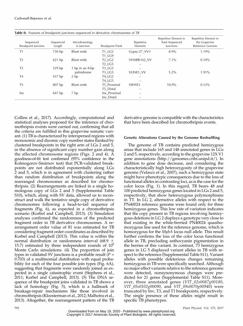
To understand the forces driving the rearrangementsoriginating the variant genome of TB, we searched forrepetitive sequences and motifs in SV junctions. In fourof them (T1–T3 and T5), one junction flank matched,with high similarity score, to fragments of Gypsy-27,
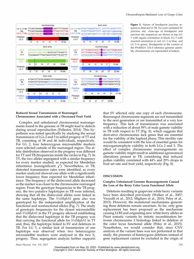
VHARB-N2, VLINE1, and VIHAT3 grapevine-specificTEs, respectively (Table II). These TE-like sequenceswere found only in one breakpoint donor site for eachof these SV junctions (Supplemental Fig. S4), preclud-ing the existence of nonallelic homologous recombina-tion in the origin of the rearrangements. However, theinvolvement of these TE sequences in TB SV through anuncertain mechanism cannot be completely ruled out,given that they appear to be enriched within the se-quenced SV junctions (Table II). On the other hand, nomatch with any repeat element deposited in RepBasewas obtained for the Inv junction. Nevertheless, a 7-bpmicrohomology was observed between both break-points of this event (Fig. 5; Supplemental Table S8).Furthermore, 1-bp microhomology in the context of an8-bp palindrome (CCTTAAGG) was present at the T3junction site, while a 2-bpmicrohomologywas found inT4. Blunt ends with no homology were detected for T1,T2, and T5 junctions (Fig. 5).
Linkage of Rearrangements in a Single Copy of theAffected Chromosomes and in Silico Reconstruction of theTB Derivative Genome
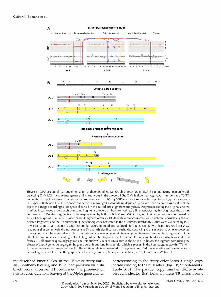
Trying to reconstruct the haplotype phasing of rear-rangements in the TB genome, we carried out high-throughput genotyping of a TT self-cross progeny (S1)comprising 78 siblings. S1 individuals were genotypedusing GrapeReSeq Illumina 18K SNP Infinium chips(Houel et al., 2015). Subsequently, the haplotype of eachTT homologous chromosome in LGs 2 and 5 wasreconstructed according to the data of 286 SNPmarkersthat were heterozygous in TT (Supplemental TableS10). The genotype of TB at these SNP positions wasinferred from our WGS data, and comparison with thetwo TT haplotypes showed that all hemizygous dele-tions detected in TBwere linked in a single homologouscopy of LGs 2 and 5 (Supplemental Table S10). In viewof that and considering detected CNV, LOH, andbreakpoint junctions (Fig. 6A), we could in silico re-construct the TB derivative chromosomes in a singlehomologous copy of the affected LGs (Fig. 6B). In thisreconstruction, we hypothesized an additional trans-location junction that was not identified in the PEManalysis to connect hemizygous deletion breakpointsdetected by the CNV analysis at LGs 5 and 9 (Figs. 2Band 4A). In fact, although not passing all the signifi-cance thresholds in the SV analysis, read pairs sup-porting this translocation join, and delimiting a deletionof an;16-kb fragment in LG 9 of TB according to CNV,were detected from sequencing read alignments. Alto-gether, 13 breakpoints and 16 chromosome fragmentswere considered to reconstruct the three derivativechromosomes of TB (Fig. 6B). According to this model,no other undetected breakpoint would be required toexplain this catastrophic rearrangement.
The rearrangement pattern detected in TB resemblesthe features described for chromothripsis events(Stephens et al., 2011; Korbel and Campbell, 2013;
Figure 4. Mapping and validation of SV junctions detected in the ge-nome of TB. A, Scheme depicting SV breakpoints detected in the TBgenome. Breakpoints detected by discordant read analysis from WGSdata are mapped on the affected chromosomes according to thePN40024 12X.0 reference genome assembly. One inversion (Inv), fourinterchromosomal translocation (T1–T4), and one intrachromosomaltranslocation (T5) junctions were predicted. Hemizygous chromosomeregions detected by LOH and/or CNV from WGS data are indicatedwith white boxes. PCR primers used for validation are represented byarrows according to their mapping positions and orientation with re-spect to the reference genome (Inv, red; T1, brown; T2, green; T3, or-ange; T4, purple; T5, blue; the arrow head indicates the 39 end of theprimer). The asterisk indicates the position of the cluster ofMybA genesat the grape color locus on LG 2. B, Agarose gel electrophoresis imageshowing the PCR amplification product obtained using primer pairsdesigned to amplify Inv and T1 to T5 SV junctions. Primer pairsdesigned for the TB junctions as well as the positive control (C) weretested for TB and TT. In the gel image, below the two electrophoresiswells corresponding to the same primer pair combination, a schemeillustrating the expected amplicon in TB is shown. Chromosome frag-ments involved in the rearrangement junction included in the ampliconare represented in the scheme. Inverted fragments are represented asarrow-ended blocks. The alignment of PCR primers colored as in A isdepicted by arrows below the amplicon. M, DNA size marker; TB, TBgDNA was used as a template; TT, TT gDNAwas used as a template.
Plant Physiol. Vol. 175, 2017 791
Chromothripsis-Mediated Loss of Grape Color
www.plantphysiol.orgon May 19, 2020 - Published by Downloaded from Copyright © 2017 American Society of Plant Biologists. All rights reserved.
Collins et al., 2017). Accordingly, computational andstatistical analyses proposed for the inference of chro-mothripsis events were carried out, confirming that allthe criteria are fulfilled in this grapevine somatic vari-ant. (1) TB is characterized by interspersed regions withmonosomic and disomic copy number states flanked byclustered breakpoints in the right arm of LGs 2 and 5,in the absence of significant copy number gain alongthe affected chromosome regions (Figs. 2 and 4). Agoodness-of-fit test confirmed (95% confidence in theKolmogorov-Smirnov test) that PCR-validated break-points are not distributed exponentially along LGs2 and 5, which is in agreement with clustering ratherthan random distribution of breakpoints along therearranged chromosomes as described for chromo-thripsis. (2) Rearrangements are linked in a single ho-mologous copy of LGs 2 and 5 (Supplemental TableS10), which, along with SV data, allowed us to recon-struct and walk the tentative single copy of derivativechromosomes following a head-to-tail sequence offragments (Fig. 6), as expected in a chromothripsisscenario (Korbel and Campbell, 2013). (3) Simulationanalyses confirmed the randomness of the predictedfragment order in TB derivative chromosomes. A re-arrangement order value of 81 was estimated for TBconsidering fragment order coordinates as described byKorbel and Campbell (2013). This value is within thenormal distribution or randomness interval (68.9 615.7) estimated by three independent rounds of 108
Monte Carlo simulations. (4) The proportion of jointypes in validated SV junctions is a probable result (P =0.703) of a multinomial distribution with equal proba-bility for each of the four possible join types (Fig. 6A),suggesting that fragments were randomly joined as ex-pected in a single catastrophic event (Stephens et al.,2011; Korbel and Campbell, 2013). (5) The DNA se-quence of the breakpoint joins validated in TB shows alack of homology (Fig. 5), which is a hallmark ofbreakage-repair mechanisms like those involved inchromothripsis (Kloosterman et al., 2012;Malhotra et al.,2013). Altogether, the rearrangement pattern of the TB
derivative genome is compatible with the characteristicsthat have been described for chromothripsis events.
Genetic Alterations Caused by the Genome Reshuffling
The genome of TB contains predicted hemizygousareas that include 165 and 148 annotated genes in LGs2 and 5, respectively, according to the grapevine 12X V1gene annotations (http://genomes.cribi.unipd.it/). Inaddition to gene dose decrease, and considering thecharacteristically high heterozygosity of the grapevinegenome (Velasco et al., 2007), such a hemizygous statemight have phenotypic consequences due to the loss offunctional alleles in contrasting loci, as is the case for thecolor locus (Fig. 1). In this regard, TB bears 48 and100 predicted hemizygous genes located in LGs 2 and 5,respectively, that show heterozygous polymorphismsin TT. In LG 2, alternative alleles with respect to thePN40024 reference genome were found only for threehemizygous genes. This low rate of variation indicatesthat the copy present in TB regions involving hemizy-gous deletions in LG 2 displays a genotype very close tothat existing in the white-berried PN40024 quasiho-mozygous line used for the reference genome, which ishomozygous for the MybA locus null allele. This resultfurther confirms the loss of the color locus functionalallele in TB, precluding anthocyanin pigmentation inthe berries of this variant. In contrast, 73 hemizygousgenes in LG 5 displayed variant alleles in TB with re-spect to the reference (Supplemental Table S11). Variantalleles with possible deleterious changes remaininghemizygous in TBwere specifically searched. Althoughnomajor effect variants relative to the reference genomewere detected, nonsynonymous changes were pre-dicted for 21 genes (Supplemental Table S11). More-over, three annotated genes (VIT_02s0087g00180,VIT_05s0102g00090, and VIT_09s0070g00940) weretruncated by Inv, T3, and T1 breakpoints, respectively.The single presence of these alleles might result inspecific TB phenotypes.
Table II. Features of breakpoint junctions sequenced in derivative chromosomes of TB
Sequenced
Breakpoint Junction
Sequenced
Length
Microhomology
in Junction Breakpoint Flank
Repetitive
Elements
Repetitive Element in
Total Sequenced
Junctions
Repetitive Element in
the Grapevine
Reference Genome
T1 730 bp Blunt ends T1_LG2 Gypsy-27_VV-I 8.9% 1.19%T1_LG9 –
T2 621 bp Blunt ends T2_LG2 VHARB-N2_VV 7.1% 0.10%T2_LG5 –
T3 529 bp 1 bp in an 8-bppalindrome
T3_LG2 –T3_LG5 VLINE1_VV 5.2% 1.91%
T4 437 bp 2 bp T4_LG2 –T4_LG5 –
T5 807 bp Blunt ends T5_Proximal VIHAT3 10.9% 0.12%T5_Distal –
Inv 647 bp 7 bp Inv_Proximal –Inv_Distal –
792 Plant Physiol. Vol. 175, 2017
Carbonell-Bejerano et al.
www.plantphysiol.orgon May 19, 2020 - Published by Downloaded from Copyright © 2017 American Society of Plant Biologists. All rights reserved.
Reduced Sexual Transmission of RearrangedChromosomes Associated with a Decreased Fruit Yield
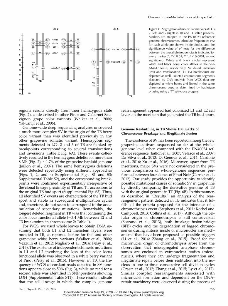
Complex and unbalanced chromosomal rearrange-ments found in the genome of TB might lead to defectsduring sexual reproduction (Pellestor, 2014). This hy-pothesis was tested specifically by studying the sexualtransmission of LGs 2 and 5 in selfed progeny of TT andTB, consisting of 38 and 46 individuals, respectively.For LG 2, four heterozygous microsatellite markerswere selected outside of the rearranged region. The al-lelic distribution observed in the progeny was differentfor TT and TB (frequencies inside the circles in Fig. 7). InTT, the two alleles segregated with a similar frequencyfor every marker studied, as expected for Mendelianinheritance (nonsignificant x2). Nevertheless, in TB,distorted transmission rates were identified, as everymarker analyzed showed one allele with a significantlylower frequency than expected for Mendelian inheri-tance. The frequency of the disfavored allele decreasedas themarkerwas closer to the chromosome rearrangedregion. From the genotype frequencies in the TB prog-eny, the two putative haplotypes in TB were inferred,showing that all the disfavored alleles were linked inthe same haplotype. The VviMybA1 gene also wasgenotyped by the independent amplification of thefunctional and nonfunctional alleles (Fig. 7). The studyof genotypic frequencies for the closest microsatelliteand VviMybA1 in the TT progeny allowed establishingthat the disfavored haplotype in the TB progeny wasthat carrying the functional allele for VviMybA1 in TTand, thus, the haplotype linked to SV and deletions inTB. For LG 5, a similar lack of transmission of onehaplotype was observed when two heterozygousmicrosatellite markers were analyzed in the two S1progeny. Thus, segregation analysis further supports
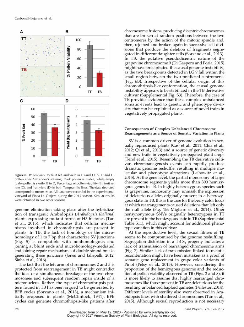
that SV affected only one copy of each chromosome.Rearranged chromosome segments are not transmittedto the next generation or are transmitted at a very lowfrequency. This lack of transmission was associatedwith a reduction of about 59% of male gamete viabilityin TB with respect to TT (Fig. 8), which suggests thatderivative chromosomes lack genes that are essentialfor the viability of the haploid phase. This sterility ratewould be consistent with the loss of essential genes formicrogametophyte viability in both LGs 2 and 5. Theeffect of complex chromosome rearrangements ongamete viability might result in additional agronomicalalterations present in TB, considering that reducedpollen viability correlated with 44% and 29% drops infruit set rate and fruit yield, respectively (Fig. 8).
DISCUSSION
Complex Unbalanced Genome Rearrangements Causedthe Loss of the Berry Color Locus Functional Allele
Deletions resulting in grapevine white berry variantshave been described previously (Walker et al., 2007;Vezzulli et al., 2012; Migliaro et al., 2014; Pelsy et al.,2015). However, the mutational mechanisms generat-ing these deletions remain uncertain. So far, only genereplacement has been proposed as the mechanismcausing LOH and originating newwhite berry alleles inPinot somatic variants by mitotic recombination be-tween chromosome 2 homologs linked to deletion ofthe color locus functional allele (Pelsy et al., 2015).Nonetheless, we would consider that, since CNVanalysis of the variant lines was not performed in thatstudy, the presence of hemizygous deletions instead ofgene replacement cannot be excluded in the origin of
Figure 5. Nature of breakpoint junction se-quences detected in TB. For each validated SVjunction site, close-ups of breakpoint andjunction site sequences are shown at top (LG2 with regular orientation in black, LG 2 withinverted orientation in red, LG 5 in blue, andLG 9 in green). Breakpoints in the context ofthe PN40024 12X.0 reference genome assem-bly chromosomes are represented at bottom.
Plant Physiol. Vol. 175, 2017 793
Chromothripsis-Mediated Loss of Grape Color
www.plantphysiol.orgon May 19, 2020 - Published by Downloaded from Copyright © 2017 American Society of Plant Biologists. All rights reserved.
the described Pinot alleles. In the TB white berry vari-ant, Southern blotting and WGS comparisons with itsblack berry ancestor, TT, confirmed the presence ofhemizygous deletions leaving at theMybA gene cluster
corresponding to the berry color locus a single copycorresponding to the null allele (Fig. 1B; SupplementalTable S11). The parallel copy number decrease ob-served indicates that LOH in these TB chromosome
Figure 6. DNA structural rearrangement graph and predicted rearranged chromosomes in TB. A, Structural rearrangement graphdepicting CNV, LOH, and rearrangement joins and types in the affected LGs. CNV is shown as log2 (copy number ratio TB/TT)calculated for eachwindowof the affected chromosomes by CNV-seq. SNP heterozygosity level is depicted as log2 (heterozygousSNPs per 100-kb ratio TB/TT). Connections between rearranged fragments are depicted by curved lines colored as indicated at thetop of the image according to join types observed in the paired-end alignment analysis. B, Diagram depicting the original and thepredicted rearranged orders of chromosome fragments affected by the chromothripsis-like restructuring that originated the variantgenome of TB. Deleted fragments in TB were predicted by LOH and CNV fromWGS data, and their extremes were confirmed byPCR of breakpoint junctions in most cases. Fragment order in TB derivative chromosomes was predicted considering the sixdeleted fragments and the six breakpoint junction sequences detected in the discordant read analysis that were validated by PCR(Inv, inversion; T, translocation). Question marks represent an additional breakpoint junction that was hypothesized from WGSread pairs that collectively did not pass all the SV analysis significance thresholds. According to this model, no other undetectedbreakpoint would be required to explain this catastrophic rearrangement. Rearrangements are represented in a single copy of theaffected chromosomes according to the linkage of deleted fragments in the same chromosome haplotype, which was inferredfrom a TT self-cross progeny segregation analysis andWGS data of TB. In purple, the asterisk indicates the segment comprising thecluster ofMybA genes belonging to the grape color locus functional allele, which is present in the heterozygous state in TTand islost after genome rearrangements in TB. The white allele is represented by the green line. Red lines denote centromeric repeatsaccording to predictions on the grapevine reference genome (Di Gaspero and Foria, 2015; Genoscope Web site).
794 Plant Physiol. Vol. 175, 2017
Carbonell-Bejerano et al.
www.plantphysiol.orgon May 19, 2020 - Published by Downloaded from Copyright © 2017 American Society of Plant Biologists. All rights reserved.
regions results directly from their hemizygous state(Fig. 2), as described in other Pinot and Cabernet Sau-vignon grape color variants (Walker et al., 2006;Yakushiji et al., 2006).Genome-wide deep sequencing analyses uncovered
a much more complex SV in the origin of the TB berrycolor variant than was identified previously in anyother grapevine somatic variant. Hemizygous seg-ments detected in LGs 2 and 5 of TB are flanked bybreakpoints corresponding to several translocationsand inversions (Table I; Fig. 6A). These events collec-tively resulted in the hemizygous deletion of more than8 Mb (Fig. 2), ;1.7% of the grapevine haploid genome(Jaillon et al., 2007). The same hemizygous deletionswere detected repeatedly using different approaches(Figs. 1, 2, and 4; Supplemental Figs. S1 and S3;Supplemental Table S2), and the corresponding break-points were confirmed experimentally irrespective ofthe clonal lineage proximity of TB and TT accessions tothe original TB bud sport (Supplemental Fig. S3). Thus,all identified SV events are characteristic of the TB budsport and stable in subsequent multiplication cyclesand, therefore, do not seem to correspond to the accu-mulation of secondary mutations. Remarkably, thelongest deleted fragment in TB was that containing thecolor locus functional allele (;3.4 Mb between T2 andT3 breakpoints in chromosome 2; Table I).For WGS, we used whole leaves to obtain DNA as-
suming that both L1 and L2 meristem layers weremutated in TB, as reported before for this and othergrapevine white berry variants (Walker et al., 2006;Vezzulli et al., 2012; Migliaro et al., 2014; Pelsy et al.,2015). The existence of independent chimeric mutationsin L1 and L2 involving the loss of the color locusfunctional allele was observed in a white berry variantof Pinot (Pelsy et al., 2015). However, in TB, the fre-quency of WGS discordant reads detected in SV junc-tions appears close to 50% (Fig. 3), while no read for asecond allele was identified in SNP positions showingLOH (Supplemental Table S11). These results indicatethat the cell lineage in which the complex genome
rearrangement appeared had colonized L1 and L2 celllayers in the meristem that generated the TB bud sport.
Genome Reshuffling in TB Shows Hallmarks ofChromosome Breakage and Illegitimate Fusion
The existence of SV has been reported among the fewgrapevine cultivars sequenced so far at the whole-genome level when compared with the PN40024 ref-erence sequence (Jaillon et al., 2007; Velasco et al., 2007;Da Silva et al., 2013; Di Genova et al., 2014; Cardoneet al., 2016; Xu et al., 2016). Moreover, apart from TEinsertions, major SVs were not considered in the pre-vious comparison of whole-genome sequences per-formed between four clones of Pinot Noir (Carrier et al.,2012). Our study provides the opportunity to identifypossible mutational causes of somatic SV in grapevineby directly comparing the derivative genome of TBwith the original genome in TT (Fig. 6B). In thismanner,as described in “Results,” an analysis of the rear-rangement pattern detected in TB indicates that it ful-fills all the criteria proposed for the inference of achromothripsis event (Stephens et al., 2011; Korbel andCampbell, 2013; Collins et al., 2017). Although the cel-lular origin of chromothripsis is still controversial(Sorzano et al., 2013), both breakage-fusion-bridge(BFB) cycles and the degradation of lagged chromo-somes during mitosis inside of micronuclei are mech-anisms that have been proposed as possible triggers(Li et al., 2014; Zhang et al., 2015). Proof for themicronuclei origin of chromothripsis arose from theobservation that missegregated anaphase chromo-somes are enclosed in extranuclear bodies (micro-nuclei), where they can undergo fragmentation andillegitimate repair before their restitution into the nu-cleus in one to three consecutive cell division cycles(Crasta et al., 2012; Zhang et al., 2015; Ly et al., 2017).Similar complex rearrangements associated withmicronuclei formation and dependent on the DNA-repair machinery were observed during the process of
Figure 7. Segregation ofmolecularmarkers at LGs2 (left) and 5 (right) in TB and TT selfed progeny.Markers are mapped to the PN40024 referencegenome chromosomes. Absolute frequencies (%)for each allele are shown inside circles, and thesignificance value of x2 tests for the differencebetween the two allele frequencies is indicated foreverymarker (*, P, 0.05; ***, P, 0.0001; ns, notsignificant). White and black circles representwhite and black berry color alleles in the Vvi-MybA1 locus, respectively. Validated inversion(Inv) and translocation (T1–T5) breakpoints aredepicted as well. Deleted chromosome segmentsdetected by CNV analysis from WGS data aredepicted as white boxes and linked in the samechromosome copy as determined by haplotypephasing using a TT self-cross progeny.
Plant Physiol. Vol. 175, 2017 795
Chromothripsis-Mediated Loss of Grape Color
www.plantphysiol.orgon May 19, 2020 - Published by Downloaded from Copyright © 2017 American Society of Plant Biologists. All rights reserved.
genome elimination taking place after the hybridiza-tion of transgenic Arabidopsis (Arabidopsis thaliana)plants expressing mutant forms of H3 histones (Tanet al., 2015), which indicates that cellular mecha-nisms involved in chromothripsis are present inplants. In TB, the lack of homology or the micro-homology of 1 to 7 bp that characterize SV junctions(Fig. 5) is compatible with nonhomologous endjoining at blunt ends and microhomology-mediatedend joining repair mechanisms of double-strand breaksgenerating these junctions (Jones and Jallepalli, 2012;Sinha et al., 2016).
The fact that the left arm of chromosomes 2 and 5 isprotected from rearrangement in TB might contradictthe idea of a simultaneous breakage of the two chro-mosomes and subsequent random repair inside of amicronucleus. Rather, the type of chromothripsis pat-tern found in TB has been argued to be generated byBFB cycles (Sorzano et al., 2013), a mechanism ini-tially proposed in plants (McClintock, 1941). BFBcycles can generate chromothripsis-like patterns after
chromosome fusions, producing dicentric chromosomesthat are broken at random positions between the twocentromeres by the action of the mitotic spindle and,then, rejoined and broken again in successive cell divi-sions that produce the deletion of fragments segre-gated in different daughter cells (Sorzano et al., 2013).In TB, the putative pseudodicentric nature of thegrapevine chromosome 9 (Di Gaspero and Foria, 2015)might have precipitated the causal genome instability,as the two breakpoints detected in LG 9 fall within thesmall region between the two predicted centromeres(Fig. 6B). Irrespective of the cellular origin of thischromothripsis-like conformation, the causal genomeinstability appears to be stabilized in the TB derivativecultivar (Supplemental Fig. S3). Therefore, the case ofTB provides evidence that these complex unbalancedsomatic events lead to genetic and phenotype diver-sity that can be exploited as a source of novel traits invegetatively propagated plants.
Consequences of Complex Unbalanced ChromosomeRearrangements as a Source of Somatic Variation in Plants
SV is a common driver of genome evolution in sex-ually reproduced plants (Cao et al., 2011; Chia et al.,2012; Qi et al., 2013) and a source of genetic diversityand new traits in vegetatively propagated plant crops(Terol et al., 2015). Resembling the TB derivative culti-var, chromoanagenesis events can rapidly producedramatic genome reshuffle, resulting in multiple mo-lecular and phenotype alterations (Leibowitz et al.,2015). At the gene level, the partial monosomy of largechromosome segments yields more than 300 hemizy-gous genes in TB. In highly heterozygous species suchas grapevine, monosomy may unmask the expressionof deleterious alleles originally present in a heterozy-gous state. In TB, this is the case for the berry color locusat which rearrangements caused deletions that left onlythe null allele (Fig. 1B; Migliaro et al., 2014). Othernonsynonymous SNVs originally heterozygous in TTare present in the hemizygous state in TB (SupplementalTable S11), which might account for additional pheno-type variation in this cultivar.
At the reproductive level, the sexual fitness of TBseems to be compromised by the genome reshuffling.Segregation distortion in a TB S1 progeny indicates alack of transmission of rearranged chromosome arms(Fig. 7). Similar lack of transmission linked to meioticrecombination might have been mistaken as a proof ofsomatic gene replacement in grape color variants ofPinot (Pelsy et al., 2015). However, considering theproportion of the hemizygous genome and the reduc-tion of pollen viability observed in TB (Figs. 2 and 8), itis more likely to assume that highly rearranged chro-mosomes like those present in TB are deleterious for theresulting unbalanced haploid gametes (Pellestor, 2014).Different levels of sterility also were observed in Ara-bidopsis lines with shattered chromosomes (Tan et al.,2015). Although sexual reproduction is not necessary
Figure 8. Pollen viability, fruit set, and yield in TB and TT. A, TTand TBpollen after Alexander’s staining. Dark pollen is viable, while empty(pale) pollen is sterile. B to D, Percentage of pollen viability (B), fruit setrate (C), and fruit yield (D) in both Tempranillo lines. The data depictedcorrespond to means 6 SD. All data were recorded in the experimentalvineyard of Finca La Grajera during the 2015 season. Similar resultswere obtained in two other seasons.
796 Plant Physiol. Vol. 175, 2017
Carbonell-Bejerano et al.
www.plantphysiol.orgon May 19, 2020 - Published by Downloaded from Copyright © 2017 American Society of Plant Biologists. All rights reserved.
for the maintenance of vegetatively propagated crops,gametophyte viability is required for proper fruit set(Iyer, 1966); thus, complex SV may have agronomicconsequences in decreasing fruit yield, as observed inTB (Fig. 8).
CONCLUSION
Awhole-genome scale approach was helpful to showthat, in the TB white grape variant, the deletion of thegrape color locus functional allele is linked to a muchmore complex genome rearrangement than was ob-served previously in any other grapevine somaticvariant. To our knowledge, this is the first studydelimiting the deletion of this allele at a base resolutionlevel. In TB, this deletion occurred associated with anunbalanced chromoanagenesis event entailing an ille-gitimate rejoining of fragments after multiple chro-mosome breakage. Taken together, our results showthat complex chromosome rearrangements with chro-mothriptic features naturally emerge and stabilizeduring plant vegetative growth. Although possiblyreducing sexual fitness and compromising fruit andseed production, these complex somatic events rapidlygenerate new phenotypes that can be selected andvegetatively propagated. Therefore, complex unbal-anced genome rearrangements that emerged duringsomatic growth might be relevant for the genetic im-provement and evolution of clonally propagated plantspecies such as woody crops.
MATERIALS AND METHODS
Plant Material
Molecular biology and viticultural experiments, respectively, were carriedout using materials collected from the two ancestral TB multiplication grape(Vitis vinifera) vineyards (TB-ICVV2 and TB-ICVV3 accessions; SupplementalTable S9). These vineyards are located in Finca Valdegón (in Agoncillo, LaRioja, Spain) and Finca LaGrajera (in Logroño, La Rioja, Spain), respectively. Asa representative accession, TT clone RJ51 (the most cultivated TT clone in theRioja DOC region) from both sites was used as respective control. Both plotsbelong to the Grapevine GermplasmCollection of the Instituto de Ciencias de laVid y del Vino (ICVV; ESP-217) and are maintained under the same agro-nomical conditions. All plants are grafted in Richter-110 rootstock, trellised in adouble cordon Royat system, and cultivated in a similar way. The genotypes ofnine microsatellite loci located in different chromosomes were obtained asdescribed elsewhere (Ibáñez et al., 2009), confirming that TB matches the gen-otype of TT (Supplemental Table S12). Another five TB and five TT accessionsfrom diverse origins were used to assess the presence of SV breakpoints(Supplemental Table S9).
Selfed progeny of TT-RJ51 and TB-ICVV2 accessionswere generated in 2008,consisting of 78 and 46 individuals, respectively. These progeny were main-tained ungrafted in pots at Finca Valdegón.
DNA Extraction
In all experiments, total gDNA was isolated from young leaves using theDNeasy Plant Mini Kit (Qiagen) according to the protocol described by themanufacturer. Additionally, mature fruits of TT-RJ51 and TB-ICVV1 wereharvested in 2016, and gDNA was obtained separately from berry skin andflesh. Independent DNA samples were used for each technique: microsatellitegenotyping, Southern blot, GrapeGen GeneChip hybridization, Illuminasequencing, and PCR amplification.
Southern Blot
DNA-blot hybridization analysis was performed as described by Sambrooket al. (1989). Briefly, gDNA fromTT and TBwas digestedwithApaI, transferredto a nylon membrane, and hybridized to a VviMybA1 probe as describedpreviously (Lijavetzky et al., 2006). The VviMybA1 probe was generated byPCR amplification with primers Ps (59-TCACGGGGTTTAGAAAGTGG-39)and Pas (59-ATCAATTGGGGAATTGGTGA-39) using TT gDNA as a template.The hybridized membrane was scanned with a STORM PhosphorImager(Molecular Dynamics).
LOH Analysis in Specific Amplicons
To analyze LOH in TB, primer pairs for 30 amplicons were designed alongLG 2 (Supplemental Table S1). PCR amplifications were carried out using TaqDNA Polymerase (Qiagen) as recommended by the manufacturer. PCR pro-ducts were purified with ExoSAP-IT (USB Products Affymetrix) according tothe manufacturer’s instructions and then used for Sanger sequencing at theGenomic Unit of Parque Científico de Madrid with the same primers used foramplification.
CNV Analysis by GeneChip Hybridization
Two biological replicates of TT and TB were hybridized to the GrapeGenGeneChip (Lijavetzky et al., 2012). For each replicate, gDNA samples (10mg each)were fragmented to an average size of 0.5 kb with a sonicator Labsonic U set at50% intensity and repeating duty cycle of 0.5, four pulses of 10 s each. Labeling ofDNA was performed as recommended in the GeneChip Whole TranscriptDouble-Stranded Target Assay (Affymetrix). Briefly, fragmented DNA was am-plified, purified, and biotin terminal labeled with the dsDNA Terminal LabelingKit (Affymetrix) following recommendations. Finally, 7.5 mg of labeled DNAwasused for the hybridization of microarrays according to the Affymetrix GeneChipExpression Analysis Technical Manual. CEL files were RMA normalized, anddifferential probe set hybridization between TB and TT was assessed using theRankProd package from Bioconductor (Breitling et al., 2004). A cutoff value of1.8-fold change absolute value and P , 0.01 were established to detect geneswith genomic CNV. To this end, the annotation of the GrapeGen GeneChipaccording to 12X V1 gene predictions from Lijavetzky et al. (2012) was used.
Genome Resequencing and ComputationalComparative Analyses
Sequencing and Preprocessing
Two gDNA libraries were built from a sample of young leaves from oneindividual of TT-RJ51 and another from TB-ICVV2 (second-round multiplica-tion plant). The gDNA of each individual was fragmented randomly. Afterelectrophoresis, DNA fragments of;470 bp were gel purified. Adapter ligationand DNA cluster preparation were then performed and subjected to sequenc-ing. Each Tempranillo line was sequenced in a different lane at Beijing Ge-nomics Institute facilities using the Illumina HiSeq 2000 sequencing system. Oneach sample, a total of ;209 3 106 paired-end reads of 90-nucleotide lengthwere obtained (;18.8 Gb of total sequence per sample; Supplemental Table S4).
Sequence data were processed by removing the adapter sequence fromreads and subsequently taking out the reads with low quality (Phred quality# 5 in 50% or more of the positions of the read) to obtain the clean data de-scribed in Supplemental Table S4. The final Q20 of each lane was above 95%.Two files containing the clean reads in FastQ format, corresponding to TT andTB samples, were submitted to an in-house pipeline composed of several bashshell scripts (available upon request to the authors). These scripts used shellcommands, Perl scripts, and open-source programs to process the reads, pro-duce the alignments, select particular subsets of alignments for the PEM study,and carry out the CNV study and the variant calling. The pipeline also includeda prediction of effects for the retrieved variants and a scanning for LOH regionsin TB. Each specific procedure is described below.
Alignment
Each FastQ file was aligned to the grapevine PN40024 12X.0 reference ge-nome assembly using BWA version 0.5.9-r16 with the option sampe (for paired
Plant Physiol. Vol. 175, 2017 797
Chromothripsis-Mediated Loss of Grape Color
www.plantphysiol.orgon May 19, 2020 - Published by Downloaded from Copyright © 2017 American Society of Plant Biologists. All rights reserved.
ends) and default parameters (Li and Durbin, 2009). Approximately 76% of thereads aligned with the reference genome (Supplemental Table S5). Redundantreads comprising the same span were then removed using the rmdup option ofSAMtools (Li et al., 2009). To improve the alignments by minimizing thenumber of artifactual mismatching bases due to the close presence of INDELswith respect to the reference genome, local realignment was then done usingGATK version 1.0.5777 (tools RealignerTargetCreator and IndelRealigner;McKenna et al., 2010). To increase the ability of SV breakpoint detection, an-other script was used to split each of the 90-nucleotide clean reads into twofragments of 45 nucleotides. The new sets of FastQ files containing the45-nucleotide split reads also were aligned as described above and used for SVjunction detection.
Variant Calling and LOH Genome Scanning
Files containing read alignments of TT and TB in BAM format were used tosearch for Tempranillo line-specific polymorphisms (SNPs and INDELs).Aligned readswere preprocessed as described above, andmoreover, readswithmapping quality, 40 after local realignment in IndelRealigner were discardedtoo. Variant calling on each line was carried out using the BCFtools utility fromSAMtools for the whole genome and the HaplotypeCaller tool from the GATKpackage for LGs 2 and 5. Initially, for both tools, VCF files independentlycomparing each Tempranillo line with the grapevine 12X.0 reference genomewere generated. In order to remove false positives, the following filters andcutoff thresholds were used: polymorphisms were considered when 15 or morereads covered the position and the variant sequence was observed in 35% ormore for heterozygous sites and 90% or more with two or fewer reads of thenonvariant allele for homozygous sites. Finally, the resulting TT and TB sets ofheterozygous polymorphisms identified in SAMtools were binned in 100-kbbins, and these bins were reported and plotted for each LG. To delimit segmentsof LOH along the TB genome, intervals of three or more consecutive hetero-zygous sites in TT (according to the filters described above) that fulfilled thecriteria of homozygous sites in TBwere considered. To estimate the genotype inTB irrespective of the variation relative to the reference genome, the sequence atthese positions was obtained from the compilation in the pileup file of thealignments. In these cases, a Perl script was constructed to detect homozygoussites in TB according to eight or more reads coverage and 90% or greater fre-quency of the most frequent allele and two or more for alleles other than themost frequent one.
GATKHaplotypeCaller also was used for variant calling in LGs 2 and 5, andthe results were cross-checked with those obtained from SAMtools + BCFtools.Although the results were highly equivalent, HaplotypeCaller was used tostudy the effect of polymorphisms involving LOH in hemizygous regions of TBby assuming that this application could perform better on INDEL detection(Hwang et al., 2015). The same filters described for the output of SAMtools wereused to detect TT polymorphic sites with LOH in TB (heterozygous in TT andhomozygous in TB). In this case, to estimate the genotype of TB for nonvariantLOH sites, the variant calling was repeated after merging the reads from thetwo lines. The genotype in the nonvariant line was inferred by deducting thereads of the variant line from the total of both. Finally, the effect of detectedpolymorphisms was predicted using SnpEff version 2.0.3 (Cingolani et al.,2012) according to the grapevine 12X V1 gene predictions from CRIBI(http://genomes.cribi.unipd.it/) and the functional annotation and classifi-cation of genes described by Grimplet et al. (2012).
Genome Resequencing CNV
CNV-seq (Xie and Tammi, 2009) was applied using TB and TT as test andreference, respectively, on the corresponding files containing the initial positionof each aligned read relative to the reference genome (called hits files by theauthors of CNV-seq). Significance values were set in CNV-seq to log2 (copynumber TB/TT) # 20.5 and P # 0.00001 for each chromosomal window andfour or more consecutive sliding windows for annotating a CNV zone. TheCNV zones detected by CNV-seq with P# 0.05 and log2 (copy number TB/TT)# 20.5 or $ 0.37 were considered as significantly decreased or increased, re-spectively, in copy number in TB relative to TT.
SV Breakpoint Search
PEM and soft-clipped reads indicative of the presence of SV junctions weresearched specifically in the two SAM files containing the TB and TT reads, re-spectively, aligned to the PN40024 12X.0 reference genome. The two alignmentsets of whole 90-nucleotide reads and reads split into two 45-nucleotide frag-ments were processed equally in parallel. First, read pairs with mapping
quality = 0 in BWA were removed, as this indicates mapping to multiple lo-cations. From the remaining set of aligned reads and using bash shell com-mands (grep and awk) and the SAMtools merge utility, putative chromosomerearrangement breakpoint sites were searched by extracting two subsets ofpaired-end reads with discordant mate alignment: (1) read pairs with increasedinsert size indicative of intrachromosomal translocations or large deletions:absolute value of insert size (TLEN field in the SAM file) . 4 times the medianTLEN value of all aligned pairs in the sample; and (2) read pairs with mates indifferent chromosomes indicative of interchromosome translocations (RNEXTfield different from = or RNAME value in the SAM file). To determine the or-ientation of the rearranged chromosome fragments comprising putativebreakpoints, paired-end reads with unexpected mate orientation alignmentwere searched from the previous two subsets. Two different discordant ori-entations were considered: (1) both mates with unexpected orientation (FLAGfield of the SAM file with values = 81, 161, 97, or 145); and (2) only onematewithunexpected orientation (FLAG with values 65, 129, 113, or 177). An additionalsubset comprising soft-clipped reads (only partially mapping to the reference inone read extreme) was searched for in genomic regions with discordant matealignment 1 or 2. A read containing CIGAR alignment operation S was thecriterion used to select soft-clipped reads.
To select breakpoints distinguishing TB andTT genomes, BEDtools (Quinlanand Hall, 2010) of TB-specific discordant reads were extracted to delimit SVcandidate breakpoints, which were inspected visually using Integrative Ge-nomics Viewer (version 2.2) software (Thorvaldsdóttir et al., 2013).
The nonclipped part of TB-specific soft-clipped reads mapping on candi-date breakpoint areas was aligned to the PN40024 12X.0 genome assemblyusing BLASTN suite (https://blast.ncbi.nlm.nih.gov/Blast.cgi?PAGE_TYPE=BlastSearch).
Analysis of Repetitive Sequences at Breakpoints
For each SV junction detected in TB, a consensus breakpoint junction se-quence was built from the sequence of discordant reads and submittedto RepeatMasker version 4.0.3 (http://www.repeatmasker.org/cgi-bin/WEBRepeatMasker). V. vinifera was the DNA source, and ABBlast/WUBlast was the search engine to query for matches with repeat elementsdeposited in RepBase database update 2013/04/22 (http://www.girinst.org/repbase), which includes all described repeat elements specific ofV. vinifera. TE-like sequences were searched by BLAST of RepBase sequences against (1) the12X.0 reference genome in regions where TB SV breakpointsmapped and (2) TBconsensus sequences at SV junctions. The structure and domains of detected TEsequences were studied with National Center for Biotechnology InformationConserved Domain Database tools (http://www.ncbi.nlm.nih.gov/cdd/).
Validation of SV Junctions
PCR primers were designed at each flank of genomically detected SVjunctions (Supplemental Table S1). The consensus breakpoint junction sequencereconstructed from discordant reads was used as a template for primer pairdesign. Primer specificity was checked by BLAST against the PN40024 12Xgrapevine reference genome assembly in the Genoscope Web site (http://www.genoscope.cns.fr/externe/GenomeBrowser/Vitis/). These primer pairswere tested for PCR amplification using as a template gDNA from different TBand TT accessions (Supplemental Fig. S3; Supplemental Table S9). PCRs werecarried out using MyTaq DNA Polymerase (Bioline, Meridian Life Science)following the manufacturer’s protocol. TheVviDXS (VIT_05s0020g02130) gene,mapping on a presumably disomic region of LG 5 in both TT and TB, was in-cluded in the experiment as a positive control using primers described else-where (Emanuelli et al., 2010). An aliquot of the amplification product wassubjected to electrophoresis through 1% agarose gels to check for the presenceand size of PCR products. PCR products were sequenced as described above.
SNP Chip Genotyping and Haplotype Phasing
Total gDNA from 78 TT S1 progeny individuals was genotyped usingthe GrapeReSeq Illumina 18K SNP Infinium as described elsewhere(Houel et al., 2015). SNP genotype data were processed as described inthe GrapeReSeq project (https://urgi.versailles.inra.fr/Species/Vitis/GrapeReSeq_Illumina_20K). To reconstruct the original phasing of the two haplo-types in TT, segregation for each pair of adjacent SNP loci represented in the chipwas studied, assuming that themost frequent genotype combination in the progenycorresponded to the parental haplotypes. The genotype of TB at SNP positions
798 Plant Physiol. Vol. 175, 2017
Carbonell-Bejerano et al.
www.plantphysiol.orgon May 19, 2020 - Published by Downloaded from Copyright © 2017 American Society of Plant Biologists. All rights reserved.
represented in the chip mapping on LGs 2 and 5 was obtained from ourWGS dataset. The haplotype remaining in TB at positions involving LOH in this variant wascompared with the two haplotypes reconstructed in TT to infer the homologouscopy affected by each deletion.
Sexual Transmission Analysis
Aseries ofmarkerswere genotyped ina set of TTandTBself-cross S1progenyto study the sexual transmission of each homologous chromosome at LGs2 and 5. Four heterozygous microsatellite markers in both Tempranillo linesmapping on LG 2 were selected: Chr2b, Chr2a, C6F1, and C5G7. Genotyping ofVviMybA1 alleles in TT, TB, and TT self-cross S1 progeny was carried out asdescribed previously (Lijavetzky et al., 2006). Primer pairs a-d3 and b-d wereused to amplify null and functional alleles, respectively. Allele segregation wasdetermined for each locus and progeny, and their deviation from expectedvalues (0.5) was evaluated through x2 tests. This statistical test was performedusing Microsoft Excel software. Genotypic segregation for each pair of adjacentloci was studied to determine microsatellite haplotypes, assuming that themostfrequent double homozygous genotypes in the progeny correspond to the pa-rental haplotypes. Joint genotypic segregation of C5G7 and VviMybA1 wasused to establish the haplotypes at LG 2. For LG 5, two microsatellite markerswere selected, VVIT68 and VMC5E11, and the procedure was similar to thatdescribed for LG 2.
Fruit Production and Pollen Viability
Experiments were carried out at Finca La Grajera during 2015. In thisvineyard plot, three rows (;50 plants each) of TB plants are interspersed bythree rows of TT plants. Each row was used as an independent biologicalreplicate, and four plants per replicate were analyzed. Pollen was collected si-multaneously from TT and TB inflorescences (one per plant) at 50% bloom.Pollen viability was analyzed using Alexander’s modified staining as describedpreviously (Royo et al., 2016). For each Tempranillo line, more than 1,000 pollengrains per biological replicate were analyzed. Fruit set percentage was calcu-lated as the rate between the number of ripened fruits per cluster at maturityand the number of flowers at flowering time in the same cluster (one cluster perplant was analyzed). The number of flowers per inflorescence was estimated bybagging the inflorescence before the onset of flowering and counting flowercaps inside the bag after fruit set. Fruit yield was estimated as the total mass ofgrape bunches per plant.
Accession Numbers
GrapeGenGeneChip andWGS data are deposited in the National Center forBiotechnology Information under Gene Expression Omnibus GSE80801 andSequence Read Archive SRP065756 (BioProject PRJNA301084) accessionnumbers, respectively.
Supplemental Data
The following supplemental materials are available.
Supplemental Figure S1. Microarray-based CNV in a TB white berry so-matic variant.
Supplemental Figure S2. Intrachromosome translocation in TB LG 5.
Supplemental Figure S3. Validation of TB SV junctions in additional Tem-pranillo accessions.
Supplemental Figure S4. Transposon and repetitive sequences detected atTB breakpoints sites.
Supplemental Table S1. Oligonucleotides used as primers for PCR ampli-fication.
Supplemental Table S2.Heterozygosity analysis in PCR-amplified regionsaround the color locus in LG 2.
Supplemental Table S3. GrapeGen GeneChip probe sets showing differ-ential CGH between TB and TT.
Supplemental Table S4. Summary of resequencing data production (cleandata).
Supplemental Table S5. Summary of resequencing read alignments.
Supplemental Table S6. Genome regions showing LOH in TB.
Supplemental Table S7. Genome windows showing copy number de-crease in TB.
Supplemental Table S8. Sanger capillary electrophoresis-obtained se-quences of PCR amplicons confirming breakpoint junctions predictedby genome resequencing in TB.
Supplemental Table S9. Origins of Tempranillo accessions.
Supplemental Table S10. Haplotype phasing of fragments deleted in LGs2 and 5 of TB.
Supplemental Table S11. List of predicted hemizygous variant sites in theTB genome and functional effect.
Supplemental Table S12. Microsatellite genotyping identification.
ACKNOWLEDGMENTS
We thank Drs. Karel H.M. van Wely and Daniel Trujillano for thoughtfulcomments and feedback and Guillermo Juárez and Silvia Hernáiz for technicalassistance; we also thank the genomics service of the CNB-Consejo Superior deInvestigaciones Científicas for running the CGH hybridizations.
Received May 31, 2017; accepted August 13, 2017; published August 15, 2017.
LITERATURE CITED
Battilana J, Emanuelli F, Gambino G, Gribaudo I, Gasperi F, Boss PK,Grando MS (2011) Functional effect of grapevine 1-deoxy-D-xylulose5-phosphate synthase substitution K284N on Muscat flavour forma-tion. J Exp Bot 62: 5497–5508
Boss PK, Davies C, Robinson SP (1996) Expression of anthocyanin bio-synthesis pathway genes in red and white grapes. Plant Mol Biol 32:565–569
Boss PK, Thomas MR (2002) Association of dwarfism and floral inductionwith a grape ‘green revolution’ mutation. Nature 416: 847–850
Breitling R, Armengaud P, Amtmann A, Herzyk P (2004) Rank products: asimple, yet powerful, new method to detect differentially regulatedgenes in replicated microarray experiments. FEBS Lett 573: 83–92
Cao J, Schneeberger K, Ossowski S, Günther T, Bender S, Fitz J, KoenigD, Lanz C, Stegle O, Lippert C, et al (2011) Whole-genome sequencingof multiple Arabidopsis thaliana populations. Nat Genet 43: 956–963
Cardone MF, D’Addabbo P, Alkan C, Bergamini C, Catacchio CR, AnaclerioF, Chiatante G, Marra A, Giannuzzi G, Perniola R, et al (2016) Inter-varietalstructural variation in grapevine genomes. Plant J 88: 648–661
Carrier G, Le Cunff L, Dereeper A, Legrand D, Sabot F, Bouchez O,Audeguin L, Boursiquot JM, This P (2012) Transposable elements are amajor cause of somatic polymorphism in Vitis vinifera L. PLoS ONE 7:e32973
Chia JM, Song C, Bradbury PJ, Costich D, de Leon N, Doebley J, ElshireRJ, Gaut B, Geller L, Glaubitz JC, et al (2012) Maize HapMap2 iden-tifies extant variation from a genome in flux. Nat Genet 44: 803–807
Cingolani P, Platts A, Wang L, Coon M, Nguyen T, Wang L, Land SJ, LuX, Ruden DM (2012) A program for annotating and predicting theeffects of single nucleotide polymorphisms, SnpEff: SNPs in the ge-nome of Drosophila melanogaster strain w1118; iso-2; iso-3. Fly (Austin)6: 80–92
Collins RL, Brand H, Redin CE, Hanscom C, Antolik C, Stone MR,Glessner JT, Mason T, Pregno G, Dorrani N, et al (2017) Defining thediverse spectrum of inversions, complex structural variation, andchromothripsis in the morbid human genome. Genome Biol 18: 36
Crasta K, Ganem NJ, Dagher R, Lantermann AB, Ivanova EV, Pan Y, NeziL, Protopopov A, Chowdhury D, Pellman D (2012) DNA breaks andchromosome pulverization from errors in mitosis. Nature 482: 53–58
D’Amato F (1997) Role of somatic mutations in the evolution of higherplants. Caryologia 50: 1–15
Da Silva C, Zamperin G, Ferrarini A, Minio A, Dal Molin A, Venturini L,Buson G, Tononi P, Avanzato C, Zago E, et al (2013) The high poly-phenol content of grapevine cultivar tannat berries is conferred pri-marily by genes that are not shared with the reference genome. PlantCell 25: 4777–4788
Plant Physiol. Vol. 175, 2017 799
Chromothripsis-Mediated Loss of Grape Color
www.plantphysiol.orgon May 19, 2020 - Published by Downloaded from Copyright © 2017 American Society of Plant Biologists. All rights reserved.
Di Gaspero G, Foria S (2015) Molecular grapevine breeding techniques. InAG Reynolds, ed, Grapevine Breeding Programs for the Wine Industry.Woodhead Publishing, Oxford, pp 23–37
Di Genova A, Almeida AM, Muñoz-Espinoza C, Vizoso P, Travisany D,Moraga C, Pinto M, Hinrichsen P, Orellana A, Maass A (2014) Wholegenome comparison between table and wine grapes reveals a compre-hensive catalog of structural variants. BMC Plant Biol 14: 7
Doligez A, Adam-Blondon AF, Cipriani G, Di Gaspero G, Laucou V,Merdinoglu D, Meredith CP, Riaz S, Roux C, This P (2006) An inte-grated SSR map of grapevine based on five mapping populations. TheorAppl Genet 113: 369–382
Emanuelli F, Battilana J, Costantini L, Le Cunff L, Boursiquot JM, This P,Grando MS (2010) A candidate gene association study on Muscat flavorin grapevine (Vitis vinifera L.). BMC Plant Biol 10: 241
Emanuelli F, Sordo M, Lorenzi S, Battilana J, Grando MS (2014) Devel-opment of user-friendly functional molecular markers for VvDXS geneconferring Muscat flavor in grapevine. Mol Breed 33: 235–241
Fernandez L, Chaïb J, Martinez-Zapater JM, Thomas MR, Torregrosa L(2013) Mis-expression of a PISTILLATA-like MADS box gene preventsfruit development in grapevine. Plant J 73: 918–928
Fernandez L, Torregrosa L, Segura V, Bouquet A, Martinez-Zapater JM(2010) Transposon-induced gene activation as a mechanism generatingcluster shape somatic variation in grapevine. Plant J 61: 545–557
Furiya T, Suzuki S, Sueta T, Takayanagi T (2009) Molecular characteri-zation of a bud sport of Pinot Gris bearing white berries. Am J Enol Vitic60: 66–73
Grimplet J, Van Hemert J, Carbonell-Bejerano P, Díaz-Riquelme J,Dickerson J, Fennell A, Pezzotti M, Martínez-Zapater JM (2012)Comparative analysis of grapevine whole-genome gene predictions,functional annotation, categorization and integration of the predictedgene sequences. BMC Res Notes 5: 213
Houel C, Chatbanyong R, Doligez A, Rienth M, Foria S, Luchaire N,Roux C, Adivèze A, Lopez G, Farnos M, et al (2015) Identification ofstable QTLs for vegetative and reproductive traits in the microvine (Vitisvinifera L.) using the 18 K Infinium chip. BMC Plant Biol 15: 205
Hwang S, Kim E, Lee I, Marcotte EM (2015) Systematic comparison ofvariant calling pipelines using gold standard personal exome variants.Sci Rep 5: 17875
Ibáñez J, Muñoz-Organero G, Zinelabidine LH, de Andrés MT, CabelloF, Martínez-Zapater JM (2012) Genetic origin of the grapevine cultivarTempranillo. Am J Enol Vitic 63: 549–553
Ibáñez J, Vélez MD, de Andrés MT, Borrego J (2009) Molecular markersfor establishing distinctness in vegetatively propagated crops: a casestudy in grapevine. Theor Appl Genet 119: 1213–1222
Iyer GS (1966) Induction of pollen sterility in grapes (Vitis vinifera). Vitis 5:433–445
Jaillon O, Aury JM, Noel B, Policriti A, Clepet C, Casagrande A, ChoisneN, Aubourg S, Vitulo N, Jubin C, et al (2007) The grapevine genomesequence suggests ancestral hexaploidization in major angiospermphyla. Nature 449: 463–467
Jones MJ, Jallepalli PV (2012) Chromothripsis: chromosomes in crisis. DevCell 23: 908–917
Kloosterman WP, Tavakoli-Yaraki M, van Roosmalen MJ, van Binsbergen E,Renkens I, Duran K, Ballarati L, Vergult S, Giardino D, Hansson K, et al(2012) Constitutional chromothripsis rearrangements involve clustered double-stranded DNA breaks and nonhomologous repair mechanisms. Cell Rep 1:648–655
Kobayashi S, Goto-Yamamoto N, Hirochika H (2004) Retrotransposon-induced mutations in grape skin color. Science 304: 982
Korbel JO, Campbell PJ (2013) Criteria for inference of chromothripsis incancer genomes. Cell 152: 1226–1236
Korbel JO, Urban AE, Affourtit JP, Godwin B, Grubert F, Simons JF, KimPM, Palejev D, Carriero NJ, Du L, et al (2007) Paired-end mappingreveals extensive structural variation in the human genome. Science 318:420–426
Laucou V, Lacombe T, Dechesne F, Siret R, Bruno JP, Dessup M, DessupT, Ortigosa P, Parra P, Roux C, et al (2011) High throughput analysis ofgrape genetic diversity as a tool for germplasm collection management.Theor Appl Genet 122: 1233–1245
Leibowitz ML, Zhang CZ, Pellman D (2015) Chromothripsis: a newmechanism for rapid karyotype evolution. Annu Rev Genet 49: 183–211
Li H, Durbin R (2009) Fast and accurate short read alignment withBurrows-Wheeler transform. Bioinformatics 25: 1754–1760
Li H, Handsaker B, Wysoker A, Fennell T, Ruan J, Homer N, Marth G,Abecasis G, Durbin R (2009) The Sequence Alignment/Map format andSAMtools. Bioinformatics 25: 2078–2079
Li XQ (2016) Natural attributes and agricultural implications of somaticgenome variation. Curr Issues Mol Biol 20: 29–46
Li Y, Schwab C, Ryan S, Papaemmanuil E, Robinson HM, Jacobs P,Moorman AV, Dyer S, Borrow J, Griffiths M, et al (2014) Constitutionaland somatic rearrangement of chromosome 21 in acute lymphoblasticleukaemia. Nature 508: 98–102
Lijavetzky D, Carbonell-Bejerano P, Grimplet J, Bravo G, Flores P, FenollJ, Hellín P, Oliveros JC, Martínez-Zapater JM (2012) Berry flesh andskin ripening features in Vitis vinifera as assessed by transcriptionalprofiling. PLoS ONE 7: e39547
Lijavetzky D, Ruiz-García L, Cabezas JA, De Andrés MT, Bravo G, IbáñezA, Carreño J, Cabello F, Ibáñez J, Martínez-Zapater JM (2006) Molec-ular genetics of berry colour variation in table grape. Mol Genet Ge-nomics 276: 427–435
Ly P, Teitz LS, Kim DH, Shoshani O, Skaletsky H, Fachinetti D, Page DC,Cleveland DW (2017) Selective Y centromere inactivation triggerschromosome shattering in micronuclei and repair by non-homologousend joining. Nat Cell Biol 19: 68–75
Malhotra A, Lindberg M, Faust GG, Leibowitz ML, Clark RA, Layer RM,Quinlan AR, Hall IM (2013) Breakpoint profiling of 64 cancer genomesreveals numerous complex rearrangements spawned by homology-independent mechanisms. Genome Res 23: 762–776
Marroni F, Pinosio S, Morgante M (2014) Structural variation and genomecomplexity: is dispensable really dispensable? Curr Opin Plant Biol 18:31–36
Martinez J, Vicente T, Martinez T, Chavarri JB, Garcia-Escudero E (2006)Una nueva variedad blanca para la D.O.Ca. Rioja: el Tempranillo Blanco[A new white variety for the DOC Rioja: ‘Tempranillo Blanco’]. In 29thOIV World Congress of Vine and Wine: 4th General Assembly of theOIV. Organisation Internationale de la Vigne et du Vin, Logrono, Spain
McClintock B (1941) The stability of broken ends of chromosomes in Zeamays. Genetics 26: 234–282
McKenna A, Hanna M, Banks E, Sivachenko A, Cibulskis K, KernytskyA, Garimella K, Altshuler D, Gabriel S, Daly M, et al (2010) The Ge-nome Analysis Toolkit: a MapReduce framework for analyzing next-generation DNA sequencing data. Genome Res 20: 1297–1303
Migliaro D, Crespan M, Muñoz-Organero G, Velasco R, Moser C, Vezzulli S(2014) Structural dynamics at the berry colour locus inVitis vinifera L. somaticvariants. Aust J Grape Wine Res 20: 485–495
Pelsy F, Dumas V, Bévilacqua L, Hocquigny S, Merdinoglu D (2015)Chromosome replacement and deletion lead to clonal polymorphism ofberry color in grapevine. PLoS Genet 11: e1005081
Pelsy F, Hocquigny S, Moncada X, Barbeau G, Forget D, Hinrichsen P,Merdinoglu D (2010) An extensive study of the genetic diversity withinseven French wine grape variety collections. Theor Appl Genet 120:1219–1231
Pellestor F (2014) Chromothripsis: how does such a catastrophic eventimpact human reproduction? Hum Reprod 29: 388–393
Qi J, Liu X, Shen D, Miao H, Xie B, Li X, Zeng P, Wang S, Shang Y, GuX, et al (2013) A genomic variation map provides insights into thegenetic basis of cucumber domestication and diversity. Nat Genet 45:1510–1515
Quinlan AR, Hall IM (2010) BEDTools: a flexible suite of utilities forcomparing genomic features. Bioinformatics 26: 841–842
Rausch T, Zichner T, Schlattl A, Stütz AM, Benes V, Korbel JO (2012)DELLY: structural variant discovery by integrated paired-end and split-read analysis. Bioinformatics 28: i333–i339
Royo C, Carbonell-Bejerano P, Torres-Pérez R, Nebish A, Martínez Ó,Rey M, Aroutiounian R, Ibáñez J, Martínez-Zapater JM (2016) De-velopmental, transcriptome, and genetic alterations associated withparthenocarpy in the grapevine seedless somatic variant Corinto bianco.J Exp Bot 67: 259–273
Sambrook J, Fritsch EF, Maniatis T (1989) Molecular Cloning: A Labora-tory Manual, Ed 2. Cold Spring Harbor Laboratory Press, Cold SpringHarbor, NY
Sinha S, Villarreal D, Shim EY, Lee SE (2016) Risky business: microhomology-mediated end joining. Mutat Res 788: 17–24
Sorzano CO, Pascual-Montano A, Sánchez de Diego A, Martínez-A C,van Wely KH (2013) Chromothripsis: breakage-fusion-bridge over andover again. Cell Cycle 12: 2016–2023
800 Plant Physiol. Vol. 175, 2017
Carbonell-Bejerano et al.
www.plantphysiol.orgon May 19, 2020 - Published by Downloaded from Copyright © 2017 American Society of Plant Biologists. All rights reserved.
Stephens PJ, Greenman CD, Fu B, Yang F, Bignell GR, Mudie LJ,Pleasance ED, Lau KW, Beare D, Stebbings LA, et al (2011) Massivegenomic rearrangement acquired in a single catastrophic event duringcancer development. Cell 144: 27–40
Tan EH, Henry IM, Ravi M, Bradnam KR, Mandakova T, Marimuthu MP,Korf I, Lysak MA, Comai L, Chan SW (2015) Catastrophic chromo-somal restructuring during genome elimination in plants. eLife 4: 4
Terol J, Ibañez V, Carbonell J, Alonso R, Estornell LH, Licciardello C,Gut IG, Dopazo J, Talon M (2015) Involvement of a citrus meiotic re-combination TTC-repeat motif in the formation of gross deletions gen-erated by ionizing radiation and MULE activation. BMC Genomics 16:69
This P, Lacombe T, Cadle-Davidson M, Owens CL (2007) Wine grape(Vitis vinifera L.) color associates with allelic variation in the domesti-cation gene VvmybA1. Theor Appl Genet 114: 723–730
This P, Lacombe T, Thomas MR (2006) Historical origins and genetic di-versity of wine grapes. Trends Genet 22: 511–519
Thorvaldsdóttir H, Robinson JT, Mesirov JP (2013) Integrative GenomicsViewer (IGV): high-performance genomics data visualization and ex-ploration. Brief Bioinform 14: 178–192
Torregrosa L, Fernandez L, Bouquet A, Boursiquot JM, Pelsy F, Martínez-Zapater JM (2011) Origins and consequences of somatic variation ingrapevine. In AF Adam-Blondon, JM Martinez-Zapater, C Kole, eds,Genetics, Genomics, and Breeding of Grapes. Science Publishers, Jersey,British Isles, pp 68–92
Velasco R, Zharkikh A, Troggio M, Cartwright DA, Cestaro A, Pruss D,Pindo M, Fitzgerald LM, Vezzulli S, Reid J, et al (2007) A high qualitydraft consensus sequence of the genome of a heterozygous grapevinevariety. PLoS ONE 2: e1326
Vezzulli S, Leonardelli L, Malossini U, Stefanini M, Velasco R, Moser C(2012) Pinot blanc and Pinot gris arose as independent somatic muta-tions of Pinot noir. J Exp Bot 63: 6359–6369
Walker AR, Lee E, Bogs J, McDavid DA, Thomas MR, Robinson SP (2007)White grapes arose through the mutation of two similar and adjacentregulatory genes. Plant J 49: 772–785
Walker AR, Lee E, Robinson SP (2006) Two new grape cultivars, budsports of Cabernet Sauvignon bearing pale-coloured berries, are theresult of deletion of two regulatory genes of the berry colour locus. PlantMol Biol 62: 623–635
Whitham TG, Slobodchikoff CN (1981) Evolution by individuals, plant-herbivore interactions, and mosaics of genetic variability: the adaptivesignificance of somatic mutations in plants. Oecologia 49: 287–292
Wong DC, Schlechter R, Vannozzi A, Höll J, Hmmam I, Bogs J, TornielliGB, Castellarin SD, Matus JT (2016) A systems-oriented analysis of thegrapevine R2R3-MYB transcription factor family uncovers new insightsinto the regulation of stilbene accumulation. DNA Res 23: 451–466
Xie C, Tammi MT (2009) CNV-seq, a new method to detect copy numbervariation using high-throughput sequencing. BMC Bioinformatics 10: 80
Xu Y, Gao Z, Tao J, Jiang W, Zhang S, Wang Q, Qu S (2016) Genome-widedetection of SNP and SV variations to reveal early ripening-relatedgenes in grape. PLoS ONE 11: e0147749
Yakushiji H, Kobayashi S, Goto-Yamamoto N, Tae Jeong S, Sueta T, MitaniN, Azuma A (2006) A skin color mutation of grapevine, from black-skinnedPinot Noir to white-skinned Pinot Blanc, is caused by deletion of the func-tional VvmybA1 allele. Biosci Biotechnol Biochem 70: 1506–1508
Zhang CZ, Spektor A, Cornils H, Francis JM, Jackson EK, Liu S,Meyerson M, Pellman D (2015) Chromothripsis from DNA damage inmicronuclei. Nature 522: 179–184
Plant Physiol. Vol. 175, 2017 801
Chromothripsis-Mediated Loss of Grape Color
www.plantphysiol.orgon May 19, 2020 - Published by Downloaded from Copyright © 2017 American Society of Plant Biologists. All rights reserved.


![36 [1,n]-sigmatropic rearrangements](https://static.fdocuments.in/doc/165x107/55504a55b4c9058f768b5083/36-1n-sigmatropic-rearrangements.jpg)


![[3,3]-Sigmatropic rearrangements - Massey Universitygjrowlan/stereo2/lecture11.pdf · 123.702 Organic Chemistry Claisen rearrangements • One of the most useful sigmatropic rearrangements](https://static.fdocuments.in/doc/165x107/5adcada77f8b9a213e8bd8b0/33-sigmatropic-rearrangements-massey-gjrowlanstereo2lecture11pdf123702.jpg)