Cataract Surgery in Eyes with Glaucoma · Cataract Surgery in Eyes with Glaucoma Malik Y. Kahook,...
48
Cataract Surgery in Eyes with Glaucoma Malik Y. Kahook, MD The Slater Family Endowed Chair in Ophthalmology Professor of Ophthalmology Vice Chair, Clinical and Translational Research University of Colorado School of Medicine Aurora, CO
Transcript of Cataract Surgery in Eyes with Glaucoma · Cataract Surgery in Eyes with Glaucoma Malik Y. Kahook,...
Cataract Surgery in Eyes with Glaucoma
Malik Y. Kahook, MDThe Slater Family Endowed Chair in Ophthalmology
Professor of Ophthalmology
Vice Chair, Clinical and Translational Research
University of Colorado School of Medicine
Aurora, CO
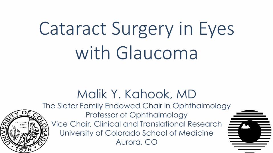
Financial DisclosureI have the following financial interests or relationships to disclose:
J&J Vision: P
Alcon Laboratories, Inc.: P
Allergan, Inc.: C
New World Medical: C,P
Equinox: C,O
IanTech Medical: P
FDA C
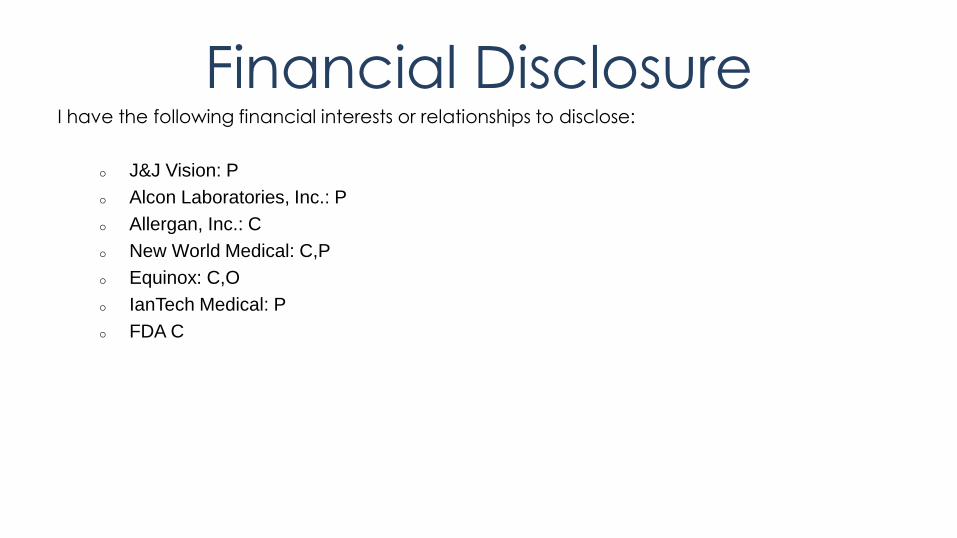
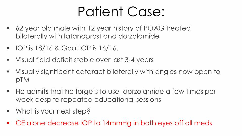
Patient Case:▪ 62 year old male with 12 year history of POAG treated
bilaterally with latanoprost and dorzolamide
▪ IOP is 18/16 & Goal IOP is 16/16.
▪ Visual field deficit stable over last 3-4 years
▪ Visually significant cataract bilaterally with angles now
open to pTM
▪ He admits that he forgets to use dorzolamide a few
times per week despite repeated educational sessions
▪ What is your next step?
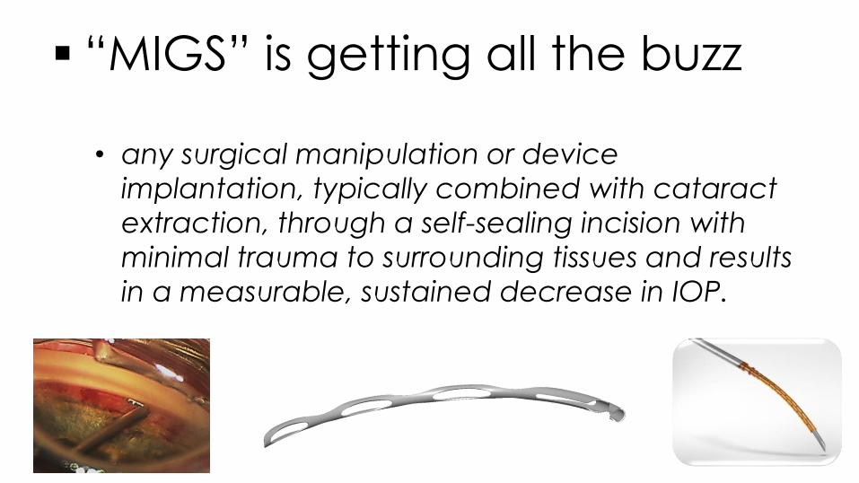
Most Recent Attempts: MIGS▪ “MIGS” is getting all the buzz
• any surgical manipulation or device
implantation, typically combined with cataract
extraction, through a self-sealing incision with
minimal trauma to surrounding tissues and results
in a measurable, sustained decrease in IOP.
without the formation of a bleb.
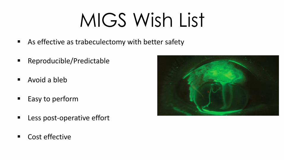
MIGS Wish List▪ As effective as trabeculectomy with better safety
▪ Reproducible/Predictable
▪ Avoid a bleb
▪ Easy to perform
▪ Less post-operative effort
▪ Cost effective
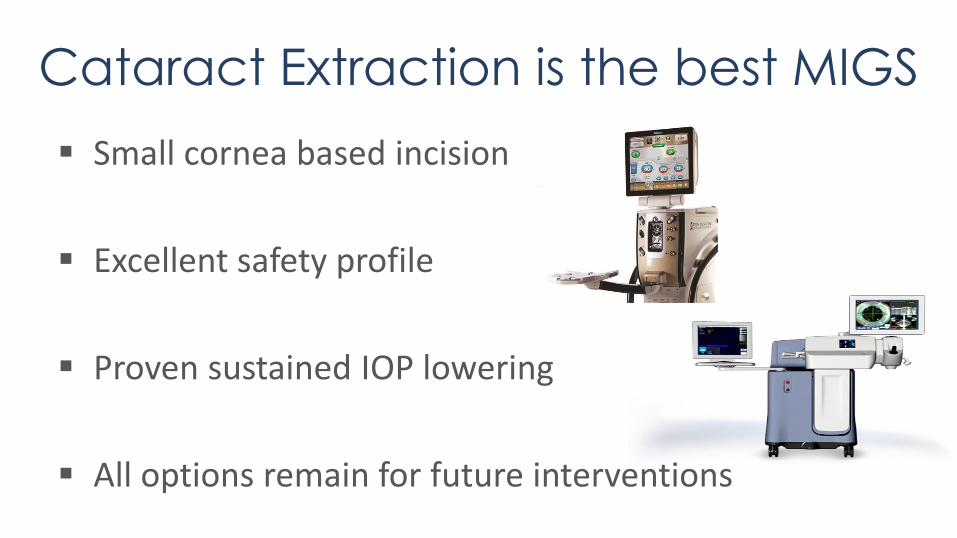
Cataract Extraction is the best MIGS
▪ Small cornea based incision
▪ Excellent safety profile
▪ Proven sustained IOP lowering
▪ All options remain for future interventions
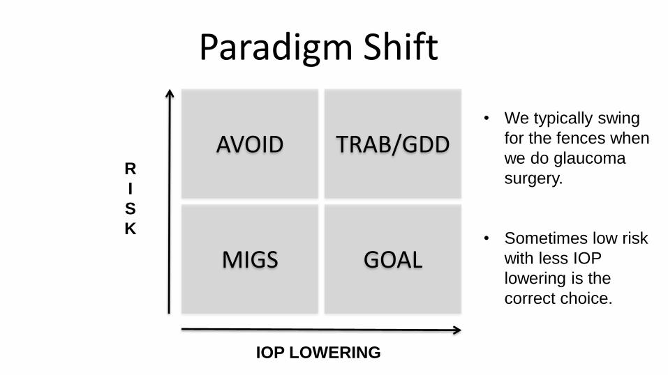
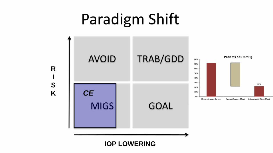
Paradigm Shift
AVOID TRAB/GDD
MIGS GOAL
R
I
S
K
IOP LOWERING
• We typically swing
for the fences when
we do glaucoma
surgery.
• Sometimes low risk
with less IOP
lowering is the
correct choice.
Paradigm Shift
AVOID TRAB/GDD
MIGS GOAL
R
I
S
K
IOP LOWERING
CE
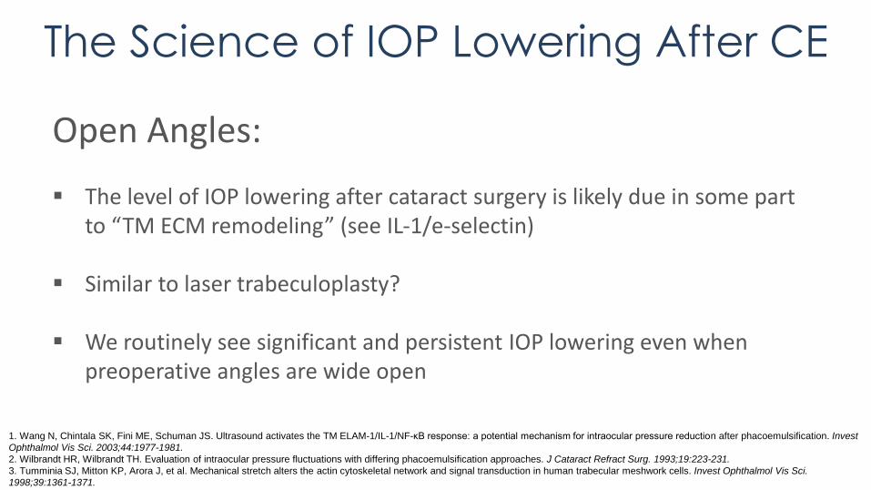
The Science of IOP Lowering After CE
Open Angles:
▪ The level of IOP lowering after cataract surgery is likely due in some part to “TM ECM remodeling” (see IL-1/e-selectin)
▪ Similar to laser trabeculoplasty?
▪ We routinely see significant and persistent IOP lowering even when preoperative angles are wide open
1. Wang N, Chintala SK, Fini ME, Schuman JS. Ultrasound activates the TM ELAM-1/IL-1/NF-κB response: a potential mechanism for intraocular pressure reduction after phacoemulsification. Invest
Ophthalmol Vis Sci. 2003;44:1977-1981.
2. Wilbrandt HR, Wilbrandt TH. Evaluation of intraocular pressure fluctuations with differing phacoemulsification approaches. J Cataract Refract Surg. 1993;19:223-231.
3. Tumminia SJ, Mitton KP, Arora J, et al. Mechanical stretch alters the actin cytoskeletal network and signal transduction in human trabecular meshwork cells. Invest Ophthalmol Vis Sci.
1998;39:1361-1371.
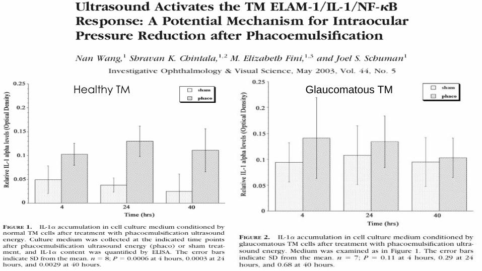
Healthy TM Glaucomatous TM
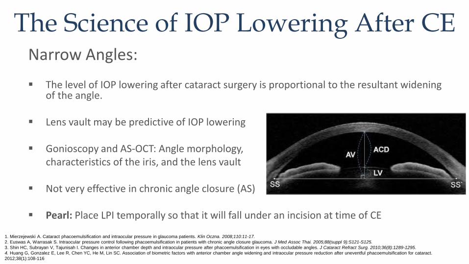
The Science of IOP Lowering After CENarrow Angles:
▪ The level of IOP lowering after cataract surgery is proportional to the resultant widening of the angle.
▪ Lens vault may be predictive of IOP lowering
▪ Gonioscopy and AS-OCT: Angle morphology, characteristics of the iris, and the lens vault
▪ Not very effective in chronic angle closure (AS)
▪ Pearl: Place LPI temporally so that it will fall under an incision at time of CE
1. Mierzejewski A. Cataract phacoemulsification and intraocular pressure in glaucoma patients. Klin Oczna. 2008;110:11-17.
2. Euswas A, Warrasak S. Intraocular pressure control following phacoemulsification in patients with chronic angle closure glaucoma. J Med Assoc Thai. 2005;88(suppl 9):S121-S125.
3. Shin HC, Subrayan V, Tajunisah I. Changes in anterior chamber depth and intraocular pressure after phacoemulsification in eyes with occludable angles. J Cataract Refract Surg. 2010;36(8):1289-1295.
4. Huang G, Gonzalez E, Lee R, Chen YC, He M, Lin SC. Association of biometric factors with anterior chamber angle widening and intraocular pressure reduction after uneventful phacoemulsification for cataract.
2012;38(1):108-116
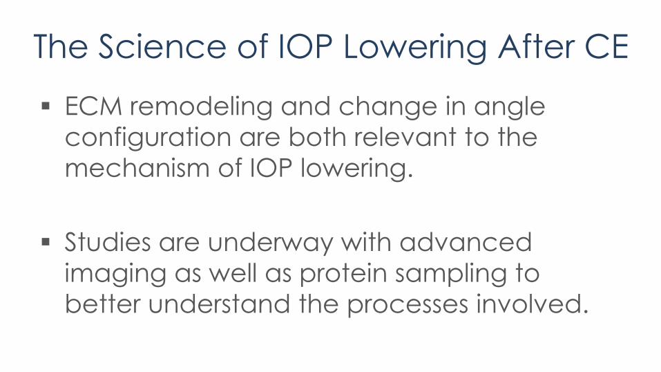
The Science of IOP Lowering After CE
▪ ECM remodeling and change in angle
configuration are both relevant to the
mechanism of IOP lowering.
▪ Studies are underway with advanced
imaging as well as protein sampling to
better understand the processes involved.
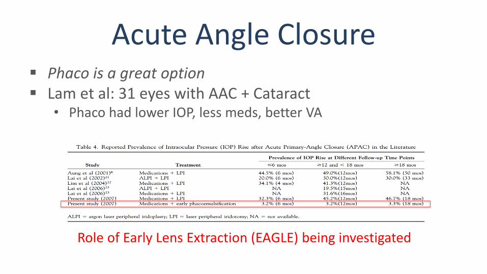
Acute Angle Closure▪ Phaco is a great option▪ Lam et al: 31 eyes with AAC + Cataract
• Phaco had lower IOP, less meds, better VA
Role of Early Lens Extraction (EAGLE) being investigated
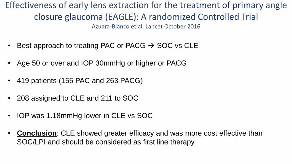
Effectiveness of early lens extraction for the treatment of primary angle closure glaucoma (EAGLE): A randomized Controlled Trial
Azuara-Blanco et al. Lancet October 2016
• Best approach to treating PAC or PACG SOC vs CLE
• Age 50 or over and IOP 30mmHg or higher or PACG
• 419 patients (155 PAC and 263 PACG)
• 208 assigned to CLE and 211 to SOC
• IOP was 1.18mmHg lower in CLE vs SOC
• Conclusion: CLE showed greater efficacy and was more cost effective than
SOC/LPI and should be considered as first line therapy
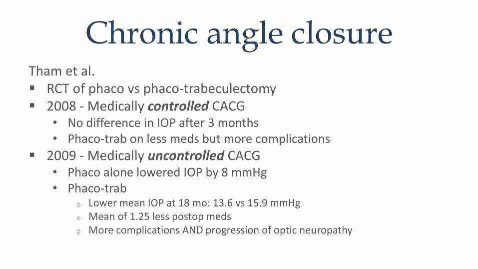
Chronic angle closureTham et al. ▪ RCT of phaco vs phaco-trabeculectomy▪ 2008 - Medically controlled CACG
• No difference in IOP after 3 months• Phaco-trab on less meds but more complications
▪ 2009 - Medically uncontrolled CACG • Phaco alone lowered IOP by 8 mmHg• Phaco-trab
Lower mean IOP at 18 mo: 13.6 vs 15.9 mmHg Mean of 1.25 less postop meds More complications AND progression of optic neuropathy
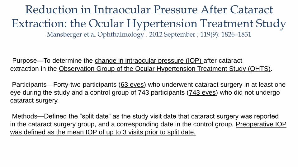
Reduction in Intraocular Pressure After Cataract Extraction: the Ocular Hypertension Treatment Study
Mansberger et al Ophthalmology . 2012 September ; 119(9): 1826–1831
Purpose—To determine the change in intraocular pressure (IOP) after cataract
extraction in the Observation Group of the Ocular Hypertension Treatment Study (OHTS).
Participants—Forty-two participants (63 eyes) who underwent cataract surgery in at least one
eye during the study and a control group of 743 participants (743 eyes) who did not undergo
cataract surgery.
Methods—Defined the “split date” as the study visit date that cataract surgery was reported
in the cataract surgery group, and a corresponding date in the control group. Preoperative IOP
was defined as the mean IOP of up to 3 visits prior to split date.
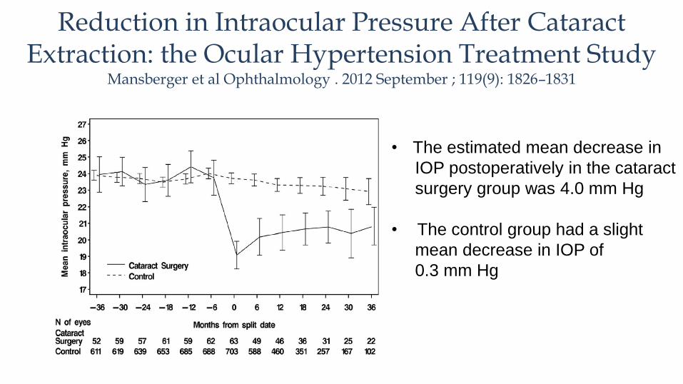
Reduction in Intraocular Pressure After Cataract Extraction: the Ocular Hypertension Treatment Study
Mansberger et al Ophthalmology . 2012 September ; 119(9): 1826–1831
• The estimated mean decrease in
IOP postoperatively in the cataract
surgery group was 4.0 mm Hg
• The control group had a slight
mean decrease in IOP of
0.3 mm Hg
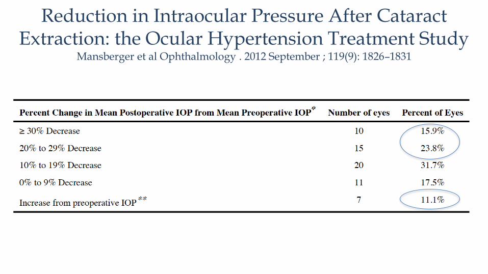
Reduction in Intraocular Pressure After Cataract Extraction: the Ocular Hypertension Treatment Study
Mansberger et al Ophthalmology . 2012 September ; 119(9): 1826–1831
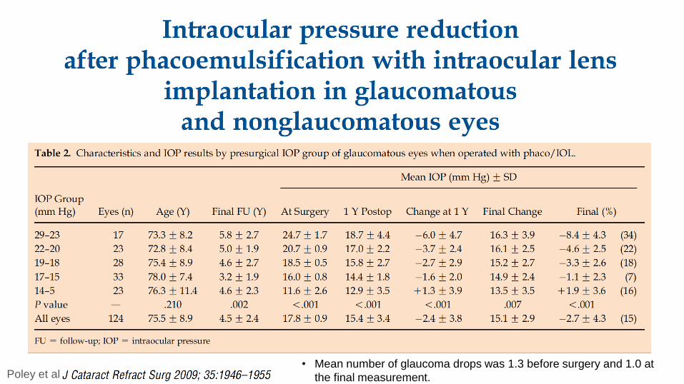
Poley et al• Mean number of glaucoma drops was 1.3 before surgery and 1.0 at
the final measurement.
Question:▪ Which mechanism has been linked with IOP lowering
in glaucoma eyes post cataract extraction
A. TM ECM changes related to ultrasound
B. Degree of lens vault preoperatively
C. Both A&B
D. None of the above
Question:▪ Which mechanism has been linked with IOP lowering
in glaucoma eyes post cataract extraction
A. TM ECM changes related to ultrasound
B. Degree of lens vault preoperatively
C. Both A&B
D. None of the above
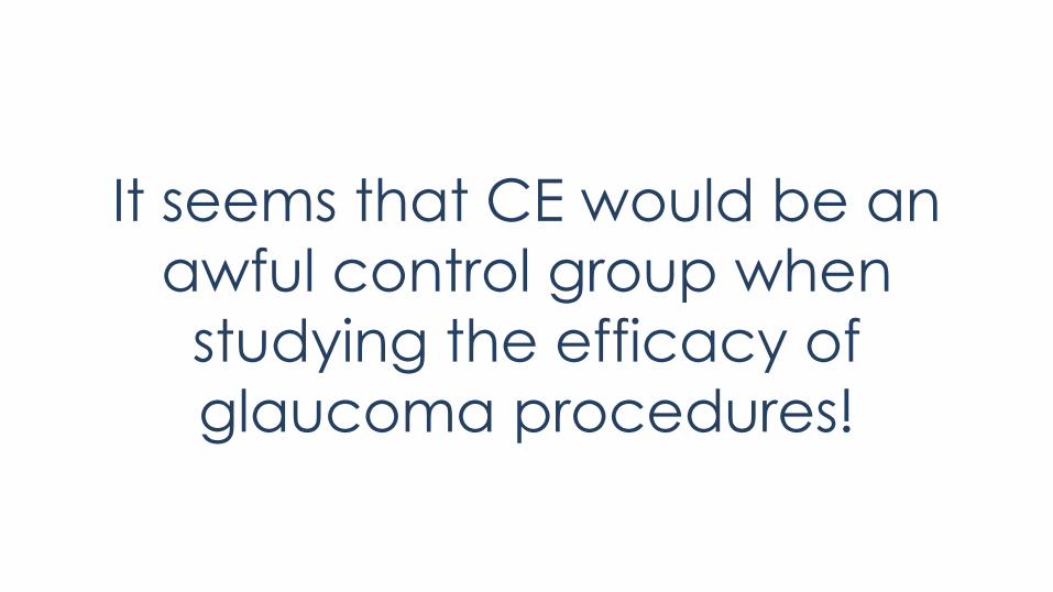
It seems that CE would be an
awful control group when
studying the efficacy of
glaucoma procedures!

Learning from MIGS Trials
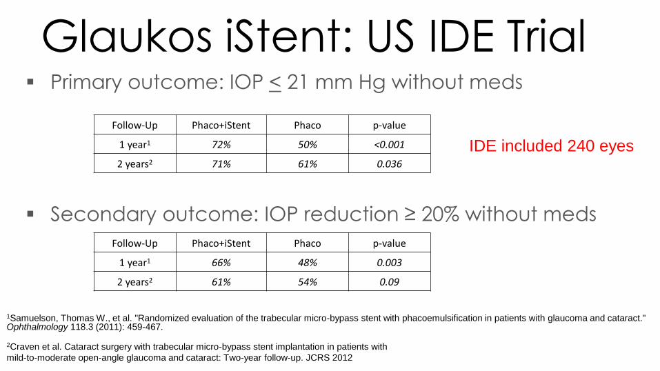
Glaukos iStent: US IDE Trial▪ Primary outcome: IOP < 21 mm Hg without meds
▪ Secondary outcome: IOP reduction ≥ 20% without meds
1Samuelson, Thomas W., et al. "Randomized evaluation of the trabecular micro-bypass stent with phacoemulsification in patients with glaucoma and cataract." Ophthalmology 118.3 (2011): 459-467.
2Craven et al. Cataract surgery with trabecular micro-bypass stent implantation in patients with
mild-to-moderate open-angle glaucoma and cataract: Two-year follow-up. JCRS 2012
Follow-Up Phaco+iStent Phaco p-value
1 year1 72% 50% <0.001
2 years2 71% 61% 0.036
Follow-Up Phaco+iStent Phaco p-value
1 year1 66% 48% 0.003
2 years2 61% 54% 0.09
IDE included 240 eyes
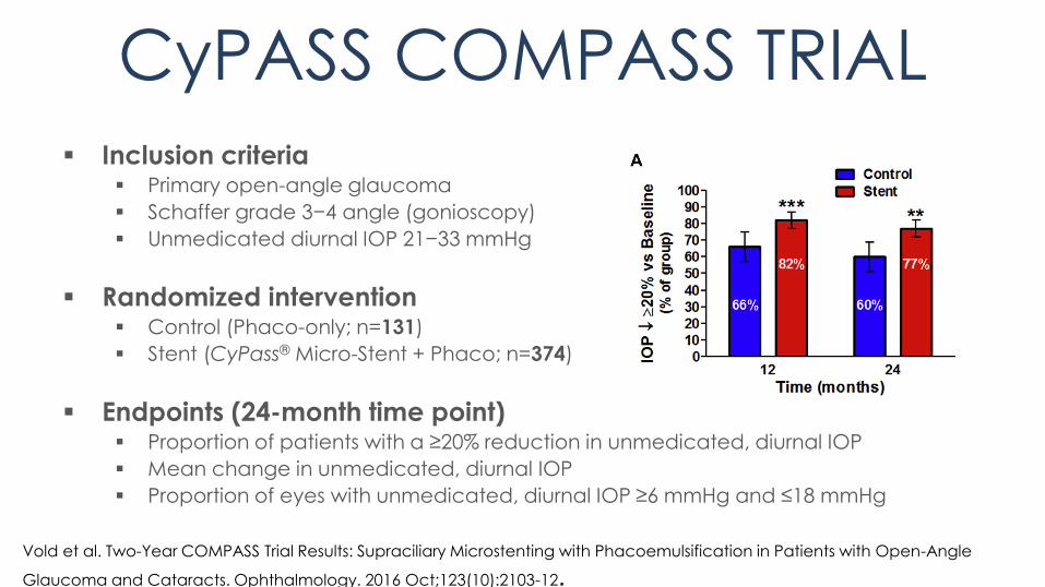
CyPASS COMPASS TRIAL
▪ Inclusion criteria▪ Primary open-angle glaucoma
▪ Schaffer grade 3−4 angle (gonioscopy)
▪ Unmedicated diurnal IOP 21−33 mmHg
▪ Randomized intervention▪ Control (Phaco-only; n=131)
▪ Stent (CyPass® Micro-Stent + Phaco; n=374)
▪ Endpoints (24-month time point)▪ Proportion of patients with a ≥20% reduction in unmedicated, diurnal IOP
▪ Mean change in unmedicated, diurnal IOP
▪ Proportion of eyes with unmedicated, diurnal IOP ≥6 mmHg and ≤18 mmHg
Vold et al. Two-Year COMPASS Trial Results: Supraciliary Microstenting with Phacoemulsification in Patients with Open-Angle
Glaucoma and Cataracts. Ophthalmology. 2016 Oct;123(10):2103-12.
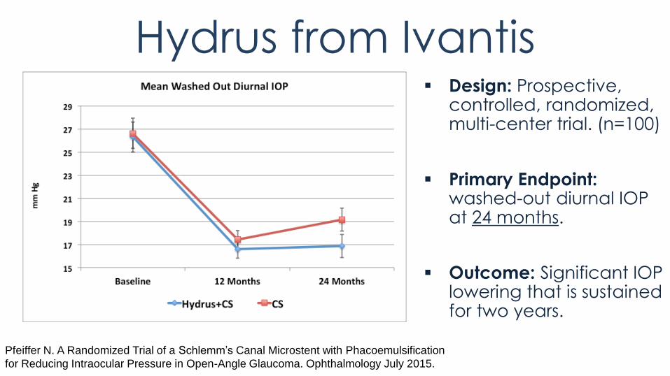
Hydrus from Ivantis▪ Design: Prospective,
controlled, randomized, multi-center trial. (n=100)
▪ Primary Endpoint: washed-out diurnal IOP at 24 months.
▪ Outcome: Significant IOP lowering that is sustained for two years.
Pfeiffer N. A Randomized Trial of a Schlemm’s Canal Microstent with Phacoemulsification
for Reducing Intraocular Pressure in Open-Angle Glaucoma. Ophthalmology July 2015.
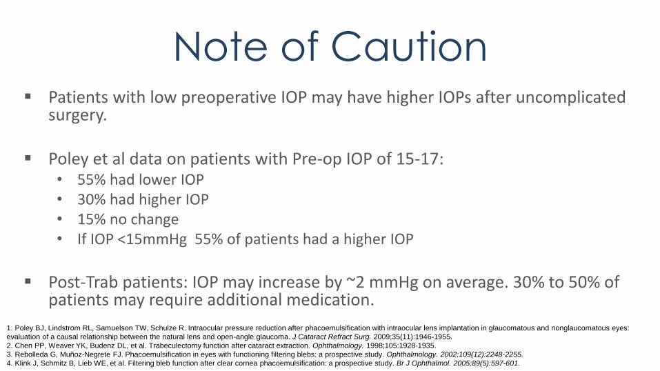
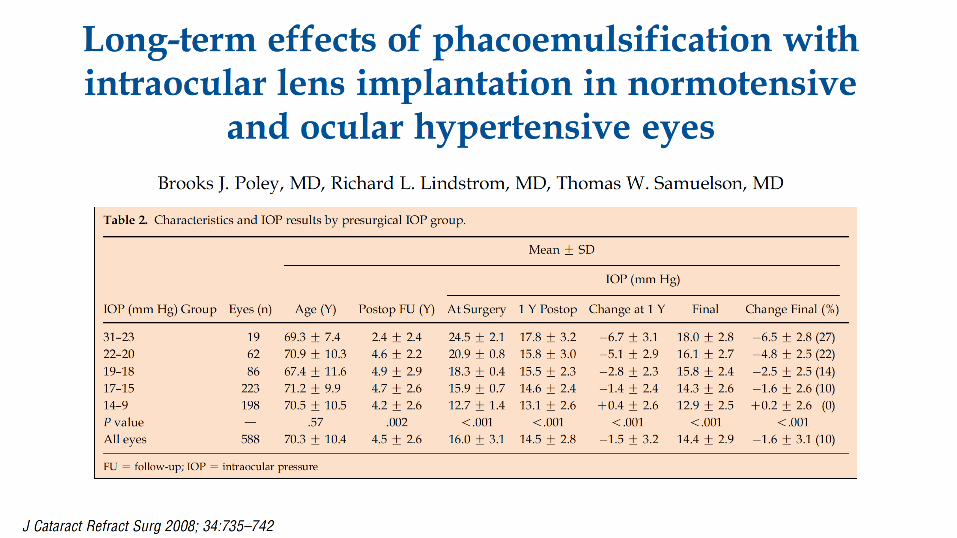
Note of Caution▪ Patients with low preoperative IOP may have higher IOPs after uncomplicated
surgery.
▪ Poley et al data on patients with Pre-op IOP of 15-17: • 55% had lower IOP • 30% had higher IOP• 15% no change• If IOP <15mmHg 55% of patients had a higher IOP
▪ Post-Trab patients: IOP may increase by ~2 mmHg on average. 30% to 50% of patients may require additional medication.
1. Poley BJ, Lindstrom RL, Samuelson TW, Schulze R. Intraocular pressure reduction after phacoemulsification with intraocular lens implantation in glaucomatous and nonglaucomatous eyes:
evaluation of a causal relationship between the natural lens and open-angle glaucoma. J Cataract Refract Surg. 2009;35(11):1946-1955.
2. Chen PP, Weaver YK, Budenz DL, et al. Trabeculectomy function after cataract extraction. Ophthalmology. 1998;105:1928-1935.
3. Rebolleda G, Muñoz-Negrete FJ. Phacoemulsification in eyes with functioning filtering blebs: a prospective study. Ophthalmology. 2002;109(12):2248-2255.
4. Klink J, Schmitz B, Lieb WE, et al. Filtering bleb function after clear cornea phacoemulsification: a prospective study. Br J Ophthalmol. 2005;89(5):597-601.
Patient Case:▪ 62 year old male with 12 year history of POAG treated
bilaterally with latanoprost and dorzolamide
▪ IOP is 18/16 & Goal IOP is 16/16.
▪ Visual field deficit stable over last 3-4 years
▪ Visually significant cataract bilaterally with angles now open to
pTM
▪ He admits that he forgets to use dorzolamide a few times per
week despite repeated educational sessions
▪ What is your next step?
▪ CE alone decrease IOP to 14mmHg in both eyes off all meds
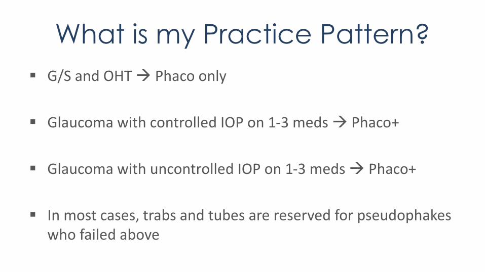
What is my Practice Pattern?
▪ G/S and OHT Phaco only
▪ Glaucoma with controlled IOP on 1-3 meds Phaco+
▪ Glaucoma with uncontrolled IOP on 1-3 meds Phaco+
▪ In most cases, trabs and tubes are reserved for pseudophakeswho failed above
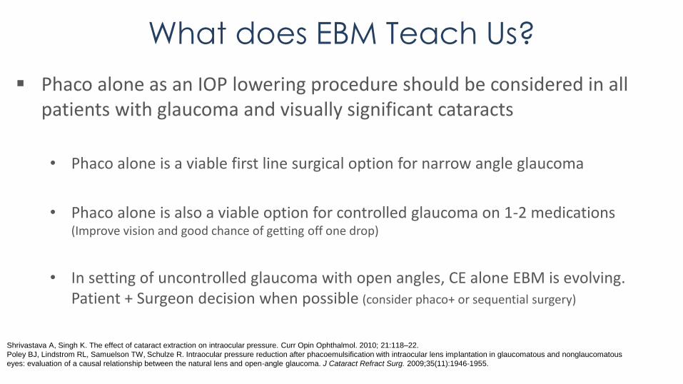
What does EBM Teach Us?
▪ Phaco alone as an IOP lowering procedure should be considered in all patients with glaucoma and visually significant cataracts
• Phaco alone is a viable first line surgical option for narrow angle glaucoma
• Phaco alone is also a viable option for controlled glaucoma on 1-2 medications (Improve vision and good chance of getting off one drop)
• In setting of uncontrolled glaucoma with open angles, CE alone EBM is evolving. Patient + Surgeon decision when possible (consider phaco+ or sequential surgery)
Shrivastava A, Singh K. The effect of cataract extraction on intraocular pressure. Curr Opin Ophthalmol. 2010; 21:118–22.
Poley BJ, Lindstrom RL, Samuelson TW, Schulze R. Intraocular pressure reduction after phacoemulsification with intraocular lens implantation in glaucomatous and nonglaucomatous
eyes: evaluation of a causal relationship between the natural lens and open-angle glaucoma. J Cataract Refract Surg. 2009;35(11):1946-1955.

In many cases, the art of
medicine matters just as
much as the science
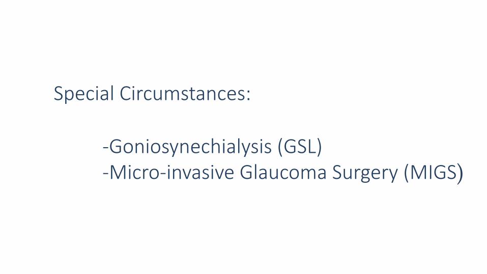
Special Circumstances:
-Goniosynechialysis (GSL)-Micro-invasive Glaucoma Surgery (MIGS)
Who is a candidate for GSL?
▪ Phakic patient with primary angle closure glaucoma (PACG): Combine CE + GSL
▪ CACG with elevated IOP and at least 50% of the angle sealed with PAS is a good candidate for CE + GSL
▪ Many have advocated for “fresh” (6-12 months) PAS as being ideal***
▪ Standalone GSL may have IOP benefits in CACG but this is controversial and likely dependent on timing***
Who is not a candidate for GSL?
▪ Eyes with very advanced cupping and central visual field defects where IOP spikes are more worrisome post-operatively
▪ Caution in patients on anti-coagulant and anti-platelet therapy
▪ Active uveitis or chronic recurrent uveitis (GSL is effective after a long period with no recurrence)
Potential Complications from GSL:
▪ Hyphema: Bleeding usually seen during IA and inflating AC with meticulous hydration or suturing of wounds is key
▪ Chronic inflammation: Minimize tugging on iris and increase use of steroids post-operatively
▪ Iridodialysis or cyclodialysis +/- Hypotony: Rare complication usually resulting from central pull rather than downward push

Question
▪ Who is a candidate for goniosynechialysis(GSL) :
A. Only pseudophakic patients
B. Only phakic patients
C. CACG with over 50% PAS
D. None of the Above
Question
▪ Who is a candidate for goniosynechialysis(GSL) :
A. Only pseudophakic patients
B. Only phakic patients
C. CACG with over 50% PAS
D. None of the Above
Evolution of Surgical Glaucoma
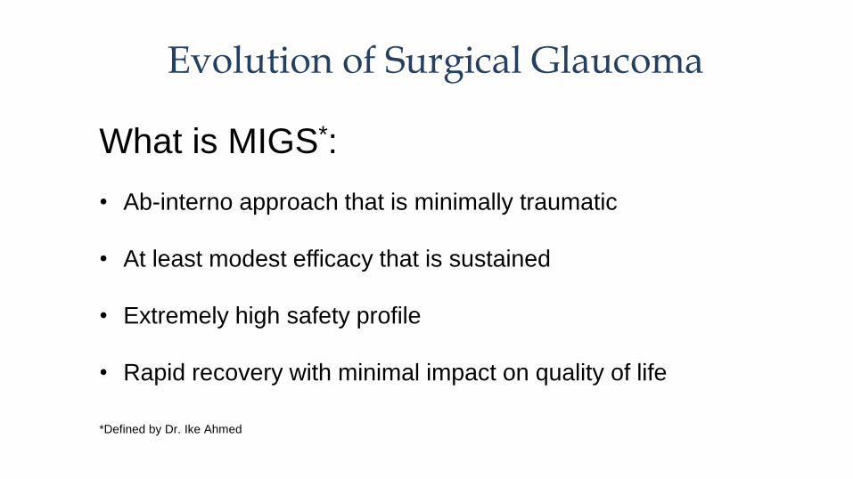
What is MIGS*:
• Ab-interno approach that is minimally traumatic
• At least modest efficacy that is sustained
• Extremely high safety profile
• Rapid recovery with minimal impact on quality of life
*Defined by Dr. Ike Ahmed
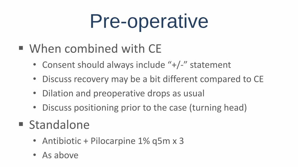
Pre-operative
▪ When combined with CE• Consent should always include “+/-” statement
• Discuss recovery may be a bit different compared to CE
• Dilation and preoperative drops as usual
• Discuss positioning prior to the case (turning head)
▪ Standalone• Antibiotic + Pilocarpine 1% q5m x 3
• As above
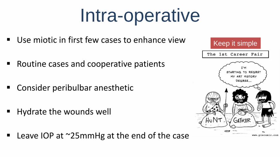
Intra-operative ▪ Use miotic in first few cases to enhance view
▪ Routine cases and cooperative patients
▪ Consider peribulbar anesthetic
▪ Hydrate the wounds well
▪ Leave IOP at ~25mmHg at the end of the case
Keep it simple
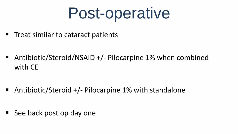
Post-operative
▪ Treat similar to cataract patients
▪ Antibiotic/Steroid/NSAID +/- Pilocarpine 1% when combined with CE
▪ Antibiotic/Steroid +/- Pilocarpine 1% with standalone
▪ See back post op day one
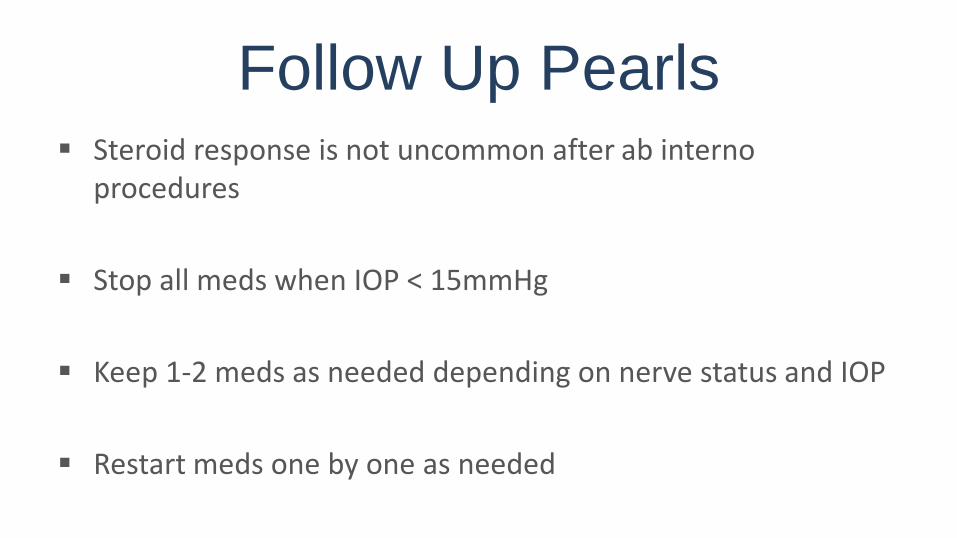
Follow Up Pearls▪ Steroid response is not uncommon after ab interno
procedures
▪ Stop all meds when IOP < 15mmHg
▪ Keep 1-2 meds as needed depending on nerve status and IOP
▪ Restart meds one by one as needed
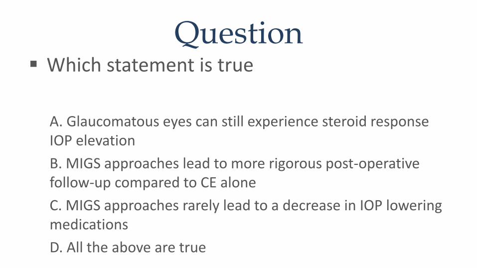
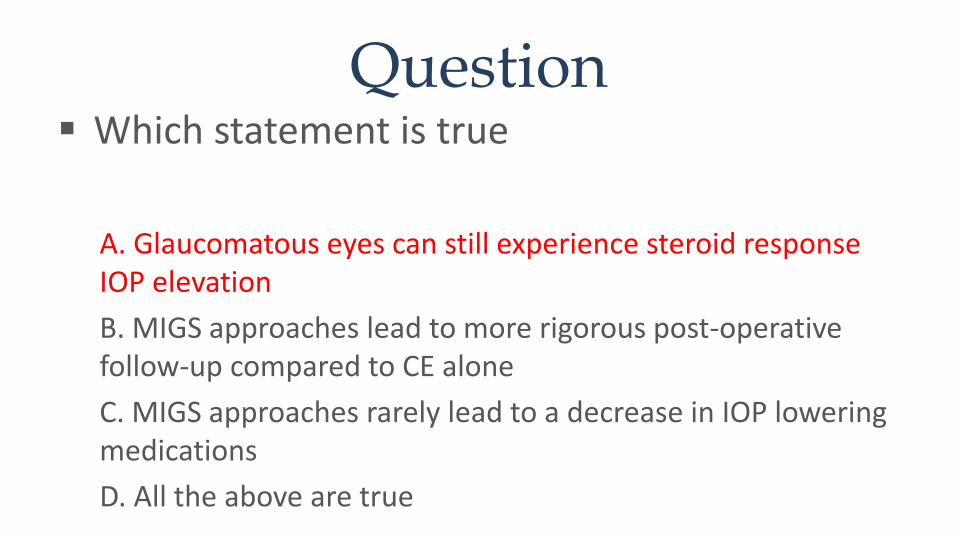
Question▪ Which statement is true
A. Glaucomatous eyes can still experience steroid response IOP elevation
B. MIGS approaches lead to more rigorous post-operative follow-up compared to CE alone
C. MIGS approaches rarely lead to a decrease in IOP lowering medications
D. All the above are true
Question▪ Which statement is true
A. Glaucomatous eyes can still experience steroid response IOP elevation
B. MIGS approaches lead to more rigorous post-operative follow-up compared to CE alone
C. MIGS approaches rarely lead to a decrease in IOP lowering medications
D. All the above are true
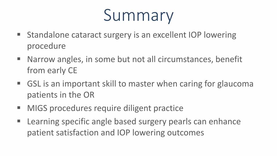
Summary▪ Standalone cataract surgery is an excellent IOP lowering
procedure
▪ Narrow angles, in some but not all circumstances, benefit from early CE
▪ GSL is an important skill to master when caring for glaucoma patients in the OR
▪ MIGS procedures require diligent practice
▪ Learning specific angle based surgery pearls can enhance patient satisfaction and IOP lowering outcomes
We continue to learn from each other and have many options compared to the not
too distant past

Thank You


![l Journal of Clinical & Experimental Ophthalmology · have resulted in higher rates of diagnosis of cataract and glaucoma [19]. In addition, the study did not identify the nature](https://static.fdocuments.in/doc/165x107/5e9f3eb80e39025c142522bc/l-journal-of-clinical-experimental-ophthalmology-have-resulted-in-higher-rates.jpg)