CATARACT SURGERY – COMPLICATIONS AND TECHNIQUES
58
Institutionen för Klinisk Neurovetenskap CATARACT SURGERY – COMPLICATIONS AND TECHNIQUES AKADEMISK AVHANDLING som för avläggande av medicine doktorsexamen vid Karolinska Institutet offentligen försvaras i Aulan, S:t Eriks Ögonsjukhus Fredagen den 11 december 2015, kl. 09.00 av Anthony Chang Leg. läkare Huvudhandledare: Docent Maria Kugelberg Karolinska Institutet Institutionen för klinisk neurovetenskap Bihandledare: Medicine doktor Alexander Fridberg Ögonläkargruppen Praktikertjänst, Stockholm Fakultetsopponent: Docent Madeleine Zetterberg Göteborgs Universitet Institutionen för klinisk neurovetenskap Betygsnämnd: Docent Eva Mönestam Umeå Universitet Institutionen för klinisk vetenskap Docent Eva Larsson Uppsala Universitet Institutionen för neurovetenskap Docent Helene Hamberg-Nyström Karolinska Institutet Institutionen för klinisk neurovetenskap Stockholm 2015
Transcript of CATARACT SURGERY – COMPLICATIONS AND TECHNIQUES

Institutionen för Klinisk Neurovetenskap
CATARACT SURGERY – COMPLICATIONS AND TECHNIQUES AKADEMISK AVHANDLING som för avläggande av medicine doktorsexamen vid Karolinska Institutet offentligen försvaras i Aulan, S:t Eriks Ögonsjukhus
Fredagen den 11 december 2015, kl. 09.00
av Anthony Chang Leg. läkare
Huvudhandledare: Docent Maria Kugelberg Karolinska Institutet Institutionen för klinisk neurovetenskap Bihandledare: Medicine doktor Alexander Fridberg Ögonläkargruppen Praktikertjänst, Stockholm
Fakultetsopponent: Docent Madeleine Zetterberg Göteborgs Universitet Institutionen för klinisk neurovetenskap Betygsnämnd: Docent Eva Mönestam Umeå Universitet Institutionen för klinisk vetenskap Docent Eva Larsson Uppsala Universitet Institutionen för neurovetenskap Docent Helene Hamberg-Nyström Karolinska Institutet Institutionen för klinisk neurovetenskap
Stockholm 2015

From THE DEPARTMENT OF CLINICAL NEUROSCIENCE
ST. ERIK EYE HOSPITAL
Karolinska Institutet, Stockholm, Sweden
CATARACT SURGERY: COMPLICATIONS AND TECHNIQUES
Anthony Chang
Stockholm 2015

All previously published studies were reproduced with permission from the publisher. Published by Karolinska Institutet. Printed by Repro Print AB, Solna © Anthony Chang, 2015 ISBN 978-91-7676-095-6
2015
Printed by

ABSTRACT
Cataract surgery is one of the most common surgical procedures performed worldwide.
Posterior capsul opacification (PCO) remains the most common postoperative complication
that can deteriorate vision. Development of glistenings in the artificial intraocular lens (IOL)
after cataract surgery is a phenomenon with the potential to reduce the outcome of an
otherwise excellent final surgical result. Phacoemulsification has been the most common
surgical technique performed to remove cataracts during the previous 25 years. The settings
controlling the fluidics in the eye intraoperatively can affect the postoperative convalescence.
Since many people undergo cataract surgery annually and all of the previously
mentioned issues can affect the final outcome, a better understanding and more studies
comparing different IOLs and phacoemulsification settings will help surgeons choose better
IOLs and surgical techniques and decrease postoperative complications.
In study I, we compared the development of PCO and glistenings associated
with two hydrophobic acrylic IOLs, the Sensar AR40e (Abbott Medical Optics) and AcrySof
SA60AT (Alcon), 5 to 7 years after cataract surgery. Both IOLs had a sharp posterior edge
design. We also evaluated if there were correlations between the amount of glistenings and
corrected distance visual acuity (CDVA) or contrast sensitivity and if subjective gradings of
glistenings were correlated with the objective quantification of glistenings with Scheimpflug
images. Eighty patients were included in this prospective randomized study. Fifty-six patients
completed the follow-up visit from 5 to 7 years postoperatively. Glistenings were graded at
the slit-lamp microscope and the amount of glistenings was quantified objectively using
Scheimpflug images with subsequent processing in computer software. There were no
significant differences in PCO area and severity or neodymium:yttrium-aluminium-garnet
(Nd:YAG) capsulotomy rates between the IOLs. Significantly more glistenings were found in
the AcrySof hydrophobic IOLs 5 to 7 years postoperatively. The glistenings were not
correlated with the CDVA or contrast sensitivity.
In study II, we evaluated in a prospective randomized trial if there were any
correlations between the amount of glistenings and CDVA or contrast sensitivity and
compared the development of glistenings in two acrylic IOLs, a hydrophilic IOL (BL27,
Bausch & Lomb) and a hydrophobic IOL (AcrySof SA60AT), 9 years after cataract surgery.
One hundred and twenty patients were recruited, 78 completed the 9-year follow-up visit.
e
The amount of glistenings was quantified objectively using Scheimpflug
images with subsequent processing in computer software. Glistenings were also subjectively
graded at the slit-lamp microscope. The hydrophobic IOL had significantly more glistenings
at the 9-year follow-up visit. The glistenings were not correlated with the CDVA or contrast
sensitivity.
In study III, we compared the PCO area, severity, and survival time without
Nd:YAG capsulotomy between a hydrophilic (BL27) and a hydrophobic (AcrySof SA60AT)
acrylic IOLs 9 years after cataract surgery. One hundred and twenty patients were recruited,
78 completed the 9-year follow-up visit. The PCO area and severity were higher in the
hydrophilic IOL. The survival time without Nd:YAG capsulotomy was longer in the
hydrophobic IOL.
In study IV, we compared low and standard fluidics settings during
phacoemulsification cataract surgery and evaluated the impact on the eye postoperatively by
measuring parameters indicating surgical trauma. Forty-three patients were recruited and
randomized into two groups, i.e., those that underwent phacoemulsification with low or
standard fluidics settings. The central corneal thickness, macular thickness, and intraocular
pressure were measured preoperatively, 1 day, 3 weeks, and 3 months postoperatively. The
CDVA was measured preoperatively, 3 weeks and 3 months after surgery. Anterior chamber
flare was measured preoperatively, 1 day and 3 weeks postoperatively. Endothelial cell
density was measured preoperatively and 3 months postoperatively. The low-settings group
had a significantly longer surgical time and higher amount of ultrasound energy used
intraoperatively, but there were no significant differences in the outcome parameters between
the two groups.
In conclusion, significantly more glistenings developed in the AcrySof
hydrophobic IOLs 5 to 7 years postoperatively compared to the hydrophobic Sensar IOL. The
glistenings were not correlated with the CDVA or contrast sensitivity. The hydrophobic
AcrySof IOL developed significantly more glistenings at the 9-year follow-up visit compared
to the hydrophilic BL27 IOL. The glistenings were not correlated with the CDVA or contrast
sensitivity. The PCO area and severity were higher in the hydrophilic IOL. The survival time
without Nd:YAG capsulotomy was longer in the hydrophobic IOL. Phacoemulsification
surgery with low fluidic settings rendered significantly longer surgical time and higher
amount of ultrasound energy used intraoperatively, but there were no significant differences
in the outcome parameters between the two groups.
LIST OF PUBLICATIONS
I. Chang A, Behndig A, Rønbeck M, Kugelberg M
Comparison of posterior capsule opacification and glistenings with 2
hydrophobic acrylic intraocular lenses: 5- to 7-year follow-up. Journal
of Cataract and Refractive Surgery 2013 May;39(5):694-698
II. Chang A, Kugelberg M
Glistenings 9 years after phacoemulsification in hydrophobic and
hydrophilic acrylic intraocular lenses. Journal of Cataract and
Refractive Surgery 2015 Jun;41(6):1199-1204
III. Chang A, Kugelberg M
Posterior capsule opacification 9 years after phacoemulcification with the
hydrophobic AcrySof SA60AT and hydrophilic BL27 intraocular lenses.
Submitted 2015-10-01
IV. Chang A, Fridberg A, Kugelberg M
Comparison of phacoemulsification cataract surgery with low versus
standard fluidic settings and the impact on postoperative parameters.
Submitted 2015-09-01
TABLE OF CONTENTS
1 Lens Glistenings .............................................................................................................. 9 1.1 Introduction ............................................................................................................ 9 1.2 Definition ............................................................................................................... 9 1.3 Glistening formation, onset, and size .................................................................... 9 1.4 Factors affecting glistening formation ................................................................ 10 1.5 Methods to assess and grade glistenings ............................................................. 11 1.6 Progression over time .......................................................................................... 13 1.7 Glistening in different IOL materials .................................................................. 13
1.7.1 Glistenings in hydrophobic IOLs ............................................................ 13 1.7.2 Glistenings in hydrophilic IOLs ............................................................. 13 1.7.3 Glistenings in PMMA IOLs .................................................................... 14 1.7.4 Glistenings in silicone IOLs .................................................................... 14
1.8 Effect on VA and contrast sensitivity ................................................................. 15 2 Posterior capsule opacification ..................................................................................... 16
2.1 Introduction .......................................................................................................... 16 2.2 Pathophysiology .................................................................................................. 17 2.3 Factors affecting PCO ......................................................................................... 17 2.4 Methods to assess PCO ....................................................................................... 18 2.5 Progression over time .......................................................................................... 19
3 Fluidics in phacoemulsification .................................................................................... 20 3.1 Introduction .......................................................................................................... 20 3.2 Parameters indicating surgical trauma ................................................................ 21 3.3 Methods to measure the parameters indicating surgical trauma in
phacoemulsification ............................................................................................. 21 3.4 Effect on vision .................................................................................................... 21
4 General aims .................................................................................................................. 22 5 Materials and methods .................................................................................................. 23
5.1 Study I .................................................................................................................. 23 5.1.1 Study design ............................................................................................ 23 5.1.2 Inclusion and exclusion criteria .............................................................. 26 5.1.3 Surgery .................................................................................................... 26 5.1.4 Statistical analysis ................................................................................... 26
5.2 Study II ................................................................................................................ 26 5.2.1 Study design ............................................................................................ 26 5.2.2 Inclusion and exclusion criteria .............................................................. 27 5.2.3 Surgery .................................................................................................... 27 5.2.4 Statistical analysis ................................................................................... 27
5.3 Study III ............................................................................................................... 28 5.3.1 Study design ............................................................................................ 28 5.3.2 Inclusion and exclusion criteria .............................................................. 28
5.3.3 Surgery .................................................................................................... 28 5.3.4 Statistical analysis ................................................................................... 28
5.4 Study IV ............................................................................................................... 28 5.4.1 Study design ............................................................................................ 28 5.4.2 Inclusion and exclusion criteria .............................................................. 29 5.4.3 Surgery .................................................................................................... 29 5.4.4 Statistical analysis ................................................................................... 29
6 Results ............................................................................................................................ 31 6.1 Study I .................................................................................................................. 31
6.1.1 Patient data .............................................................................................. 31 6.1.2 PCO and Nd:YAG capsulotomy ............................................................ 31 6.1.3 Glistenings ............................................................................................... 31
6.2 Studies II and III .................................................................................................. 31 6.2.1 Patient data .............................................................................................. 31 6.2.2 Glistenings ............................................................................................... 32 6.2.3 PCO ......................................................................................................... 32
6.3 Study IV ............................................................................................................... 33 6.3.1 Patient data .............................................................................................. 33 6.3.2 Fluidics and the impact on postoperative parameters ............................ 33
7 Discussion ...................................................................................................................... 35 7.1 Studies I-III .......................................................................................................... 35
7.1.1 Lens glistenings ....................................................................................... 35 7.1.2 PCO ......................................................................................................... 36
7.2 Study IV ............................................................................................................... 38 8 Main conclusions ........................................................................................................... 39 9 Future perspectives ........................................................................................................ 40 10 Acknowledgements ....................................................................................................... 42 11 References ..................................................................................................................... 44
LIST OF ABBREVIATIONS
BSS Balanced saline solution
CCT Central corneal thickness or computer compatible tape
CDE Cumulative dissipated energy
CDVA Corrected distance visual acuity
CME Cystoid macular edema
ECD Endothelial cell density
IOL Intraocular lens
IOP Intraocular pressure
Nd:YAG Neodymium:yttrium-aluminium-garnet
OCT Optical coherence tomography
PMMA Polymethyl methacrylate
PCO Posterior capsule opacification
POCOman Posterior capsule opacification software

9
1 LENS GLISTENINGS
1.1 INTRODUCTION
Glistenings were first reported in polymethyl methacrylate (PMMA) intraocular lenses
(IOLs)1. However, it was after the introduction of the popular hydrophobic acrylic AcrySof
IOL (Alcon) in 1994 that glistenings caught the attention of clinical researchers, who then
began investigating if this phenomenon had any substantial clinical impact on vision.
1.2 DEFINITION
Glistenings are defined as fluid-filled microvacuoles that form within the IOL when it is in an
aqueous environment2.
1.3 GLISTENING FORMATION, ONSET, AND SIZE
The formation process of lens glistenings remains controversial. Two theories have been
proposed. The first theory suggests that formation of microvoids inside the IOL material
occurs during the polymerization process in one of the IOL production steps. The microvoids
slowly absorb water when the IOL is in the aqueous environment. When water vapor
detaches from the surrounding matter inside the microvoids, a reaction called phase
separation occurs. Because there are differences in the refractive indices between water and
the surrounding IOL material, light scatters when it passes between the two media and
appears as sparkling dots, hence, the term glistenings2.
The water absorption rate differs between different IOLs and the surrounding
environment regarding temperature3 and osmotic level4. Different IOLs have different glass
transition temperatures (Tgs). When the temperature is above the Tg, the IOL absorbs water
faster and is soft and flexible. Below the Tg, the IOL is rigid and the water absorption rate is
slower. Hydrophobic acrylic IOLs have Tgs close to room temperature, 20 Cº for the
hydrophobic AcrySof IOL. A temperature below 15°C has not been associated with
glistening formation in the most commonly implanted IOLs5.
In vitro experiments in which IOLs were exposed to temperature variations,
mimicking accelerated glistening formation that could take years in vivo, showed that when
10
the IOLs are suspended in an aqueous environment and heated, the IOLs become
oversaturated with water. However, after cooling, causing phase separation in the microvoids
of the IOLs, glistenings are observed because of the difference in the refractive indices
between water and the surrounding IOL material as mentioned previously.
The second theory suggests that as hydrophilic impurities enter the microvoids
inside the IOL material, the osmotic gradient inside the voids increases from the surrounding
aqueous environment, causing an influx of water through diffusion with subsequent
expansion of the microvoids. When a critical level of expansion is reached, probably causing
cracks and tears in the IOL material surrounding the microvoids, they become permanent4.
Repeated heating and cooling of the IOL showed that glistenings appear at the same locations
in the IOL6.
Glistenings can develop as soon as 1 week after cataract surgery7 in sizes
ranging from 1 to 20 microns6 8-10, often 1 to 10 microns in clinical cases. In vitro studies with
more extreme environmental variations can generate glistenings with sizes up to 20 micron or
larger.
1.4 FACTORS AFFECTING GLISTENING FORMATION
Different IOL materials, hydrophilic, hydrophobic, PMMA, and silicone IOLs have been
identified to develop glistenings to different degrees7 11-13. Time is also crucial; the longer
time that has passed since IOL implantation the more likely it is that glistenings will develop
or the number of existing glistenings will increase13-16.
The IOL dioptric power17, IOL packaging18, IOL manufacturing technique19,
temperature changes6, glaucoma20, uveitis and other conditions with breakdown of the blood-
aqueous barrier21, use of antiglaucoma eye drops,20 22 and anti-inflammatory eye drops
containing surfactant may have a role in glistenings formation23 24.
When the AcrySof hydrophobic acrylic IOL was introduced to the market in
1994, it was delivered packed in the AcryPak system, containing both the IOL and a folder in
a plastic case that was sterilized in the plastic case. One of the first reports of glistenings in
AcrySof IOLs was associated with this IOL packaging system. The increased glistenings in
the IOLs in the AcryPak compared to the same IOLs in the so-called Wagon Wheel packages
led to the conclusion that the microenvironment in the AcryPak system was changed and
hence the development of glistenings increased18.
11
The production of IOLs is divided into two principal techniques: cast molding
and lathe-cutting. The former is suitable for large quantity production and involves
polymerization of IOL monomer mixtures in casting molds. Parts of heterogeneous unreacted
monomers can still be present in the molds and may explain why in some studies there are
more glistenings in IOLs produced with cast molding compared to lathe-cutting19.
The latter, lathe-cut IOLs, are produced from mixtures of monomers
polymerized into large acrylic sheets and IOLs from homogenous parts of the sheet are
subsequently cut out from the sheet and polished.
1.5 METHODS TO ASSESS AND GRADE GLISTENINGS
Glistenings can be graded in two ways. Most commonly subjective grading systems of 0 to
325 and 0 to 4 have been used7 14 26 27, with 0 representing no glistenings, 1 trace, 2 minor, 3
moderate, and 4 severe. The examiner grades the glistenings based on how many glistenings
the IOL appears to have under slit-lamp microscopy. To grade glistenings objectively and
facilitate quantitative comparisons between studies and reproduction of the results, Professor
Behndig invented a method of analyzing glistenings performed with Scheimpflug images13 14
23 28 29.
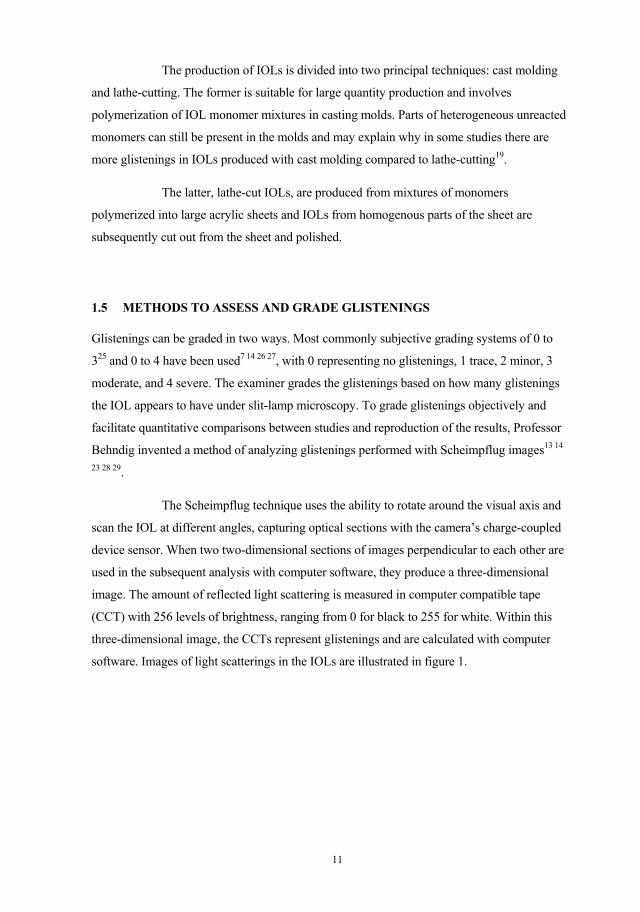
The Scheimpflug technique uses the ability to rotate around the visual axis and
scan the IOL at different angles, capturing optical sections with the camera’s charge-coupled
device sensor. When two two-dimensional sections of images perpendicular to each other are
used in the subsequent analysis with computer software, they produce a three-dimensional
image. The amount of reflected light scattering is measured in computer compatible tape
(CCT) with 256 levels of brightness, ranging from 0 for black to 255 for white. Within this
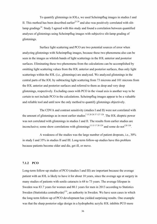
three-dimensional image, the CCTs represent glistenings and are calculated with computer
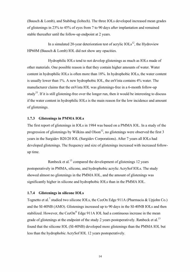
software. Images of light scatterings in the IOLs are illustrated in figure 1.
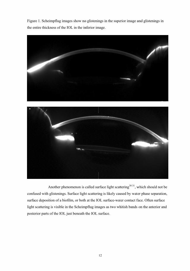
Figur
the e
confu
surfa
light
poste
re 1. Scheim
ntire thickn
An
used with gl
ace depositio
scattering i
erior parts o
mpflug imag
ness of the IO
nother phen
listenings. S
on of a biofi
s visible in
f the IOL ju
ges show no
OL in the in
omenon is c
Surface light
ilm, or both
the Scheimp
ust beneath t
12
o glistenings
nferior imag
called surfac
t scattering
h at the IOL
pflug image
the IOL surf
s in the supe
e.
ce light scat
is likely cau
surface-wat
es as two wh
face.
erior image a
ttering30 31, w
used by wate
ter contact f
hitish bands
and glistenin
which shoul
ter phase sep
face. Often s
on the ante
ngs in
ld not be
paration,
surface
erior and

13
1.6 PROGRESSION OVER TIME
Several studies, some of which were long term, have reported that glistenings progress over
time in all IOLs7 13, but especially in the hydrophobic acrylic AcrySof IOLs7 12-15. However,
some studies have reported that glistenings tend to stabilize after a certain time10 20.
In a study7 with up to a 2-year follow-up, the earliest glistenings were observed
as early as 1 week after cataract surgery and increased thereafter up to 90 days. After 180
days, the amount of glistenings in all IOLs included in the study (2 silicone, 3 hydrophilic
acrylic, and 2 hydrophobic acrylic) stabilized; however, in the AcrySof (hydrophobic acrylic)
and CeeOn Edge 911A (Pharmacia & Upjohn Co.) (silicone) IOLs, the glistenings continued
to increase.
Another study15 that followed patients up to 50 months postoperatively also
confirmed increasing amounts of glistenings with time. Two other studies21 32 also reported
that glistenings increase with time.
In one in vivo study20 the incidence and severity of glistenings did not increase
up to 24 months postoperatively. One in vitro study10 showed that glistenings stabilizes after
a certain time after the IOL was immersed in an aqueous environment.
1.7 GLISTENING IN DIFFERENT IOL MATERIALS
1.7.1 Glistenings in hydrophobic IOLs
Hydrophobic acrylic IOLs, especially AcrySof IOLs, have received most attention in
previous studies of glistenings. The AcrySof MA60BM (Alcon), Sensar AR40e (AMO),
Acryfold VA-60BB (HOYA), Nex-Acri N4-18B (NiDEK), and Avansee AU (Kowa Co.)
were studied in vitro for accelerated glistening formation in a laboratory test simulating 20-
year deterioration of acrylic IOLs12. All IOLs but the Avansee AU6 developed glistenings.
Two other IOLs, XACT X60 (Advanced Vision Science) and enVista MX60
(Bausch & Lomb), both made of the same material, claimed that no glistenings developed in
2-year and 6-month follow-up studies33-35.
1.7.2 Glistenings in hydrophilic IOLs
There are few studies on hydrophilic acrylic IOLs and glistenings development. Tognetto et
al.7 evaluated three hydrophilic IOLs: the ACR6D (Corneal Laboratories), Hydroview
14
(Bausch & Lomb), and Stabibag (Ioltech). The three IOLs developed increased mean grades
of glistenings in 23% to 45% of eyes from 7 to 90 days after implantation and remained
stable thereafter until the follow-up endpoint at 2 years.
In a simulated 20-year deterioration test of acrylic IOLs12, the Hydroview
HP60M (Bausch & Lomb) IOL did not show any opacities.
Hydrophilic IOLs tend to not develop glistenings as much as IOLs made of
other materials. One possible reason is that they contain higher amounts of water. Water
content in hydrophilic IOLs is often more than 18%. In hydrophobic IOLs, the water content
is usually lower than 1%. A new hydrophobic IOL, the enVista contains 4% water. The
manufacturer claims that the enVista IOL was glistenings-free in a 6-month follow-up
study33. If it is still glistening-free over the longer run, then it would be interesting to discuss
if the water content in hydrophilic IOLs is the main reason for the low incidence and amount
of glistenings.
1.7.3 Glistenings in PMMA IOLs
The first report of glistenings in IOLs in 1984 was based on a PMMA IOL. In a study of the
progression of glistenings by Wilkins and Olson32, no glistenings were observed the first 3
years in the Surgidev B20/20 IOL (Surgidev Corporations). After 7 years all IOLs had
developed glistenings. The frequency and size of glistenings increased with increased follow-
up time.
Rønbeck et al.13 compared the development of glistenings 12 years
postoperatively in PMMA, silicone, and hydrophobic acrylic AcrySof IOLs. The study
showed almost no glistenings in the PMMA IOL, and the amount of glistenings was
significantly higher in silicone and hydrophobic IOLs than in the PMMA IOL.
1.7.4 Glistenings in silicone IOLs
Tognetto et al.7 studied two silicone IOLs, the CeeOn Edge 911A (Pharmacia & Upjohn Co.)
and the SI-40NB (AMO). Glistenings increased up to 90 days in the SI-40NB IOLs and then
stabilized. However, the CeeOn® Edge 911A IOL had a continuous increase in the mean
grade of glistenings at the endpoint of the study 2 years postoperatively. Rønbeck et al.13
found that the silicone IOL (SI-40NB) developed more glistenings than the PMMA IOL but
less than the hydrophobic AcrySof IOL 12 years postoperatively.
15
1.8 EFFECT ON VA AND CONTRAST SENSITIVITY
It seems logical that glistening should adversely affect the CDVA and contrast sensitivity
because the tiny microvacuoles in the IOL scatter the light passing through the IOL and
therefore cause deteriorated CDVA or glare. However, no studies up to now support any
significant correlations between diminished CDVA and glistenings.
For the examiner, it is obvious when performing neodymium:yttrium-
aluminium-garnet (Nd:YAG) capsulotomy in a patient with an IOL with severe glistenings,
that it is more difficult to target the laser beam at the posterior lens capsule in the Nd:YAG
slit-lamp.
It remains controversial if glistenings affect contrast sensitivity. Most studies
have not reported any correlation20 28 36 37. However, some studies have shown that contrast
sensitivity is affected25 and some only at high spatial frequencies27 38.
16
2 POSTERIOR CAPSULE OPACIFICATION
2.1 INTRODUCTION
Posterior capsule opacification (PCO) is the most common complication after
phacoemulsification cataract surgery. The incidence rates range from less than 10% to 50%39-
41. PCO was observed in the early days of IOL implantation at the end of the 1940s. With a
successively better understanding of the pathogenesis and treatment options for PCO, we can
now with modern cataract surgery gradually decrease the incidence of PCO with the help of
better tools39, such as continuous development of safer phacoemulsification machines
facilitating more thorough cortical removal of lens material, better IOL materials and designs
that inhibit PCO, and even surgical laser systems to create customized repeatable and perfect
capsulorhexis sizes, avoiding the unnecessary risk of making too large or off-center
capsulorhexes and hence decreasing the risk of PCO development42-44.
Studies have been done with immunotherapy, gene therapy, chemical therapy,
and physical techniques to eliminate lens epithelial cells (LECs)45-47.
PCO is still far from eradicated. The only treatment is outpatient Nd:YAG laser
capsulotomy for cooperative patients. However, because of the total number of cataract
surgeries annually, it is a societal economic burden48 and Nd:YAG lasers are not readily
available everywhere, especially in rural areas in developing parts of the world. For the
patient, Nd:YAG capsulotomy is associated with several sight-threatening complications such
as retinal detachment49 50, macular edema51, intraocular inflammation50, transient increased
intraocular pressure (IOP)51 52, increased vitreous opacities53, IOL damage50 54 and IOL
dislocation55.
PCO causes symptoms when light passes through the opacified posterior lens
capsule and is forward-scattered to the retinal fundus. Symptoms such as decreased VA and
contrast sensitivity, increased glare, diplopia, and blurred images are typical56 57. The
symptoms may cause disabilities in daily life, for example, when driving a car and especially
when driving at night. The amount and density of PCO may not be experienced or expressed
similarly regarding severity by different patients58. The CDVA also can be better or worse
than expected compared to the PCO status observed by the physician.
17
2.2 PATHOPHYSIOLOGY
Phacoemulsification cataract surgery removes the opacified lens nucleus and cortex. Two
types of remnant LECs proliferate and migrate from the equatorial zone of the anterior lens
capsule (A-cells) or equatorial lens bow (E-cells) toward the central optical zone with time48.
When LECs are in the central parts of the posterior lens capsule, they scatter light entering
the posterior segment of the eye causing deteriorated CDVA, glare, and distorted images. A-
cells transformed into myofibroblasts cause fibrosis and shrinking of the anterior lens capsule
and may lead to IOL tilting and decentration. E-cells have a high capacity for mitosis and
differentiation into balloon-like bladder cells (Wedl cells) and can form Elschnig’s pearls, a
regenerative form of lens capsule opacification and the most common PCO, characterized
clinically as having a pearl-like appearance. A special form of regenerative PCO is
Soemmerring’s ring, a doughnut-shaped opacity described as early as 1828 with formation of
Elschnig’s pearls retained at the periphery of the anterior-posterior capsule fuse as a ring-
shaped lesion59.
2.3 FACTORS AFFECTING PCO
Earlier studies have suggested that age60, uveitis, diabetes mellitus61-65, retinitis pigmentosa66,
surgery techniques43 44 67, IOL material68-72, and IOL design53 73-77 are PCO-related factors.
The sharp-edge design of IOLs is probably the most important factor inhibiting
PCO development74. In a comparative study between two single-piece hydrophilic acrylic
IOLs78, one IOL was a standard model with a sharp posterior edge except at the optic-haptic
junctions. The other IOL had an enhanced edge with a peripheral ridge around the lens optic
360º circumferentially. The latter developed significantly less PCO than the standard model.
Round-edge IOLs were shown in previous studies to be inferior to sharp-edge
IOLs in inhibiting PCO74 78-84. The sharp posterior IOL edge acts as a barrier by bending in
the posterior lens capsule against the sharp posterior square-edge IOL and thus inhibiting
migration of LECs from the equator of the lens capsule to the central parts of the IOL optic75
77.
Nishi et al.74 compared development of PCO between a round-edge and a
sharp-edge AcrySof IOL and reported that the round-edge IOL developed significantly more
PCO. The authors concluded that the sharp-edge IOL design was the main PCO inhibitory
factor and the IOL material instead has a complimentary role in inhibiting PCO
18
development74. Angulation of the IOL haptics enhances the force exerted on the bending
between the posterior lens capsule and the sharp IOL edge and increases the integrity of the
barrier85. However, the sharpness of the square-edge IOL can vary between IOLs of different
materials81 or even the same materials86. Generally, most hydrophilic IOLs have rounder
edges than hydrophobic IOLs probably because during the manufacturing process
hydrophilic IOLs are lathe-cut from dehydrated IOL blocks and then polished leading to loss
of the sharp edge81. However, hydrophilic IOLs with sharp edges also exist.
Comparisons of IOL materials have shown that hydrophilic IOLs develop PCO
at higher rates than hydrophobic IOLs, 50.3% in a hydrophilic IOL compared with 4.9% in a
hydrophobic IOL in a 1-year follow-up study by Heatley et al68. Other studies with 1- to 3-
year follow-ups also have reported similar results69 87 88. Linnola et al.89 90 argued that
fibronectin bindings between the posterior lens capsule and the IOL surface of hydrophobic
acrylic IOLs seem to have an important role in inhibiting migration of LECs by increasing the
barrier effect. However, in studies with more than 3 years of follow-up postoperatively that
compared hydrophobic acrylic with silicone and PMMA IOLs, the PCO incidence and
Nd:YAG capsulotomy rates increase in the hydrophobic IOLs are equal to or even surpass the
rates for silicone IOLs91-93. One theory is that the barrier effect of the sharp posterior edge
design in acrylic IOLs that inhibit PCO development gradually may be compromised when
increasing amounts of slowly migrating LECs physically force the reopening of the barrier
between the IOL edge and posterior lens capsule91 92.
2.4 METHODS TO ASSESS PCO
Most earlier studies have analyzed PCO with retroillumination images. Several image
computer software packages are available with their respective advantages and
disadvantages. Evaluation of Posterior Capsule Opacification software (Augentagesklinik
Spreebogen) evaluates PCO subjectively. Posterior Capsule Opacification software
(POCOman, Kings College) evaluates PCO semiobjectively, Posterior Capsule Opacification
software and Automated Quantification of After-Cataract (Medical University of Vienna)
processes the PCO images in the computer software to calculate the size of the area at the
posterior capsule that is affected by PCO and determine the PCO density94-96.
Nd:YAG laser capsulotomy frequency is considered another important
parameter for evaluating PCO. This parameter correlates well with the CDVA but has a
19
weaker correlation with the contrast sensitivity97. Scheimpflug images have also been used in
PCO studies98.
Patients who already underwent Nd:YAG capsulotomy cannot be evaluated by any of
the above-mentioned computer software since there is no posterior capsule. This problem was
addressed previously99. These patients received the highest scores for PCO area and severity
in studies I and III. We assumed if they have not had Nd:YAG capsulotomy earlier they
would have had a high grade of PCO.
Long-term studies also have an increased number of patient dropouts with increased
follow-up time. Patients get older, some die, and some get ill during the follow-ups.
Information about Nd:YAG capsulotomy frequency becomes uncertain when this
information only comes from patients living long enough or those healthy enough to
complete the follow-up examination. It is possible that some dropouts would have had
Nd:YAG capsulotomy at the follow-up visit if they had arrived for the examination.
However, the capsulotomy data are only obtained from patients at the follow-up visits. To
collect as much information as possible from all patients, we used survival analysis, which
has been used in earlier similar studies13 99. The analysis has the advantage of considering the
data from patients until they are lost to follow-up.
2.5 PROGRESSION OVER TIME
PCO frequency has been reported with great variability from 10% to 60% in different studies.
Since the introduction of IOLs with a sharp posterior edge profile, the PCO frequency can be
as low as about 10% 5 years postoperatively39 40.
Development of PCO in different IOL materials shows different patterns. Two
follow-up studies100 101 have reported that the Nd:YAG capsulotomy rates increased from the
2-year to the 5-year follow-up for three different IOLs, a PMMA, a silicone, and a
hydrophobic acrylic. The Nd:YAG rate increased from 20% to 61% for the PMMA (HSM
809C) IOL (Pharmacia & Upjohn), 22% to 33% for the silicone (SI-40NB) IOL, and 8% to
22% for the hydrophobic acrylic (AcrySof MA60BM) IOL.
20
3 FLUIDICS IN PHACOEMULSIFICATION
3.1 INTRODUCTION
Modern cataract surgery is performed using the phacoemulsification technique102 103. This
technique has the advantages of small incision wounds, faster sight rehabilitation, less
damage to the intraocular tissues, and minimal intraoperative and postoperative complications
compared to previous surgical techniques104-107.
To extract the opacified lens with a diameter of about 10 millimeters through a
2- to 3-millimeter incision, the lens is divided into smaller fragments. This is done with the
phacoemulsification tip inserted through the incisional wound to divide the lens into smaller
fragments and aspirate the lens fragments through the tip opening. The tip uses ultrasound
energy to emulsify the lens, builds up heat in and around the tip, and can damage the
surrounding tissues. The tip also has two other functions, i.e., irrigation and aspiration.
Irrigation helps cool the heat around the tip by producing an inflow of saline solution that
dilutes the heat near the tip. Aspiration also maintains stabilized fluid volume in the anterior
chamber and drains excess heat from the eye. Lens fragments follow the irrigating fluid flow
to the opening of the tip. When lens material fully occludes the tip opening, the machine then
uses vacuum to crush and suck (i.e., aspirate) the material into a waste bag in the machine
through the hand-piece connected to the tip. Modifying the irrigation and aspiration settings
in the phacoemulsification machine can create different effects to enhance and facilitate a
smooth surgical procedure. The surgeon may ensure a surgery with a stable anterior chamber
and good followability by changing the settings for different anatomic variations in eyes or
different hardnesses of the lens nucleus. Most surgeons have their own settings during the
different surgical phases: sculpting, lens consumption and removal of remaining lens cortex.
However, some questions remain unanswered. Some surgeons claim that minimizing fluid
turbulence intraoperatively may cause less surgical trauma to the eye compared to the
standard settings most surgeons use. However, few studies have been conducted in this
field108 109.
21
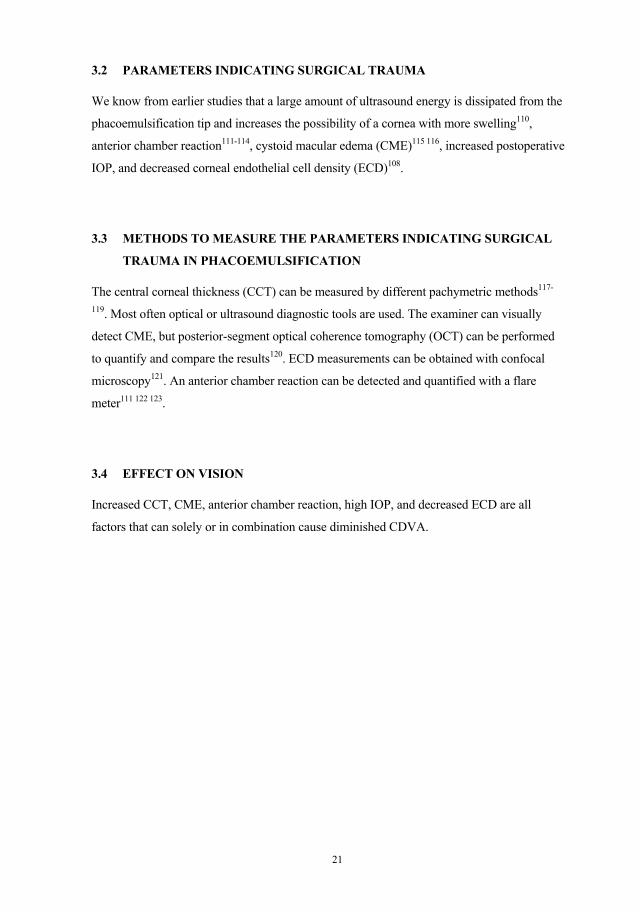
3.2 PARAMETERS INDICATING SURGICAL TRAUMA
We know from earlier studies that a large amount of ultrasound energy is dissipated from the
phacoemulsification tip and increases the possibility of a cornea with more swelling110,
anterior chamber reaction111-114, cystoid macular edema (CME)115 116, increased postoperative
IOP, and decreased corneal endothelial cell density (ECD)108.
3.3 METHODS TO MEASURE THE PARAMETERS INDICATING SURGICAL
TRAUMA IN PHACOEMULSIFICATION
The central corneal thickness (CCT) can be measured by different pachymetric methods117-
119. Most often optical or ultrasound diagnostic tools are used. The examiner can visually
detect CME, but posterior-segment optical coherence tomography (OCT) can be performed
to quantify and compare the results120. ECD measurements can be obtained with confocal
microscopy121. An anterior chamber reaction can be detected and quantified with a flare
meter111 122 123.
3.4 EFFECT ON VISION
Increased CCT, CME, anterior chamber reaction, high IOP, and decreased ECD are all
factors that can solely or in combination cause diminished CDVA.
22
4 GENERAL AIMS
In study I, our goal was to determine if two hydrophobic acrylic IOLs develop PCO and
glistenings differently and if glistenings affected VA and contrast sensitivity 5 to 7 years after
phacoemulsification cataract surgery.
In study II, we evaluated a hydrophobic and a hydrophilic acrylic IOL and
compared the development of glistenings and the impact on VA and contrast sensitivity in the
IOLs 9 years after phacoemulsification cataract surgery.
In study III, we evaluated the same hydrophobic and hydrophilic acrylic IOLs
as in study II and compared the development of PCO and survival without Nd:YAG
capsulotomy in the IOLs 9 years after phacoemulsification cataract surgery.
In study IV, we compared two different fluidic settings in phacoemulsification
cataract surgery and the impact on postoperative parameters.
23
5 MATERIALS AND METHODS
5.1 STUDY I
5.1.1 Study design
The 80 patients who underwent cataract surgery from 2002 to 2005 in this prospective study
were randomized into two equal groups of 40 patients each. One group received the three-
piece Sensar AR40e IOL and the other group received the one-piece AcrySof SA60AT IOL;
both IOLs are made of a hydrophobic acrylic material with a sharp posterior IOL edge
design. The patients were contacted for a follow-up visit during 2010. The CDVA
measurements obtained with the 100% and 2.5% Early Treatment Diabetic Retinopathy
Study (ETDRS) charts were recorded in the logarithm of the minimum angle of resolution
scale. We obtained retroillumination images of PCO with a fundus camera and Scheimpflug
images of glistenings with the Pentacam HR (OCULUS Inc.).
The retroillumination images of PCO were loaded into a posterior capsule
opacity computer software system (POCOman) and calculated semiobjectively, because
some steps in the analysis required manual interactions. The program applied a grid pattern
on the retroillumination images and divided the area into 56 approximately equal sectors. The
area outside the capsulorhexis was excluded from analysis. The examiner marked any sector
with PCO covering more than 50% of the area and graded the severity of PCO as 0 indicating
no PCO, 1 mild, 2 moderate, and 3 severe. The software then calculated the overall severity
of the PCO ranging from 0 to 3 and the total area of the PCO as a fraction of the affected area
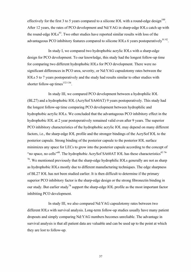
divided by the total area within the capsulorhexis (figure 2).
24
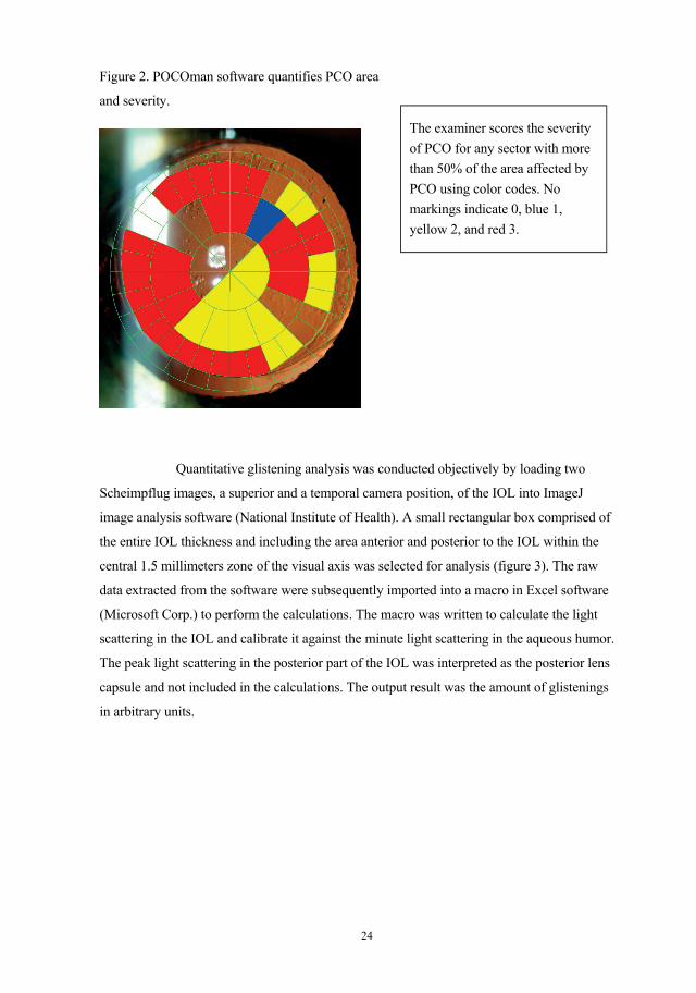
Figure 2. POCOman software quantifies PCO area
and severity.
Quantitative glistening analysis was conducted objectively by loading two
Scheimpflug images, a superior and a temporal camera position, of the IOL into ImageJ
image analysis software (National Institute of Health). A small rectangular box comprised of
the entire IOL thickness and including the area anterior and posterior to the IOL within the
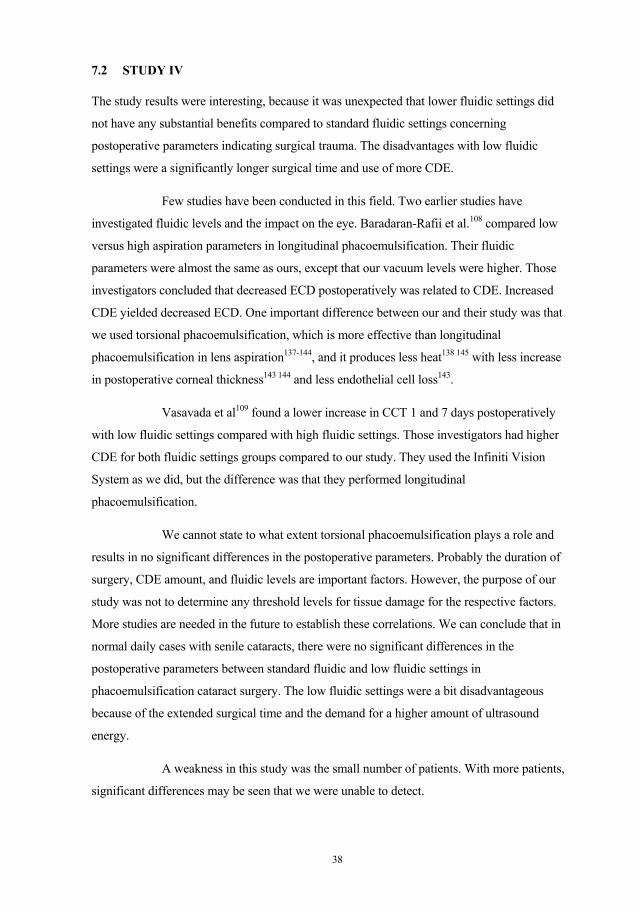
central 1.5 millimeters zone of the visual axis was selected for analysis (figure 3). The raw
data extracted from the software were subsequently imported into a macro in Excel software
(Microsoft Corp.) to perform the calculations. The macro was written to calculate the light
scattering in the IOL and calibrate it against the minute light scattering in the aqueous humor.
The peak light scattering in the posterior part of the IOL was interpreted as the posterior lens
capsule and not included in the calculations. The output result was the amount of glistenings
in arbitrary units.
The examiner scores the severity of PCO for any sector with more than 50% of the area affected by PCO using color codes. No markings indicate 0, blue 1, yellow 2, and red 3.
25
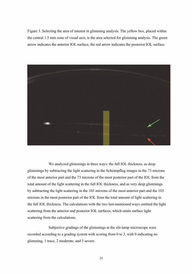
Figure 3. Selecting the area of interest in glistening analysis. The yellow box, placed within
the central 1.5 mm zone of visual axis, is the area selected for glistening analysis. The green
arrow indicates the anterior IOL surface; the red arrow indicates the posterior IOL surface.
We analyzed glistenings in three ways: the full IOL thickness, as deep
glistenings by subtracting the light scattering in the Scheimpflug images in the 73 microns
of the most anterior part and the 73 microns of the most posterior part of the IOL from the
total amount of the light scattering in the full IOL thickness, and as very deep glistenings
by subtracting the light scattering in the 103 microns of the most anterior part and the 103
microns in the most posterior part of the IOL from the total amount of light scattering in
the full IOL thickness. The calculations with the two last-mentioned ways omitted the light
scattering from the anterior and posterior IOL surfaces, which omits surface light
scattering from the calculations.
Subjective gradings of the glistenings at the slit-lamp microscope were
recorded according to a grading system with scoring from 0 to 3, with 0 indicating no
glistening, 1 trace, 2 moderate, and 3 severe.
26
5.1.2 Inclusion and exclusion criteria
Patients 39 to 86 years old with cataracts were included. Patients were excluded who had
moderate or advanced age-related macular degeneration, corneal pathology, earlier retinal
photocoagulation, diabetes mellitus, glaucoma, exfoliation syndrome, and uveitis, and those
who received preoperative oral steroid therapy or underwent a previous intraocular surgery.
5.1.3 Surgery
One experienced surgeon performed the cataract surgery using phacoemulsification with a
clear corneal incision. The surgical procedure was initiated with topical and intracameral
anesthesia. Sodium hyaluronate was used as an ophthalmic viscosurgical device (OVD).
Continuous capsulorhexis was followed by hydrodissection with balanced saline solution
(BSS) and phacoemulsification in the capsular bag. Irrigation and aspiration of the remaining
cortex were performed and one of the two IOLs was folded and injected into the capsular bag.
The procedure ended with an intracameral injection of cephalosporin as antibiotic
prophylaxis, and the corneal wound was hydrated with BSS using a blunt injection needle.
The patients instilled topical dexamethasone in a tapering dose during the first 3
postoperative weeks.
5.1.4 Statistical analysis
Quantitative comparisons of the PCO area and severity and the amount of glistenings were
analyzed with the Mann-Whitney U-test. Nd:YAG capsulotomy rates to compare the two
IOLs were calculated using Fisher’s exact two-tailed test. All calculations for correlations
were conducted with the Spearman rank-order correlation.
5.2 STUDY II
5.2.1 Study design
This was a prospective randomized study that included 120 patients, who had
phacoemulsification cataract surgery between 2002 and 2004. The patients were randomized
to one of two groups and received either the hydrophilic BL27 IOL or the hydrophobic
AcrySof SA60AT IOL, both of which were one-piece acrylic IOLs with sharp posterior
edges. Nine year after surgery the patients were contacted for a follow-up examination.
27
Lens glistening analysis was conducted in the same way as in study I with
Scheimpflug images with subsequent data processing using computer software for objective
quantification and grading at the slit-lamp for subjective scoring.
The CDVA and contrast sensitivity were measured using the Optec® 6500
Vision Tester (Stereo Optical Co., Inc.), with an ETDRS chart for the CDVA
measurements and Functional Acuity Contrast Test (F.A.C.T, Stereo Optical Co., Inc.) for
the contrast sensitivity measurements. The F.A.C.T is a sine-wave grating chart that tests
the functional VA in five spatial frequencies (1.5, 3, 6, 12, and 18 cycles per degree) and
nine levels of contrast. The patient determined the minimal contrast grating level seen for
each spatial frequency. The last correct grating level identified for each spatial frequency
was plotted on a contrast sensitivity curve. The area under the curve value was used in the
statistical analysis to determine any correlation with the amount of glistenings. This
method of measuring the contrast sensitivity is more accurate than other available contrast
sensitivity measurements124. The contrast sensitivity measurements were conducted with
and without glare. When testing with glare, 12 light-emitting diode lamps were arranged in
an oval arc around the testing field and lighted.
5.2.2 Inclusion and exclusion criteria
Patients 60 to 90 years old with senile cataracts were included. Patients were excluded who
had a dilated pupil less than 6 millimeters; a previous history of intraocular surgery, corneal
endothelial damage, or ocular trauma; traumatic cataract; pseudoexfoliation syndrome;
uveitis; diabetic retinopathy; glaucoma; or advanced macular degeneration; and those
receiving long-term anti-inflammatory treatment.
5.2.3 Surgery
One of three experienced surgeons performed the phacoemulsification surgeries. The surgical
procedures were the same as described for study I.
5.2.4 Statistical analysis
The follow-up time between the two IOLs was compared using the Student’s t-test. The
comparison of the amount of glistenings associated with the two IOLs was calculated with
the Wilcoxon rank-sum test. The correlations between glistenings, CDVA, and contrast
sensitivity were analyzed using the Spearman rank-order correlations.
28
5.3 STUDY III
5.3.1 Study design
The study design was the same as described for study II.
The PCO area and severity analyses were conducted with POCOman software in the same
way as in study I. The survival time without Nd:YAG laser capsulotomy was recorded and
defined as the time from the date of surgery to that of Nd:YAG laser capsulotomy.
5.3.2 Inclusion and exclusion criteria
The inclusion and exclusion criteria were the same as in study II.
5.3.3 Surgery
The surgical procedures were the same as described for study II.
5.3.4 Statistical analysis
The comparisons of the follow-up time and average age at surgery between the two groups
were calculated with the Student’s t-test. The Mann-Whitney U-test was used to compare the
PCO area and severity. The Gehan-Wilcoxon test and log-rank test were used to calculate the
survival rate without Nd:YAG capsulotomy.
5.4 STUDY IV
5.4.1 Study design
This prospective randomized study included 43 patients who underwent phacoemulsification
cataract surgery from 2012 to 2015 at St. Erik Eye Hospital. Patients were randomized to
torsional phacoemulsification performed using the Infiniti Vision System (Alcon Inc.); the
stop-and-chop technique was used with either low or standard fluidic settings. The low-
settings group had the bottle height and aspiration parameters of the phacoemulsification
machine at about half the standard settings and vacuum at 73% of the standard settings,
which diminishes the fluid turbulence in the anterior chamber and the IOP levels
intraoperatively. The amount of saline used intraoperatively, the duration of surgery, and the
cumulative dissipated energy (CDE) were recorded. The parameters indicating surgically
induced trauma were measured preoperatively as reference values and compared with the
29
values at the postoperative follow-up visits on 1 day, 3 weeks, and 3 months postoperatively.
The measurements included the CDVA tested with the ETDRS chart, IOP measured by
Goldmann applanation tonometry, macular thickness measured on posterior-segment OCT
images, CCT measured on anterior-segment OCT images, ECD using confocal microscopy,
and anterior chamber flare using a laser flare meter.
5.4.2 Inclusion and exclusion criteria
Patients 50 to 85 years old with cataracts were included. The exclusion criteria were the same
as in studies I-III with addition of traumatic, extremely dense cataract or subluxated lenses;
an anterior depth shallower than 2.1 millimeters, pupillary dilation with cyclopentolate-
phenylephrine less than 5 millimeters, previous retinal photocoagulation, ECD less than
1,500 cells/mm2 and medical treatment with corticosteroids or non-steroidal anti-
inflammatory drugs.
5.4.3 Surgery
One cataract surgeon performed standard torsional phacoemulsification using the Infiniti
Vision System. The procedure began with creation of a clear corneal incision, followed by
administration of intracameral anesthesia and a cohesive OVD (1.5% sodium hyaluronate, Z-
HYALIN plus, Carl Zeiss Medical AG). A continuous capsulorhexis and subsequent
hydrodissection with BSS and phacoemulsification in the capsular bag and irrigation and
aspiration of the remaining lens cortex with BSS using an instrument tip were performed. An
acrylic hydrophobic IOL, the AcrySof IQ SN60WF, was folded and injected into the capsular
bag followed by OVD removal. The corneal wound was hydrated with BSS using a blunt
injection needle. The procedure ended with an intracameral injection of moxifloxacin as
antibiotic prophylaxis. The patients instilled topical dexamethasone three times daily in a
tapering dose during the first 3 postoperative weeks.
5.4.4 Statistical analysis
Normally distributed data were analyzed with the Student’s t-test to compare the groups and
the paired t-test to compare within the groups. Non-parametric data analysis was conducted
with the Wilcoxon rank-sum test to compare two groups, and the postoperative differences
were compared to the preoperative values within the group with repeated measure analysis of
variance (Friedman) and the Wilcoxon signed-rank test.

30
The means and SDs in parametric data and medians with lower and upper
quartiles for the measured non-parametric data were calculated for both groups.

31
6 RESULTS
6.1 STUDY I
6.1.1 Patient data
The study included 80 patients divided evenly between two groups. Fifty-six patients
completed a follow-up visit between 5 to 7 years after phacoemulsification cataract surgery.
The average age of the patients at surgery was 68.2 years ± 9.7 (SD) (range, 39-86 years).
6.1.2 PCO and Nd:YAG capsulotomy
There were no significant (P>0.05 for all comparisons) differences between the groups in
PCO area and severity or Nd:YAG capsulotomy.
6.1.3 Glistenings
The AcrySof SA60AT IOL developed significantly more glistenings detected by quantitative
Scheimpflug image analysis compared to the Sensar AR40e IOL not only when the full
thickness of the IOL was analyzed but also when deep glistenings and very deep glistenings
were compared between the two IOLs (P<0.001 for all 3 comparisons) . Similar results were
also observed when obtaining the data based on the subjective gradings of glistenings at the
slit-lamp microscope. No glistenings were seen in 24 of 27 IOLs and mild glistenings in the
remaining three IOLs in the Sensar AR40e IOL group. The AcrySof IOL was characterized
by even distribution of glistening severity, but more of these IOLs had mild glistenings. The
quantitative analysis of glistenings using Scheimpflug images was correlated with the
subjective gradings of glistenings (R=0.61; P<0.05). The IOL power, contrast sensitivity, or
CDVA were not correlated with the amount of glistenings (R=0.13, P>0.05; R=0.16, P>0.05;
and R=0.1, P>0.05, respectively).
6.2 STUDIES II AND III
6.2.1 Patient data
Study II included 120 patients divided evenly into two groups. The mean patient age was
72.8 years ± 6.7 years (SD) (range, 60-84 years) at surgery. At the 9-year follow-up visit, 78
patients, 42 in the hydrophilic BL27 group and 36 in the hydrophobic AcrySof SA60AT
group, completed the examination.
32
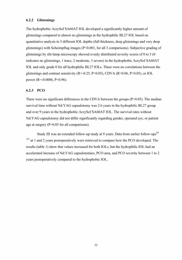
6.2.2 Glistenings
The hydrophobic AcrySof SA60AT IOL developed a significantly higher amount of
glistenings compared to almost no glistenings in the hydrophilic BL27 IOL based on
quantitative analysis in 3 different IOL depths (full thickness, deep glistenings and very deep
glistenings) with Scheimpflug images (P<0.001, for all 3 comparisons). Subjective grading of
glistenings by slit-lamp microscopy showed evenly distributed severity scores of 0 to 3 (0
indicates no glistenings, 1 trace, 2 moderate, 3 severe) in the hydrophobic AcrySof SA60AT
IOL and only grade 0 for all hydrophilic BL27 IOLs. There were no correlations between the
glistenings and contrast sensitivity (R=-0.25, P>0.05), CDVA (R=0.06, P>0.05), or IOL
power (R=-0.0086, P=0.96).
6.2.3 PCO
There were no significant differences in the CDVA between the groups (P>0.05). The median
survival time without Nd:YAG capsulotomy was 2.6 years in the hydrophilic BL27 group
and over 9 years in the hydrophobic AcrySof SA60AT IOL. The survival rates without
Nd:YAG capsulotomy did not differ significantly regarding gender, operated eye, or patient
age at surgery (P>0.05 for all comparisons).
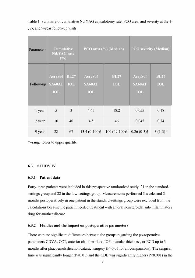
Study III was an extended follow-up study at 9 years. Data from earlier follow-ups69
125 at 1 and 2 years postoperatively were retrieved to compare how the PCO developed. The
results (table 1) show that values increased for both IOLs, but the hydrophilic IOL had an
accelerated increase of Nd:YAG capsulotomies, PCO area, and PCO severity between 1 to 2
years postoperatively compared to the hydrophobic IOL.
33
Table 1. Summary of cumulative Nd:YAG capsulotomy rate, PCO area, and severity at the 1-
, 2-, and 9-year follow-up visits.
Parameters
Cumulative Nd:YAG rate
(%)
PCO area (%) (Median)
PCO severity (Median)
Follow-up
AcrySof
SA60AT
IOL
BL27
IOL
AcrySof
SA60AT
IOL
BL27
IOL
AcrySof
SA60AT
IOL
BL27
IOL
1 year 5 3 4.65 18.2 0.055 0.18
2 year 10 40 4.5 46 0.045 0.74
9 year 28 67 13.4 (0-100)† 100 (49-100)† 0.26 (0-3)† 3 (1-3)†
†=range lower to upper quartile
6.3 STUDY IV
6.3.1 Patient data
Forty-three patients were included in this prospective randomized study, 21 in the standard-
settings group and 22 in the low-settings group. Measurements performed 3 weeks and 3
months postoperatively in one patient in the standard-settings group were excluded from the
calculations because the patient needed treatment with an oral nonsteroidal anti-inflammatory
drug for another disease.
6.3.2 Fluidics and the impact on postoperative parameters
There were no significant differences between the groups regarding the postoperative
parameters CDVA, CCT, anterior chamber flare, IOP, macular thickness, or ECD up to 3
months after phacoemulsification cataract surgery (P>0.05 for all comparisons). The surgical
time was significantly longer (P<0.01) and the CDE was significantly higher (P<0.001) in the

34
group with low fluidic settings. The median surgical time in the standard-settings group was 7
minutes and 20 seconds and 8 minutes and 28 seconds in the low-settings group. The mean
CDE was 7.61 ± 3.61 (SD) in the standard-settings group and 15.14 ± 5.0 (SD) in the low-
settings group.
35
7 DISCUSSION
7.1 STUDIES I-III
7.1.1 Lens glistenings
Different hydrophobic IOLs do not develop glistenings to the same extent as we
showed that the AcrySof IOL did in study I. The hydrophobic AcrySof IOL developed much
more glistenings as Tognetto et al.7 reported earlier in a glistening study that evaluated
AcrySof IOLs compared to another hydrophobic IOL. The investigators compared
development of glistenings in seven different IOLs, two (AcrySof and Sensar) were
hydrophobic acrylic IOLs. The AcrySof IOL showed a continuous increase in glistenings
over 2 years and more glistenings than the Sensar IOL.
An in vitro study12 that simulated 20-year deterioration of IOLs included six
different hydrophobic acrylic IOLs, AcrySof MA60BM, AcrySof SA60AT, Sensar AR40e,
Acryfold VA-60BB, Nex-Acri N4-18B, and Avansee AU6, showed that only the last IOL did
not develop glistenings. The authors described the characteristics of glistening development
in each IOLs, but they did not rank them.
Study II compared one hydrophobic IOL with a hydrophilic IOL 9 years
postoperatively and showed that the hydrophilic IOL was not prone to glistening
development even over the long term. This agreed with an earlier short-term follow-up study
conducted for hydrophilic IOLs7. No other in vivo study of glistening development in
hydrophilic IOLs has been conducted until now.
The severity of glistenings development in one in vitro study simulating
accelerated 20-year IOL deterioration included a series of hydrophobic IOLs12. The tendency
was that IOLs with higher water content developed fewer glistening-like opacities.
Another issue is the differences in refractive indices between the hydrophobic
IOLs we studied, 1.55 for the AcrySof IOL and 1.47 for the Sensar AR40e IOL. Aqueous
humor has a refractive index of 1.336, which is very close to that of water (1.333). When
differences in refractive indices between two media (i.e., IOL material and water) increase,
the amount of reflected light rays also increases according to Snell’s law126. The higher
refractive index in the AcrySof IOL compared to the refractive index of the Sensar IOL also
may make glistenings more visible at the slit-lamp and in the Scheimpflug images.
36
To quantify glistenings in IOLs, we used Scheimpflug images in studies I and
II. This method has been described earlier23 29 and also was positively correlated with slit-
lamp gradings14. Study I agreed with this study and found a correlation between quantified
analyses of glistenings using Scheimpflug images with subjective slit-lamp grading of
glistenings.
Surface light scattering and PCO are two potential sources of error when
analyzing glistenings with Scheimpflug images, because these two phenomena also can be
seen in the images as whitish bands of light scatterings in the IOL anterior and posterior
surfaces. Eliminating these two phenomena from the calculations can be accomplished by
omitting light scattering values from the IOL anterior and posterior surfaces, thus only light
scatterings within the IOL (i.e., glistenings) are analyzed. We analyzed glistenings in the
central parts of the IOL by subtracting light scattering from 73 microns and 101 microns from
the IOL anterior and posterior surfaces and referred to them as deep and very deep
glistenings, respectively. Excluding cases with PCO in the visual axis is another way to be
certain to not include PCO in the calculations. Scheimpflug images appear to be a valuable
and reliable tool and until now the only method to quantify glistenings objectively.
The CDVA and contrast sensitivity (studies I and II) were not correlated with
the amount of glistenings as in most earlier studies7 15 20 28 37 127 128. The IOL dioptric power
was not correlated with glistenings in studies I and II. The results from earlier studies are
inconclusive; some show correlations with glistenings13 15 17 20 129 and some do not128 130.
A weakness of the studies was the large number of patient dropouts, i.e., 30%
in study I and 35% in studies II and III. Long-term follow-up studies have this problem
because patients become older and die, get ill, or move.
7.1.2 PCO
Long-term follow-up studies of PCO (studies I and III) are important because the average
patient with an IOL is likely to have it for about 10 years, since the average age at surgery in
many studies of patients with senile cataracts is 68 to 73 years. The average lifespan in
Sweden was 83.7 years for women and 80.1 years for men in 2013 according to Statistics
Sweden (Statistiska centralbyrån)131, an authority in Sweden. We have seen cases in which
the long-term follow-up of PCO development has yielded surprising results. One example
was that the sharp posterior edge design in a hydrophobic acrylic IOL inhibits PCO more
37
effectively for the first 3 to 5 years compared to a silicone IOL with a round-edge design100.
After 12 years, the rates of PCO development and Nd:YAG in sharp-edge IOLs catch up with
the round-edge IOLs93. Two other studies have reported similar results with loss of the
advantageous PCO inhibitory features compared to silicone IOLs 6 years postoperatively91 92.
In study I, we compared two hydrophobic acrylic IOLs with a sharp-edge
design for PCO development. To our knowledge, this study had the longest follow-up time
for comparing two different hydrophobic IOLs for PCO development. There were no
significant differences in PCO area, severity, or Nd:YAG capsulotomy rates between the
IOLs 5 to 7 years postoperatively and the study had results similar to other studies with
shorter follow-up times132-136.
In study III, we compared PCO development between a hydrophilic IOL
(BL27) and a hydrophobic IOL (AcrySof SA60AT) 9 years postoperatively. This study had
the longest follow-up time comparing PCO development between hydrophilic and
hydrophobic acrylic IOLs. We concluded that the advantageous PCO inhibitory effect in the
hydrophobic IOL at 2 year postoperatively remained valid even after 9 years. The superior
PCO inhibitory characteristics of the hydrophobic acrylic IOL may depend on many different
factors, i.e., the sharp-edge IOL profile and the stronger bindings of the AcrySof IOL to the
posterior capsule. Strong binding of the posterior capsule to the posterior IOL surface
minimizes any space for LECs to grow into the posterior capsule according to the concept of
“no space, no cells”48. The hydrophobic AcrySof SA60AT IOL has these characteristics43 74-
76. We mentioned previously that the sharp-edge hydrophilic IOLs generally are not as sharp
as hydrophobic IOLs mostly due to different manufacturing techniques. The edge sharpness
of BL27 IOL has not been studied earlier. It is then difficult to determine if the primary
superior PCO inhibitory factor is the sharp-edge design or the strong fibronectin binding in
our study. But earlier study74 support the sharp-edge IOL profile as the most important factor
inhibiting PCO development.
In study III, we also compared Nd:YAG capsulotomy rates between two
different IOLs with survival analysis. Long-term follow-up studies usually have many patient
dropouts and simply comparing Nd:YAG numbers becomes unreliable. The advantage in
survival analysis is that all patient data are valuable and can be used up to the point at which
they are lost to follow-up.
38
7.2 STUDY IV
The study results were interesting, because it was unexpected that lower fluidic settings did
not have any substantial benefits compared to standard fluidic settings concerning
postoperative parameters indicating surgical trauma. The disadvantages with low fluidic
settings were a significantly longer surgical time and use of more CDE.
Few studies have been conducted in this field. Two earlier studies have
investigated fluidic levels and the impact on the eye. Baradaran-Rafii et al.108 compared low
versus high aspiration parameters in longitudinal phacoemulsification. Their fluidic
parameters were almost the same as ours, except that our vacuum levels were higher. Those
investigators concluded that decreased ECD postoperatively was related to CDE. Increased
CDE yielded decreased ECD. One important difference between our and their study was that
we used torsional phacoemulsification, which is more effective than longitudinal
phacoemulsification in lens aspiration137-144, and it produces less heat138 145 with less increase
in postoperative corneal thickness143 144 and less endothelial cell loss143.
Vasavada et al109 found a lower increase in CCT 1 and 7 days postoperatively
with low fluidic settings compared with high fluidic settings. Those investigators had higher
CDE for both fluidic settings groups compared to our study. They used the Infiniti Vision
System as we did, but the difference was that they performed longitudinal
phacoemulsification.
We cannot state to what extent torsional phacoemulsification plays a role and
results in no significant differences in the postoperative parameters. Probably the duration of
surgery, CDE amount, and fluidic levels are important factors. However, the purpose of our
study was not to determine any threshold levels for tissue damage for the respective factors.
More studies are needed in the future to establish these correlations. We can conclude that in
normal daily cases with senile cataracts, there were no significant differences in the
postoperative parameters between standard fluidic and low fluidic settings in
phacoemulsification cataract surgery. The low fluidic settings were a bit disadvantageous
because of the extended surgical time and the demand for a higher amount of ultrasound
energy.
A weakness in this study was the small number of patients. With more patients,
significant differences may be seen that we were unable to detect.
39
8 MAIN CONCLUSIONS
In study I, the AcrySof SA60AT IOL developed significantly more glistenings than the
Sensar AR40e IOL, both hydrophobic acrylic IOLs, 5 to 7 years after phacoemulsification
cataract surgery. Development of PCO area and severity and the frequency of Nd:YAG laser
capsulotomy did not differ between the AcrySof SA60AT and Sensar AR40e IOLs 5 to 7
years postoperatively.
In study II, the AcrySof SA60AT IOL, a hydrophobic acrylic IOL, developed
significantly more glistenings compared to the BL27 IOL, a hydrophilic acrylic IOL, 9 years
after phacoemulsification cataract surgery.
In study III, the AcrySof SA60AT IOL had a significantly longer survival time
without Nd:YAG laser capsulotomy compared to the BL27 IOL, 9 years after
phacoemulsification cataract surgery.
In study IV, there were no significant differences in the postoperative
parameters indicating surgical trauma between the standard and low fluidic settings in
phacoemulsification cataract surgery. However, the surgical time was longer in the low-
fluidic-settings group, and more phacoemulsification energy was needed to aspirate the lens.
40
9 FUTURE PERSPECTIVES
Thus far, glistenings have not been a big issue in relation to symptomatic complaints from
patients who have undergone phacoemulsification cataract surgery. Is it necessary to further
investigate this matter? In my opinion, it is certainly worth the effort, because according to
the studies, glistenings may increase with time. The average lifespan has been increasing for
many years in Sweden and worldwide, and the average patient age for IOL implantation
during cataract surgery is slightly lower than before, i.e., 76 years in 1999 compared to 74
years in 2013 (Swedish National Cataract Registry annual report 2013)146. This means that
the average patient is going to have their IOL for a longer period and the possibility for
developing glistenings and PCO also increases.
In most cases, patients only undergo IOL implantation once because of the risk
of complications when trying to explant the old IOL and re-implant a new IOL. Therefore, it
is important that we implant the most satisfactory IOLs initially. That includes lowering the
rates or if possible even eradicating glistenings and PCO to maximize the chances of the best
postoperative outcomes.
In our studies, the average patient age was about 70 years. However, there is an
increasing market for presbyopic refractive surgery due to the popularity and potential with
gradually safer phacoemulsification machines, improved surgical techniques, and better IOL
materials. These patients are often younger than most patients in the studies of glistenings and
PCO and they are still in the workforce, tend to have better CDVA and contrast sensitivity
preoperatively, and are more demanding of perfect results. If the contrast sensitivity decreases
only slightly in this group, it may be enough to lead to visual symptoms and complaints. It is
not rare that the implanted IOLs in these patients are premium IOLs with toric and multifocal
characteristics. Performing Nd:YAG capsulotomy in patients with these lenses is best
avoided if possible. Potential complications of Nd:YAG capsulotomy such as IOL dislocation
and tilting with IOL decentration may cause more pronounced visual symptoms compared to
monofocal IOLs.
Multifocal IOLs decrease contrast sensitivity by about 20% compared to
standard monofocal IOLs147-151. Could this reduction in contrast sensitivity in a younger
group of patients together with development of glistenings reach a threshold level at which
visual symptoms occur and is this a new group of patients we should expect to see in the
41
ophthalmology wards in a few years? I think it would be interesting to investigate the
development of glistenings in premium IOLs and their impact on CDVA and contrast
sensitivity.
Many different methods have been used to decrease the PCO rates. However, is
it possible to eradicate it or further decrease PCO rates? Studies by Tassignon et al.152 153,
using the so-called bag-in-the-lens technique with a posterior capsulorhexis, have shown
promising results, but creating a posterior capsulorhexis in the center and with almost the
same diameter every time can be challenging. Could the new femtosecond laser surgical
technique be helpful? Study of PCO development using the bag-in-the-lens technique with a
femtosecond laser also may be worth considering, making an eventual second visit for
Nd:YAG capsulotomy obsolete. With even better knowledge in these fields of research, we
can provide feedback to the IOL and surgery machine manufacturers and they in turn can
develop even better IOLs and machines and minimize intraoperative and postoperative
complications.
There are new commercially available phacoemulsification machines that claim
to control the anterior chamber depth and an IOP more stable than before. It would be
interesting to study the fluidic settings and the impact on postoperative parameters when the
IOP can be maintained at a stable and almost fix level throughout the entire surgery, allowing
further study of different factors and their respective impact on the eye.
42
10 ACKNOWLEDGEMENTS
I want to thank everybody who made this thesis possible. I am expressing my sincere
gratitude to the following individuals.
Maria Kugelberg, my supervisor, for your knowledge and guidance. You showed me the joy
of doing scientific research and how important it is to have a fantastic supervisor. You have
always been supportive and available when I needed your help.
Alexander Fridberg, my co-supervisor, my first teacher in phacoemulsification cataract
surgery, and the person who introduced me to clinical research.
Professor Jan Ygge, head of the division of ophthalmology and vision at the Institute of
Clinical Neuroscience, for allowing me to be a PhD student at the department.
Helene Hamberg-Nyström for introducing me to my supervisor and Dr. Kugelberg’s research
team.
My co-authors, Anders Behndig for sharing the glistening analysis method with Scheimpflug
images and Margrethe Rønbeck for showing me how to use POCOman analysis software.
Mikaela Taube, Kerstin Jurelius, Laurence Querat, and Christina Åkerstedt for your help with
the administrative and clinical work with the research patients.
Members of my research team: Behrad Samadi, Magnus Grimfors, Daniel Beckung-
Farhoudi, Kaziwe Mollazadegan, Tiina Konradsen, Pia Agervi, Ulla Kugelberg, and
Christina Persson.
Gunilla Högberg, my principal teacher in anterior segment surgery and the person who made
me an accomplished cataract surgeon.
Örjan Wallin, who taught me glaucoma surgery, and Ditte Artzén, who taught me pterygium
surgery.
Steffen Schulz-Key and Luai Alissa, my roommates at the clinic for sharing your views of
life and discussions about difficult clinical cases.
My colleagues, Annemari Koivula, Annika Grunge, Branka Samolov, Carl-Gustaf Laurell,
Emma Nivenius, Emmanuel Kai-Lewis, Gisela Wejde, Gysbert van Setten, Helena Gartzios,
Ioannis Sidiropoulus, Laura Armonaite, Mario Economou, Per Montan, and Talal Ali for

43
your supportive attitude and care of my patients during periods when I was conducting
research.
St. Erik Eye Hospital and CEO Anders Boman for giving me the opportunity to develop as an
ophthalmologist and PhD student and making me a better physician to serve my patients.
My parents for your tireless support and encouragement. My brother Robert for always being
a beacon in the dark and my brother Johnny for sharing many interests and joys.
My wife, Emily for your patience, understanding, and never-ending trust and confidence in
me. My boys Nicholas and Alexander for being my sunshine. You are growing up so quickly,
and making me always trying to remember that I must embrace every moment we are
together and make the most of it.
44
11 REFERENCES
1. Ballin N. Glistenings in injection-molded lens. J Am Intraocul Implant Soc 1984;10(4):473.
2. Werner L. Glistenings and surface light scattering in intraocular lenses. J Cataract Refract Surg 2010;36(8):1398-420.
3. Miyata A, Yaguchi S. Equilibrium water content and glistenings in acrylic intraocular lenses. J Cataract Refract Surg 2004;30(8):1768-72.
4. Saylor DM, Coleman Richardson D, Dair BJ, Pollack SK. Osmotic cavitation of elastomeric intraocular lenses. Acta Biomater 2010;6(3):1090-8.
5. Shiba T, Mitooka K, Tsuneoka H. In vitro analysis of AcrySof intraocular lens glistening. Eur J Ophthalmol 2003;13(9-10):759-63.
6. Kato K, Nishida M, Yamane H, Nakamae K, Tagami Y, Tetsumoto K. Glistening formation in an AcrySof lens initiated by spinodal decomposition of the polymer network by temperature change. J Cataract Refract Surg 2001;27(9):1493-8.
7. Tognetto D, Toto L, Sanguinetti G, Ravalico G. Glistenings in foldable intraocular lenses. J Cataract Refract Surg 2002;28(7):1211-6.
8. Gregori NZ, Spencer TS, Mamalis N, Olson RJ. In vitro comparison of glistening formation among hydrophobic acrylic intraocular lenses(1). J Cataract Refract Surg 2002;28(7):1262-8.
9. Miura M, Osako M, Elsner AE, Kajizuka H, Yamada K, Usui M. Birefringence of intraocular lenses. J Cataract Refract Surg 2004;30(7):1549-55.
10. Miyata A, Uchida N, Nakajima K, Yaguchi S. Clinical and Experimental Observation of Glistening in Acrylic Intraocular Lenses. Jpn J Ophthalmol 2000;44(6):693.
11. Allers A, Baumeister M, Steinkamp GW, Ohrloff C, Kohnen T. [Intra-individual comparison of intraocular lenses of highly refractive silicone (Allergan SI40NB) and hydrophobic acrylate (Alcon Acrysof MA60BM). 1-year follow-up]. Ophthalmologe 2000;97(10):669-75.
12. Kawai K, Hayakawa K, Suzuki T. Simulation of 20-year deterioration of acrylic IOLs using severe accelerated deterioration tests. Tokai J Exp Clin Med 2012;37(3):62-5.
13. Ronbeck M, Behndig A, Taube M, Koivula A, Kugelberg M. Comparison of glistenings in intraocular lenses with three different materials: 12-year follow-up. Acta Ophthalmol 2013;91(1):66-70.
14. Behndig A, Monestam E. Quantification of glistenings in intraocular lenses using Scheimpflug photography. J Cataract Refract Surg 2009;35(1):14-7.
15. Moreno-Montanes J, Alvarez A, Rodriguez-Conde R, Fernandez-Hortelano A. Clinical factors related to the frequency and intensity of glistenings in AcrySof intraocular lenses. J Cataract Refract Surg 2003;29(10):1980-4.
16. Peetermans E, Hennekes R. Long-term results of wagon wheel packed acrylic intra-ocular lenses (AcrySof). Bull Soc Belge Ophtalmol 1999;271:45-8.
17. Monestam E, Behndig A. Impact on visual function from light scattering and glistenings in intraocular lenses, a long-term study. Acta Ophthalmol 2011;89(8):724-8.
45
18. Omar O, Pirayesh A, Mamalis N, Olson RJ. In vitro analysis of AcrySof intraocular lens glistenings in AcryPak and Wagon Wheel packaging. J Cataract Refract Surg 1998;24(1):107-13.
19. Nishihara H KT, Ohnishi T, Koike M, Imai M,, Shibuya A YS. [Glistenings in lathe-cut acrylic intraocular lens]. [Japanese]. Ganka Shujutsu 2000;13:227-30.
20. Colin J, Orignac I, Touboul D. Glistenings in a large series of hydrophobic acrylic intraocular lenses. J Cataract Refract Surg 2009;35(12):2121-6.
21. Dick HB, Olson RJ, Augustin AJ, Schwenn O, Magdowski G, Pfeiffer N. Vacuoles in the Acrysof intraocular lens as factor of the presence of serum in aqueous humor. Ophthalmic Res 2001;33(2):61-7.
22. Miyake K, Ota I, Maekubo K, Ichihashi S, Miyake S. Latanoprost accelerates disruption of the blood-aqueous barrier and the incidence of angiographic cystoid macular edema in early postoperative pseudophakias. Arch Ophthalmol 1999;117(1):34-40.
23. Ayaki M, Nishihara H, Yaguchi S, Koide R. Surfactant induced glistenings: surface active ingredients in ophthalmic solutions may enhance water entry into the voids of implanted acrylic intraocular lenses. J Long Term Eff Med Implants 2006;16(6):451-7.
24. Ayaki M, Nishihara H, Yaguchi S, Koide R. Effect of ophthalmic solution components on acrylic intraocular lenses. J Cataract Refract Surg 2007;33(1):122-6.
25. Dhaliwal DK, Mamalis N, Olson RJ, Crandall AS, Zimmerman P, Alldredge OC, et al. Visual significance of glistenings seen in the AcrySof intraocular lens. J Cataract Refract Surg 1996;22(4):452-7.
26. Cisneros-Lanuza A, Hurtado-Sarrio M, Duch-Samper A, Gallego-Pinazo R, Menezo-Rozalen JL. Glistenings in the Artiflex phakic intraocular lens. J Cataract Refract Surg 2007;33(8):1405-8.
27. Gunenc U, Oner FH, Tongal S, Ferliel M. Effects on visual function of glistenings and folding marks in AcrySof intraocular lenses. J Cataract Refract Surg 2001;27(10):1611-4.
28. Chang A, Behndig A, Ronbeck M, Kugelberg M. Comparison of posterior capsule opacification and glistenings with 2 hydrophobic acrylic intraocular lenses: 5- to 7-year follow-up. J Cataract Refract Surg 2013;39(5):694-8.
29. Klos KM, Richter R, Schnaudigel O, Ohrloff C. Image analysis of implanted rigid and foldable intraocular lenses in human eyes using Scheimpflug photography. Ophthalmic Res 1999;31(2):130-3.
30. Nishihara H, Ayaki M, Watanabe T, Ohnishi T, Kageyama T, Yaguchi S. [Comparison of surface light scattering of acrylic intraocular lenses made by lathe-cutting and cast-molding methods--long-term observation and experimental study]. Nihon Ganka Gakkai Zasshi 2004;108(3):157-61.
31. Yaguchi S, Chida M, Nishihara H, Ohnishi T, Ayaki M. [Light scattering observed on the surface of acrylic intraocular lenses ten years after implantation]. Nihon Ganka Gakkai Zasshi 2002;106(2):109-11.
32. Wilkins E, Olson RJ. Glistenings with long-term follow-up of the Surgidev B20/20 polymethylmethacrylate intraocular lens. Am J Ophthalmol 2001;132(5):783-5.
46
33. Packer M, Fry L, Lavery KT, Lehmann R, McDonald J, Nichamin L, et al. Safety and effectiveness of a glistening-free single-piece hydrophobic acrylic intraocular lens (enVista). Clin Ophthalmol 2013;7:1905-12.
34. Packer M, Rajan M, Ligabue E, Heiner P. Clinical properties of a novel, glistening-free, single-piece, hydrophobic acrylic IOL. Clin Ophthalmol 2014;8:421-7.
35. Tetz MR WL, Schwahn-Bendig S, Batlle JF. Prospective clinical study to quantify glistenings in new hydrophobic acrylic IOL. Paper presented at the ASCRS Symposium on Cataract, IOL and Refractive Surgery. San Francisco, CA, April 2009, 2009.
36. Hayashi K, Hirata A, Yoshida M, Yoshimura K, Hayashi H. Long-term effect of surface light scattering and glistenings of intraocular lenses on visual function. Am J Ophthalmol 2012;154(2):240-51 e2.
37. Chang A, Kugelberg M. Glistenings 9 years after phacoemulsification in hydrophobic and hydrophilic acrylic intraocular lenses. J Cataract Refract Surg 2015;41(6):1199-204.
38. Waite A, Faulkner N, Olson RJ. Glistenings in the single-piece, hydrophobic, acrylic intraocular lenses. Am J Ophthalmol 2007;144(1):143-4.
39. Apple DJ, Peng Q, Visessook N, Werner L, Pandey SK, Escobar-Gomez M, et al. Eradication of posterior capsule opacification: documentation of a marked decrease in Nd:YAG laser posterior capsulotomy rates noted in an analysis of 5416 pseudophakic human eyes obtained postmortem. Ophthalmology 2001;108(3):505-18.
40. Apple DJ, Peng Q, Visessook N, Werner L, Pandey SK, Escobar-Gomez M, et al. Surgical prevention of posterior capsule opacification. Part 1: Progress in eliminating this complication of cataract surgery. Journal of cataract and refractive surgery 2000;26(2):180-7.
41. Thompson AM, Sachdev N, Wong T, Riley AF, Grupcheva CN, McGhee CN. The Auckland Cataract Study: 2 year postoperative assessment of aspects of clinical, visual, corneal topographic and satisfaction outcomes. Br J Ophthalmol 2004;88(8):1042-8.
42. Smith SR, Daynes T, Hinckley M, Wallin TR, Olson RJ. The effect of lens edge design versus anterior capsule overlap on posterior capsule opacification. Am J Ophthalmol 2004;138(4):521-6.
43. Meacock WR, Spalton DJ, Hollick EJ, Barman S, Boyce JF. The effect of polymethylmethacrylate and acrysof intraocular lenses on the posterior capsule in patients with a large capsulorrhexis. Jpn J Ophthalmol 2001;45(4):348-54.
44. Wejde G, Kugelberg M, Zetterstrom C. Position of anterior capsulorhexis and posterior capsule opacification. Acta Ophthalmol Scand 2004;82(5):531-4.
45. Mamalis N, Grossniklaus HE, Waring GO, 3rd, Werner L, Brubaker J, Davis D, et al. Ablation of lens epithelial cells with a laser photolysis system: histopathology, ultrastructure, and immunochemistry. J Cataract Refract Surg 2010;36(6):1003-10.
46. Meacock WR, Spalton DJ, Hollick EJ, Boyce JF, Barman S, Sanguinetti G. Double-masked prospective ocular safety study of a lens epithelial cell antibody to prevent posterior capsule opacification. J Cataract Refract Surg 2000;26(5):716-21.
47
47. Fernandez V, Fragoso MA, Billotte C, Lamar P, Orozco MA, Dubovy S, et al. Efficacy of various drugs in the prevention of posterior capsule opacification: experimental study of rabbit eyes. J Cataract Refract Surg 2004;30(12):2598-605.
48. Apple DJ, Solomon KD, Tetz MR, Assia EI, Holland EY, Legler UF, et al. Posterior capsule opacification. Surv Ophthalmol 1992;37(2):73-116.
49. Koch DD, Liu JF, Gill EP, Parke DW, 2nd. Axial myopia increases the risk of retinal complications after neodymium-YAG laser posterior capsulotomy. Arch Ophthalmol 1989;107(7):986-90.
50. Burq MA, Taqui AM. Frequency of retinal detachment and other complications after neodymium:Yag laser capsulotomy. J Pak Med Assoc 2008;58(10):550-2.
51. Ari S, Cingu AK, Sahin A, Cinar Y, Caca I. The effects of Nd:YAG laser posterior capsulotomy on macular thickness, intraocular pressure, and visual acuity. Ophthalmic Surg Lasers Imaging 2012;43(5):395-400.
52. Holweger RR, Marefat B. Intraocular pressure change after neodymium:YAG capsulotomy. J Cataract Refract Surg 1997;23(1):115-21.
53. Buehl W, Findl O. Effect of intraocular lens design on posterior capsule opacification. J Cataract Refract Surg 2008;34(11):1976-85.
54. Newland TJ, McDermott ML, Eliott D, Hazlett LD, Apple DJ, Lambert RJ, et al. Experimental neodymium:YAG laser damage to acrylic, poly(methyl methacrylate), and silicone intraocular lens materials. J Cataract Refract Surg 1999;25(1):72-6.
55. Charles S. Vitreoretinal complications of YAG laser capsulotomy. Ophthalmol Clin North Am 2001;14(4):705-10.
56. Meacock WR, Spalton DJ, Boyce J, Marshall J. The effect of posterior capsule opacification on visual function. Invest Ophthalmol Vis Sci 2003;44(11):4665-9.
57. Jose RM, Bender LE, Boyce JF, Heatley C. Correlation between the measurement of posterior capsule opacification severity and visual function testing. J Cataract Refract Surg 2005;31(3):534-42.
58. Cheng CY, Yen MY, Chen SJ, Kao SC, Hsu WM, Liu JH. Visual acuity and contrast sensitivity in different types of posterior capsule opacification. J Cataract Refract Surg 2001;27(7):1055-60.
59. Guha GS. Soemmering's ring and its dislocations. Br J Ophthalmol 1951;35(4):226-31.
60. Dana MR, Chatzistefanou K, Schaumberg DA, Foster CS. Posterior capsule opacification after cataract surgery in patients with uveitis. Ophthalmology 1997;104(9):1387-93; discussion 93-4.
61. Ebihara Y, Kato S, Oshika T, Yoshizaki M, Sugita G. Posterior capsule opacification after cataract surgery in patients with diabetes mellitus. J Cataract Refract Surg 2006;32(7):1184-7.
62. Hayashi K, Hayashi H, Nakao F, Hayashi F. Posterior capsule opacification after cataract surgery in patients with diabetes mellitus. Am J Ophthalmol 2002;134(1):10-6.
63. Hayashi Y, Kato S, Fukushima H, Numaga J, Kaiya T, Tamaki Y, et al. Relationship between anterior capsule contraction and posterior capsule opacification after cataract surgery in patients with diabetes mellitus. J Cataract Refract Surg 2004;30(7):1517-20.
48
64. Praveen MR, Vasavada AR, Shah GD, Shah AR, Khamar BM, Dave KH. A prospective evaluation of posterior capsule opacification in eyes with diabetes mellitus: a case-control study. Eye (Lond) 2014;28(6):720-7.
65. Zaczek A, Zetterstrom C. Posterior capsule opacification after phacoemulsification in patients with diabetes mellitus. J Cataract Refract Surg 1999;25(2):233-7.
66. Dikopf MS, Chow CC, Mieler WF, Tu EY. Cataract extraction outcomes and the prevalence of zonular insufficiency in retinitis pigmentosa. Am J Ophthalmol 2013;156(1):82-88 e2.
67. Peng Q, Apple DJ, Visessook N, Werner L, Pandey SK, Escobar-Gomez M, et al. Surgical prevention of posterior capsule opacification. Part 2: Enhancement of cortical cleanup by focusing on hydrodissection. J Cataract Refract Surg 2000;26(2):188-97.
68. Heatley CJ, Spalton DJ, Kumar A, Jose R, Boyce J, Bender LE. Comparison of posterior capsule opacification rates between hydrophilic and hydrophobic single-piece acrylic intraocular lenses. J Cataract Refract Surg 2005;31(4):718-24.
69. Kugelberg M, Wejde G, Jayaram H, Zetterstrom C. Two-year follow-up of posterior capsule opacification after implantation of a hydrophilic or hydrophobic acrylic intraocular lens. Acta Ophthalmol 2008;86(5):533-6.
70. Ursell PG, Spalton DJ, Pande MV, Hollick EJ, Barman S, Boyce J, et al. Relationship between intraocular lens biomaterials and posterior capsule opacification. J Cataract Refract Surg 1998;24(3):352-60.
71. Hollick EJ, Spalton DJ, Ursell PG, Pande MV. Lens epithelial cell regression on the posterior capsule with different intraocular lens materials. Br J Ophthalmol 1998;82(10):1182-8.
72. Fong CS, Mitchell P, Rochtchina E, Cugati S, Hong T, Wang JJ. Three-year incidence and factors associated with posterior capsule opacification after cataract surgery: The Australian Prospective Cataract Surgery and Age-related Macular Degeneration Study. Am J Ophthalmol 2014;157(1):171-79 e1.
73. Nishi O, Nishi K. Preventing posterior capsule opacification by creating a discontinuous sharp bend in the capsule. Journal of cataract and refractive surgery 1999;25(4):521-6.
74. Nishi O, Nishi K, Akura J, Nagata T. Effect of round-edged acrylic intraocular lenses on preventing posterior capsule opacification. J Cataract Refract Surg 2001;27(4):608-13.
75. Nishi O, Nishi K, Sakanishi K. Inhibition of migrating lens epithelial cells at the capsular bend created by the rectangular optic edge of a posterior chamber intraocular lens. Ophthalmic Surg Lasers 1998;29(7):587-94.
76. Nishi O, Nishi K, Wickstrom K. Preventing lens epithelial cell migration using intraocular lenses with sharp rectangular edges. Journal of cataract and refractive surgery 2000;26(10):1543-9.
77. DW S. Beobachtungen über die organischen Veränderungen an Auge mit Staaroperationen. Frankfurt/Main: Wesche, 1828.
49
78. Werner L, Mamalis N, Pandey SK, Izak AM, Nilson CD, Davis BL, et al. Posterior capsule opacification in rabbit eyes implanted with hydrophilic acrylic intraocular lenses with enhanced square edge. J Cataract Refract Surg 2004;30(11):2403-9.
79. Buehl W, Findl O, Menapace R, Sacu S, Kriechbaum K, Koeppl C, et al. Long-term effect of optic edge design in an acrylic intraocular lens on posterior capsule opacification. Journal of cataract and refractive surgery 2005;31(5):954-61.
80. Findl O, Menapace R, Sacu S, Buehl W, Rainer G. Effect of optic material on posterior capsule opacification in intraocular lenses with sharp-edge optics: randomized clinical trial. Ophthalmology 2005;112(1):67-72.
81. Nanavaty MA, Spalton DJ, Boyce J, Brain A, Marshall J. Edge profile of commercially available square-edged intraocular lenses. Journal of cataract and refractive surgery 2008;34(4):677-86.
82. Sacu S, Menapace R, Findl O, Kiss B, Buehl W, Georgopoulos M. Long-term efficacy of adding a sharp posterior optic edge to a three-piece silicone intraocular lens on capsule opacification: five-year results of a randomized study. American journal of ophthalmology 2005;139(4):696-703.
83. Hayashi K, Hayashi H. Posterior capsule opacification in the presence of an intraocular lens with a sharp versus rounded optic edge. Ophthalmology 2005;112(9):1550-6.
84. Shah A, Spalton DJ, Gilbert C, Vasavada A, Boyce JF, Minassian D, et al. Effect of intraocular lens edge profile on posterior capsule opacification after extracapsular cataract surgery in a developing country. J Cataract Refract Surg 2007;33(7):1259-66.
85. Spalton D. Posterior capsule opacification: have we made a difference? Br J Ophthalmol 2013;97(1):1-2.
86. Hancox J, Spalton D, Cleary G, Boyce J, Nanavaty MA, Thyagarajan S, et al. Fellow-eye comparison of posterior capsule opacification with AcrySof SN60AT and AF-1 YA-60BB blue-blocking intraocular lenses. Journal of cataract and refractive surgery 2008;34(9):1489-94.
87. Li Y, Wang J, Chen Z, Tang X. Effect of hydrophobic acrylic versus hydrophilic acrylic intraocular lens on posterior capsule opacification: meta-analysis. PLoS One 2013;8(11):e77864.
88. Vasavada AR, Raj SM, Shah A, Shah G, Vasavada V. Comparison of posterior capsule opacification with hydrophobic acrylic and hydrophilic acrylic intraocular lenses. J Cataract Refract Surg 2011;37(6):1050-9.
89. Linnola RJ, Werner L, Pandey SK, Escobar-Gomez M, Znoiko SL, Apple DJ. Adhesion of fibronectin, vitronectin, laminin, and collagen type IV to intraocular lens materials in pseudophakic human autopsy eyes. Part 1: histological sections. Journal of cataract and refractive surgery 2000;26(12):1792-806.
90. Linnola RJ, Werner L, Pandey SK, Escobar-Gomez M, Znoiko SL, Apple DJ. Adhesion of fibronectin, vitronectin, laminin, and collagen type IV to intraocular lens materials in pseudophakic human autopsy eyes. Part 2: explanted intraocular lenses. Journal of cataract and refractive surgery 2000;26(12):1807-18.
91. Vock L, Crnej A, Findl O, Neumayer T, Buehl W, Sacu S, et al. Posterior capsule opacification in silicone and hydrophobic acrylic intraocular lenses with sharp-edge optics six years after surgery. Am J Ophthalmol 2009;147(4):683-90 e2.
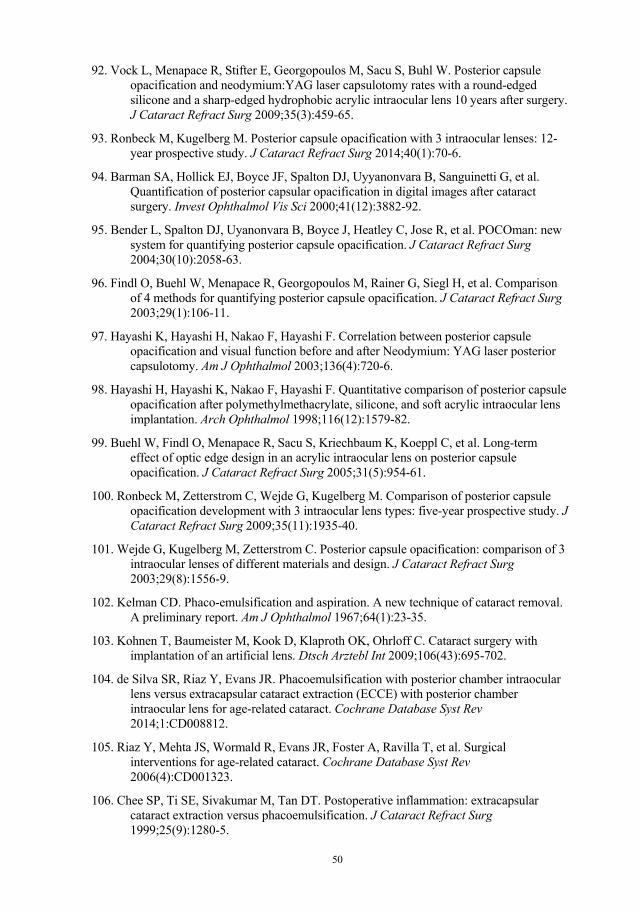
50
92. Vock L, Menapace R, Stifter E, Georgopoulos M, Sacu S, Buhl W. Posterior capsule opacification and neodymium:YAG laser capsulotomy rates with a round-edged silicone and a sharp-edged hydrophobic acrylic intraocular lens 10 years after surgery. J Cataract Refract Surg 2009;35(3):459-65.
93. Ronbeck M, Kugelberg M. Posterior capsule opacification with 3 intraocular lenses: 12-year prospective study. J Cataract Refract Surg 2014;40(1):70-6.
94. Barman SA, Hollick EJ, Boyce JF, Spalton DJ, Uyyanonvara B, Sanguinetti G, et al. Quantification of posterior capsular opacification in digital images after cataract surgery. Invest Ophthalmol Vis Sci 2000;41(12):3882-92.
95. Bender L, Spalton DJ, Uyanonvara B, Boyce J, Heatley C, Jose R, et al. POCOman: new system for quantifying posterior capsule opacification. J Cataract Refract Surg 2004;30(10):2058-63.
96. Findl O, Buehl W, Menapace R, Georgopoulos M, Rainer G, Siegl H, et al. Comparison of 4 methods for quantifying posterior capsule opacification. J Cataract Refract Surg 2003;29(1):106-11.
97. Hayashi K, Hayashi H, Nakao F, Hayashi F. Correlation between posterior capsule opacification and visual function before and after Neodymium: YAG laser posterior capsulotomy. Am J Ophthalmol 2003;136(4):720-6.
98. Hayashi H, Hayashi K, Nakao F, Hayashi F. Quantitative comparison of posterior capsule opacification after polymethylmethacrylate, silicone, and soft acrylic intraocular lens implantation. Arch Ophthalmol 1998;116(12):1579-82.
99. Buehl W, Findl O, Menapace R, Sacu S, Kriechbaum K, Koeppl C, et al. Long-term effect of optic edge design in an acrylic intraocular lens on posterior capsule opacification. J Cataract Refract Surg 2005;31(5):954-61.
100. Ronbeck M, Zetterstrom C, Wejde G, Kugelberg M. Comparison of posterior capsule opacification development with 3 intraocular lens types: five-year prospective study. J Cataract Refract Surg 2009;35(11):1935-40.
101. Wejde G, Kugelberg M, Zetterstrom C. Posterior capsule opacification: comparison of 3 intraocular lenses of different materials and design. J Cataract Refract Surg 2003;29(8):1556-9.
102. Kelman CD. Phaco-emulsification and aspiration. A new technique of cataract removal. A preliminary report. Am J Ophthalmol 1967;64(1):23-35.
103. Kohnen T, Baumeister M, Kook D, Klaproth OK, Ohrloff C. Cataract surgery with implantation of an artificial lens. Dtsch Arztebl Int 2009;106(43):695-702.
104. de Silva SR, Riaz Y, Evans JR. Phacoemulsification with posterior chamber intraocular lens versus extracapsular cataract extraction (ECCE) with posterior chamber intraocular lens for age-related cataract. Cochrane Database Syst Rev 2014;1:CD008812.
105. Riaz Y, Mehta JS, Wormald R, Evans JR, Foster A, Ravilla T, et al. Surgical interventions for age-related cataract. Cochrane Database Syst Rev 2006(4):CD001323.
106. Chee SP, Ti SE, Sivakumar M, Tan DT. Postoperative inflammation: extracapsular cataract extraction versus phacoemulsification. J Cataract Refract Surg 1999;25(9):1280-5.
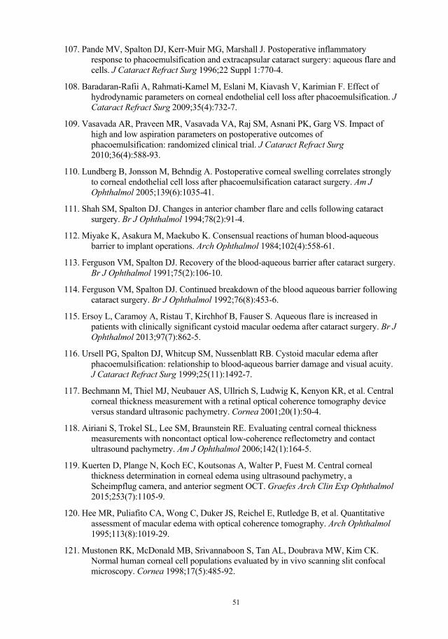
51
107. Pande MV, Spalton DJ, Kerr-Muir MG, Marshall J. Postoperative inflammatory response to phacoemulsification and extracapsular cataract surgery: aqueous flare and cells. J Cataract Refract Surg 1996;22 Suppl 1:770-4.
108. Baradaran-Rafii A, Rahmati-Kamel M, Eslani M, Kiavash V, Karimian F. Effect of hydrodynamic parameters on corneal endothelial cell loss after phacoemulsification. J Cataract Refract Surg 2009;35(4):732-7.
109. Vasavada AR, Praveen MR, Vasavada VA, Raj SM, Asnani PK, Garg VS. Impact of high and low aspiration parameters on postoperative outcomes of phacoemulsification: randomized clinical trial. J Cataract Refract Surg 2010;36(4):588-93.
110. Lundberg B, Jonsson M, Behndig A. Postoperative corneal swelling correlates strongly to corneal endothelial cell loss after phacoemulsification cataract surgery. Am J Ophthalmol 2005;139(6):1035-41.
111. Shah SM, Spalton DJ. Changes in anterior chamber flare and cells following cataract surgery. Br J Ophthalmol 1994;78(2):91-4.
112. Miyake K, Asakura M, Maekubo K. Consensual reactions of human blood-aqueous barrier to implant operations. Arch Ophthalmol 1984;102(4):558-61.
113. Ferguson VM, Spalton DJ. Recovery of the blood-aqueous barrier after cataract surgery. Br J Ophthalmol 1991;75(2):106-10.
114. Ferguson VM, Spalton DJ. Continued breakdown of the blood aqueous barrier following cataract surgery. Br J Ophthalmol 1992;76(8):453-6.
115. Ersoy L, Caramoy A, Ristau T, Kirchhof B, Fauser S. Aqueous flare is increased in patients with clinically significant cystoid macular oedema after cataract surgery. Br J Ophthalmol 2013;97(7):862-5.
116. Ursell PG, Spalton DJ, Whitcup SM, Nussenblatt RB. Cystoid macular edema after phacoemulsification: relationship to blood-aqueous barrier damage and visual acuity. J Cataract Refract Surg 1999;25(11):1492-7.
117. Bechmann M, Thiel MJ, Neubauer AS, Ullrich S, Ludwig K, Kenyon KR, et al. Central corneal thickness measurement with a retinal optical coherence tomography device versus standard ultrasonic pachymetry. Cornea 2001;20(1):50-4.
118. Airiani S, Trokel SL, Lee SM, Braunstein RE. Evaluating central corneal thickness measurements with noncontact optical low-coherence reflectometry and contact ultrasound pachymetry. Am J Ophthalmol 2006;142(1):164-5.
119. Kuerten D, Plange N, Koch EC, Koutsonas A, Walter P, Fuest M. Central corneal thickness determination in corneal edema using ultrasound pachymetry, a Scheimpflug camera, and anterior segment OCT. Graefes Arch Clin Exp Ophthalmol 2015;253(7):1105-9.
120. Hee MR, Puliafito CA, Wong C, Duker JS, Reichel E, Rutledge B, et al. Quantitative assessment of macular edema with optical coherence tomography. Arch Ophthalmol 1995;113(8):1019-29.
121. Mustonen RK, McDonald MB, Srivannaboon S, Tan AL, Doubrava MW, Kim CK. Normal human corneal cell populations evaluated by in vivo scanning slit confocal microscopy. Cornea 1998;17(5):485-92.
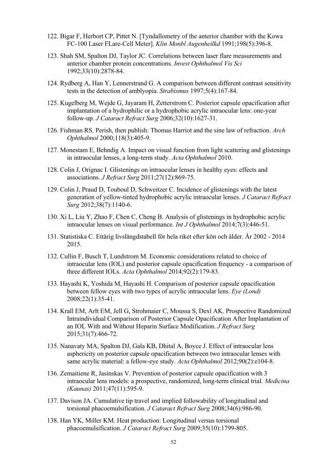
52
122. Bigar F, Herbort CP, Pittet N. [Tyndallometry of the anterior chamber with the Kowa FC-100 Laser FLare-Cell Meter]. Klin Monbl Augenheilkd 1991;198(5):396-8.
123. Shah SM, Spalton DJ, Taylor JC. Correlations between laser flare measurements and anterior chamber protein concentrations. Invest Ophthalmol Vis Sci 1992;33(10):2878-84.
124. Rydberg A, Han Y, Lennerstrand G. A comparison between different contrast sensitivity tests in the detection of amblyopia. Strabismus 1997;5(4):167-84.
125. Kugelberg M, Wejde G, Jayaram H, Zetterstrom C. Posterior capsule opacification after implantation of a hydrophilic or a hydrophobic acrylic intraocular lens: one-year follow-up. J Cataract Refract Surg 2006;32(10):1627-31.
126. Fishman RS. Perish, then publish: Thomas Harriot and the sine law of refraction. Arch Ophthalmol 2000;118(3):405-9.
127. Monestam E, Behndig A. Impact on visual function from light scattering and glistenings in intraocular lenses, a long-term study. Acta Ophthalmol 2010.
128. Colin J, Orignac I. Glistenings on intraocular lenses in healthy eyes: effects and associations. J Refract Surg 2011;27(12):869-75.
129. Colin J, Praud D, Touboul D, Schweitzer C. Incidence of glistenings with the latest generation of yellow-tinted hydrophobic acrylic intraocular lenses. J Cataract Refract Surg 2012;38(7):1140-6.
130. Xi L, Liu Y, Zhao F, Chen C, Cheng B. Analysis of glistenings in hydrophobic acrylic intraocular lenses on visual performance. Int J Ophthalmol 2014;7(3):446-51.
131. Statistiska C. Ettårig livslängdstabell för hela riket efter kön och ålder. År 2002 - 2014 2015.
132. Cullin F, Busch T, Lundstrom M. Economic considerations related to choice of intraocular lens (IOL) and posterior capsule opacification frequency - a comparison of three different IOLs. Acta Ophthalmol 2014;92(2):179-83.
133. Hayashi K, Yoshida M, Hayashi H. Comparison of posterior capsule opacification between fellow eyes with two types of acrylic intraocular lens. Eye (Lond) 2008;22(1):35-41.
134. Krall EM, Arlt EM, Jell G, Strohmaier C, Moussa S, Dexl AK. Prospective Randomized Intraindividual Comparison of Posterior Capsule Opacification After Implantation of an IOL With and Without Heparin Surface Modification. J Refract Surg 2015;31(7):466-72.
135. Nanavaty MA, Spalton DJ, Gala KB, Dhital A, Boyce J. Effect of intraocular lens asphericity on posterior capsule opacification between two intraocular lenses with same acrylic material: a fellow-eye study. Acta Ophthalmol 2012;90(2):e104-8.
136. Zemaitiene R, Jasinskas V. Prevention of posterior capsule opacification with 3 intraocular lens models: a prospective, randomized, long-term clinical trial. Medicina (Kaunas) 2011;47(11):595-9.
137. Davison JA. Cumulative tip travel and implied followability of longitudinal and torsional phacoemulsification. J Cataract Refract Surg 2008;34(6):986-90.
138. Han YK, Miller KM. Heat production: Longitudinal versus torsional phacoemulsification. J Cataract Refract Surg 2009;35(10):1799-805.
53
139. Rekas M, Montes-Mico R, Krix-Jachym K, Klus A, Stankiewicz A, Ferrer-Blasco T. Comparison of torsional and longitudinal modes using phacoemulsification parameters. J Cataract Refract Surg 2009;35(10):1719-24.
140. El-Moatassem Kotb AM, Gamil MM. Torsional mode phacoemulsification: effective, safe cataract surgery technique of the future. Middle East Afr J Ophthalmol 2010;17(1):69-73.
141. Kim DH, Wee WR, Lee JH, Kim MK. The comparison between torsional and conventional mode phacoemulsification in moderate and hard cataracts. Korean J Ophthalmol 2010;24(6):336-40.
142. Miyoshi T, Yoshida H. Emulsification action of longitudinal and torsional ultrasound tips and the effect on treatment of the nucleus during phacoemulsification. J Cataract Refract Surg 2010;36(7):1201-6.
143. Vasavada AR, Raj SM, Patel U, Vasavada V. Comparison of torsional and microburst longitudinal phacoemulsification: a prospective, randomized, masked clinical trial. Ophthalmic Surg Lasers Imaging 2010;41(1):109-14.
144. Christakis PG, Braga-Mele RM. Intraoperative performance and postoperative outcome comparison of longitudinal, torsional, and transversal phacoemulsification machines. J Cataract Refract Surg 2012;38(2):234-41.
145. Jun B, Berdahl JP, Kim T. Thermal study of longitudinal and torsional ultrasound phacoemulsification: tracking the temperature of the corneal surface, incision, and handpiece. J Cataract Refract Surg 2010;36(5):832-7.
146. Zetterstrom C, Lundström M, Serring I, Montan P, Behndig A, Kugelberg M. Svensk Kataraktkirurgi Årsrapport 2013 baserad på data från Nationella Kataraktregistret. 2014.
147. Blaylock JF, Si Z, Aitchison S, Prescott C. Visual function and change in quality of life after bilateral refractive lens exchange with the ReSTOR multifocal intraocular lens. J Refract Surg 2008;24(3):265-73.
148. Sen HN, Sarikkola AU, Uusitalo RJ, Laatikainen L. Quality of vision after AMO Array multifocal intraocular lens implantation. J Cataract Refract Surg 2004;30(12):2483-93.
149. Kaushik S, Kamlesh. A clinical evaluation of an aspheric multifocal intraocular lens and its implications for the developing world. Ophthalmic Surg Lasers 2002;33(4):298-303.
150. Steinert RF, Post CT, Jr., Brint SF, Fritch CD, Hall DL, Wilder LW, et al. A prospective, randomized, double-masked comparison of a zonal-progressive multifocal intraocular lens and a monofocal intraocular lens. Ophthalmology 1992;99(6):853-60; discussion 60-1.
151. Post CT, Jr. Comparison of depth of focus and low-contrast acuities for monofocal versus multifocal intraocular lens patients at 1 year. Ophthalmology 1992;99(11):1658-63; discussion 63-4.
152. Tassignon MJ, De Groot V, Vrensen GF. Bag-in-the-lens implantation of intraocular lenses. J Cataract Refract Surg 2002;28(7):1182-8.

54
153. Tassignon MJ, De Veuster I, Godts D, Kosec D, Van den Dooren K, Gobin L. Bag-in-the-lens intraocular lens implantation in the pediatric eye. J Cataract Refract Surg 2007;33(4):611-7.