Cataract
35
Cataract Edwin Dermody Sirait I11109069
-
Upload
yusufharkian -
Category
Documents
-
view
4 -
download
2
description
y
Transcript of Cataract

CataractEdwin Dermody Sirait
I11109069

Opacities Crystalline lens of the eye.◦ Extensive opacities of the lens will interfere light
passing through the crystalline lens cause distortion of, or considerable reduction in light rays falling on retina.
Defintion
In 2006, WHO estimated that the number of visul impaired people worldwide was 314 million. Which 45 million among them was blindness.
Age releted cataract was 48% causes of blindness in world wide. Which 58% of cases happen in Africa and South east asia region.
Epidemiology

Anatomy of the lens

Genetics Nutrition, health, diabetics Antioxidants Sunlight and irradiation Age, education
Risk Factor
The transparncy of the lens is dependent on the regular organization of the lens cells and intracellular lens proteins.
Patofisiology

The lens maintains ion differentials between intra- and extracellular fluids (high potassium and low sodium internally; low potassium and high sodium externally) via the action of the sodium-potassium ATPase pump.
Pump inactivation causes increased intracellular osmolality, which with membrane leakiness results in localized water accumulation and light scatter.
Metabolic Disturbance and Osmotic Regulation Failure

Calpains are a group of intracellular cysteine proteases, which are activated by Ca2+.
Calpains can contribute to cataract in two ways. First, a lack of calpains can lead to
pathologically elevated levels of damaged proteins, reduced optical performance, and cause cataract.
Second, excessive stimulation of calpain activity by Ca2+ can also lead to unregulated proteolysis and cataract.
Calpain inhibitors could therefore be useful in the nonsurgical treatment of cataract.
Calpains

Oxidation is a key feature in the pathogenesis of most cataracts and low oxygen levels (O2) are important for maintaining a clear lens.
There is a steep oxygen gradient from the outer part of the lens to the center. Mitochondria in the lens cortex remove most of the oxygen, thus keeping nuclear O2 levels low.
However, in older people mitochondrial function diminishes and superoxide production by the mitochondria increases resulting in increased nuclear oxygen and superoxide levels.
Oxidation
Primary defenses are provided by antioxidant enzymes and antioxidants such as ascorbate, glutathione, tocopherols, and carotenoids, which maintain lens proteins in the reduced state.
Secondary defenses include proteolytic and repair processes, which degrade and eliminate damaged proteins, UV filters, and other molecules such as glutathione reductase and free radical scavenging systems.
Failure of these protective mechanisms, a shortage of antioxidants, and increased free radicals result in cell membrane and protein damage.
Defensive Mechanisms

Crystallins may have a number of functions.
Decreased crystallin levels cause proteins to precipitate, which leads to cataract formation.
Other Factors
Age Trauma Systemic disorder Ocular disease and cataract Toxic cause
Causes of cataract

Age-related diseases:a. Congenitalb. Juvenile c. Senile
Complication Diabetic Secondary
Classification

A cataract present at birth or in aged less than 1 year.
It can affect one eye, which is known as 'unilateral cataract' or both eyes, which is known as 'bilateral cataracts'.
Most children with cataract in only one eye usually have good vision in the other.
Congenital Cataract

To know the exact cause of congenital cataract it needs a prenatal history of infection (example: rubella) during the first tri-semester of pregnancy and medicine used during pregnancy.
Cataract with positive urine reduction test might be cause by galactosemia
Almost 50% of congenital cataract is sporadic and have an unknown cause
Gray or white cloudiness of the pupil (which is normally black)
Infant doesn't seem to be able to see (if cataracts are in both eyes)
"Red eye" glow of the pupil is missing in photos, or is different between the two eyes
Unusual rapid eye movements (nystagmus)
Symptoms of Congenital Cataract
Surgical may be present if the fundus reflex is not shown
Surgical acts for congenital cataract are lens incision, linear extraction, and extraction with aspiration
Treatment

A mature cataract with a poorly developed nucleus in a child or young adult, it developed by the age less than 9 years and more than 3 months. (usually it’s a continuation of congenital cataract)
A juvenile cataract cause by manifestation of systemic or metabolic disorder
Juvenile Cataract
Senile cataract is one that forms as a result of the ageing process and one most commonly found in the elderly. The word senile comes from the Latin word for "old" and has nothing to do with mental faculties or behavior.
Senile Cataract
There are four clinical stages in the senile cataract:
a. Insipient cataractb. Intumescent cataractc. Mature cataract d. Hypermature cataract
Clinical Stages
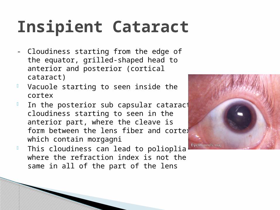
Insipient Cataract- Cloudiness starting from the edge of
the equator, grilled-shaped head to anterior and posterior (cortical cataract)
- Vacuole starting to seen inside the cortex
- In the posterior sub capsular cataract cloudiness starting to seen in the anterior part, where the cleave is form between the lens fiber and cortex which contain morgagni
- This cloudiness can lead to polioplia where the refraction index is not the same in all of the part of the lens

- Cloudiness start and acommpanying by swollen of the lens, where the degenerative lens absorbed water
- The swollen pushing the iris and make the eye chamber became shallow than normal
- Intumescent cataract usually have a rapid progress which can lead to lenticular miophy, where this condition can cause cortex hydration until the lens have a convex shape and the refraction level will increase than normal (known as miopisation)
Intumesent Cataract

- Only part of the cloudiness found in the lens
(not affected all the part),- The volume of the degenerative lens will
increase due to the osmotic pressure’s rise- In a convex lens condition, the difficulties in
pupil will cause secondary glaucoma
Immature cataract
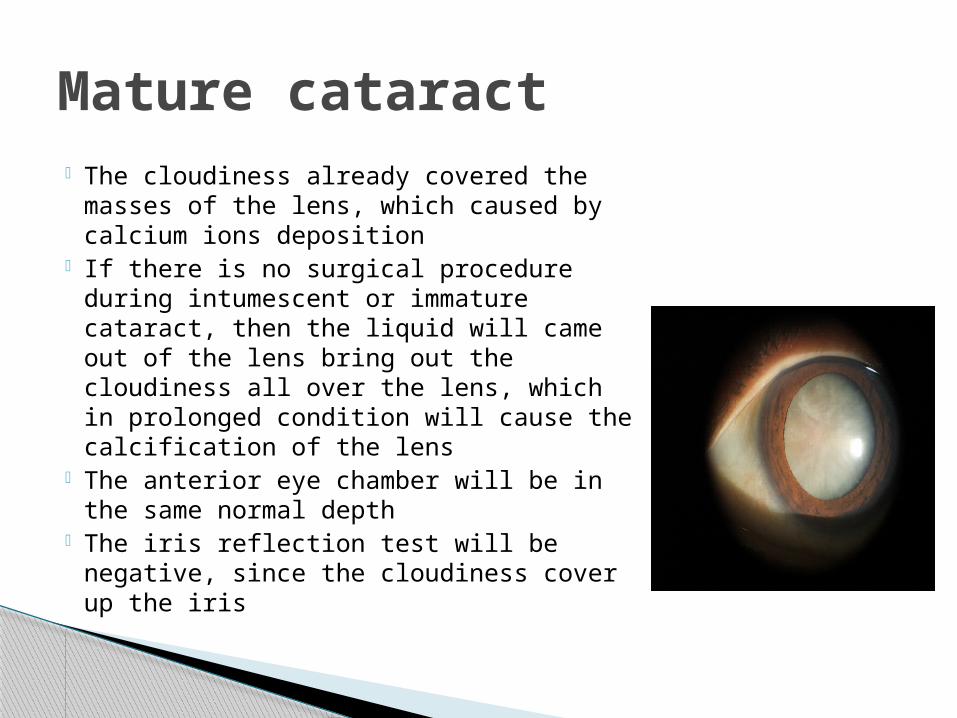
- The cloudiness already covered the masses of the lens, which caused by calcium ions deposition
- If there is no surgical procedure during intumescent or immature cataract, then the liquid will came out of the lens bring out the cloudiness all over the lens, which in prolonged condition will cause the calcification of the lens
- The anterior eye chamber will be in the same normal depth
- The iris reflection test will be negative, since the cloudiness cover up the iris
Mature cataract

- A continuation of degenerative stage of cataract , will result in hard, soft, or melting
- The degenerative lens mass will be out of the lens capsule and the lens will be shrink, yellow and dry.
- Sometime the shrink process will continue and make the zonular of zinn got loosen.
- If the cataract process continue, accompanied by thick capsule, the degenerative cortex will shown like a box of milk-shape with a drowning heavy nucleus inside the cortex lens (condition knows as Morgagni cataract)
Hypermature cataract

insipen immature Mature Hypermature
opacities Mild moderate All of the lense
massive
Lense fluid Normal increase normal Decrease
Iris Normal Pushed Normal Tremulans
Anterior chamber
Normal Pushed Normal Deep
Angle Normal Narrow Normal open
Shadow test negative positive negative pseudopos
Complicated cataract
- glaucoma - Uveitis+galucoma

Iodium liquid tear-drops, topical meds, iontophoresis,
Calcium cysteine, Immunization in purpose to fix the lens
metabolic disorder Lentokalin and cataractolysin made from
fish lens and high dose of vitamin Recent treatment is surgical procedure
(with several condition; glaukoma, uveitis, or the visual acuity is decreasing gradually and disturb the daily activities)
Treatments been used for Senile Cataract

Cataract cause by other eye’s diseases manifestation like inflammation, or the degenerative processes such as, retinal detachment, retinitis pigmentosa, glaucoma, okular ischemic, and etc
Other than eye diseases, it can caused by systemic endocrine disease
Complication cataract
The cataract in diabetic happen in 3 forms:a. Patient with heavy dehydration, acidosis,
hyperglycemia the lens will shown a line-cloudiness result from a folding lens
b. Old and uncontrollable patient with diabetic juvenile, where the cataract happen in both eyes at the same time in 48 hours (snow-flake shape or sub-capsular plate)
c. Mature diabetic patient, where the histological and biochemical views are the same withy non-diabetic cataract patient
Diabetic Cataract

Result from the fibroses made from the remain of the lens , the result will came up two days after Extra Capsular Cataract Extraction.
Secondary Cataract
Two types of surgical processes for cataract:a. Extra Capsular Cataract Extraction
Surgical procedure to the cataract lens where the action is to do the incision or tearing the lens in order to pulling out the contain of the lens
Phacoemulsification. It utilizes a handheld ultrasonic vibrator to disintegrate the hard nucleus such that the nuclear material and cortex can be aspirated through an incision of approximately 3 mm.
b. Intra Capsular Cataract Extraction Surgical procedure where the lens is being pulled
out with the capsule
Surgical Processes
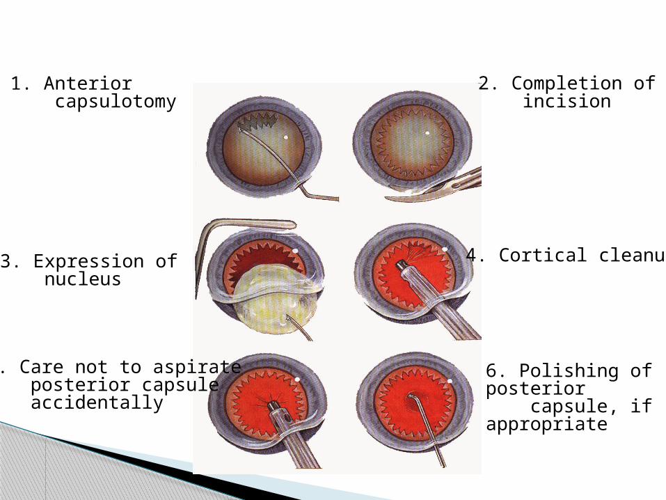
1. Anterior capsulotomy
2. Completion of incision
3. Expression of nucleus
4. Cortical cleanup
5. Care not to aspirate posterior capsule accidentally
6. Polishing of posterior capsule, if appropriate

7. Injection of viscoelastic substance
8. Grasping of IOL and coating with viscoelastic substance
10. Insertion of superior haptic
9. Insertion of inferior haptic and optic
11. Placement of haptics into capsular bag and not into ciliary sulcus
12. Dialling of IOL into horizontal position

1. Capsulorrhexis 2. Hydrodissection
3. Sculpting of nucleus 4. Cracking of nucleus
5. Emulsification of each quadrant 6. Cortical cleanup and
insertion of IOL


![Overview of Congenital, Senile and Metabolic Cataractrelated cataract [7] and metabolic cataract [8]. Congenital & Senile Cataract Cataract is a clouding of the eye’s natural lens](https://static.fdocuments.in/doc/165x107/5f361b7a353bcc123d74d127/overview-of-congenital-senile-and-metabolic-cataract-related-cataract-7-and-metabolic.jpg)