Catalytic Significance of the Specificity of Divalent Cations as K S * and k cat * Cofactors for...
12
Catalytic Significance of the Specificity of Divalent Cations as K S * and k cat * Cofactors for Secreted Phospholipase A 2 ² Bao-Zhu Yu, ‡ Joseph Rogers, ‡ Gordon R. Nicol, ‡ Klaus H. Theopold, ‡ K. Seshadri, § S. Vishweshwara, § and Mahendra Kumar Jain* ,‡ Department of Chemistry and Biochemistry, UniVersity of Delaware, Newark, Delaware 19716, and Molecular Biophysics Unit, Indian Institute of Science, Bangalore 560012, India ReceiVed NoVember 21, 1997; ReVised Manuscript ReceiVed June 30, 1998 ABSTRACT: Calcium is required for the substrate binding and for the chemical step of the interfacial catalytic turnover cycle of pancreatic phospholipase A 2 (PLA2), but not for the binding of the enzyme to the interface. The role of calcium and other divalent cations (C) is analyzed for the effect on the substrate binding and k cat * for the chemical step. The cofactor role of 3d-cations(II) (C) for the hydrolysis of dimyristoylphosphatidylmethanol (DMPM) vesicles is characterized as an equilibrium dissociation constant for the interfacial binary (E*C) and ternary (E*CL) complexes of PLA2 and substrate mimics (L). Of the cations(II) that promote the binding of a mimic to the enzyme at the interface (E*), only a subgroup supports the chemical step. For example, Cd, Zn, and Cu form ternary E*CL complexes with k cat * of <1 s -1 , compared to the rate of >100 s -1 with Ca, Fe, Mn, Co, and Ni. Oxygen exchange from H 2 18 O to the products of hydrolysis of DMPM incorporates one 18 O in myristate. Incorporation of the first and second 18 O occurs during the incubation of both the products of hydrolysis in H 2 18 O with PLA2 and Ca, but not with Zn. The cation-dependent changes in the UV difference spectrum, associated with the formation of E*C and E*CL, suggest that the changes are mainly due to catalytic His-48, and possibly Tyr-52 and Tyr-73, and are different with Ca as opposed to Zn. These results and simulations suggest considerable plasticity in the calcium binding and catalytic site environment. It is proposed that the higher ground state stability of the E*CS complex with the inhibitory cations increases the effective activation energy. For the chemical step, calcium coordinated with a nucleophilic water and the ester carbonyl oxygen facilitates the near-attack geometry in the E*CaS, and the His-48‚Asp-99 pair acts as a proton acceptor. As a prelude to establishing the catalytic mechanism, factors controlling the energetically demanding transition state are also discussed. Calcium is a cofactor for the catalysis by secreted 14 kDa phospholipase A 2 (PLA2) 1 (1-3). As a part of the highly conserved active site (Figure 1A), calcium is present in a pentagonal bipyramidal first coordination shell (Figure 1B) formed by seven oxygen ligands (4-9). In pancreatic PLA2, two equatorial bidentate ligands are from the carboxylate of Asp-49, and three other ligands are from the backbone carbonyl oxygen of highly conserved Tyr-28 (axial or apical), Gly-30 (equatorial), and Gly-32 (equatorial) residues. The five ligands provided by the protein are a part of the helix- loop-helix (EF hand) motif in which calcium is exposed to the active site pocket from one side. In the free enzyme, two other ligands are water molecules: W12 in the apical position and W5 in the equatorial position. In addition, there are several other water molecules in the “second shell”; W6 connects W5 to δNH of the His-48‚Asp-99 catalytic diad, and W7 is also within 4 Å of W12, W6, and the diad. In cocrystals of PLA2 with substrate mimics, the pentagonal bipyramidal geometry is retained, yet W5 is replaced by the oxygen of the sn-2-carbonyl or an equivalent substituent, and W12 is replaced by the pro-S oxygen of the sn-3-phosphate in the mimic (10, 11) as also suggested by studies with chiral thiophosphate analogues of the substrate (12). On the other hand, in the complex of PLA2 with MJ33, a sn-2-tetrahedral mimic without a sn-3-phosphoester group of the substrate, calcium is six-coordinated with pentagonal monopyramidal geometry (9). The sn-3-phosphate of substrate is not ² This work was supported by U.S. Public Health Service Grant GM29703. The hospitality of the Nehru Center during the visits of M.K.J. to Bangalore is gratefully acknowledged. ‡ University of Delaware. § Indian Institute of Science. Current address: INBRI, Bangalore, India. 1 Abbreviations: AM3, 6,9,12-linolenoylamide; BAPTA, 1,2-bis- (O-aminophenoxy)ethane-N,N,N′,N′-tetraacetic acid; DC7PC, 1,2-di- heptanoyl-sn-glycero-3-phosphocholine; DC8PX, 1,2-dioctanoyl-sn- glycero-3-X (where As is arsonate, PA phosphatidic acid, PM phosphomethanol, and PhM phosphonomethanol); deoxy-LPC, 1-hexa- decylpropanediol-3-phosphocholine; dithio-DMPM, 1,2-dimyristoyl- 1,2-dithio-sn-glycero-3-phosphomethanol; DMPC, 1,2-dimyristoyl-sn- glycero-3-phosphocholine; DMPM, 1,2-dimyristoyl-sn-glycero-3- phosphomethanol; DTPC, 1,2-ditetradecyl-sn-glycero-3-phosphocholine; DTPM, 1,2-ditetradecyl-sn-glycero-3-phosphomethanol; LPM, 1-myris- toyl-sn-glycero-3-phosphomethanol; MA, myristate; MG14, 1-octyl- 2-(phosphonoheptyl)-sn-glycero-3-phosphoethanolamine; MJ33, 1-hexa- decyl-3-(trifluoroethyl)-rac-glycero-2-phosphomethanol; PLA2, phospho- lipase A 2 from pig pancreas unless indicated otherwise; Quin-2, 2-[[2,2- bis(carboxymethyl)amino-5-methylphenoxyl]methyl]-6-methoxy-8,8- bis(carboxymethyl)aminoquinoline; RM2, 1-thiooctadecyl-2-acetamido- sn-glycero-3-phosphocholine; T*, tetrahedral-like transition state or intermediate. All kinetic and equilibrium parameters are defined in Scheme 1. 12576 Biochemistry 1998, 37, 12576-12587 S0006-2960(97)02860-2 CCC: $15.00 © 1998 American Chemical Society Published on Web 08/15/1998
-
Upload
mahendra-kumar -
Category
Documents
-
view
212 -
download
0
Transcript of Catalytic Significance of the Specificity of Divalent Cations as K S * and k cat * Cofactors for...

Catalytic Significance of the Specificity of Divalent Cations asKS* and kcat*Cofactors for Secreted Phospholipase A2
†
Bao-Zhu Yu,‡ Joseph Rogers,‡ Gordon R. Nicol,‡ Klaus H. Theopold,‡ K. Seshadri,§ S. Vishweshwara,§ andMahendra Kumar Jain*,‡
Department of Chemistry and Biochemistry, UniVersity of Delaware, Newark, Delaware 19716, and Molecular Biophysics Unit,Indian Institute of Science, Bangalore 560012, India
ReceiVed NoVember 21, 1997; ReVised Manuscript ReceiVed June 30, 1998
ABSTRACT: Calcium is required for the substrate binding and for the chemical step of the interfacial catalyticturnover cycle of pancreatic phospholipase A2 (PLA2), but not for the binding of the enzyme to theinterface. The role of calcium and other divalent cations (C) is analyzed for the effect on the substratebinding andkcat* for the chemical step. The cofactor role of 3d-cations(II) (C) for the hydrolysis ofdimyristoylphosphatidylmethanol (DMPM) vesicles is characterized as an equilibrium dissociation constantfor the interfacial binary (E*C) and ternary (E*CL) complexes of PLA2 and substrate mimics (L). Ofthe cations(II) that promote the binding of a mimic to the enzyme at the interface (E*), only a subgroupsupports the chemical step. For example, Cd, Zn, and Cu form ternary E*CL complexes withkcat* of <1s-1, compared to the rate of>100 s-1 with Ca, Fe, Mn, Co, and Ni. Oxygen exchange from H2
18O to theproducts of hydrolysis of DMPM incorporates one18O in myristate. Incorporation of the first and second18O occurs during the incubation of both the products of hydrolysis in H2
18O with PLA2 and Ca, but notwith Zn. The cation-dependent changes in the UV difference spectrum, associated with the formation ofE*C and E*CL, suggest that the changes are mainly due to catalytic His-48, and possibly Tyr-52 andTyr-73, and are different with Ca as opposed to Zn. These results and simulations suggest considerableplasticity in the calcium binding and catalytic site environment. It is proposed that the higher groundstate stability of the E*CS complex with the inhibitory cations increases the effective activation energy.For the chemical step, calcium coordinated with a nucleophilic water and the ester carbonyl oxygenfacilitates the near-attack geometry in the E*CaS, and the His-48‚Asp-99 pair acts as a proton acceptor.As a prelude to establishing the catalytic mechanism, factors controlling the energetically demandingtransition state are also discussed.
Calcium is a cofactor for the catalysis by secreted 14 kDaphospholipase A2 (PLA2)1 (1-3). As a part of the highlyconserved active site (Figure 1A), calcium is present in a
pentagonal bipyramidal first coordination shell (Figure 1B)formed by seven oxygen ligands (4-9). In pancreatic PLA2,two equatorial bidentate ligands are from the carboxylate ofAsp-49, and three other ligands are from the backbonecarbonyl oxygen of highly conserved Tyr-28 (axial or apical),Gly-30 (equatorial), and Gly-32 (equatorial) residues. Thefive ligands provided by the protein are a part of the helix-loop-helix (EF hand) motif in which calcium is exposed tothe active site pocket from one side. In the free enzyme,two other ligands are water molecules: W12 in the apicalposition and W5 in the equatorial position. In addition, thereare several other water molecules in the “second shell”; W6connects W5 toδNH of the His-48‚Asp-99 catalytic diad,and W7 is also within 4 Å of W12, W6, and the diad. Incocrystals of PLA2 with substrate mimics, the pentagonalbipyramidal geometry is retained, yet W5 is replaced by theoxygen of thesn-2-carbonyl or an equivalent substituent, andW12 is replaced by thepro-Soxygen of thesn-3-phosphatein the mimic (10, 11) as also suggested by studies with chiralthiophosphate analogues of the substrate (12). On the otherhand, in the complex of PLA2 with MJ33, asn-2-tetrahedralmimic without asn-3-phosphoester group of the substrate,calcium is six-coordinated with pentagonal monopyramidalgeometry (9). The sn-3-phosphate of substrate is not
† This work was supported by U.S. Public Health Service GrantGM29703. The hospitality of the Nehru Center during the visits ofM.K.J. to Bangalore is gratefully acknowledged.
‡ University of Delaware.§ Indian Institute of Science. Current address: INBRI, Bangalore,
India.1 Abbreviations: AM3, 6,9,12-linolenoylamide; BAPTA, 1,2-bis-
(O-aminophenoxy)ethane-N,N,N′,N′-tetraacetic acid; DC7PC, 1,2-di-heptanoyl-sn-glycero-3-phosphocholine; DC8PX, 1,2-dioctanoyl-sn-glycero-3-X (where As is arsonate, PA phosphatidic acid, PMphosphomethanol, and PhM phosphonomethanol); deoxy-LPC, 1-hexa-decylpropanediol-3-phosphocholine; dithio-DMPM, 1,2-dimyristoyl-1,2-dithio-sn-glycero-3-phosphomethanol; DMPC, 1,2-dimyristoyl-sn-glycero-3-phosphocholine; DMPM, 1,2-dimyristoyl-sn-glycero-3-phosphomethanol; DTPC, 1,2-ditetradecyl-sn-glycero-3-phosphocholine;DTPM, 1,2-ditetradecyl-sn-glycero-3-phosphomethanol; LPM, 1-myris-toyl-sn-glycero-3-phosphomethanol; MA, myristate; MG14, 1-octyl-2-(phosphonoheptyl)-sn-glycero-3-phosphoethanolamine; MJ33, 1-hexa-decyl-3-(trifluoroethyl)-rac-glycero-2-phosphomethanol; PLA2, phospho-lipase A2 from pig pancreas unless indicated otherwise; Quin-2, 2-[[2,2-bis(carboxymethyl)amino-5-methylphenoxyl]methyl]-6-methoxy-8,8-bis(carboxymethyl)aminoquinoline; RM2, 1-thiooctadecyl-2-acetamido-sn-glycero-3-phosphocholine; T*, tetrahedral-like transition state orintermediate. All kinetic and equilibrium parameters are defined inScheme 1.
12576 Biochemistry1998,37, 12576-12587
S0006-2960(97)02860-2 CCC: $15.00 © 1998 American Chemical SocietyPublished on Web 08/15/1998
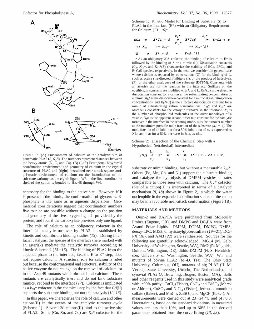
necessary for the binding to the active site. However, if itis present in the mimic, the conformation of glycero-sn-3-phosphate is the same as in aqueous dispersions. Geo-metrical considerations suggest that coordination numbersfive to nine are possible without a change on the positionand geometry of the five oxygen ligands provided by theprotein, and four if the carboxylate provides only one ligand.
The role of calcium as an obligatory cofactor in theinterfacial catalytic turnover by PLA2 is established bykinetic and equilibrium binding studies (13). During inter-facial catalysis, the species at the interface (here marked withan asterisk) mediate the catalytic turnover according tokinetic Scheme 2 (13-16). The binding of PLA2 from theaqueous phase to the interface, i.e., the E to E* step, doesnot require calcium. A structural role for calcium is ruledout because the conformational and structural stability of thenative enzyme do not change on the removal of calcium, orin the Asp-49 mutants which do not bind calcium. Thesemutants are catalytically inert and do not bind substratemimics, yet bind to the interface (17). Calcium is implicatedas akcat* cofactor in the chemical step by the fact that Cd(II)supports the substrate binding but not the catalysis (12, 13).
In this paper, we characterize the role of calcium and othercations(II) in the events of the catalytic turnover cycle(Scheme 1). Several 3d-cations(II) bind to the active siteof PLA2. Some (Cu, Zn, and Cd) areKS* cofactor for the
substrate or mimic binding, but without a measurablekcat*.Others (Fe, Mn, Co, and Ni) support the substrate bindingand catalyze the hydrolysis of DMPM vesicles at ratescomparable to those seen with calcium. Thekcat* cofactorrole of a cation(II) is interpreted in terms of a catalyticmechanism (8, 18) shown in Figure 2, in which the waternucleophile in the expanded coordination sphere of the cationmay be in a favorable near-attack conformation (Figure 1B).
MATERIALS AND METHODS
Quin-2 and BAPTA were purchased from MolecularProbes (Eugene, OR), and DMPC and DC8PA were fromAvanti Polar Lipids. DMPM, DTPM, DMPG, DMPS,deoxy-LPC, MJ33, dimyristoylglycerosulfate (19-21), DC8-PX (18), and AM3 (22) were synthesized. Sources for thefollowing are gratefully acknowledged: MG14 (M. Gelb,University of Washington, Seattle, WA), RM2 (R. Magolda,DuPont, Wilmington, DE), dithio-DMPM (H. S. Hendrick-son, University of Washington, Seattle, WA), WT andmutants of bovine PLA2 (M.-D. Tsai, The Ohio StateUniversity, Columbus, OH), mutants of pig PLA2 (H. M.Verheij, State University, Utrecht, The Netherlands), andsynovial PLA2 (J. Browning, Biogen, Boston, MA). Saltsand other reagents used in this study were analytical gradewith >99% purity: CaCl2 (Fisher), CoCl2 and CdSO4 (Merckor Aldrich), CuSO4 and NiCl2 (Fisher), ferrous ammoniumsulfate (Baker), and MnCl2, ZnSO4, and MgCl2 (Baker). Allmeasurements were carried out at 23-24 °C and pH 8.0.Uncertainties, based on the standard deviations, in measuredvalues are less than 10%, and up to 30% in the derivedparameters obtained from the curve fitting (13, 23).
FIGURE 1: (A) Environment of calcium at the catalytic site ofpancreatic PLA2 (3, 6, 8). The numbers represent distances betweenthe heavy atoms (N, C, and Ca). (B) (Left) Pentagonal bipyramidcoordination environment and geometry of calcium in the crystalstructure of PLA2 and (right) postulated near-attack square anti-prismatic environment of calcium on the introduction of thesubstrate carbonyl as the eighth ligand. W5 in the first coordinationshell of the cation is bonded to His-48 through W6.
Scheme 1: Kinetic Model for Binding of Substrate (S) toPLA2 in the Interface (E*) with an Obligatory Requirementfor Calcium (13-16)a
a As an obligatoryKS* cofactor, the binding of calcium to E* isfollowed by the binding of S or a mimic (L). Dissociation constantsKCa, KCa*, and KCa*(S) characterize the stability of ECa, E*Ca, andE*CaS species, respectively. In the text, we consider the general casewhere calcium is replaced by other cations (C) for the binding of L,such as active site-directed inhibitors (I), or the product of hydrolysis(P), or the ether analogues of the substrate (DTPM). Constants withan asterisk are for the reaction in the interface. Suffixes on theequilibrium constants are modified with C and L.KC*(L) is the effectivedissociation constant for a cation at the subsaturating concentration ofa mimic.KL* is the dissociation constant for a mimic at saturating cationconcentrations. andKL*(C) is the effective dissociation constant for amimic at subsaturating cation concentrations.KM* and kcat* areMichaelis constants for the catalytic turnover in the interface.NS isthe number of phospholipid molecules in the outer monolayer of avesicle.NSki is the apparent second-order rate constant for the catalyticturnover in the interface in the scooting mode.Vo is the turnover numberat the maximum possible mole fraction of the substrate (XS ) 1). Themole fraction of an inhibitor for a 50% inhibition ofVo is expressed asXI50 and that for a 50% decrease inNSki as nI50.
Scheme 2: Dissection of the Chemical Step with aHypothetical (tetrahedral) Intermediate
Cofactor for Phospholipase A2 Biochemistry, Vol. 37, No. 36, 199812577

Kinetic and Equilibrium Parameters. All equilibrium andkinetic constants, defined in Scheme 1, were determined asdescribed previously (13-16, 23). Detailed studies werecarried out with pig pancreatic PLA2, and PLA2 from othersources and site-directed mutants were used for specificpurposes. The reaction progress for the hydrolysis of smallsonicated vesicles of DMPM was fitted to the integratedinterfacial Michaelis-Menten equation to obtain the apparentsecond-order rate constant,NSki (14). The affinity fordivalent cations atXS ) 1, KC*(S), was obtained from thedependence ofVo on the divalent ion concentration (13). Theequilibrium dissociation constantsKC, KC*, and KL* weredetermined by the protection method (13, 23, 24). InterfacialKM* values were obtained from a combination of suchparameters, including XI50 for competitive inhibitors (13, 14,18). Inhibition of the zero-order initial rate of hydrolysis ofDMPM by Cd(II), Cu(II), and Zn(II) is expressed as IC50,the concentration required for a 50% decrease inVo at a givencalcium concentration.
Kinetic Measurements. Reaction progress was recordedby pH-stat (Radiometer) titration under a stream of nitrogenat pH 8.0 and 23°C in a 4 mLreaction mixture containing1 mM NaCl. Before the addition of divalent cation solutions,all salt solutions and buffers were filtered through a columnof Chelex (Sigma) to remove trace amounts of multivalentions. The background concentration of calcium was esti-mated to be less than 5µM on the basis of the observed rateof hydrolysis of DMPM in the absence of added Ca(II) orby titration with Quin-2 (described later). Typically, lessthan 0.1 mL of stock solution of divalent cations was addedbefore adding the substrate, and the reaction mixture wasallowed to equilibrate for about 3 min before initiating thereaction progress by PLA2.Vo values atXS ) 1 for DMPMvesicles are most conveniently measured in the presence of
20 µg of polymyxin B (25) which promotes rapid substratereplenishment through the peptide contact (26).
Precautions Necessary for the Kinetic Measurements inthe Presence of 3d-Cations(II).Background calcium levelsof the solutions without added calcium interfere with thelower-limit estimates of rates in the presence of other cations.Titration with BAPTA or Quin-2 (27) with aKD of <50 nMfor calcium was used to estimate these low levels. The limitestimates were necessary for quantifying the cation effects.Controls showed that the probes do not interact with theenzyme. Kinetic effects of cations were characterized insolutions prepared in HPLC grade water. Background rates,which could be totally eliminated by the addition of BAPTA,showed that with such precautions the background calciumconcentration in the reaction vessel was variable, largelyoriginating from the anionic lipid samples. Attempts tocompletely eliminate the background levels of calcium werenot entirely satisfactory; however, the levels were routinelyreduced to less than 5µM. Our best method for removingcalcium from the lipid solutions included the passage of amethanol solution of phospholipid through a column of CM-Sephadex cation exchanger, which was preconditioned with0.25 M HCl and then with 4 M NaCl filtered through Chelexand finally equilibrated in methanol.
Complications in the kinetic and equilibrium measure-ments, due to nonspecific interactions of cations on theanionic substrate interface, could be avoided by using 0.5mM cations. Cation concentrations were kept below 0.2 mMwith dianionic lipids to prevent precipitation and flocculation.The choice of the salt of a cation used for detailedcharacterization was governed by its stability under thereaction conditions. Around neutral pH, certain metal ionsform essentially insoluble hydroxides with solubility productswell below 10-14 M. Anomalous time-dependent behavioris expected from the hydrolysis of salts and from the rate ofprecipitation of the hydroxides and oxides. In some cases,such as Fe(II), spontaneous oxidation is also a problem. Theobserved rates were corrected only if the background driftwas less than 20% of the rate; more pronounced drifts wereconsidered intractable.
Analysis of Products of18O Exchange Reactions by FAB-MS. The product analysis of the PLA2-catalyzed incorpora-tion of 18O from H2
18O was carried out by fast atombombardment mass spectrometry (FAB-MS) on a MicromassAutospecQ apparatus (Manchester, U.K.) equipped with aCs ion gun. Typically, a sample of lipid dispersion, withother components (Table 7) in buffer with 80 at. %18O inwater, was applied at appropriate time intervals to the FABprobe containing glycerol acidified with HCl as a matrix (28).The spectra were obtained in the negative ion mode. TheCs ion voltage was set at 20 kV. The mass range was from100 to 900 Da.
UV Difference Spectra. Spectra in the 210-360 nm rangewere recorded on a HP8452 diode array spectrophotometer(Hewlett-Packard) in 10 mM Tris at pH 8.0 and 24°C withabout 10µM PLA2 in the presence of other additives asindicated in the figure legends. Corrections for dilution,calculation of the difference spectra, and turbidity correctionby extrapolation from the baseline in the 325-360 nm regionby a monotonic logarithmic function were carried out withthe software package provided with the instrument (32, 33).Thus, spectral signatures, characterized as the difference
FIGURE 2: Proposed sequence of events during the chemical stepof the catalytic cycle of PLA2. It emphasizes a role for the cationcofactor. Several key features of this model are developed in thetext. Initial binding of the substrate carbonyl occurs with anexpansion of the Ca coordination shell (first step) where W5 couldbecome the nucleophile. Conversion of the substrate tetrahedralintermediate to the one with the product (ES and EP in Scheme 2)is initiated as the oxygen of thesn-3-phosphate displaces W12.The rate-limiting transition state is postulated in the second step.
12578 Biochemistry, Vol. 37, No. 36, 1998 Yu et al.

spectra in the linear concentration-dependent range, ratherthan the absolute intensities, are used for comparisons. Dueto potential problems from excess turbidity,>0.3 OD at 280nm, results in the presence of mimics are often not atsaturating levels of the neutral diluent. However, the metalion and inhibitor concentration dependence of the spectralchanges is consistent with a two-state equilibrium.
RESULTS
3d-Cations(II) Differ as KS* and kcat* Cofactors for theHydrolysis of DMPM by PLA2. The kinetic effects of several3d-cations(II) are analyzed in terms of the primary constantswith established relationships, eqs 1-3 (13).
For cations that support hydrolysis, the values for theapparent kinetic dissociation constant for the cation,KC*-(DMPM) in column 7 of Table 1, were obtained from thehyperbolic cation concentration dependence of the initial rateof hydrolysis of DMPM atXS ) 1 (13). For example, atsaturating calcium, the initial rate,Vo, is 300 s-1, andKCa*-(DMPM) equals 100µM. The rate of hydrolysis in thepresence of Ni(II) and Co(II) is also significant. Thehyperbolic concentration dependence gives aVo of 130 s-1
with a KCo*(DMPM) of 140 µM and aVo of 150 s-1 with aKNi*(DMPM) of 610 µM (Table 1, column 4). These arelimit estimates, because Ni(II) at higher concentrations (>300µM) produced a modest background drift. Although Fe(II)and Mn(II) also support hydrolysis of DMPM vesicles,detailed analysis could not be carried out at pH 8 due to pHdrifts associated with their oxidation, hydrolysis, and pre-cipitation as hydroxides. As a control, the rate of hydrolysisof DMPM by PLA2 is<0.1 s-1 in the presence of chelating
agents such as EGTA or BAPTA. In the absence of addedcalcium or a chelating agent, the observed rate was typically5 s-1, corresponding to about a 1µM background level ofcalcium in the reaction mixture. Thus, the effective workingrange for monitoring the catalytic role of added cations thatsupport catalysis is for rates above 5 s-1.
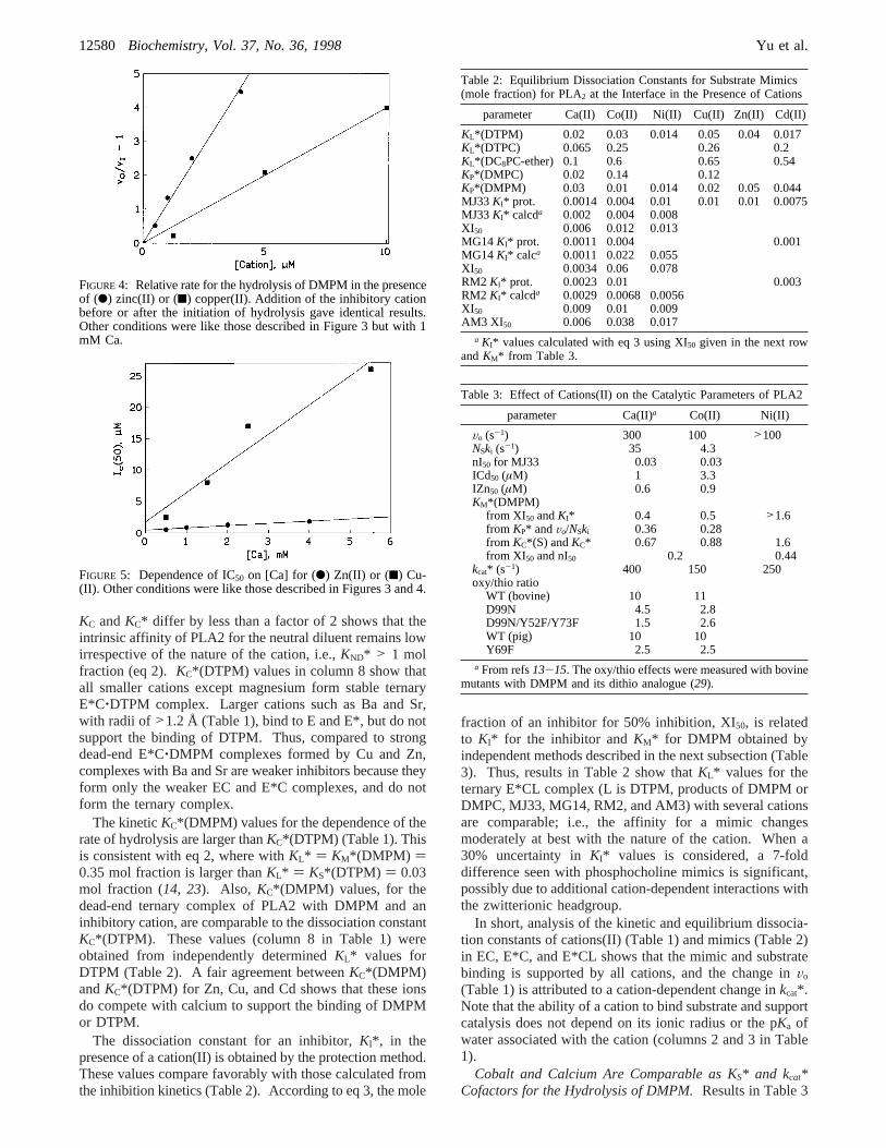
Not all cations(II) that bind to PLA2 support catalysis.Our upper limit estimate is 0.1 s-1 for the rates in thepresence of Cd, Cu, or Zn in the absence of added calcium.In such cases,KC*(DMPM) was obtained from competitiveinhibition studies (eq 1) and analyzed in terms independentlyobtained equilibrium constants (eqs 2 and 3). The cationconcentration for 50% inhibition of the calcium-mediatedhydrolysis of DMPM (IC50, column 9 in Table 1) is obtainedas 1/slope from the plot in Figure 4. According to eq 1,IC50 is related toKC*(DMPM) for the dead-end E*C(DMPM)complex. By this criterion, several 3d-cations(II) (Table 1)and 4d-cations(III) (results not shown) compete with calciumfor the binding to PLA2, and are potent inhibitors. As shownin Figure 5, IC50 for Zn(II) or Cu(II) increases with thecalcium concentration. According to eq 1,KC*(DMPM) isthey-intercept in this plot. Since the uncertainty in the valueof the y-intercept is large,KC*(DMPM) values in Table 1(column 7) are calculated from the slope and the kineticallydeterminedKCa*(DMPM) of 0.1 mM (13).
E, E*, and E*L (Table 1) forms have a higher affinity forthe inhibitory cations. Dissociation constantsKC and KC*show a 1000-fold range for the 3d-cations(II). The fact that
Table 1: Effect of Cations on the Hydrolysis of DMPM Vesicles by PLA2a
cationradius(Å)
pKa
(s-1)Vo
(s-1)KC
(µM)KC*(µM)
KC*(DMPM)(µM)
KC*(DTPM)(µM)
IC50
(µM)
Ca(II) 1.20 12.7 300 350 250 100 5Mg(II) 0.95 11.4 <1 >2000 >2000Mn(II) 1.04 10.7 >30 >500Fe(II) 1.00 10.1 >30Co(II) 0.96 9.6 130 500 300 140 14Ni(II) 0.90 9.4 150 >1000 1000 610 14Cu(II) 0.94 7.5 <1 1.5 1.5 0.045 0.12 2.5Zn(II) 0.96 9.6 <1 1 0.6 0.034 0.027 0.6Cd(II) 1.07 11.7 <1 160 65 0.5 1 1Sr(II) 1.35 100 100 680Ba(II) 1.52 300 300 850
a The radii and pKa values are for the coordination number seven (74, 75); some estimates are based on the assumption that the radius increasesabout 6% as the coordination number increases by one.KC, KC*, and KC*(DTPM) were obtained by the protection method; for the inhibitorycations,KC*(DMPM) or KC*(DTPM) was also calculated with eq 2.
IC50 ) KC*(L) +KC*(L)
KCa*(S)[Ca] (1)
KC*(L) )KC*
1 + 1KL*
(2)
Vo
VI) 1 + ( 1 + 1
KI*
1 + 1KM*
)( XI
1 - XI) (3)
FIGURE 3: Dependence of the initial rate of hydrolysis of DMPMvesicles by pig pancreatic PLA2 on the (b) cobalt(II) or (O) nickel-(II) concentration. The reaction mixture contained DMPM (700µgor 0.3 mM), polymyxin B (20µg or 3µM), and pig PLA2 (2 pmol)at pH 8.0 and 25°C in 4 mL of 1 mM Na.
Cofactor for Phospholipase A2 Biochemistry, Vol. 37, No. 36, 199812579
KC andKC* differ by less than a factor of 2 shows that theintrinsic affinity of PLA2 for the neutral diluent remains lowirrespective of the nature of the cation, i.e.,KND* > 1 molfraction (eq 2). KC*(DTPM) values in column 8 show thatall smaller cations except magnesium form stable ternaryE*C‚DTPM complex. Larger cations such as Ba and Sr,with radii of >1.2 Å (Table 1), bind to E and E*, but do notsupport the binding of DTPM. Thus, compared to strongdead-end E*C‚DMPM complexes formed by Cu and Zn,complexes with Ba and Sr are weaker inhibitors because theyform only the weaker EC and E*C complexes, and do notform the ternary complex.
The kineticKC*(DMPM) values for the dependence of therate of hydrolysis are larger thanKC*(DTPM) (Table 1). Thisis consistent with eq 2, where withKL* ) KM*(DMPM) )0.35 mol fraction is larger thanKL* ) KS*(DTPM) ) 0.03mol fraction (14, 23). Also, KC*(DMPM) values, for thedead-end ternary complex of PLA2 with DMPM and aninhibitory cation, are comparable to the dissociation constantKC*(DTPM). These values (column 8 in Table 1) wereobtained from independently determinedKL* values forDTPM (Table 2). A fair agreement betweenKC*(DMPM)andKC*(DTPM) for Zn, Cu, and Cd shows that these ionsdo compete with calcium to support the binding of DMPMor DTPM.
The dissociation constant for an inhibitor,KI*, in thepresence of a cation(II) is obtained by the protection method.These values compare favorably with those calculated fromthe inhibition kinetics (Table 2). According to eq 3, the mole
fraction of an inhibitor for 50% inhibition, XI50, is relatedto KI* for the inhibitor andKM* for DMPM obtained byindependent methods described in the next subsection (Table3). Thus, results in Table 2 show thatKL* values for theternary E*CL complex (L is DTPM, products of DMPM orDMPC, MJ33, MG14, RM2, and AM3) with several cationsare comparable; i.e., the affinity for a mimic changesmoderately at best with the nature of the cation. When a30% uncertainty inKI* values is considered, a 7-folddifference seen with phosphocholine mimics is significant,possibly due to additional cation-dependent interactions withthe zwitterionic headgroup.
In short, analysis of the kinetic and equilibrium dissocia-tion constants of cations(II) (Table 1) and mimics (Table 2)in EC, E*C, and E*CL shows that the mimic and substratebinding is supported by all cations, and the change inVo
(Table 1) is attributed to a cation-dependent change inkcat*.Note that the ability of a cation to bind substrate and supportcatalysis does not depend on its ionic radius or the pKa ofwater associated with the cation (columns 2 and 3 in Table1).
Cobalt and Calcium Are Comparable as KS* and kcat*Cofactors for the Hydrolysis of DMPM.Results in Table 3
FIGURE 4: Relative rate for the hydrolysis of DMPM in the presenceof (b) zinc(II) or (9) copper(II). Addition of the inhibitory cationbefore or after the initiation of hydrolysis gave identical results.Other conditions were like those described in Figure 3 but with 1mM Ca.
FIGURE 5: Dependence of IC50 on [Ca] for (b) Zn(II) or (9) Cu-(II). Other conditions were like those described in Figures 3 and 4.
Table 2: Equilibrium Dissociation Constants for Substrate Mimics(mole fraction) for PLA2 at the Interface in the Presence of Cations
parameter Ca(II) Co(II) Ni(II) Cu(II) Zn(II) Cd(II)
KL*(DTPM) 0.02 0.03 0.014 0.05 0.04 0.017KL*(DTPC) 0.065 0.25 0.26 0.2KL*(DC8PC-ether) 0.1 0.6 0.65 0.54KP*(DMPC) 0.02 0.14 0.12KP*(DMPM) 0.03 0.01 0.014 0.02 0.05 0.044MJ33KI* prot. 0.0014 0.004 0.01 0.01 0.01 0.0075MJ33KI* calcda 0.002 0.004 0.008XI50 0.006 0.012 0.013MG14KI* prot. 0.0011 0.004 0.001MG14KI* calca 0.0011 0.022 0.055XI50 0.0034 0.06 0.078RM2 KI* prot. 0.0023 0.01 0.003RM2 KI* calcda 0.0029 0.0068 0.0056XI50 0.009 0.01 0.009AM3 XI 50 0.006 0.038 0.017
a KI* values calculated with eq 3 using XI50 given in the next rowandKM* from Table 3.
Table 3: Effect of Cations(II) on the Catalytic Parameters of PLA2
parameter Ca(II)a Co(II) Ni(II)
Vo (s-1) 300 100 >100NSki (s-1) 35 4.3nI50 for MJ33 0.03 0.03ICd50 (µM) 1 3.3IZn50 (µM) 0.6 0.9KM*(DMPM)
from XI50 andKI* 0.4 0.5 >1.6from KP* and Vo/NSki 0.36 0.28from KC*(S) andKC* 0.67 0.88 1.6from XI50 and nI50 0.2 0.44
kcat* (s-1) 400 150 250oxy/thio ratio
WT (bovine) 10 11D99N 4.5 2.8D99N/Y52F/Y73F 1.5 2.6WT (pig) 10 10Y69F 2.5 2.5
a From refs13-15. The oxy/thio effects were measured with bovinemutants with DMPM and its dithio analogue (29).
12580 Biochemistry, Vol. 37, No. 36, 1998 Yu et al.

show that the interfacial catalytic parameters (KM*, kcat*, andNSki) for the hydrolysis of DMPM in the presence of Ca,Co, and Ni are comparable, and so are theIZn(50) andICd-(50) values for the inhibition of the hydrolysis of DMPMsupported by Ca or Co. The most significant difference isseen inNSki, which is consistent (14) with lowerKP*(DMPM)(Table 2) and lowerkcat* values (see below) in the presenceof Co(II). KM* values for DMPM, obtained by fourindependent methods, are not significantly different withcobalt or calcium (Table 3). The averageKM* was used tocalculatekcat* [ )Vo(1 + KM*)], which is moderately lowerwith cobalt (Table 3). The oxy/thio ratio of 10 with bothCa and Co suggests that the chemical step remains rate-limiting; i.e., hydrolysis of the thio analogue of DMPM byWT PLA2 proceeds at 10% of the rate seen with DMPM(29).
Co(II) Supports DMPM Hydrolysis by EVolutionarilyDiVergent PLA2. Results in Table 4 show that the cobalt-mediated rates of hydrolysis of DMPM by bovine and pigpancreatic (type I), human synovial (type II), and bee (typeIII) PLA2 are 5-100% of the rate with Ca. The Ca versusCo discrimination inVo is unchanged for Lys-53 and Lys-56 mutants with a change in the interfacial recognition region.Mutation of the active site residues in pancreatic PLA2minimizes the Ca versus Co difference inVo. Ile-9 and Leu-31, which make contact with the hydrophobic region of thesubstrate, show an effect that depends on the nature of thesubstituent. With a change in the cation binding ligand,D49E does not support hydrolysis in the presence of Ca orCo, yet it binds calcium with 10% affinity (17).
The oxy/thio (Table 3) and Ca/Co (Table 4) ratio inVo islower for D99N and Y69F. As a part of the catalytic diad,a modest effect of D99N mutation onkcat* (8) is attributedto a modest change in the stability of the intermediate in thechemical step (Figure 2). The role of Tyr-69 in catalysis(30, 76) is supported by results in Table 5, which show thatY69F iskcat*-impaired with a less than 3-fold change in theequilibrium binding parameters for the cations and mimics.This is particularly significant because the hydroxyl groupof Tyr-69 is within H-bond distance of thepro-R oxygen ofthesn-3-phosphate of thesn-2-amide analogues (11) and thesn-2-ester oxygen of MJ33 (9). These results imply a weakrole for the Tyr-69 hydroxyl in the chemical step.
The Cofactor Role of Cobalt(II) Depends on the Structureof the Substrate. Hydrolysis of phosphatidylcholines isconsiderably slower with cobalt than with calcium (Table6). This extends the earlier conclusion that the specificityfor the sn-3 substituent of the substrate resides in thechemical step of the turnover cycle (18), and it is consistentwith the observation thatKL* for phosphocholine mimics(Table 2) is 3-7-fold higher in the presence of Co(II) thanin the presence of calcium. If there is a comparable changein KM*, the rate will change at the most by a factor of 3. Amore than 100-fold Ca versus Co rate difference seen withphosphatidylcholines suggests a large cation-dependent effectof the sn-3 substituent onkcat*. In short, results in Tables3-6 show that the cation in E*CS modulates eventsassociated withkcat*.
Calcium Remains Bound during the Chemical Step.Binding of the substrate and products to the active site ofPLA2 requires calcium. Analysis of the products of the18Oexchange reaction of H218O with E*CS or E*CP providessignificant insight into the partial reactions of the chemicalstep, operationally dissected in Scheme 2 and Figure 2 byinvoking formation and decomposition of T*. A FAB mass
Table 4: Rates of Hydrolysis of DMPM (Vo in s-1) by PLA2 withCa(II) or Co(II) as a Cofactor at pH 8 and 23°C
source Vo for Ca Vo for Co
synovial 90 4.5bee 115 6bovine
WT 330 200K53M 240 200K56E 230 100K56M 280 150I9A 50 50D99N 20 17D99N/Y52F/Y73F 11 10D49E <0.1 <0.1
pigWT 300 105L31A 270 270L31R 125 110L31S 190 40Y69F 30 27Y69K 140 110
Table 5: Equilibrium Binding and Catalytic Parameters for Y69FPig PLA2
parameter Y69F WT
inactivation time for E (min) 3.5 2.8KL*(deoxy-LPC) mole fraction 0.5 >1Vo (s-1) for DMPM/Ca 30 300Vo (s-1) for DMPM/Co 27 100IZn50 (µM) 1 0.6NSki (s-1) 13 35KCa (mM) for ECa 0.7 0.35KCa* (mM) for E*Ca 0.3 0.25KCa*(S) (mM) (with DMPM) 0.17 0.10KS* for E*DTPM 0.04 0.025KS* for E*DTPC 0.8 0.067KP*(DMPM) 0.1 0.03KP*(DMPC) 0.03 0.02KI* for E*MJ33 0.0033 0.0014XI50 for MJ33 0.0064 0.006KM*(DMPM) from KI* and XI50 1 0.4KM*(DMPM) from KCa*(S) andKCa* 0.9 0.67KM*(DMPM) from Vo/NSki andKP* 0.35 0.36kcat* (s-1) 50 400
Table 6: Initial Rates (Vo in s-1) and ICu50 of Hydrolysis ofPhospholipid with Different Headgroups by PLA2a
phospholipid Vo for Ca(II) Vo for Co(II) ICu50 (µM)
dimyristoylglycero--phosphate 35 6 23-phosphomethanol 300 100 0.5-sulfate 40 9 40-phosphoserine 270 35 130-phosphoglycerol 240 138 2-phosphocholine/DOCb 40 3 1.4
diheptanoylGPC 130 1 15dioctanoylglycero-
-phosphomethanol 1000 800-phosphate 340 135-arsonate 8 1-choline 600 5a Assays were carried out in the presence of 100 mM NaCl at pH
8.0 and 24°C and 1 mM cations; the cation concentration in thepresence of phosphate and phosphoserine was 0.1 mm to avoidcomplications due to precipitation of the lipid. The inhibition resultsare for the Ca cofactor.b DOC, 7-deoxycholate.
Cofactor for Phospholipase A2 Biochemistry, Vol. 37, No. 36, 199812581

spectrum of DMPM shows the parent peak atm/z 605 andtwo major fragments:m/z227 from the myristate (MA) anda m/z 395 fragment corresponding to LPM resulting fromthe loss of ketene from DMPM (28). MA alone gives them/z 227 peak, and LPM givesm/z 395 and 227 peaks.Exchange of one or both oxygens in MA with18O givesm/z229 and 231, respectively. Besides the relative abundance(concentration), the FAB-MS peak intensities depend on ahost of factors: apparent sensitivity (LPM> MA), differinginteractions with the matrix, and differing intrinsic rates forthe parallel reactions which produce individual fragment ions.For these reasons, it is not possible to quantify the concentra-tions of the individual species from the FAB-MS resultsalone. However, the singly and doubly18O exchanged
species of MA, which are only enzymatically produced,reflect changes in the product levels with the reactionconditions. Thus, the relativem/z 229 and 231 peakintensities are proportional to the changes in concentrationsof these species with time, reaction conditions, and the18Oexchange reaction.
At short incubation times (A and B in Table 7), only oneO18 is incorporated in the initially produced myristate product(m/z227 and 229), as also reported by Ghomaschi et al. (31).The O18-labeled substrate (e.g.,m/e607) or lysophospholipid(m/z 397) is not detected under any of the conditions, withan upper limit of 1% for the O18 exchange with the substrate.These results are consistent with the mechanism in Figure2, where the two oxygens of T*, one from the carbonyl
Table 7: Peak (m/e) Intensities of Phospholipid Incubated with H218Oa
relative intensity (%)
no. lipidE
(mol %) M2+time(min) m/z 227 m/z 2 m/z 4 m/z 395 m/z 605
Aa S 0 Ca 10 67.9 0 0 28.1 100Ab S 0 Ca 2400 67.8 0 0 28.1 100Ac S 0.01 Ca 1 80.1 0 0 43.1 100Ad S 0.01 Ca 6 87.4 0 0 46.1 100Ae S 0.01 Ca 12 59 4.1 0 52.8 100Ai S 0.01 Ca 31 89.5 5.3 0 61.8 100Ak S 0.01 Ca 50 92.7 6.7 0 70.7 100Ao S 0.01 Ca 86 83 5.6 0 91.8 100Av S 0.01 Ca 185 77.7 8.2 2.0 76.9 100Aac S 0.01 Ca >3000 15.5 5.4 3.8 100 0Bf S 0.3 Ca 1.2 41.7 10.4 100 27.2Bg S 0.3 Ca 7 26.2 7.3 100 10.8Bh S 0.3 Ca 11 35.4 12.2 100 8.5Bj S 0.3 Ca 22 28.4 8.3 100 6.4Bl S 0.3 Ca 40 33.4 9.2 100 4.7Bn S 0.3 Ca 58 30.3 9.9 100 2.6Bw S 0.3 Ca 174 23.2 7.0 100 0Cm S 2 Ca 2 16.5 7.2 9.2 100 0Cr S 2 Ca 40 16.6 6.5 12.1 100 0Cx S 2 Ca 135 22.8 6.4 13.4 100 0Dp S 2 bkg 1 28.1 15.3 100 12.5Dt S 2 bkg 20 14.2 4.5 8.0 100 0Dy S 2 bkg 110 16.0 8.2 13.7 100 0Eq S 2 Zn 0.1 100 7.5 0 65.1 100Es S 2 Zn 12 32.6 12.3 3.3 100 24.5Eu S 2 Zn 24 22.5 13 4.1 100 6.2Ez S 2 Zn 115 19.5 11.6 4.1 100 0Eaf S 2 Zn 180 17.5 8.9 4.3 100 0Faa MA 0 Ca 2 100 0 0 0Faw MA 0 Ca 1150 100 0 0 0Gab MA 2 Ca 2 100 0 0 0Gav MA 2 Ca 1150 100 0 0 0Hae MA/LPM 0 Ca 0 22.3 0 100 0Hag MA/LPM 0 Ca 3000 23.4 0 100 0Has MA/LPM 2 Ca 3000 42.5 80.4 100Hax MA/LPM 2 bkg 3000 54.7 72.8 100Hay MA/LPM 2 Zn 3000 100 80.7 51.6Jah S 4 10Zn 0 48.8 13.6 100 76.7Jai S 4 10Zn 8 27.3 11.1 100 18.1Jaj S 4 10Zn 10 17.6 15.6 100 12.0Jak S 4 10Zn 12 24.7 12.9 100 4.2Jal S 4 10Zn 16 21.7 13.1 100 3.3Kam S 0.4 10Zn 0 60.8 5.5 57.9 100Kan S 0.4 10Zn 2 79.4 11.6 90.5 100Kao S 0.4 10Zn 5 70 15.1 100 79.8Kap S 0.4 10Zn 7 53.3 14.2 100 63.3Kaq S 0.4 10Zn 13 34.3 7.1 100 51.4Kar S 0.4 10Zn 17 42.1 10.6 100 33.4Kat S 0.4 10Zn 26 23 8.5 100 13.4Kau S 0.4 10Zn 66 18.0 7.8 100 5.7Kay S 0.4 10Zn 1100 17.7 9.1 2.2 100 0a bkg, background level. [Ca]) 0.5 mM. [Zn] ) 0.02 or 0.2 mM.
12582 Biochemistry, Vol. 37, No. 36, 1998 Yu et al.

oxygen and the other from the nucleophile, cannot changetheir positions before leaving the enzyme in the forward orthe reverse direction.
At longer periods of incubation of E*Ca‚DMPM (Av vsAac), the m/e 229 intensity begins to decrease with theappearance ofm/z 231 due to the exchange of the second18O in myristate. Them/z 231 peak is also seen if thereaction is carried out with E*CaP (Has and Hax in Table7). Controls show that18O exchange in MA is seen only ifboth the products, as well as PLA2 and calcium, are presentin the reaction mixture. The rates of appearance anddisappearance ofm/z229 show a dependence on the amountof enzyme and calcium. These results show that E*CaPexchanges18O in myristate through the formation of T*(Figure 2).
The rate of hydrolysis of DMPM monitored as the changein m/z 605, 395, and 227 is considerably slower in thepresence of zinc (E and J in Table 7), even slower comparedto that with background calcium (D). Since the rate ofhydrolysis in the presence of the background levels ofcalcium is significant on the time scale of these measure-ments, it is not possible to unequivocally ascertain whetherzinc promotes hydrolysis. On the basis of the observationthat in the presence of Zn the18O exchange is considerablybelow the background calcium level (Hey, J, and K), wesuspect that E*ZnS or E*ZnP do not participate in theformation of any of the reaction intermediates required forthe 18O exchange from water.
UV Difference Spectral Signatures Induced by MimicsDepend on the Nature of the Cation(II). Evidence that theground state of E*CS complexes is different for calcium andinhibitory Zn is provided by the UV absorbance of PLA2.The spectral signatures, determined as the difference spectra,result from a shift in the spectral mass and distribution; i.e.,the peak positions in the difference spectra do not necessarilycorrespond to a change in the peak position in the absorptionspectrum from a chromophore. Absorbance changes in the210-310 nm range are induced upon the binding of cationsand mimics to PLA2 (Figures 6-9). The changes in the280-300 nm region are attributed to a change in the onlytryptophan (Trp-3) present at the interfacial recognition faceof PLA2. These changes are small with a molar extinctioncoefficient of <1000 M-1 cm-1, and they are largely
associated with the changes in the N terminus segment dueto the binding of a mimic to the active site of the enzyme atthe interface (32-34). The changes in the 220-250 nmregion are assigned mostly to His-48. The changes are notobserved with the H48A mutant, and the changes are lessprominent in the Y52F/Y73F double mutant. The spatialrelationship between these residues is shown in Figure 1A.
The spectral changes in the 220-250 nm region dependon the nature of the cation and the mimic in the active site.Changes induced by the binding of calcium to the E form isappreciably different from that with the E* form (Figure 6).Also, the difference spectra in E* induced by Ca, Ba, andZn are different. The calcium-induced changes in the E*form of pig (Figure 6) and the bovine PLA2 (Figure 7) arecomparable. These two enzymes differ in>20% residues;however, their architecture is virtually identical (6, 35). Alsoas compared in Figure 7, the 230 nm region is considerably
FIGURE 6: Change in the UV difference spectrum of pig pancreaticPLA2 (10µM) in the aqueous phase (E form) induced by calcium(dots). Also shown are the difference spectra for PLA2 bound to4.3 mM deoxy-LPC (E*) induced by calcium (s), barium (- -),and zinc (-‚-). Conditions were 10 mM Tris at pH 8.0 with 1mM Ca or Ba or 0.2 mM Zn.
FIGURE 7: UV difference spectra induced by 1 mM calcium addedto bovine pancreatic PLA2 WT (s), H48A (- -), and Y52F/Y73F(‚‚‚) bound to 4.3 mM deoxy-LPC. Other conditions were like thosedescribed in Figure 6.
FIGURE 8: (A) UV difference spectra induced on the binding ofMJ33 to pig pancreatic PLA2 bound to 4.3 mM deoxy-LPC in thepresence of calcium (s) and zinc (‚‚‚). (B) UV spectral changeinduced by MJ33 added to bovine PLA2 WT (s), H48A (- -),or Y52F/Y73F (‚‚‚) in the presence of calcium. Other conditionswere like those described in Figure 6.
Cofactor for Phospholipase A2 Biochemistry, Vol. 37, No. 36, 199812583

depressed in the difference spectrum from the E* form ofH48A, and less so with Y52F/Y73F.
Binding of MJ33 to form the ternary E*CI complex alsoinduces a significant change in the 210-260 nm region(Figure 8). The changes induced by the binding of MJ33 tothe E*Ca and E*Zn forms of pig PLA2 differ significantlyin the 245 nm region with a modest difference in the 290nm region (Figure 8A). Also, the binding of MJ33 to theE*Ca form of bovine WT or the H48A or Y52/73F mutantshows a major change near 230 nm, and a minor effect near245 nm (Figure 8B). Differential perturbation of His-48 andTrp-3 is also induced by four competitive inhibitors (Figure9A) and DC8PM (Figure 9B) added to the E*Ca form ofpig PLA2.
Collectively, remarkable plasticity of the active site regionof PLA2 is indicated by the kinetic and binding results. UVdifference spectra show that the binding of Ca or Zn to E,E*, or E*I forms of PLA2 induces characteristic spectralperturbations. Crystallographic evidence suggests that thebackbone conformation is not significantly altered on thebinding of the cation and the mimic; however, the headgroupof bound mimics interacts with His-48, and the chaininteracts with Trp-3 (6-11). The dependence of the spectralsignatures on the nature of the cation and mimic also suggeststhat side chains are involved in the cation and mimic binding.Among other factors, ionization, H-bonding, and tautomericstates of the imidazole ring could change with the nature ofthe cation and the hydrogen bonding tendencies of thefunctional groups on the mimic. Thus, the changes in the230 nm region are assigned to the His-48 chromophorecoupled to Tyr-52 and -73 through Asp-99 (Figure 1A). Theinhibitory cations form dead-end complexes with spectro-scopic signatures that suggest a change in residues connectingthe catalytic His-48‚Asp-99 pair to Tyr-52 and -73, and theN-terminal helix. How these interactions control the stabilityof the Michaelis complex and its partitioning for the chemicalstep remains to be determined.
DISCUSSION
Calcium regulates numerous processes in living organisms.It is found in crystal structures of several hundred proteins,where a general pattern of its coordination behavior isbeginning to emerge (36, 37). Yet a role of calcium as acofactor in the catalytic turnover cycle is apparently estab-
lished, to the best of our knowledge, only forR-amylase (38),staphylococcal nuclease (39, 40), and trypanosomal nucleo-side hydrolase (41). For the nuclease, a convincing case ismade for the nucleophilic catalytic role of a water moleculein the first or second coordination sphere of calcium (42),where cobalt acts as a competitive inhibitor by binding in avestibular site adjacent to the calcium site (43).
Results with PLA2 show a role for calcium in the substratebinding and the chemical step, and a remarkable plasticityof the active site environment is also indicated. Ca, Co, andNi show comparablekcat* values with anionic substrates, butnot with zwitterionicsn-3-phosphatidylcholines (Table 6).We attribute such effects to differences in the coordinationgeometry of the cation in E*CS; i.e., only the catalytic cationsfavor a near-attack conformation (Figure 1B). The signifi-cance of key results related to the chemical step is discussedbelow in the context of the mechanism in Figure 2.
Near-Attack Conformation for Esterolysis by PLA2. PLA2-catalyzed esterolysis is>1010-fold faster than uncatalyzedesterolysis (44-46). Within the paradigm of a nucleophilicattack (47, 48), formation of a near-tetrahedral transition stateor intermediate (T*) in the chemical step of PLA2 is oftenassumed (3, 4, 6, 10, 49, 50). According to the mechanismin Figure 2, during the attack of W5 on the ester carbonylcoordinated to calcium, the His-48‚Asp-99 pair accepts theproton through W6, and during the decomposition, the protonis transferred to the leaving group.
Factors favoring catalysis by enzymes involve both groundstate and transition state features, including the effective pKa
of the general acid (51, 52), the relative orientation andposition of the reactant species (53-55), and the entropiccontributions from freezing motions (56). In a remarkablesynthesis of intramolecular catalysis, Lightstone and Bruice(55) have suggested that the catalytic advantage comesmainly from the near-attack ground state conformers on theway to the enthalpy-dominated factors in the transition state.In our mechanism, the movement of proton from thenucleophilic water to His-48 and then to the leaving groupoccurs through W6 located between Ca,δNH, and thecarboxylic ester oxygens. In addition, the expanded eight-coordinate square antiprism geometry (Figure 1B) has asuitable near-attack conformation. The angle of approachof the nucleophile is within a 20° cone from the perpendicularto the ester O-CdO plane. Also, the distance between W5and the carbonyl carbon, 3.26 Å, is nearly ideal for anucleophilic attack (54, 57). This conformation may besufficiently activated, but not much strained, along thereaction coordinates with a minimum change in the position,orientation, or charge distribution around the cation.
Plasticity of the active site is indicated by the range ofglycerophospholipid headgroups that are accommodated inthe active site, and factors that stabilize the anionic characteron the ester oxygens are indicated in crystal structures ofPLA2 with substrate mimics. With MG14 or RM-2, calciumis seven-coordinate and the NH of Gly-30 is hydrogenbonded to the carbonyl oxygen of the substrate (10, 11). Inthe MJ33 complex, calcium is six-coordinate and the NH ofGly-30 is H-bonded to the other oxygen on the tetrahedralphosphate; also, the OH of Tyr-69 is a short hydrogen bond(2.8 Å) away from thesn-2-ester oxygen, where it couldstabilize a charge (9).
FIGURE 9: (A) UV spectral change induced by RM2 (s), MG14(- -), MJ33 (‚‚‚), and AM3 (-‚-) added to the E*Ca form ofpig PLA2 on deoxy-LPC. Under these conditions, about 50% ofthe enzyme would be converted to the E*I form. (B) UV spectralchange induced by addition of dioctanoylphosphatidylmethanol tothe E* form of pig PLA2 on deoxy-LPC in the presence of calcium(s) or zinc (- -). The spectra in the presence of the substratewere taken 5 s after the addition of the substrate, and a sufficientexcess is likely to be present.
12584 Biochemistry, Vol. 37, No. 36, 1998 Yu et al.

Expansion of the Cation Coordination Shell in PLA2. Wepropose that during the chemical step the coordinationgeometry of calcium(II) changes from pentagonal bipyramidto a square antiprism (Figure 1B). Calcium with eightligands is found inR-amylase (38, 60), proteinase K (52),and nucleoside hydrolase (41). A change in the coordinationnumber from seven to eight is observed upon sugar bindingto the C-type mannose binding protein (61). Such a changein the coordination number is permitted for the electrostaticcomplexes of calcium (36, 62, 63) with a small energypenalty (37), and the stability of complexes increases fromH-bonding between ligand in the same polyhedron or theneighboring groups (62, 64). Simulations (65; also see theSupporting Information) show that the coordination numberchange from seven to eight in PLA2 (Figure 1B) occurs withvery favorable hydrogen-bonding relationships, and withoutany unfavorable steric contacts or perturbation of the proteinside chains or backbone. A particularly striking feature ofthe coordination environment of the cation that emerges fromsuch docking exercises is that six-coordinate octahedral andeight-coordinate antiprismatic geometries are generated fromthe seven-coordinate pentagonal bipyramidal geometry ofcalcium in PLA2 without a shift in the position of the fiveligands provided by PLA2 (Figure 1B). A four-coordinategeometry, even if somewhat strained, preferred by Cd, Zn,and Cu, could be achieved if the carboxylate of Asp-49provides a single ligand. These readily interconvertiblegeometries are closely related, and are preferred for therespective coordination numbers (66, 67).
It is likely that 3d-cations(II) form ionic complexes withPLA2. Virtually all 3d-cations(II) with a radius of less than1.1 Å form stable EC, E*C, and E*CL complexes. Sinceonly five oxygen ligands are provided by the protein,differences in the partial desolvation tendencies of the cations(71) or the ligand substitution energies could account forsome of the differences in the dissociation constants. Theextent of ligand substitution associated with the formationof these complexes with Cu, Zn, or even Co for that matteris difficult to predict, and must await crystallographic results.
The ability to accommodate a range of geometries in theactive site is also evident from the structural diversity of theinhibitors that compete with the substrate and bind to theactive site (24, 68). Such complexes of PLA2 are formedwithout a change in the backbone structure (6, 9-11);however, changes in the His-48 chromophore (Figures 6-9)suggest significant changes in the side chains. The bindingof inhibitors to the active site of E* is driven primarily, ifnot exclusively, by headgroup interactions (15, 22, 24) afterthe partitioning of the mimic into the interface is taken intoaccount (69). This is not surprising because during theformation of the E*CaL complex at the interface the acylchains are transferred from the hydrophobic region of thebilayer to the hydrophobic environment of the active sitecavity. The importance of the headgroup interactions isemphasized further by the fact that primary acylamides, suchas AM3 (Table 2), are potent calcium-dependent active site-directed inhibitors of PLA2, whereas theirN-methyl deriva-tives are less potent by a factor of 50 (22). This is asurprising result in light of the fact that secondary acyl-amides, such as thesn-2-amidophospholipid analogues, arepotent inhibitors (70; also see earlier papers of the series).This difference is best rationalized by invoking expanded
eight-coordinate geometry (see ref74 and the SupportingInformation).
Dynamics of the Catalytic Water. With expanded coor-dination (Figure 1B), the carbonyl carbon is 3.4 Å from W5and 4.1 Å from W6. Both of these potential nucleophilesare held between Ca andδNH-H48 (Figure 1B). The angleof approach of W6 (90° from the trigonal CdO plane) tothe carbonyl carbon is somewhat more favorable comparedto 60° for W5. W6 is a better proximal proton acceptorbecause it is H-bonded to H48‚D99. Direct coordination ofW5 to the cation makes it a stronger proton donor, andadditional activation is expected because the dicationiccalcium is coordinated to ligands with only one net negativecharge. Also, H-bonding of W5 to the more basic electronpair of O1 (64, 72) places Asp-49 in an excellent positionto assist in the deprotonation of W5.
It is difficult to predict the pKa of water coordinated to ametal ion in general terms, especially if the metal is held bystructurally different ligands (73). The net charge and aproton from W5-W6 could be shared by the carbonyloxygen, ester oxygen, the imidazole ring, and possiblycarboxylate of D99. An effectively lower pKa of W5-W6is expected due to the coupling to calcium and to His-48. Itis also consistent with the observation that the pKa of His-48 decreases from 7 to 5.7 in the presence of the boundcalcium, andKCa decreases from 2.5 mM at pH 6 to 0.3 mMat pH 8 (3).
The role of W5-W6 as the catalytic water is alsosuggested by molecular dynamic simulation of the ternaryE*CaS complex of PLA2 (50). As the initial condition, onlyW6 was included as the “catalytic water” and W5 wassubstituted by the carbonyl oxygen of the substrate in thepentagonal bipyramidal geometry. During the 48 ps simula-tion, W6 occupied alternating positions occupied by W6 andW5. Toward the end of the simulation, W6 and anotherwater molecule joined in the expanded coordination shell ofcalcium, thus making up a total of nine ligands. Since W5was not modeled, this simulation is equivalent to a situationwhere two water molecules coexist in positions occupied byW5 and W6 with the carbonyl oxygen in the expandedcoordination shell. In short, not only is there ample spacefor simultaneous placement of carbonyl oxygen, W5, andW6, but they can also be suitably placed for a nucleophilicattack without a large movement and reorientation of anyof the groups involved.
Factors Controlling the Energetically Demanding Transi-tion State. A range of effects onkcat* imply a role formultiple interactions in the chemical step. Ultimately, thefollowing observations must be accommodated in anyformulation of the reaction coordinates.
(a) On the basis of the oxy/thio ratio of 10, the chemicalstep is rate-limiting (29) with kcat* equal tok2 which is 400s-1 for DMPM (14) and about 3000 s-1 for DC8PC (15).These effects onkcat* (Table 3; also see ref29) are consistentwith the participation of thesn-2-oxygen in the transitionstate.
(b) The12C/14C carbonyl carbon isotope effect is 1.01 forthe hydrolysis of DMPM by PLA2 (31). It suggests a strongcommitment for catalysis without equilibration of the isotopicsubstrate species in E*CS, which is consistent with experi-mentally observedk-1* , kcat* (14).
Cofactor for Phospholipase A2 Biochemistry, Vol. 37, No. 36, 199812585

(c) D99N PLA2 is modestly (25-fold)kcat*-impaired (8).As is the case with WT, the amide oxygen of N99 isH-bonded toε-NH of His-48. Thus, a moderate effect ofAsp to Asn substitution is expected on the tautomericdeprotonatedδN for the proton transfer.
(d) Initial incorporation of a single18O in the myristateproduct, and the lack of18O exchange in the substrate,suggest formation of a species in the chemical step wherethe carbonyl oxygen and the one from the nucleophile donot become equivalent.
(e) The headgroup specificity of the substrate, withdifferent sn-3 substituents, lies inkcat*; e.g., the substratespecificity forsn-3-phosphate, -sulfate, or -arsonate substit-uents is inkcat* (18, 30, 58, 59, 76), and not inKS* (22, 24).The charge onpro-S oxygen of the phosphate also plays arole inkcat* because the phosphomethyl ester of DMPM bindsto the active site (24) and the rate of hydrolysis is low (58).
(f) The approach of the anionicpro-S oxygen of thesn-3-phosphate to Ca is aided by the interaction of thepro-Roxygen with the hydroxyl of Tyr-69; however, Y69F ismodestlykcat*-impaired.
(g) Hydrogen bonding of thesn-2-OH of the lysophos-pholipid product is suggested by a more than 100-fold loweraffinity of PLA2 for the 2-deoxy analogue, deoxy-LPC (24).
(h) TheKM*/KI* ratio for tetrahedral mimics is about 100.TheKS*/KI* ratio is about 10 becausek2* . k-1*. It impliesthat thesn-2 tetrahedral geometry does not resemble thetransition state, yet a tetrahedral intermediate may be partof the chemical step.
In our formulation of the reaction mechanism (Figure 2),interactions of the substrate ester oxygen with OH of Tyr-69 and of NH of Gly-30 with the carbonyl oxygen (9) assistin the trigonal to tetrahedral conversion of the carbonylcarbon, and subsequent rotation around the ester C-O bondinfluences the glycerol backbone. Torsional rotation of theC-O-C2(glycerol) single bond is followed by changes thatrequire a shift in the position of atoms that result in thesubstitution of the apical water (W12) by thepro-Soxygenof the sn-3-phosphate. Besides depolarizing the anioniccharge on the oxyanion hole, anionic phosphate could alsopush W12 in the position originally occupied by W5 in thefirst coordination shell of calcium. Although the magnitudeof each of these effects is modest, the total contributionwould be significant.
ACKNOWLEDGMENT
We gratefully acknowledge numerous discussions with Dr.Jenny Glusker on the coordination environment of calciumin proteins. We express appreciation for numerous insightfulcomments from an anonymous reviewer. We dedicate thiswork to Prof. Bert Verheij, who contributed so much to thefield.
SUPPORTING INFORMATION AVAILABLE
Further discussion of the mechanism, a table showing themolecular dynamics parameters for acylamide docked inPLA2, and a figure showing the calculated distances for theenergy-minimized coordination environment of calcium inthe acitve site of PLA2 (8 pages). Ordering information isgiven on any current masthead page.
REFERENCES
1. de Haas, G). H., Bonsen, P. P. M., Pieterson, W. A., andVan Deenen, L. L. M. (1971)Biochim. Biophys. Acta 239,252-266.
2. Verheij, H. M., Volwerk, J. J., Jansen, E. H. J. M., Puijk, W.C., Dijkstra, B. W., Drenth, J., and de Haas, G. H. (1980)Biochemistry 19, 743-740.
3. Verheij, H. M., Slotboom, A. J., and de Haas, G. H. (1981)ReV. Physiol. Biochem. Pharmacol. 91, 91-203.
4. Dijkstra, B. W., Drenth, J., and Kalk, K. H. (1981)Nature289, 604-606.
5. Dijkstra, B. W., Kalk, K. H., Hol, W. G. J., and Drenth, J.(1981)J. Mol. Biol. 147, 97-123.
6. Scott, D., and Sigler, P. (1994)AdV. Protein Chem. 45, 53-88.
7. Kumar, A., Sekharudu, C., Dupureur, C. M., Zhu, H., Tsai,M. D., and Sundaralingam, M. (1994)Protein Sci. 3, 2073-2081.
8. Sekar, K., Yu, B.-Z., Rogers, J., Lutton, J., Liu, X., Chen,X., Tsai, M.-D., Jain, M. K., and Sundaralingam, M. (1997)Biochemistry 36, 3104-3114.
9. Sekar, K., Eswaramoorthy, S., Jain, M. K., and Sundaralin-gam, M. (1997)Biochemistry 36, 14186-14191.
10. Scott, D., White, S. P., Otwinowski, Z., Yuan, W., Gelb, M.H., and Sigler, P. B. (1990)Science 250, 1541-1546.
11. Thunnissen, M. M. G. M., Ab, E., Kalk, K. H., Drenth, J.,Dijkstra, B. W., Kuipers, O. P., Dijkman, R., de Haas, G. H.,and Verheij, H. M. (1990)Nature 347, 689-691.
12. Tsai, T. C., Hart, J., Jiang, R., Bruzik, K., and Tsai, M. D.(1985)Biochemistry 24, 3180-3188.
13. Yu, B.-Z., Berg, O. G., and Jain, M. K. (1993)Biochemistry32, 6485-6492.
14. Berg, O. G., Yu, B.-Z., Rogers, J., and Jain, M. K. (1991)Biochemistry 30, 7283-7297.
15. Berg, O. G., Rogers, J., Yu, B.-Z., Yao, J., Romsted, L. S.,and Jain, M. K. (1997)Biochemistry 36, 14512-14530.
16. Jain, M. K., Gelb, M. H., Rogers, J., and Berg, O. G. (1995)Methods Enzymol. 249, 567-614.
17. Li, Y., Yu, B.-Z., Zhu, H., Jain, M. K., and Tsai, M. D. (1994)Biochemistry 33, 14714-14722.
18. Rogers, J., Yu, B.-Z., Serves, S. V., Tsivgoulis, G. M.,Sotiropoulos, D. N., Ioannou, P. V., and Jain, M. K. (1996)Biochemistry 35, 9375-9384.
19. Jain, M. K., Rogers, J., Jahagirdar, D. V., Marecek, J. F.,and Ramirez, F. (1986)Biochim. Biophys. Acta 860, 435-447.
20. Jain, M. K., Maliwal, B. P., de Haas, G. H., and Slotboom,A. J. (1986)Biochim. Biophys. Acta 860, 448-461.
21. Jain, M. K., Rogers, J., Marecek, J. F., Ramirez, F., and Eibl,H. (1986)Biochim. Biophys. Acta 860, 462-474.
22. Jain, M. K., Ghomashchi, F., Yu, B. Z., Bayburt, T., Murphy,D., Houck, D., Brownell, J., Reid, J. C., Solwiej, J. E., Wong,S. M., Mocek, U., Jarrell, R., Sasser, M., and Gelb, M. H.(1992)J. Med. Chem. 35, 3584-3586.
23. Jain, M. K., Yu, B.-Z., Rogers, J., Ranadive, G. N., and Berg,O. G. (1991)Biochemistry 30, 7306-7317.
24. Jain, M. K., Tao, W., Rogers, J., Arenson, C., Eibl, H., andYu, B.-Z. (1991)Biochemistry 30, 10256-10268.
25. Jain, M. K., Rogers, J., Berg, O. G., and Gelb, M. H. (1991)Biochemistry 30, 7340-7348.
26. Cajal, Y., Rogers, J., Berg, O. G., and Jain, M. K. (1996)Biochemistry 35, 299-308.
27. . Tsien, R., and Pozzan, T. (1989)Methods Enzymol. 172,230-262.
28. Murphy, R. C., and Harrison, K. A. (1994)Mass Spectrom.ReV. 13, 57-75.
29. Jain, M. K., Rogers, J., Gelb, M. G., Tsai, M.-D., Hendrickson,E. K., and Hendrickson, S. (1992)Biochemistry 31, 7841-7847.
30. Cleland, W. W., and Kreevoy, M. M. (1994)Science 264,1887-1890.
31. Ghomashchi, F., O’Hare, T., Clary, D., and Gelb, M. H. (1991)Biochemistry 30, 7298-7305.
12586 Biochemistry, Vol. 37, No. 36, 1998 Yu et al.

32. Dupureur, C. M., Yu, B.-Z., Jain, M. K., Noel, J. P., Deng,T., Li, Y., Byeon, I. L., and Tsai, M. D. (1992)Biochemistry31, 6402-6413.
33. Dupureur, C. M., Yu, B.-Z., Mamone, J. A., Jain, M. K., andTsai, M. D. (1992)Biochemistry 31, 10576-10583.
34. Jain, M. K., and Maliwal, B. P. (1993)Biochemistry 32,11838-11846.
35. Dijkstra, B. W., Renetseder, R., Kalk, K. H., Hol, W. G. J.,and Drenth, J. (1983)J. Mol. Biol. 168, 163-179.
36. McPhalan, C. A., Strynadka, N. C. J., and James, M. N. G.(1991)AdV. Protein Chem. 42, 77-144.
37. Katz, A. K., Glusker, J. P., Beebe, S. A., and Bock, C. W.(1996)J. Am. Chem. Soc. 118, 5752-5763.
38. Boel, E., Brady, L., Brzozowski, A. M., Derewenda, Z.,Dodson, G. G., Jensen, V. J., Petersen, S. B., Swift, H., Thim,L., and Woldike, H. F. (1990)Biochemistry 29, 6244-6249.
39. Serpersu, E. H., Shortle, D., and Mildvan, A. S. (1986)Biochemistry 25, 68-77.
40. Serpersu, E. H., Shortle, D., and Mildvan, A. S. (1987)Biochemistry 26, 1289-1300.
41. Degano, M., Almo, S. C., Sacchettini, J. C., and Schramm,V. L. (1998) Biochemistry 37, 6277-6285.
42. Weber, D. J., Libson, A. M., Gittis, A. G., Lebowitz, M. S.,and Mildvan, A. S. (1994)Biochemistry 33, 8017-8028.
43. Loll, P. J., Quirk, S., Lattman, E. E., and Gravito, R. M. (1995)Biochemistry 34, 4316-4324.
44. Bruice, T. C., and Benkovic, S. J. (1966)BioorganicMechanisms, Vol. 1, W. A. Benjamin, Inc., New York.
45. Guthrie, J. P. (1983)Acc. Chem. Res. 16, 122-129.46. Guthrie, J. P., and Cullimore, P. A. (1980)Can. J. Chem. 58,
1281.47. Kirsch, J. F., and Jencks, W. P. (1964)J. Am. Chem. Soc.
86, 837-843.48. Johnson, S. L. (1967)AdV. Phys. Org. Chem. 5, 237-330.49. Waszkowycs, B., Hiller, I. H., Gensmatel, N., and Polying,
D. W. (1990)J. Chem. Soc., Perkin Trans. 2, 1259-1268.50. Jones, S. T., Ahlstram, P., Berendsen, H. J. C., and Pickersgill,
R. W. (1993)Biochim. Biophys. Acta 1162, 135-142.51. Gerlt, J. A., and Gassman, P. G. (1993)Biochemistry 32,
11943-11952.52. Bajorath, J., Hinrichs, W., and Sanger, W. (1988)Eur. J.
Biochem. 176, 441-447.53. Bruice, T. C. (1976)Annu. ReV. Biochem. 45, 331.54. Lighthouse, F. C., and Bruice, T. C. (1996)J. Am. Chem.
Soc. 118, 2595-2605.
55. Lighthouse, F. C., and Bruice, T. C. (1997)J. Am. Chem.Soc. 119, 9103-9113.
56. Page, M. I., and Jencks, W. P. (1971)Proc. Natl. Acad Sci.U.S.A. 68, 1678.
57. Burgi, H. B., and Dunitz, J. D. (1983)Acc. Chem. Res. 16,153-161.
58. Kuipers, O. P., Vincent, M., Brochon, J., Verheij, H. M., deHaas, G. H., and Gallay, J. (1991)Biochemistry 30, 8771-8785.
59. Bonsen, P. P. M., de Haas, G. H., Pieterson, W. A., and VanDeenen, L. L. M. (1972)Biochim. Biophys. Acta 270, 364-382.
60. Qian, M., Haser, R., and Payan, F. (1993)J. Mol. Biol. 231,785.
61. Weis, W. I., Drickamer, K., and Hendrickson, W. A. (1992)Nature 360, 127-134.
62. Glusker, J. P. (1991)AdV. Protein Chem. 42, 1-76.63. Villafranca, J. J., and Nowak, T. (1992)Enzymes 20, 63-94.64. Glusker, J. P. (1995)Acta Crystallogr. D51, 418-427.65. Seshadri, K., Vishveshwara, S., and Jain, M. K. (1994)Proc.
Ind. Acad. Sci. 106, 1177-1189.66. Kepert, D. L. (1978)Prog. Inorg. Chem. 24, 179-249.67. Kepert, D. L. (1979)Prog. Inorg. Chem. 25, 41-144.68. Gelb, M. H., Jain, M. K., and Berg, O. G. (1994)FASEB J.
8, 916-924.69. Jain, M. K., Yu, B.-Z., and Berg, O. G. (1993)Biochemistry
32, 11319-11329.70. de Haas, G. H., Dijkman, R., Lugtigheid, R. B., Dakker, N.,
Van den Berg, L., Egmond, M. R., and Verheij, H. M. (1993)Biochim. Biophys. Acta 1167, 281-288.
71. Frey, C. M., and Stuehr, J. (1974) inMetal Ions in BiologicalSystems(Sigel, H., Ed.) Vol. 1, p 69, Mercel Dekker, Inc.,New York.
72. Gandour, R. D. (1981)Bioorg. Chem. 10, 169-176.73. Tsubouchi, A., and Bruice, T. C. (1995)J. Am. Chem. Soc.
117, 7399-7411.74. Huheey, J. E. (1983)Inorganic Chemistry, 3rd ed., pp 73-
76, Harper & Row, New York.75. Shannon, R. D. (1976)Acta Crystallogr. A32, 751-767.76. Kuipers, P., Dekker, N., Verheij, H. M., and de Haas, G. H.
(1990)Biochemistry 29, 6094-6102.
BI9728607
Cofactor for Phospholipase A2 Biochemistry, Vol. 37, No. 36, 199812587