Catabolism of amino acids Ammonia detoxification ... · of certain peptide sequence . 6 ......
82
1 Catabolism of amino acids Ammonia detoxification Biosynthesis of non-essential amino acids Department of Biochemistry (J.D.) 2013
Transcript of Catabolism of amino acids Ammonia detoxification ... · of certain peptide sequence . 6 ......

1
Catabolism of amino acids
Ammonia detoxification
Biosynthesis of non-essential amino acids
Department of Biochemistry (J.D.) 2013

2
Anabolic and catabolic conversions of AA
AA pool
endogenous
proteins
food
proteins
FA TAG
CO2 + energy
glucose
signal molecules
(hormone, neurotransmiter)
purine / pyrimidine bases
porphyrines heme
creatine creatinine
arginine NO
and other ...
NH3 NH4+
synthesis of
non-essential AA
urea glutamine
CAC
GI tract
intracellular
degradation
nitrogen compound
compound without N

3
Amino acid pool Three sources of AA pool:
1) Proteolysis of dietary proteins (food)
2) Proteolysis of tissue proteins (physiological turnover, more in starvation)
3) Synthesis of non-essential AA (11)
Three utilizations of AA pool:
1) Synthesis of tissue and blood plasma proteins (liver)
2) Synthesis of low-molecular nitrogen compounds (with specific functions)
3) Catabolism: deamination + utilization of carbon skeleton
Three utilizations of AA carbon skeleton
1) Gluconeogenesis (in starvation, most AA are glucogenic)
2) Synthesis of FA and TAG (in AA excess)
3) Metabolic fuel = gain of energy (minor utilization)
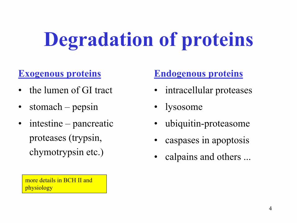
4
Degradation of proteins
Exogenous proteins
• the lumen of GI tract
• stomach – pepsin
• intestine – pancreatic
proteases (trypsin,
chymotrypsin etc.)
Endogenous proteins
• intracellular proteases
• lysosome
• ubiquitin-proteasome
• caspases in apoptosis
• calpains and others ...
more details in BCH II and
physiology
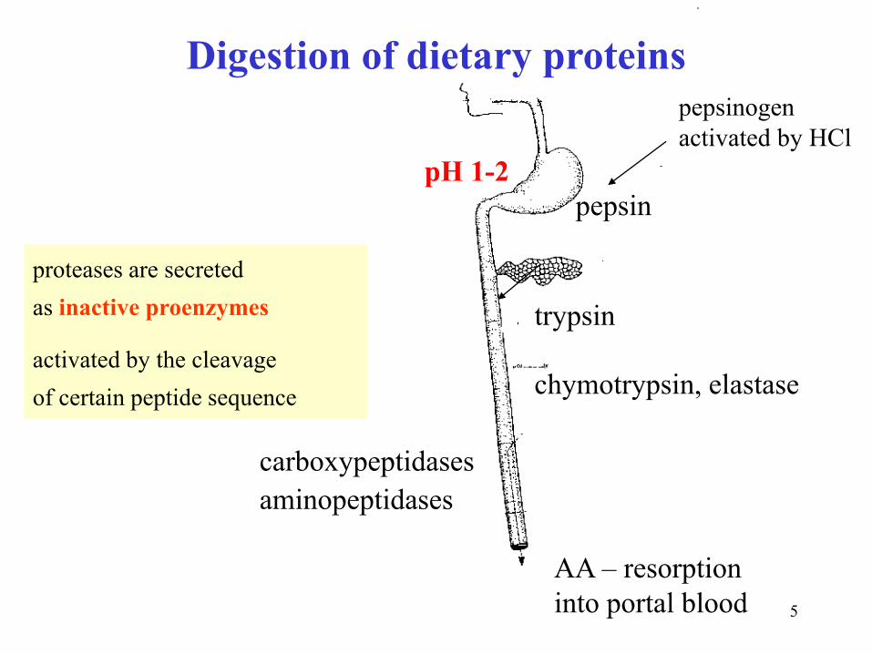
5
pepsin
trypsin
pepsinogen
activated by HCl
carboxypeptidases
aminopeptidases
AA – resorption
into portal blood
pH 1-2
chymotrypsin, elastase
Digestion of dietary proteins
proteases are secreted
as inactive proenzymes
activated by the cleavage
of certain peptide sequence

6
Enteropeptidase secreted by the mucosa of duodenum initiates
the activation of the pancreatic proenzymes

7
Proteolytic enzymes exhibit the preference for particular types of peptide bonds
Proteinases preferentially attacks the bond after:
Pepsin aromatic (Phe, Tyr) and acidic AA (Glu, Asp)
Trypsin basic AA (Arg, Lys)
Chymotrypsin hydrophobic (Phe, Tyr, Trp, Leu) and acidic AA (Glu, Asp)
Elastase AA with a small side chain (Gly, Ala, Ser)
Peptidases:
Carboxypeptidase A nearly all AA (not Arg and Lys)
Carboxypeptidase B basic AA (Arg, Lys)
aminopeptidase nearly all AA
Prolidase proline
Dipeptidase only dipeptides

8
Na+
L-amino acids:
about seven specific transporters, symport with Na+
D-amino acids (trace amounts):
nonspecific diffusion, hydrophilic pores in membranes,
D-AA cannot be utilized in the body, they are only catabolized to gain energy
Also small oligopeptides (symport with H+)
Transcellular transport of AA from intestine to portal blood
9
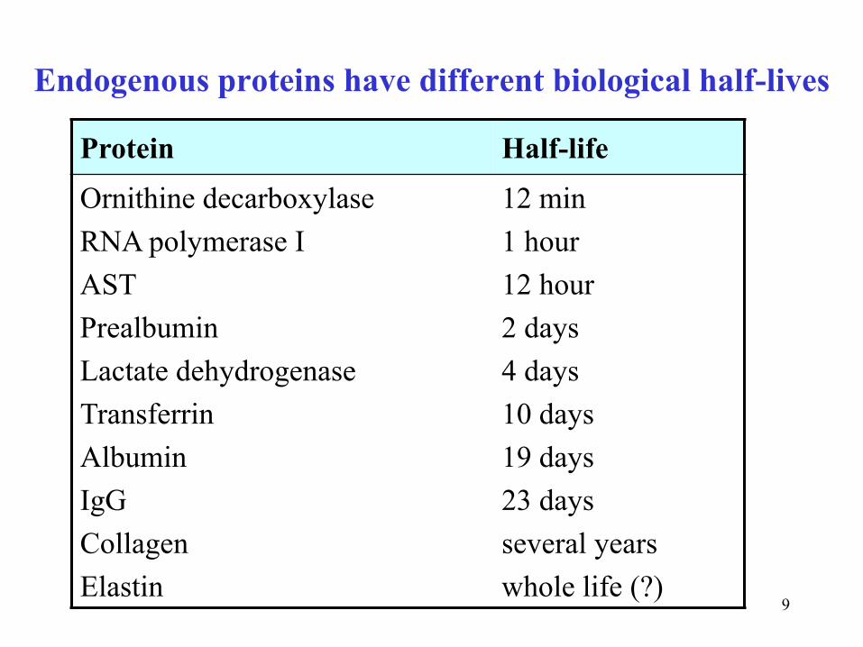
Endogenous proteins have different biological half-lives
Protein Half-life
Ornithine decarboxylase
RNA polymerase I
AST
Prealbumin
Lactate dehydrogenase
Transferrin
Albumin
IgG
Collagen
Elastin
12 min
1 hour
12 hour
2 days
4 days
10 days
19 days
23 days
several years
whole life (?)
10
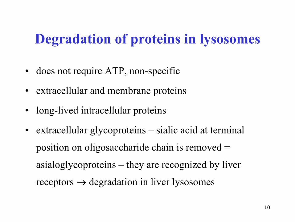
Degradation of proteins in lysosomes
• does not require ATP, non-specific
• extracellular and membrane proteins
• long-lived intracellular proteins
• extracellular glycoproteins – sialic acid at terminal
position on oligosaccharide chain is removed =
asialoglycoproteins – they are recognized by liver
receptors degradation in liver lysosomes

11
Examples of lysosomal hydrolases
Hydrolase Type of bond
Glucosidase
Galactosidase
Hyaluronidase
Arylsulfatase
Lysozyme
Cathepsin
Collagenase
Elastase
Ribonuclease
Lipase
Phosphatase
Ceramidase
glycoside
glycoside
glycoside
sulfoester
glycoside
peptide
peptide
peptide
phosphodiester
ester
phosphoester
amide

12
Ubiquitin (Ub) targets proteins for proteasome degradation
• small protein, in all cells - ubiquitous
• C-terminus binds Lys of proteins to be degraded (kiss of death)
• binding Ub to protein has three phases, with three enzymes E1,E2,E3
• binding Ub to E1-SH requires ATP
• more Ub molecules are attached - polyubiquitination
• Ub-tagged protein is directed to proteasome

13
The targeting of proteins
E1 ubiquitin-activating enzyme (ATP)
E2 ubiquitin-conjugating enzyme
E3 ubiquitin-protein ligase
The N-terminal rule
Stabilizing AA (long life):
• Met, Ser, Ala, Thr, Val, Gly, Cys
Destabilizing AA (short life):
• Phe, Leu, Asp, Lys, Arg
• PEST proteins: segments rich in Pro, Glu, Ser, Thr

14
Proteasome
• hollow cylindric supramolecule, 28 polypeptides
• four cyclic heptamers (4 7 = 28)
• the caps on the ends regulate the entry of proteins into
destruction chamber, upon ATP hydrolysis
• inside the barrel, differently specific proteases hydrolyze
target protein into short (8 AA) peptides
• ubiquitin is not degraded, it is released intact

15
Proteasomes degrade regulatory proteins (short half-life)
and abnormal or misfolded proteins
Ub + short peptides
Protein-Ub
AA
cytosolic
peptidases
important in regulation of cell cycle, growth, differentiation, apoptosis

16
Spatial model of proteasome
28 subunits in 4 rings (4 x 7),
yellow chains in beta subunits contain proteolytic active sites on N-terminals
17
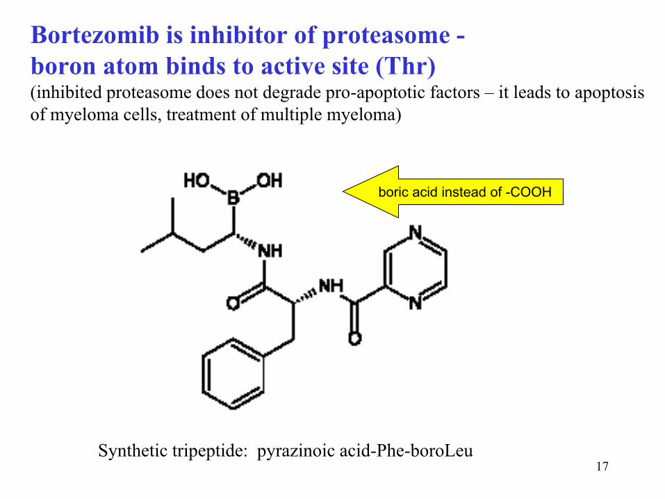
Bortezomib is inhibitor of proteasome -
boron atom binds to active site (Thr) (inhibited proteasome does not degrade pro-apoptotic factors – it leads to apoptosis
of myeloma cells, treatment of multiple myeloma)
Synthetic tripeptide: pyrazinoic acid-Phe-boroLeu
boric acid instead of -COOH

18
Caspases trigger apoptosis
• caspase (cysteinyl aspartate-specific proteinase)
• hydrolyse proteins near aspartate
• degradation of cellular proteins during apoptosis
• formed as inactive precursors (procaspases), activated by the actions of other caspase
• initiator caspases start the apoptic pathway
• after receiving stimulus they activate effector caspases – cascade of caspases
(amplification of the process)
• accidentally acivated caspases are neutralized by specific inhibitors
Calpains – cytosolic proteases activated by Ca2+ ions. They occur in all cells, participate in many
cell processes, e.g. the metabolism of cytoskeletal proteins, cell cycle progression etc.

19
Proteins in nutrition:
Biological value (BV) of proteins refers to how well the body can utilize
the proteins we consume
Relative amount of nitrogen (%) used to synthesis of endogenous
proteins from total protein nitrogen absorbed from food.
BV depends on:
• total content of essential AA
• mutual ratios of essential AA
• protein digestibility
BVanimal prot > BVplant prot
wheat – deficit in Lys, Trp, Thr, Met
legumes – deficit in Met, Cys
Daily intake of proteins: 0.8 g/kg
20
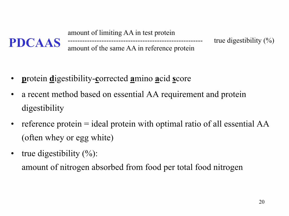
PDCAAS
• protein digestibility-corrected amino acid score
• a recent method based on essential AA requirement and protein
digestibility
• reference protein = ideal protein with optimal ratio of all essential AA
(often whey or egg white)
• true digestibility (%):
amount of nitrogen absorbed from food per total food nitrogen
amount of limiting AA in test protein -------------------------------------------------------- true digestibility (%) amount of the same AA in reference protein

21
Essential (9) and semiessential (3) amino acids
• valine, leucine, isoleucine (BCAA)
• threonine (two C*)
• lysine, histidine (basic)
• phenylalanine, tryptophan (aromatic)
• methionine (-S-CH3)
Semiessential AA
• arginine – in childhood
• alanine, glutamine – in metabolic stress (Ala-gluconeogenesis, Gln – ammonia detoxification)
-----------------------------------------------------------------------------
• about 30 % of methionine need can be substituted by cysteine
• about 50 % of phenylalanine need can be substituted by tyrosine

22
Quality of some proteins
Protein BV (%) PDCAAS (%)
Egg white
Whey
Milk casein
Beef
Beans
Wheat flour
Gelatin
100
100
80
80
49
54
25
100
100
100
92
68
40
8

23
Egg white, whey, and gluten
Egg white is a viscous solution of globular proteins (ovalbumin,
ovotransferrin, ovomucoid, ovomucin, ovoglobulins, avidine etc.)
Whey is a by-product in cottage chesse (curd) production
a yellowish liquid (riboflavin), after precipitation of casein contains high
quality proteins (lactoalbumin, lactoglobulins), B-vitamins, and lactose
Gluten is protein fraction in wheat and other cereals, containing mainly
gliadin (high content of Pro and Gln). In genetically predisposed people,
it may cause autoimmune celiac disease.
GF (gluten free)
BL (bezlepkový)

24
Quantity of proteins in foodstuffs (%)
Parmesan cheese
Emmental cheese
Curd
Beans
Meat
Eggs
Yeast
Cereals, rice
Milk
Potatoes
Fruits, vegetables
40
30
25
25
20
13
11
8
4
2
1

25
Alternative protein sources
Food Protein
content Commentary
Šmakoun
Robi
Seitan
Hemp seeds
Tofu
Tempeh
13 %
22 %
25 %
30 %
16 %
20 %
processed egg white, Czech product
rostlinné bílkoviny, cereal + rice proteins
isolated wheat proteins
good content of essential AA
coagulated soy milk proteins
fermented soybeans by Rhizopus oligosporus

26
Protein supplements
• high content of proteins (20 – 90%)
• mainly derived from dried whey
• and/or free AA (BCAA = Val, Leu, Ile)
• it is a metabolic load for:
• digestion ( putrefaction in large intestine, correlates with
some types of cancer)
• liver ( urea synthesis), kidneys (excretion of urea, NH4+,
free AA)
• may be adulterated with anabolic steroids !!!
27
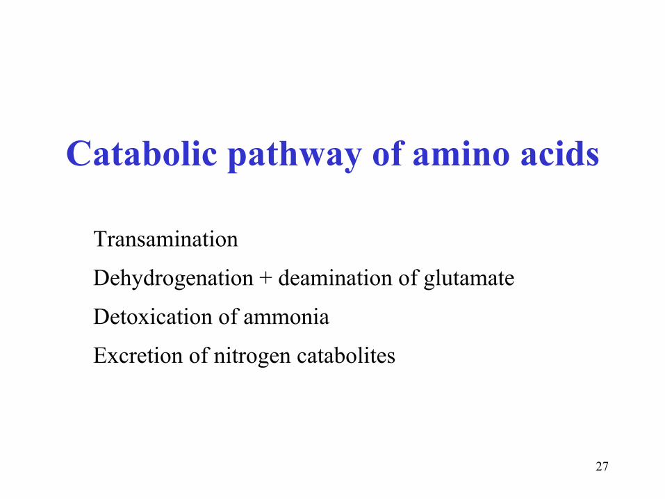
Catabolic pathway of amino acids
Transamination
Dehydrogenation + deamination of glutamate
Detoxication of ammonia
Excretion of nitrogen catabolites
28
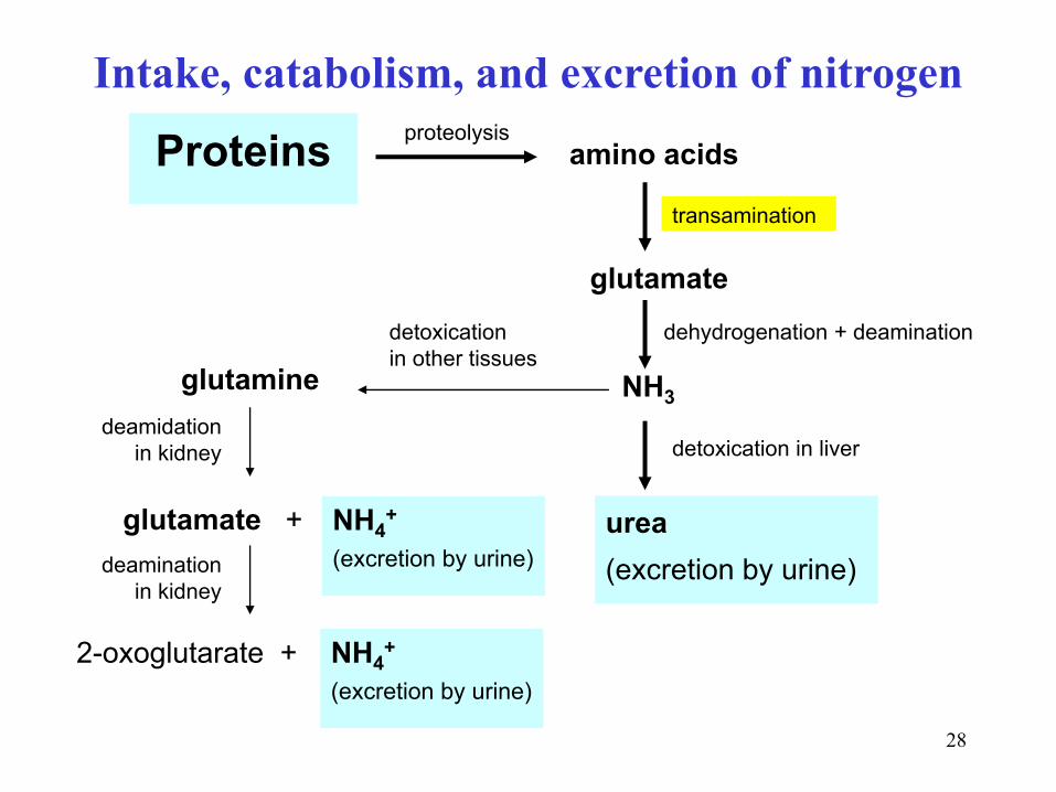
Proteins
NH3
glutamate
glutamate + urea
(excretion by urine)
2-oxoglutarate +
glutamine
proteolysis
dehydrogenation + deamination
detoxication in liver deamidation
in kidney
amino acids
transamination
detoxication
in other tissues
NH4+
(excretion by urine)
NH4+
(excretion by urine)
deamination
in kidney
Intake, catabolism, and excretion of nitrogen

29
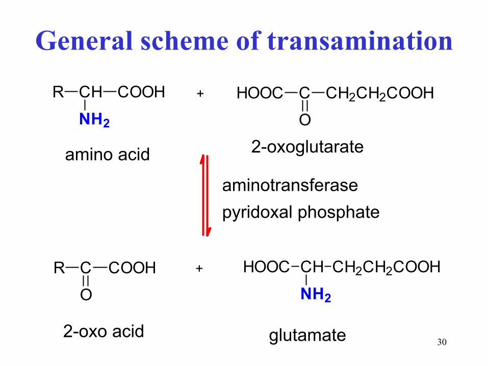
Transamination transfer of -NH2 group from one substrate to other
• most AA (not Lys, Thr, Pro, His, Trp, Arg, Met)
• amino group is transferred from AA to 2-oxoglutarate
• cofactor – pyridoxal phosphate ( Schiff bases)
• reversible reaction important for synthesis of AA
30
General scheme of transamination
CH2CH2COOH
O
CHOOC+R CH
NH2
COOH
aminokyselina 2-oxoglutarát
HOOC CH CH2CH2COOH
NH2
+R C
O
COOH
glutamát2-oxokyselina
aminotransferasa
pyridoxalfosfát
amino acid
2-oxo acid
2-oxoglutarate
glutamate
aminotransferase
pyridoxal phosphate

31
Pyridoxal phosphate has a reactive aldehyde group
N
CH2O
H3C
HO
COH
P
O
O
O
reaktivní skupinaaldehyde group covalently
linked to enzyme
(Lys)

32
1. Phase of transamination
Schiffova báze
CH
NH2
COOHR R C COOH
CH2
N
R CH COOH
CH
N H2O
R C
O
COOH
CH2NH2
COH - H2O
aminokyselina
pyridoxamin-Ppyridoxal-P iminokyselina
oxokyselina
izomerace
aldimin pyridoxalu ketimin oxokyseliny
AA oxoacid
pyridoxal-P pyridoxamine-P
amino acid oxo acid
Schiff base
aldimine of pyridoxal
Schiff base
iminoacid
isomeration

33
2. Phase of transamination
2-oxoglutarát glutamát
pyridoxamin-Ppyridoxal-P
COH
CH2NH2
- H2O
H2O
CH2
CH2
COOH
CHOOC
O
CH2
CH2
COOH
CHOOC
N
CH2
ketiminoxokyseliny
CH2
CH2
COOH
CHHOOC
N
CH
aldiminpyridoxalu
CH2
CH2
COOH
CHHOOC
NH2
2-oxoglutarate glutamate
pyridoxamine-P pyridoxal-P
Schiff base
aldimine of pyridoxal
Schiff base
iminoacid
2-oxoglutarate glutamate

34
In transaminations, nitrogen of most
AA is concentrated in glutamate
Glutamate then undergoes
dehydrogenation + deamination
and releases free ammonia NH3

35
Dehydrogenation + deamination of glutamate
is reversible reaction
HOOC CH CH2CH2COOH
NH2
CH2CH2COOH
NH
CHOOC- 2H
H2O
glutamát 2-iminoglutarát
2-oxoglutarát
NH3 CH2CH2COOH
O
CHOOC+
GMD
NAD(P)+
main source
of ammonia
in tissues
glutamate 2-iminoglutarate
2-oxoglutarate

36
Glutamate dehydrogenase (GMD, GD, GDH)
• requires pyridine cofactor NAD(P)+
• GMD reaction is reversible: dehydrogenation with NAD+,
hydrogenation with NADPH+H+
• two steps:
• dehydrogenation of CH-NH2 to imino group C=NH
• hydrolysis of imino group to oxo group and ammonia

37
1. Deamination of glutamate (GD reaction) in tissues
2. Bacterial putrefaction of proteins in the large intestine
produces nitrogen catabolites (e.g. biogenic amines + ammonia),
ammonia diffuses freely into portal blood portal blood has high
concentration of NH4+ eliminated by liver
Two main sources of ammonia in organism

38
Other sources of ammonia:
deaminations of various substrates
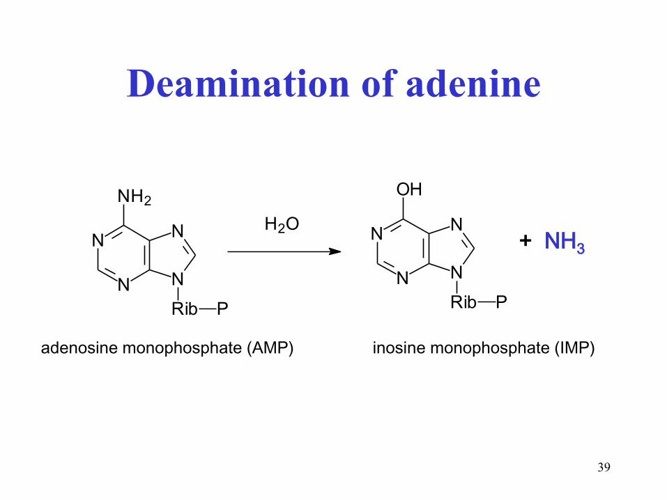
• deamination of adenine
• oxidative deamination of some AA ( H2O2)
• desaturation deamination of histidine urocanic acid + NH3
• oxidative deamination of terminal –NH2 in lysine
lysyl oxidase(Cu2+): Lys + O2 NH3 + allysine + H2O
• dehydratation deamination of serine (see next lecture)
• oxidative deamination of biogenous amines, MAO monoamine oxidase
( H2O2, see also Med. Chem. II, p. 60)
39
Deamination of adenine
N
N
N
N
NH2
Rib P
N
N
N
N
OH
Rib P
H2O+ NH3
adenosine monophosphate (AMP) inosine monophosphate (IMP)

40
Oxidative deamination of some AA
uses flavine cofactor and dioxygen
R CH
NH2
COOH
FAD FADH2
R C COOH
NH
O2H2O2
katalasa
H2O + O2
H2OR C COOH
O
NH3
iminokyselina
• typical for glycine
• D-amino acids
• side product is H2O2
H2O + ½ O2
2-imino acid
catalase

41
Oxidative deamination of biogenous amines
R CH2 NH2
FAD FADH2
H2O2 O2
R CH NHH2O
R CH O
NH3
biogenní amin imin aldehyd
monoaminoxidasa
R-COOH
acid
biogenous amine
monoamine oxidase
imine aldehyde

42
Desaturation type of deamination in histidine
CH CH
NH2
COOH
H
N
NH
CH CH COOH
N
NH
- NH3
urocanic acid
(urocanate)
43
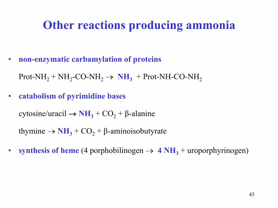
Other reactions producing ammonia
• non-enzymatic carbamylation of proteins
Prot-NH2 + NH2-CO-NH2 NH3 + Prot-NH-CO-NH2
• catabolism of pyrimidine bases
cytosine/uracil NH3 + CO2 + β-alanine
thymine NH3 + CO2 + β-aminoisobutyrate
• synthesis of heme (4 porphobilinogen 4 NH3 + uroporphyrinogen)

44
Hydrolysis of amide group in glutamine releases ammonia
(deamidation). In kidneys, NH4+ ions are released into urine.
COOH
CH
CH2
H2N
CH2
CO NH2
H2O
COOH
CH
CH2
H2N
CH2
CO OH
+ NH3
glutamin glutamátglutamate glutamine
glutaminase
Glutamine is non-toxic transport form of ammonia

45
Ammonia production under pathological conditions
• bleeding in GIT increased NH3 in portal blood
• uroinfections – bacterial urease catalyzes the hydrolysis of urea
H2N-CO-NH2 + H2O 2 NH3 + CO2
NH3 + H2O NH4+ + OH-
alkaline urine (pH ~ 8) phosphate stones

46
Acide-base properties of NH3
pKB (NH3) = 4.75 (weak base)
NH3 + H2O NH4+ + OH-
pKA (NH4+) = 14 – 4.75 = 9.25 (very weak acid)
under physiological pH values in ICF and ECF (~ 7.40):
98 % NH4+
2 % NH3

47
Body fluid Concentration
of NH4+ ions
Metabolic origin of NH4+
Urine
Saliva
Portal blood
Venous blood
10 – 40 mmol/l
2 – 3 mmol/l
100 – 300 μmol/l
5 – 30 μmol/l
hydrolysis of Gln, deamination of Glu (tubules)
hydrolysis of urea by oral microflora
protein putrefaction (GIT), Gln/Glu catabolism (enterocyte)
catabolism of AA in tissues
Compare: Ammonium ions in body fluids

48
1. Low-protein diet (especially important in liver diseases)
2. Alteration of colon microflora by the ingestion of:
• Probiotics – live bacteria stimulating saccharolytic (fermentative)
processes in large intestine instead of putrefactive ones (Lactobacillus,
Bifidobacterium) – yoghurt, kefir milk, sauerkraut etc.
• Prebiotics – non-digestible food ingredients (polysaccharides) that
stimulate the growth probiotics in the colon (dietary fibre, inulin)
How to decrease ammonia production in body?

49
Three ways of ammonia detoxification
Feature Urea Glutamine (Gln) Glutamate (Glu)
Relevance
Compound type
Reaction(s)
Enzyme
Energy needs
Organelle(s)
Organ(s)
H2CO3 diamide
urea cycle
5 enzymes
4 ATP
mitoch. + cytosol
only liver
γ-amide of Glu
Glu + NH3
Gln-synthetase
1 ATP
cytosol
liver, brain, other
α-amino acid
hydrog. amin. 2-OG
GMD
1 NADPH+H+
mitochondria
(brain)

50
Ureasynthesis in liver
five reactions
1. and 2. in mitochondria
3. - 5. in cytosol

51
1. Carbamoyl phosphate (matrix)
CO2 NH4++
2 ATP 2 ADP + 1 P
H2N OC
O
P
O
O
O
• carbamoyl phosphate synthetase (activated by N-acetylglutamate)
• matrix of mitochondria
• two moles of ATP
• amide bond + mixed anhydride
• macroergic compound
carbamoyl phosphate synthetase
52
Carbamoyl is the acyl of carbamic acid
H2NC
O
OH H2NC
O
carbamic acid
(carbonic acid monoamide)
does not exist
carbamoyl
53
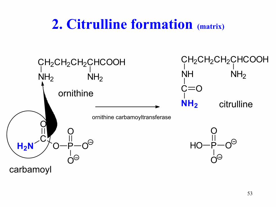
2. Citrulline formation (matrix)
CH2CH2CH2CHCOOH
NH2 NH2
H2N OC
O
P
O
O
O
CH2CH2CH2CHCOOH
NH NH2
C
NH2
O
HO P
O
O
O
citrulin
ornitin
karbamoyl
citrulline
carbamoyl
ornithine
ornithine carbamoyltransferase
54
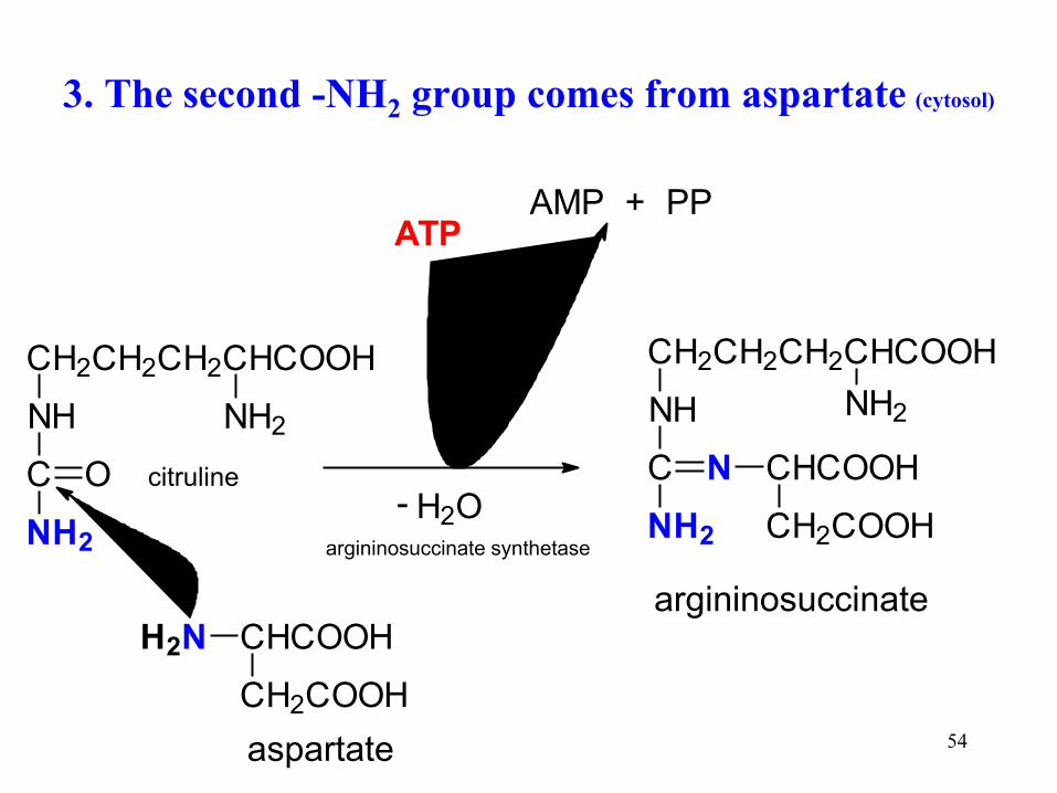
3. The second -NH2 group comes from aspartate (cytosol)
CH2CH2CH2CHCOOH
NH NH2
C
NH2
O
CHCOOH
CH2COOH
H2N
aspartát
CH2CH2CH2CHCOOH
NH NH2
C
NH2
N CHCOOH
CH2COOH H2O-
argininsukcinát
ATPAMP + PP
citruline
aspartate
argininosuccinate
argininosuccinate synthetase

55
Less usual hydrolysis of ATP means
that two ATP are consumed
ATP + H2O AMP + PPi
PPi + H2O 2 Pi (diphosphatase, pyrophosphatase)
AMP + ATP 2 ADP (adenylate kinase)
--------------------------------------------------
summary:
2 ATP + 2 H2O 2 ADP + 2 Pi

56
4. The cleavage of argininosuccinate
CH2CH2CH2CHCOOH
NH NH2
C
NH2
N CHCOOH
CH2COOH
argininsukcinát
CH2CH2CH2CHCOOH
NH NH2
C
NH2
N H
arginin
C
CCOOH
H
H
HOOC
fumarát
argininosuccinate
arginine
fumarate
argininosuccinate lyase
57
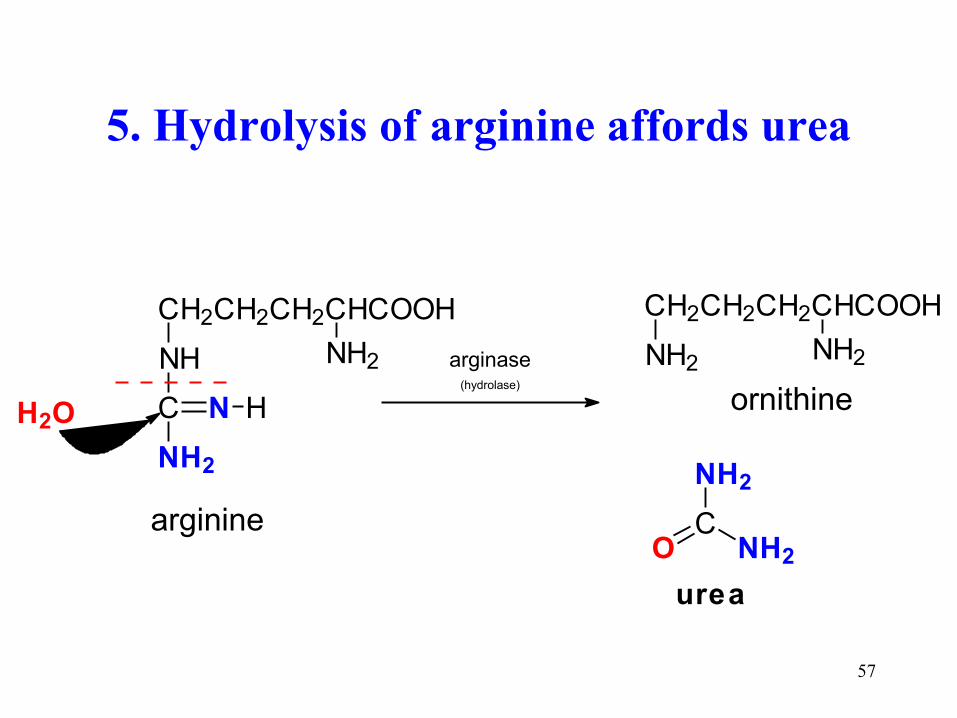
5. Hydrolysis of arginine affords urea
CH2CH2CH2CHCOOH
NH NH2
C
NH2
N H
arginin
H2O
CH2CH2CH2CHCOOH
NH2NH2
OC
NH2
NH2
ornitin
urea
arginine
ornithine
arginase (hydrolase)

58
H2NC
O
NH2free ammonia aspartate
Metabolic origin of nitrogen in urea

59
glutamate + NAD+ ammonia + 2-OG + NADH+H+
GMD
UREA
oxalacetate
AST
2-oxoglutarate aspartate
Dual function of glutamate in AA catabolism

60
CO2 + NH4+ + aspartate urea + fumarate + H2O + 2 H+
CO(NH2)2 + -OOC-CH=CH-COO- + H2O + 2 H+
Urea synthesis is proton-productive reaction
OOC CH
NH3
CH2 COO CO2 + NH4+ +

61
Urea is non-electrolyte
• carbonic acid diamide
• polar compound (dipole) well soluble in water
• diffuses easily through cell membranes (hydrophilic pores)
• contributes to blood plasma osmolality
osmolality 2 [Na+] + [glucose] + [urea] mmol/kg H2O
• synthesis only in liver
• excretion by urine depends on the amount of food proteins
330-600 mmol/day (20-35 g/day)
62
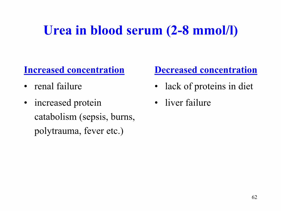
Urea in blood serum (2-8 mmol/l)
Increased concentration
• renal failure
• increased protein
catabolism (sepsis, burns,
polytrauma, fever etc.)
Decreased concentration
• lack of proteins in diet
• liver failure

63
Compare and distinguish
urea uric acid
!

64
Compare
Feature Urea Uric acid
Chemical name
Latin name
In water
Solubility in water
Reducing property
Salt formation
Catabolite of
carbonic ac. diamide
urea
non-electrolyte
excelent
no
no
amino acids
2,6,8-trihydroxypurine
acidum uricum
weak diprotic acid
poor, depeds on pH
yes antioxidant
yes (two types)
adenine and guanine
Organe location liver only liver and other tissues
Subcellular location mitochondria + cytosol cytosol
Serum concentration 2 - 8 mmol/l 150 - 400 μmol/l
Urine excretion 20 - 35 g/day 0.5 - 1 g/day
Catabolic nitrogen 80 - 90 % 1- 2 %
H2N NH2
C
O
N
N
N
N
OH
HO
OH
H

65
Glutamine synthesis
glutamin
COOH
CH
CH2
H2N
CH2
CO NH2
COOH
CH
CH2
H2N
CH2
CO OH
+ NH3
glutamát
ATP ADP + P
- H2O
glutamine synthetase
glutamate glutamine
2. way of detoxification
66
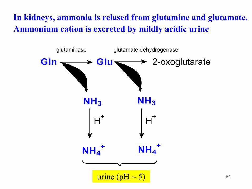
In kidneys, ammonia is relased from glutamine and glutamate.
Ammonium cation is excreted by mildly acidic urine
Gln Glu 2-oxoglutarát
NH3 NH3
H+
H+
NH4+ NH4
+
Moč
glutamátdehy drogenasaglutaminasa
urine (pH ~ 5)
2-oxoglutarate
glutaminase glutamate dehydrogenase

67
Multiple functions of glutamine
• Synthesis of proteins
• Metabolic fuel – for some tissues: enterocytes, lymphocytes, macrophages,
fibroblasts, kidneys
• Source of nitrogen in synthesis – purine, pyrimidines, NAD+, aminosugars
• Source of glutamate – glutathione (GSH), GABA,
Glu ornithine, Glu proline
• Source of ammonium ions in urine
• detoxification of ammonia in tissues and non-toxic transport form of ammonia
from tissues to liver
!

68
Glutamate dehydrogenase reaction is reversible
dehydrogenation deamination of glutamate
hydrogenation amination of 2-oxoglutarate
COOH
CHH2N
CH2
CH2
COOH
NAD+
NADH H+
+
COOH
CHN
CH2
CH2
COOH
COOH
CO
CH2
CH2
COOH
H2O
NH3
ammonia formation
3. way of ammonia detoxication

69
Subcellular location of AA conversions
transamination (ALT) glutamate
NH3 glutamate
synthesis
of urea
mitochondria
cytosol
GMD
Glu + NH3 Gln transamination (AST)
cytosol

70
Synthesis of non-essencial
amino acids

71
Synthesis of glycine
CH2
OH
CH
NH2
COOH + FH4CH2
NH2
COOH + HOCH2 FH4
serin glycin
1. The reverse of transamination
2. From serine
CH2
NH2
COOH
CH
NH2
COOHCH2CH2HOOCC
O
COOHCH2CH2HOOC
C
O
COOHH
+ +
2-oxoglutarát
glyoxalát
glutamát2-oxoglutarate glutamate
glyoxalate
serine glycine

72
Formation of serine from the glycolysis intermediate
COOH
CH OH
CH2 O P
NAD+
COOH
C
CH2 O P
O
NADH H+
COOH
CH
CH2 O P
H2Ntransaminace
H2O
COOH
CH
CH2 OH
H2N
glucose
gly
coly
sis
3-P-glycerate 3-P-hydroxypyruvate
3-P-serine
transamination

73
Synthesis of alanine from pyruvate and glutamate
(ALT = alanine aminotransferase)
COOHCHH3C
NH2
COOHCH3C
O
Glu
2-oxoglutarát
ALT
alanin pyruvátalanine pyruvate
glutamate
2-oxoglutarate

74
Aspartate from oxaloacetate and glutamate (AST = aspartate aminotransferase)
COOHCHCH2
NH2
CH2HOOC
Asp
oxalacetát
AST
COOHCCH2
O
CH2HOOC
glutamát 2-oxoglutarát
AST reaction produces aspartate for urea synthesis
aspartate
glutamate 2-oxoglutarate
oxaloacetate
75
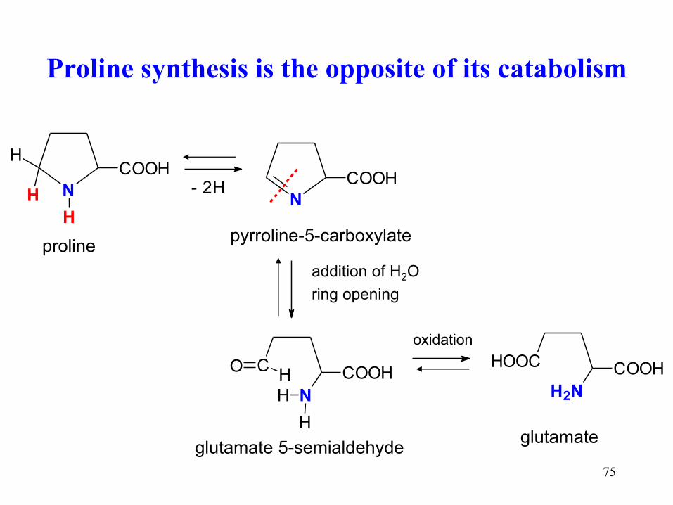
Proline synthesis is the opposite of its catabolism
N
COOH
H
H
H - 2H
prolin
N
COOH
pyrrolin-5-karboxylát
adice H2O
otevření kruhu
N
C COOHOH
H
H
glutamát-5-semialdehyd
H2N
HOOC COOH
glutamát
oxidace
proline
glutamate 5-semialdehyde glutamate
pyrroline-5-carboxylate
oxidation
addition of H2O
ring opening

76
COOH
CHH2N
CH2
CH2
COOH
NAD+
NADH H+
+
COOH
CHN
CH2
CH2
COOH
COOH
CO
CH2
CH2
COOH
H2O
NH3
Glutamate is formed by the reductive amination
of 2-oxoglutarate (GMD reaction)
L-glutamate 2-iminoglutarate 2-oxoglutarate

77
Hydroxylation of essential phenylalanine
gives non-essential tyrosine
COOH
CHH2N
CH2
H
+ O O + BH4
COOH
CHH2N
CH2
OH
+ +H2O BH2
fenylalanin tyrosin
tetrahydrobiopterine (BH4) is a donor of 2H to form water
from the second oxygen atom
phenylalanine tyrosine

78
Glutamine from glutamate and ammonia
glutamin
COOH
CH
CH2
H2N
CH2
CO NH2
COOH
CH
CH2
H2N
CH2
CO OH
+ NH3
glutamát
ATP ADP + P
- H2O
glutamine synthetase
glutamate glutamine
Similarly: asparagine from aspartate

79
Cysteine is made from methionine
HOOC CH
NH2
CH2CH2 SH
homocystein
kondenzace se serinem
HOOC CH
NH2
CH2CH2 SCH2 CH
NH2
COOH
H2O
cystathionin
odštěpení cysteinu
homoserin
HOOC CH
NH2
CH2CH2 OH
methionine
CH
NH2
COOHCH2
SH
homocysteine
cystathionine
homoserine
cysteine
condensation with serine
cysteine release

80
Arginine from glutamate via ornithine
COOH
CHNH2
COOH
CH2
CH2
COOH
CHNH2
CH2
CH2
CH2
NH2
glutamate ornithine arginine urea cycle
oxidative deamination
reductive amination
C
COOH
NH
NH
CH
NH2
NH2
CH2
CH2
CH2

81
Selenocysteine arises co-translationally
from serine and selenophosphate
Seryl-tRNA + selenophosphate selenocysteyl-tRNA + phosphate
Selenophosphate is made from selenide (food) and ATP
Se2- + ATP + H2O AMP + Pi + P
O
O
OSeH
COOHCH
NH2
CH2
Se
H
few enzymes (redox reactions) contain selenocysteine
Glutathione peroxidase (2 GSH + H2O2 2 H2O + G-S-S-G)
Deiodase of thyronines (thyroxine T4 triiodothyronine T3)
Thioredoxin reductase (ribose deoxyribose)
82
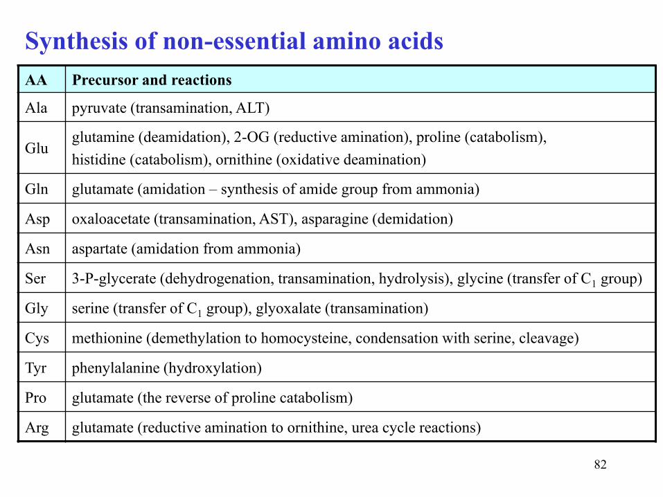
Synthesis of non-essential amino acids
AA Precursor and reactions
Ala pyruvate (transamination, ALT)
Glu glutamine (deamidation), 2-OG (reductive amination), proline (catabolism),
histidine (catabolism), ornithine (oxidative deamination)
Gln glutamate (amidation – synthesis of amide group from ammonia)
Asp oxaloacetate (transamination, AST), asparagine (demidation)
Asn aspartate (amidation from ammonia)
Ser 3-P-glycerate (dehydrogenation, transamination, hydrolysis), glycine (transfer of C1 group)
Gly serine (transfer of C1 group), glyoxalate (transamination)
Cys methionine (demethylation to homocysteine, condensation with serine, cleavage)
Tyr phenylalanine (hydroxylation)
Pro glutamate (the reverse of proline catabolism)
Arg glutamate (reductive amination to ornithine, urea cycle reactions)