CAT Critically Appraised Topic Fetal RHD genotyping … · CAT Critically Appraised Topic Fetal RHD...
18
Katinka De Vreese pagina 1/18 CAT Critically Appraised Topic Fetal RHD genotyping in maternal blood using cell-free fetal DNA Author: Dr. Katinka De Vreese Supervisor: Prof. Dr. Marie-Paule Emonds Date: 13/3/2012 CLINICAL BOTTOM LINE The discovery of cell-free fetal DNA (cffDNA) in maternal plasma has opened up new possibilities for non-invasive prenatal diagnosis (NIPD). Real-time PCR protocols as well as MALDI-TOF mass spectrometry techniques have been developed to determine fetal RHD genotype on maternal plasma. In several European centers, NIPD for fetal RHD has become the standard of care for evaluation of anti-D allo-immunized pregnant women. Several authors have also suggested the feasibility of fetal RHD genotyping in non-immunized women to determine the need for antenatal and perinatal anti-D immunoglobulin. However, up until now there are insufficient data of the cost-effectiveness of large- scale implementation of this test. Most studies were organized in a research/experimental setting in which several replicates were analyzed and inconclusive or unobtainable results have systematically been excluded. In case of large-scale implementation, the number of analyzed replicates should be limited, and inconclusive or unobtainable results need to be taken into consideration. Moreover, up until today, there’s still no universal internal control for the presence of cell-free fetal DNA available. This is necessary to avoid false negative results. International guideline providers such as NICE, ICSI and BCSH await further studies on NIPD using cell-free fetal DNA to issue new guidelines considering the pre- and postnatal management of RhD negative non-immunized pregnant women. CLINICAL/DIAGNOSTIC SCENARIO The Rh blood group system is the largest and most complex of the human blood group systems. Of the 49 known antigens, the most important is the Rhesus D (RhD) antigen. In the Caucasic population, about 17% is RhD negative. About 60% of RhD-negative pregnant women carry an RhD positive fetus. These women are at risk of anti-D alloimmunization, which is the most common cause of hemolytic disease of the fetus and newborn (HDFN) (13,30). During pregnancy, small amounts of fetal blood may enter the maternal circulation, which could cause an immune response in the RhD-negative mother. (5). This is most common in the third trimester and during childbirth, however, it can happen at any time during pregnancy, with higher risk in case of medical intervention, abdominal trauma, miscarriage or antepartum hemorrhages (5). Sensitization has no immediate effects on the mothers health, and doesn’t usually affect the pregnancy during which it’s caused, but leads to a risk of HDFN in case of a subsequent pregnancy with a RhD positive fetus (5). Circulating maternal IgG anti-D-antibodies can cross the placenta and may bind to the RhD-antigen on the surface of fetal red blood cells (RBC), with subsequent destruction of these antibody-coated RBC (5,15). During pregnancy, this may lead to fetal anemia and, in severe cases, development of fetal heart failure, fluid retention, hydrops and intrauterine death (5). The bilirubin produced as a result of RBC lysis is cleared by the placenta before birth, but after birth the neonatal liver is not capable to clear this excess production of bilirubin, leading to jaundice (5). High bilirubin levels can result in severe brain damage (kernicterus) with a range of neurodevelopmental problems (5). Fetal anemia is treated with intrauterine transfusion; postnatal jaundice can be treated with phototherapy and/or exchange transfusion (5).
Transcript of CAT Critically Appraised Topic Fetal RHD genotyping … · CAT Critically Appraised Topic Fetal RHD...
Katinka De Vreese pagina 1/18
CAT
Critically Appraised Topic
Fetal RHD genotyping in maternal blood using cell-free fetal DNA
Author: Dr. Katinka De Vreese
Supervisor: Prof. Dr. Marie-Paule Emonds
Date: 13/3/2012
CLINICAL BOTTOM LINE
The discovery of cell-free fetal DNA (cffDNA) in maternal plasma has opened up new possibilities for
non-invasive prenatal diagnosis (NIPD). Real-time PCR protocols as well as MALDI-TOF mass
spectrometry techniques have been developed to determine fetal RHD genotype on maternal plasma. In
several European centers, NIPD for fetal RHD has become the standard of care for evaluation of anti-D
allo-immunized pregnant women. Several authors have also suggested the feasibility of fetal RHD
genotyping in non-immunized women to determine the need for antenatal and perinatal anti-D
immunoglobulin. However, up until now there are insufficient data of the cost-effectiveness of large-
scale implementation of this test. Most studies were organized in a research/experimental setting in
which several replicates were analyzed and inconclusive or unobtainable results have systematically
been excluded. In case of large-scale implementation, the number of analyzed replicates should be
limited, and inconclusive or unobtainable results need to be taken into consideration. Moreover, up until
today, there’s still no universal internal control for the presence of cell-free fetal DNA available. This is
necessary to avoid false negative results.
International guideline providers such as NICE, ICSI and BCSH await further studies on NIPD using
cell-free fetal DNA to issue new guidelines considering the pre- and postnatal management of RhD
negative non-immunized pregnant women.
CLINICAL/DIAGNOSTIC SCENARIO
The Rh blood group system is the largest and most complex of the human blood group systems. Of the
49 known antigens, the most important is the Rhesus D (RhD) antigen. In the Caucasic population,
about 17% is RhD negative. About 60% of RhD-negative pregnant women carry an RhD positive fetus.
These women are at risk of anti-D alloimmunization, which is the most common cause of hemolytic
disease of the fetus and newborn (HDFN) (13,30). During pregnancy, small amounts of fetal blood may
enter the maternal circulation, which could cause an immune response in the RhD-negative mother. (5).
This is most common in the third trimester and during childbirth, however, it can happen at any time
during pregnancy, with higher risk in case of medical intervention, abdominal trauma, miscarriage or
antepartum hemorrhages (5).
Sensitization has no immediate effects on the mothers health, and doesn’t usually affect the pregnancy
during which it’s caused, but leads to a risk of HDFN in case of a subsequent pregnancy with a RhD
positive fetus (5). Circulating maternal IgG anti-D-antibodies can cross the placenta and may bind to the
RhD-antigen on the surface of fetal red blood cells (RBC), with subsequent destruction of these
antibody-coated RBC (5,15). During pregnancy, this may lead to fetal anemia and, in severe cases,
development of fetal heart failure, fluid retention, hydrops and intrauterine death (5). The bilirubin
produced as a result of RBC lysis is cleared by the placenta before birth, but after birth the neonatal
liver is not capable to clear this excess production of bilirubin, leading to jaundice (5). High bilirubin
levels can result in severe brain damage (kernicterus) with a range of neurodevelopmental problems (5).
Fetal anemia is treated with intrauterine transfusion; postnatal jaundice can be treated with phototherapy
and/or exchange transfusion (5).
Fetal RHD genotyping on maternal blood using cffDNA
Katinka De Vreese pagina 2/18
National and international guidelines recommend to determine the blood group, the RhD status and the
presence or absence of irregular antibodies in the maternal circulation, before pregnancy or at the first
prenatal visit (3,5,6-8,9). Repeat D antibody testing is recommended for all unsensitized RhD-negative
women at 28 weeks gestation (3,6,8,9). The risk of anti-D alloimmunization between first trimester and
week 28 is low (0.18%) but anti-D might not be detected in the first screening (below threshold) but
develop during pregnancy and cause HDFN (9).
In non-immunized women, the risk of sensitization can be reduced by administering one dose of 1500
IU anti-D immunoglobulin within 72 hours after a potentially sensitizing event, including child-birth
(3,6-8). A similar dose of anti-D immunoglobulin can be administered as a prophylactic treatment in the
third trimester of pregnancy (3,5-8,9,30). This last therapy is known as routine antenatal anti-D
prophylaxis (RAADP) (3,5,7,8). RAADP neutralizes the fetal RhD antigen in the maternal circulation,
which leads to a further reduction of the immunization risk from 1.2% to 0.3% (5,6,24,27). But since
only 60% of RhD negative women are pregnant with an RhD-positive fetus, 40% will receive
unnecessarily anti-D prophylaxis (30). In Belgium, no national policy about RAADP exists. Due to the
limited availability of anti-D immunoglobulin, treatment strategy in the University Hospitals of Leuven
is limited to antenatal administration in case of a potentially sensitizing event and postnatal
administration to mothers giving birth to an RhD-positive newborn.
When an anti-D-immunized woman is pregnant, intensive follow-up is recommended. Rising anti-D
titers or titers over 4 IU/ml are considered to be at risk for HDFN. In that case, Doppler measurement of
the middle cerebral artery peak velocity flow is performed to evaluate the degree of fetal anemia
(11,26).
Another possible approach is the determination of the fetal RHD genotype on amniotic fluid. In case of
an RHD negative fetus, no further investigations are required. An RHD positive fetus requires intensive
follow-up. Until recently, the only manner to obtain fetal DNA was through invasive procedures such as
amniocentesis or chorionic villus sampling. However, recent developments in noninvasive prenatal
diagnosis have changed this.
RHD genotyping using maternal plasma as a source of fetal DNA is introduced in many European
laboratories as a noninvasive prenatal test in anti-D allo-immunized women (15). The International
Blood Group Reference Laboratory (IBGRL) in Bristol, UK, has been offering this test to immunized
women with heterozygous partners since 2001 (13,24,35). In Belgium, non-invasive fetal RHD
genotyping is proposed to all RhD-negative pregnant women at the University Hospital of Liege, with
proposition of RAADP at 28 weeks gestation for all patients bearing RhD-positive fetuses (26).
Starting July first, 2011, the Dutch Sanquin blood supply foundation and the Dutch National Institute
for Public Health and Environment (RIVM) have implemented systematic screening of RhD-negative
pregnant women in the 27th week of pregnancy with PCR genotyping of the fetal RHD on maternal
blood (2). Only RhD negative women carrying an RHD positive fetus will receive RAADP and
postnatal RhIg, providing that the screening for irregular antibodies is negative (2). A pilot study, with
parallel testing of RhD serology on umbilical cord blood samples after birth, will run until October
2012. In case of postnatal RhD positivity, anti-D Ig is still offered (2).
In this document, we will review the current knowledge and diagnostic accuracy of fetal RHD
genotyping on maternal blood. Based on literature data, we will also explore its cost-effectiveness and
its potential in clinical practice.
QUESTION(S)
PICO
P: RhD-negative pregnant women
I: RHD genotyping on maternal blood using cell-free fetal DNA
C: Serologic RhD phenotyping or RHD genotyping on amniotic fluid or chorionic villus sample
O: Determination of fetal RHD genotype in sensitized women / Reducing RAADP in non-sensitized
women

Fetal RHD genotyping on maternal blood using cffDNA
Katinka De Vreese pagina 3/18
Questions
1. What is the current knowledge of fetal RHD genotyping on maternal blood, using cell-free fetal
DNA?
2. Is it useful to implement fetal RHD genotyping on maternal blood in routine clinical practice?
SEARCH TERMS
1) MeSH Database (PubMed): MeSH term: “(("Rh-Hr Blood-Group System"[Mesh]) AND
"Genotype"[Mesh]) AND "Blood"[Mesh]; (("Fetus"[Mesh]) AND "Genotype"[Mesh]) AND "Rh-Hr
Blood-Group System"[Mesh] ”
2) PubMed Clinical Queries (from 1966; http://www.ncbi.nlm.nih.gov/entrez/query.fcgi): Systematic
Reviews; Clinical Queries using Research Methodology Filters (diagnosis + specific, diagnosis +
sensitive, prognosis + specific)
3) Pubmed (Medline; from 1966), SUMSearch (http://sumsearch.uthscsa.edu/), National Guideline
Clearinghouse (http://www.ngc.org/), Institute for Clinical Systems Improvement
(http://www.icsi.org), The National Institute for Clinical Excellence (http://www.nice.org.uk/),
Cochrane (http://www.update-software.com/cochrane, Health Technology Assessment Database
(http://www.york.ac.uk/inst/crd/htahp.htm)
4) UpToDate Online version 19.3 (2012)
RELEVANT EVIDENCE/REFERENCES (most recent topics on top)
1) Guidelines and Recommendations
(1) CBO (2011)
CBO richtlijn bloedtransfusie, 11 oktober 2011.
(2) RIVM (2011)
Rijksinstituut voor Volksgezondheid en Milieu. Draaiboek Prenatale Screening Infectieziekten
en Erytrocytenimmunisatie; versie 3.0; 1 juli 2011.
(3) ICSI (2010)
Institute for Clinical Systems Improvement. Health Care Guideline: Routine Prenatal Care.
Fourteenth Edition; Juli 2010.
(4) HGR (2010)
Hoge Gezondheidsraad. Goede transfusiepraktijken in ziekenhuizen. Brussel: HGR; 2010.
Advies nr. 8381.
(5) NICE (2008)
National Institute for Health and Clinical Excellence Technology Appraisal Guidance 156.
Routine antenatal anti-D prophylaxis for women who are rhesus D negative. NICE TA-156.
(6) VVOG (2007)
Vlaamse Vereniging voor Obstetrie en Gynaecologie. Erythrocyten alloimmunisatie en
zwangerschap. VVOG-aanbeveling 20/11/2007.
(7) BCSH (2006)
British Committee for Standards in Haematology. Guidelines for the use of prophylactic anti-D
immunoglobulin. Parker J, Wray J, Gooch A, Robson S, Qureshi H.
(8) KCE (2004)
Federaal Kenniscentrum voor de Gezondheidszorg. Nationale richtlijn prenatale zorg: een basis
voor een klinisch pad voor de opvolging van zwangerschappen. KCE report vol. 6A.
(9) Practice guidelines for prenatal and perinatal immunohematology, revisited. Judd WJ; Scientific
Section Coordinating Committee of the AABB. Transfusion. 2001 Nov;41(11):1445-52. 2) Systematic Reviews and Meta-analyses
(10) Non-invasive fetal RHD genotyping tests: a systematic review of the quality of reporting of
diagnostic accuracy in published studies. Freeman K, Szczepura A, Osipenko L. Eur J Obstet
Gynecol Reprod Biol. 2009 Feb;142(2):91-8. Review.
Fetal RHD genotyping on maternal blood using cffDNA
Katinka De Vreese pagina 4/18
(11) Diagnostic accuracy of noninvasive fetal Rh genotyping from maternal blood – a meta-
analysis. Geifman-Holtzman O, Grotegut CA, Gaughan JP. Am J Obstet Gynecol. 2006
Oct;195(4):1163-73. 3) Reviews
(12) Management of red cell alloimmunisation in pregnancy: the non-invasive monitoring of the
disease. Illanes S, Soothill P. Prenat Diagn. 2010 Jul;30(7):668-73.Review.
(13) The use of maternal plasma for prenatal RhD blood group genotyping. Finning K, Martin P,
Daniels G. Methods Mol Biol. 2009;496:143-57.Review.
(14) The SAFE project: towards non-invasive prenatal diagnosis. Maddocks DG, Alberry MS,
Attilakos G, Madgett TE, Choi K, Soothill PW, Avent ND. Biochem Soc Trans. 2009
Apr;37(Pt2):460-5.Review.
(15) RHD genotyping from maternal plasma: guidelines and technical challenges. Avent ND.
Methods Mol Biol. 2008;444:185-201.Review.
(16) Recent developments in fetal nucleic acids in maternal plasma: implications to noninvasive
prenatal fetal blood group genotyping. Lo YM. Transfus Clin Biol. 2006 Mar-Apr;13(1-2):50-
2. Review.
(17) Non-invasive antenatal RHD typing. Van der Schoot CE, Soussan AA, Koelewijn J, Bonsel G,
Paget-Christiaens LG, de Haas M. Transfus Clin Biol. 2006 Mar-Apr;13(1-2):53-7.Review.
(18) Circulating fetal DNA: its origin and diagnostic potential – a review. Bianchi DW. Placenta.
2004 Apr;25 Suppl A:S93-S101.Review.
(19) Fetal DNA in maternal plasma: application to non-invasive blood group genotyping of the
fetus. Lo YM. Transfus Clin Biol. 2001 Jun;8(3):306-10.Review. 4) Original Articles
(20) Non-invasive prenatal diagnosis. Meaney C, Norbury G. Methods Mol Biol. 2011;688:155-72.
(21) Noninvasive fetal blood group genotyping of rhesus D, c, E and of K in alloimmunised
pregnant women; evaluation of a 7-year clinical experience. Scheffer PG, van der Schoot CE,
Page-Christiaens GC, de Haas M. BJOG. 2011 Oct;118(11):1340-8.
(22) Fetal RHD genotype detection from circulating cell-free fetal DNA in maternal plasma in non-
sensitized RhD negative women. Bombard AT, Akolekar R, Farkas DH, VanAgtmael AL,
Aquino F, Oeth P, Nicolaides KH. Prenat Diagn. 2011 Aug;31(8):802-8.
(23) Fetal RHD genotyping in maternal plasma at 11-13 weeks of gestation. Akolekar R, Finning K,
Kuppusamy R, Daniels G, Nicolaides KH. Fetal Diagn Ther. 2011;29(4):301-6.
(24) A new fetal RHD genotyping test: costs and benefits of mass testing to target antenatal anti-D
prophylaxis in England and Wales. Szczepura A, Osipenko L, Freeman K. BMC Pregnancy
Childbirth. 2011 Jan 18;11:5.
(25) Maldi-TOF mass spectrometry for analyzing cell-free fetal DNA in maternal plasma. Ding C.
Methods Mol Biol. 2008;444:253-67.
(26) Routine fetal RHD genotyping with maternal plasma: a four-year experience in Belgium.
Minon JM, Gerard C, Senterre JM, Schaaps JP, Foidart JM. Transfusion. 2008 Feb;48(2):373-
81.
(27) Effect of high throughput RHD typing of fetal DNA in maternal plasma on use of anti-RhD
immunoglobulin in RhD negative pregnant women: prospective feasibility study. Finning K,
Martin P, Summers J, Massey E, Poole G, Daniels G. BMJ. 2008 Apr 12;336(7648):816-8.
(28) Noninvasive genotyping fetal Kell blood group (KEL1) using cell-free fetal DNA in maternal
plasma by MALDI-TOF mass spectrometry. Li Y, Finning K, Daniels G, Hahn S, Zhong X,
Holzgreve W. Prenat Diagn. 2008 Mar;28(3):203-8
(29) Noninvasive fetal RHD genotyping from maternal plasma. Use of a new developed Free DNA
Fetal Kit RhD. Rouillac-Le Sciellour C, Sérazin V, Brossard Y, Oudin O, Le Van Kim C,
Colin Y, Guidicelli Y, Menu M, Cartron JP. Transfus Clin Biol. 2007 Dec;14(6):572-7.
(30) Fetal RhD genotyping: a more efficient use of anti-D immunoglobulin. Daniels G, Finning K,
Martin P, Summers J. Transfus Clin Biol. 2007 Dec;14(6):568-71.
(31) Workshop report on the extraction of foetal DNA from maternal plasma. Legler TJ, Liu Z,
Mavrou A, Finning K, Hromadnikova I, Galbiati S, Meaney C, Hultén MA, Crea F, Olsson
ML, Maddocks DG, Huang D, Fisher SA, Sprenger-Haussels M, Soussan AA, van der Schoot
CE. Prenat Diagn. 2007 Sep;27(9):824-9
(32) Hypermethylated RASSF1A in maternal plasma: A universal fetal DNA marker that improves
the reliability of noninvasive prenatal diagnosis. Chan KC, Ding C, Gerovassili A, Yeung SW,

Fetal RHD genotyping on maternal blood using cffDNA
Katinka De Vreese pagina 5/18
Chiu RW, Leung TN, Lau TK, Chim SS, Chung GT, Nicolaides KH, Lo YM. Clin Chem. 2006
Dec;52(12):2211-8.
(33) Maldi-tof mass spectrometry compared with real-time PCR for detection of fetal cell-free DNA
in maternal plasma. Li Y, Holzgreve W, Kiefer V, Hahn S. Clin Chem. 2006 Dec;52(12):2311-
2.
(34) Large-scale pre-diagnosis study of fetal RHD genotyping by PCR on plasma DNA from RhD-
negative pregnant women. Rouillac-Le Sciellour C, Puillandre P, Gillot R, Baulard C, Métral
S, Le Van Kim C, Cartron JP, Colin Y, Brossard Y. Mol Diagn. 2004;8(1):23-31.
(35) A clinical service in the UK to predict fetal Rh (Rhesus) D blood group using free fetal DNA
in maternal plasma. Finning K, Martin P, Daniels G. Ann N Y Acad Sci. 2004 Jun;1022:119-
23.
(36) Prenatal diagnosis of fetal RhD status by molecular analysis of maternal plasma. Lo YM,
Hjelm NM, Fidler C, Sargent IL, Murphy MF, Chamberlain PF, Poon PM, Redman CW,
Wainscoat JS. N Eng J Med. 1998 Dec 10;339(24):1734-8.
(37) Quantitative analysis of fetal DNA in maternal plasma and serum: implications for noninvasive
prenatal diagnosis. Lo YM, Tein MS, Lau TK, Haines CJ, Leung TN, Poon PM, Wainscoat JS,
Johnson PJ, Chang AM, Hjelm NM. Am J Hum Genet. 1998 Apr;62(4):768-75.
(38) Presence of fetal DNA in maternal plasma and serum. Lo YM, Corbetta N, Chamberlain PF,
Rai V, Sargent IL, Redman CW, Wainscoat JS. Lancet. 1997 Aug 16;350(9076):485-7. 5) Reference Works, Handbooks and Databases
(39) Cell-free fetal nucleic acids for non-invasive prenatal diagnosis. Report of the UK expert
working group. Caroline Wright. January 2009. www.phgfoundation.org.
6) Posters, “grey literature”, presentations
(40) A noninvasive test for fetal RHD genotype. www.clinicaltrials.gov.
(41) Mass Screening for Fetal RhD Type. Edwin Massey. International Blood Group Reference
Laboratory (IBGRL), NHS Blood and Transplant, Filton, Bristol, UK. Presentation.
(42) Free DNA Fetal Kit® RhD Leaflet – Institut de Biotechnologies Jacques Boy
(43) Free DNA Fetal Kit® RhD Leaflet – BioRad (DiaMed).
(44) Focus Diagnostica. Nomenclatuur 2010.

Fetal RHD genotyping on maternal blood using cffDNA
Katinka De Vreese pagina 6/18
APPRAISAL
a) Genetic base of Rh phenotypes
Rh phenotypes are controlled by RHD and RHCE, two homologous genes located on
chromosome 1, which have ten exons each and share about 95% identity (13,15,29,30). The
RhD-positive phenotype is seen in the majority of the population and is caused by the
homozygous or hemizygous presence of the RHD gene (13,27,30). The RhD-negative
phenotype is quite prevalent in Caucasians (15-17%), moderately prevalent in Africans (3-5%)
and rare in Asian populations (<1%) (26,27).
In Caucasians, the RhD-negative phenotype almost always results from homozygosity for a
complete deletion of RHD (13,15,26,27,30). RhD antigen negative, RHD gene positive
haplotypes are rare (0.2-1% of all D negative Caucasians) and known to preferentially occur in
the Cde and cdE haplotypes. The molecular base is mostly the presence of hybrid RHD-CE-D
genes with an intact exon 10 (26).
In Africans and Asians, the most common cause of an RhD-negative phenotype is the presence
of an inactive RHD gene (13,26). 67% of the RhD negative Africans carry the silent RHD
pseudogene, RHDψ, and 15% carry an inactive hybrid RHD-CE-DS gene (26,30). Only 18% are
homozygous for a RHD deletion (30).
RHDψ contains all exons, but is characterized by two inactivating mutations: a 37 bp
duplication in exon 4 and a nonsense mutation in exon 6 (13,30). There are also four
characteristic single nucleotide changes in exons 4 and 5 (13). Due to these mutations, no RhD
antigens are expressed.
The RHD-CE-DS hybrid gene contains exons 1, 2, part of 3, 9 and 10 from RHD but part of
exon 3 and exons 4 to 8 from RHCE (30). This hybrid gene doesn’t express RhD epitopes either
(30).
Next to RHDψ and RHD-CE-DS, many other rare variant RHD haplotypes exist (13,30). These
RHD variants result either from single nucleotide polymorphisms (SNPs) in RHD, encoding
amino acid substitutions, or from hybrid RH genes (30). Several of these variants may produce
anti-D alloantibodies after immunization by normal RhD-positive cells (26,30).
b) Cell-free fetal DNA (cffDNA)
The past years, molecular biology has proven to be a powerful tool for the prenatal investigation
of genetic disorders. However, most methods rely on the use of fetal material which has been
obtained through invasive procedures, such as amniocentesis or chorionic villus sampling
(19,21,30,34). These procedures are associated with a certain risk (0.5-1%) of fetal loss and a
significant risk (17%) of fetomaternal bleeding which could boost the maternal immune
response to fetal red blood cell antigens, with possible worsening of HDFN (12,19,21,30,34).
It has been well established that during pregnancy, a small number of fetal nucleated cells
passes from the fetus into the maternal circulation (19). This phenomenon makes use of these
cells for NIPD possible (37). However, there are two major problems with this strategy,
reducing its accuracy. Fetal nucleated cells are only present in maternal blood in extremely low
concentrations (19). The average number of fetal cells in maternal blood during the second
trimester of a normal pregnancy is 1.2 cells/ml (37). Therefore, this requires very sensitive
methods of detection or use of fetal cell isolation procedures (19). Since most techniques are
time-consuming and labor-intensive, they are difficult to implement on a large scale (19,37).
The second problem is that fetal cells may persist post partum for many years, sometimes as
long as 27 years after delivery (14,17-19,36).
In 1997, Lo et al. started to focus on the acellular fraction of maternal blood, inspired by
workers in the field of oncology who discovered the presence of tumor DNA in plasma and
1) What is the current knowledge on fetal RHD genotyping on maternal
blood, using cell-free fetal DNA?

Fetal RHD genotyping on maternal blood using cffDNA
Katinka De Vreese pagina 7/18
serum of cancer patients (19,38). They demonstrated the presence of cell-free fetal DNA
(cffDNA) in plasma and serum of pregnant women by demonstrating the presence of fetus-
derived Y sequences (38). Physiological and clinical information suggest that the majority of
circulating fetal nucleic acids are derived from the placenta, most probably from apoptotic
syncytiotrophoblast (12,17,18).
One year later, the same group demonstrated that the amount of circulating fetal DNA is
considerably higher than the amount of fetal nucleated cells in maternal blood. Fetal DNA
comprises 3.4% of total plasma DNA in early pregnancy, rising to 6.2% of total plasma DNA in
late pregnancy. Fetal cells only comprise 0.0035% in early pregnancy and 0.008% in late
pregnancy (37). Cell-free fetal DNA can be detected as early as the 5th week of gestation and in
contrast to fetal cells, cffDNA is cleared from the maternal circulation within hours after child-
birth (14,17-19,36,37).
The same year, Lo et al. also developed a real-time Taqman PCR assay for determination of
fetal RHD genotype on maternal plasma (36).
c) Fetal RHD genotyping on maternal blood using cffDNA
1. Analytical performance
1.1. Pre-analytical considerations
1.1.1 Patient variables
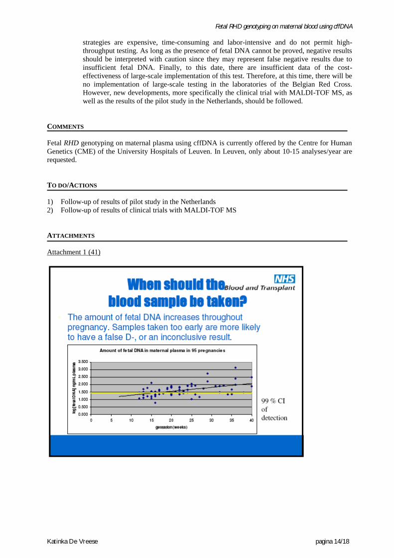
There is no consensus on the optimal gestational age for blood sampling. There
is also a difference in approach of sensitized and non-sensitized pregnancies.
As stated above, fetal DNA can already be detected at the 5th week of gestation
and increases with gestational age (attachment 1). In sensitized women, early
diagnosis is desirable in order to plan further diagnostic procedures and
therapy. However, if sampling takes place too early in pregnancy, a false
negative or inconclusive result is possible due to low levels of cffDNA. The
leaflets of the commercially available kits advise a minimal gestational age of
12 weeks with, in case of a negative result, confirmation with a second sample
collected at least two weeks later (42,43).
In non-sensitized women, the result of the fetal RHD genotyping test will be
decisive on whether or not administering RAADP. Therefore, sampling can be
delayed until later in pregnancy. Since RAADP is administered at week 28, the
sample may be taken one week earlier, as it’s done in the Netherlands (2)
1.1.2 Sample requirements
When comparing plasma and serum samples, Lo et al. discovered that the
absolute concentration of fetal DNA in maternal plasma is similar to that in
maternal serum, but the main difference is the presence of a larger quantity of
background maternal DNA in serum as compared to plasma, possibly caused by
liberation of DNA during the clotting process (attachment 2) (14,37). Most
studies are performed with EDTA plasma but citrate plasma may also be used
(13,20,21,23,25,26,29,34,35,38,42). As it is generally known, heparin samples
are not useful given the inhibitory effect on the PCR assay.
Some centers require a paternal sample as well to determine specific markers
which can be used as internal controls (21).
1.1.3 Sample stability and processing
Samples should be processed within 48 hours of venipuncture to reduce the
amount of maternal DNA in the plasma resulting from breakdown of maternal
white cells (13,21,26,29,34,35). Samples must be centrifuged, followed by
careful removal of the plasma. The plasma may be stored at -20°C or lower
pending further processing (20,21,26,29,34,35). The buffy coat can also be
removed and frozen for determination of silent maternal RHD variants
(26,29,34).

Fetal RHD genotyping on maternal blood using cffDNA
Katinka De Vreese pagina 8/18
1.2. Analytical considerations
1.2.1 (Lack of) standardization
As stated above, cffDNA extracted from maternal plasma is now recognized as
a potential source for prenatal diagnosis, but the methodology is currently not
well standardized (29,31). There are considerable differences in the used
techniques for DNA extraction, primer choice, PCR conditions and detection of
amplified products, resulting in considerable variations in diagnostic
performance between different laboratories (29).
The SAFE network (Special Advances in Fetal and Neonatal Evaluation,
funded by the European Community network of excellence), proposes the
QIAamp DSP Virus Kit (Qiagen, Hilden, Germany) for cffDNA extraction,
since this kit has proven to have the highest yield of cffDNA (14,17,31).
There is a commercial kit available, offered by L’Institut de Biotechnologies
Jacques Boy and BioRad/DiaMed, namely the Free DNA Fetal Kit® RhD
(42,43). Rouillac-Le Sciellour et al. compared the commercial kit with their in-
house developed PCR on 300 RhD negative women and found 100%
concordance (29).
1.2.2 Size fractionation
Fetal DNA fragments are smaller than maternal DNA, so size-fractionation is a
possible approach to reduce background maternal DNA (14). Most of
circulating fetal DNA molecules are 145-201 bp long whereas most maternal
DNA is over 300 bp long (17). This might be used to enrich fetal DNA (17,25).
1.2.3 Primer choice
Primers should be chosen so that only RHD and not RHCE is amplified (30).
Based on the knowledge that the majority of RhD negative phenotypes is
caused by a deletion of the RHD gene, first generation genotyping tests were
based on a single RHD gene region amplification, most often exon 10
(13,17,26,34,36). The discovery of variant RHD genes has hampered the test
interpretation. Nonfunctional RHD alleles such as RHDψ and RHD-CE-DS are
serologically typed as RhD negative. However, since they contain an intact
RHD exon 10, this inactive maternal RHD gene will be amplified, leading to
false positive results (13,26). Two or more ‘diagnostic sites’, with at least
inclusion of an RHD-specific PCR that is negative on the RHD pseudogene,
should be tested to limit the rate of false positives (17,26,30). This often
requires multiplex PCR assays in which several regions of the RHD gene are
amplified (17).
Several combinations have been proposed by the different groups. Details can
be found in attachment 3.
1.2.4 Test principle
Real-Time quantitative PCR using Taqman chemistry is a possible diagnostic
approach because of its ease of use and ability to automate and thus avoid
contamination (15,34). Taqman RT PCR relies upon PCR primers, to define the
specificity of the reaction, and a probe with reporter and quencher dyes
attached (13). If the target DNA sequence is present, the increase in PCR
product formation is monitored by the increase in probe reporter dye
fluorescence throughout each cycle and converted by the software into an
amplification plot (increase in reporter dye fluorescence vs. PCR cycle)
(13,20). The cycle at which the reporter dye reaches a threshold level of
fluorescence (Ct) is dependent on the starting amount of target DNA present
(13). The more target DNA is present in the sample at the start of the PCR, the
lower the Ct value (13,26).
MALDI-TOF MS has recently emerged as a new platform for highly sensitive
and accurate analysis of DNA, especially cell-free DNA (cf-DNA) (33). This
Fetal RHD genotyping on maternal blood using cffDNA
Katinka De Vreese pagina 9/18
technique combines flexibility, accuracy, automated analysis and high-
throughput data generation (15,22,25,28). Sequenom Inc. (San Diego, CA)
developed a MassARRAY genotyping platform combining PCR, base
extension reaction (SABER) and MALDI-TOF MS and offering all necessary
software for efficient and accurate analysis (25,28,40). After initial DNA
amplification by PCR, an additional linear amplification step called single
allele base extension reaction is performed with an extension primer designed
to anneal to the region immediately upstream of the mutation site (25). SABER
has been shown to improve the detection of fetal-derived subtle mutations (25).
The MassARRAY platform can be used for SNP genotyping, methylation
detection and quantitative gene expression analysis. Sequenom also
manufactures clinical tests, such as the SensiGeneTM
Fetal RHD genotyping
test. They are currently conducting a clinical trial for the evaluation of the
performance of a noninvasive first trimester fetal RHD genotyping test, using
MALDI-TOF mass spectrometry (40). Estimated study completion date is
March 2012 (40).
1.2.5 Internal controls
One issue that has been repeatedly discussed is the need for an internal control
to demonstrate the presence of circulating cell-free fetal DNA in the maternal
plasma sample when PCR results are negative (10,16,34). Up until now, there is
no such universal control available (10,29). False-negative PCR results caused
by a low level of fetal DNA at time of blood sampling is especially a problem
in early pregnancy (< 13 weeks) (29).
An overview of possible internal controls for the presence of cffDNA is given
in attachment 4, with the SRY gene, epigenetic markers such as RASSF1A
methylation and SNP detection by Maldi-TOF MS being the most promising.
1.2.6 Number of replicates and test interpretation
A recurring item in all studies is the poor repeatability of this test. Minon et al.
found that, of 360 women with an RhD-positive fetus, 77 (21.4%) had at least
one negative replicate (26). This emphasizes the importance of performing
several replicates from each maternal sample and of testing several RHD-
specific sequences to increase the probability of fetal DNA detection (26).
The number of replicates, analyzed by the different study groups, varied from 1
to 4 replicates per exon and 2 to 12 replicates per sample
(13,17,21,23,26,34,35). Each group developed different interpretation schemes
based on the type and number of positive replicates (13,23,26,29,34,35).
However, this approach is labor intensive and expensive and thus not suitable
for mass screening (23,27).
Considering the low total amount of fetal DNA present in the maternal plasma,
Ct values for positive fetal RHD are high (range of 32-40) (29). Low Ct values,
< 30 cycles, indicate a very high amount of DNA and thus suggest the presence
of a silent variant RHD gene in the maternal genome (29). This can be
confirmed by RHD genotyping of genomic DNA extracted from maternal
leukocytes, isolated from the buffy coat (22,29).
2. Diagnostic performance
2.1. False positive results
False-positive results have been attributed to the presence of afunctional/dysfunctional
maternal or fetal genes, resulting in a serologically RhD-negative status or, in one case,
to a mother who previously received an organ transplantation from an RhD-positive
donor (11,15,17,26,34).
In case of false-positive results, the mother will receive unnecessary anti-D
immunoglobulin which is the ‘preferred’ mistake since now, 40% of RhD-negative
mothers receive unnecessary prophylaxis (11).

Fetal RHD genotyping on maternal blood using cffDNA
Katinka De Vreese pagina 10/18
2.2. False negative results
False-negative results were mainly caused by an insufficient amount of fetal DNA in
the maternal sample, often due to early gestation (<13W) (10,11,15,17,34). This
emphasizes the need for a universal internal control for the presence of fetal DNA.
Other potential causes of false-negative results are suboptimal test sensitivity or genetic
mutations in the RHD gene, localized within the PCR primer or probe binding sites
(10,11,15,17).
2.3. Sensitivity, Specificity, PPV, NPV, accuracy
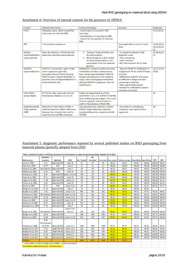
Attachment 5 shows a detailed overview of diagnostic performance characteristics
found by 24 different study groups. Data were partially adopted from the table
published in the meta-analysis by Geifman-Holtzman (11). The mean reported
diagnostic accuracy was 97.9% (range 80.8%-100%). Lowest diagnostic accuracy was
reported in two small studies from 2003, performed on 28 and 31 patients respectively.
Sensitivity and specificity values ranged from 82.4%-100% and 66.7%-100%
respectively, with positive and negative predictive values ranging from 89.5%-100%
and 57.1%-100% respectively.
Several of these studies were carried out on a small patient population. The total
number of included patients ranged from 20 to 1869.
Geifman-Holtzman et al. concluded from their analysis that best diagnostic accuracy
was reached when obtaining the sample during the first trimester (11). However, only 1
out of 3 published studies reported gestational age (11). Opposite results were reported
by Akolekar et al., who evaluated 591 samples from first trimester pregnancies (23).
They noticed a significantly higher false negative rate at 11-13 weeks (3.5%) than the
rate of < 0.2% at 26-32 weeks, reported by Finning et al. (23,27). This is most likely
due to the failure to detect cffDNA in early pregnancy (23). A potential solution is a
repeat test at 26 weeks to reassess the need for prophylactic anti-D (23). This implies
the additional cost of an extra test, but women with a false negative result will not be
deprived of receiving prophylactic anti-D (23).
In 2009, Freeman et al. performed a systematic review of the quality of reporting of
diagnostic accuracy in published studies, an item which was not considered by
Geifman-Holtzman et al. (10). 27 studies were assessed using the STARD (standards
for reporting studies of diagnostic accuracy) checklist (attachment 6) (10). Scores
ranged from 5/25 to 13/25 points, illustrating a generally poor quality of reporting
(attachment 7) (10). There is a widespread exclusion of inconclusive and unobtainable
results due to insufficient fetal material or maternal variant genes. Both will have
implications for population wide implementation (10). In clinical practice, all such
samples excluded from analysis would represent individuals for whom the test cannot
produce a result (10). Inconclusive/unobtainable results should be managed as RHD
positive since statistically, the majority will indeed be RHD positive (23).
The systematic exclusion of inconclusive or unobtainable results continues to exist in
recent publications. In 2011, Bombard et al. reported a diagnostic accuracy of 97.1%,
decreasing to 85.2% if the 29 excluded samples (12% of all samples) were taken into
account (22). Akolekar et al. found a diagnostic accuracy of 98.8% when performing
the PCR on 591 samples obtained during the first trimester of pregnancy (11-13 weeks
gestational age) (23). However, when inconclusive or unobtainable results were
included, accuracy decreased to a meager 84.6%. As many as 15% of all performed
PCRs produced inconclusive results, which the authors attributed to the relatively high
proportion of African women (19.3%) (23).
Another issue is the lack of large-scale peer-reviewed studies reporting on high-
throughput testing of non-sensitized women (10). The proposal for the large-scale
implementation of this test is based on results from small-scale studies (10,21). Most
data from studies have been obtained in a research setting rather than a clinical setting
and lacked a control for the presence of fetal DNA in case of negative results. However,
the cost of the test, as well as its reliability, will be determined by the number of retests
(10). Finning et al. were the only group that published a report on the effect of high

Fetal RHD genotyping on maternal blood using cffDNA
Katinka De Vreese pagina 11/18
throughput RHD typing (27). A total of 1869 patients was included, with the majority
(92%) having a gestational age of 26-30 weeks (range 8-38W) (27). Accuracy was
determined at 95.7% with a false negative rate of 0.16% (27). The authors concluded
that conclusive negative results were obtained in 36% of the women tested, which, in
England and Wales, would represent 35.000 to 40.000 mothers per year (attachment 8)
(27,30).
Sanquin Diagnostic Services offers noninvasive fetal blood group genotyping for rhesus
D in maternal plasma for alloimmunized pregnant women since the beginning of 2003,
using a stringent diagnostic algorithm with the inclusion of fetal DNA identifiers to
exclude false-negative results (21). Through the analysis of both exon 5 and 7, false
positive results due to variant genes could be avoided.
Bombard et al. evaluated analysis of cffDNA using MALDI-TOF MS (22). They used
two cohorts of patients: in the first cohort of 236 patients, they compared the result with
serological RhD determination on cord blood; in the second cohort of 205 patients, they
compared the result with PCR determined RHD genotype (22).
The reported diagnostic accuracy was 97.1% in cohort 1 and 99.5% in cohort 2 (22).
But again, in this study, accuracy was calculated after exclusion of inconclusive or
unobtainable results. After correction for this, diagnostic accuracy decreased to 85.2%
in cohort 1 and 96.6% in cohort 2. Positive and negative predictive values were 98.6%
and 94% respectively in cohort 1 and 99.3% and 100% respectively in cohort 2,
demonstrating an excellent concordance between the two methods (22)

Fetal RHD genotyping on maternal blood using cffDNA
Katinka De Vreese pagina 12/18
1. Clinical impact
A. Diagnostic aspect
The use of cffDNA avoids invasive procedures such as amniocentesis and chorionic
villus sampling to determine fetal RHD status in anti-D sensitized women
(13,24,36). If the fetus is RHD negative, it is not at risk for HDFN and no further
antenatal monitoring or invasive procedure is required (12,23). In cases where the
fetus is RHD positive, the woman should be referred to a tertiary fetal medicine unit
for appropriate management. Serial assessment of maternal antibody titers and
monitoring for fetal anemia are advisable (12,15,21,23).
The lack of a universal internal control to demonstrate the presence of cffDNA in
the maternal plasma is the main cause for concern. Negative results should be
interpreted with caution since a small proportion will be labeled false negative.
Therefore, it is suggested to analyze a repeat sample 2-4 weeks later in case of a
negative result, especially when the sample was obtained at less than 12 weeks of
gestation.
Next to fetal RHD typing, the use of cffDNA has also opened new possibilities for
testing other blood group antigens, such as Kell, RhC/c and RhE/e (12,21).
B. Treatment
NIPD for RHD has no real therapeutic impact, since the decision to treat for HDFN
is based on clinical or echographic signs of fetal anemia or HDN. If significant
anemia is detected, intra-uterine transfusion by cordocentesis will be performed
using antigen negative blood (1,4,15). After birth, phototherapy or exchange
transfusion may be necessary (1,4).
C. Prevention
Introduction of postnatal anti-D prophylaxis within 72h of delivery in the late 1960s
reduced sensitization and HDFN rates considerably internationally (10,24,29). A
further reduction was obtained by the introduction of RAADP in non-sensitized
RhD negative women (10,24,27). However, 40% of RhD-negative women carry a
RhD negative fetus, leading to unnecessary administration of anti-D prophylaxis
(10,23,26,27,29). Fetal RHD genotyping on maternal plasma can reduce the number
of unnecessary anti-D injections.
Advantages of avoidance of unnecessary RAADP are listed below
(8,11,24,26,27,30,34).
- No unnecessary injections
- Avoidance of exposure to blood products / ethical considerations:
Anti-D Ig is a blood-derived product, obtained by immunization of RhD
negative male volunteers. Although considered safe for routine antenatal
use, administration of this biological substance is associated with a
theoretical risk of transferring (unrecognized) viruses or prions. Scientists
are currently working on recombinant anti-D Ig.
- Allergic reactions to RAADP administration are described
- Low supplies of anti-D immunoglobulin: Promotes efficient use of the
limited pool of anti-D immunoglobulin.
- Reduced costs
2. Organizational impact
Mass testing will require an accurate high-throughput automated laboratory procedure with
a very high test sensitivity (24). Up to date, there is still no universal control to confirm the
presence of fetal DNA available. The current available methods have a low throughput and
are labor intensive, so mass screening is not feasible unless development of highly
automated procedures which require as little replicates as possible (24).
2) Is it useful to implement fetal RHD genotyping in routine clinical
practice?

Fetal RHD genotyping on maternal blood using cffDNA
Katinka De Vreese pagina 13/18
3. Cost impact
A. Intellectual property rights
The use of cell-free fetal nucleic acids from maternal plasma has been patent-
protected by European patent EP 0994963B1 and the international US patent
6,258,540. The patents were granted in 2001 (US) and 2003 (UK) and are owned by
the University of Oxford’s ISIS Innovations Ltd, with Dennis Lo as one of the
inventors (39). The patents include all detection methods in prenatal diagnosis,
performed on maternal serum or plasma samples from a pregnant female, which
comprise detection of the presence of a nucleic acid of fetal origin (39). In 2005,
ISIS granted an exclusive license to the US-based company Sequenom Inc., giving
it rights to control the technology claimed in both patents, with exception of the use
of QF-PCR for RhD genotyping in Europe, which had previously been exclusively
licensed to L’Institut de Biotechnologies Jacques Boy in France (39).
In most European countries, there are rules that no liability arises if the patent
infringement is done privately for purposes which are not commercial, or for
experimental purposes (39). If the technology would be offered as a service,
however, this exception would no longer apply and, in the absence of a permission
or license, the service provider would be infringing the patent (39).
B. Commercial situation
The largest and most dominant company in this area is Sequenom Inc, based in San
Diego. Studies with their Mass-ARRAY platform are ongoing.
Other companies offering commercial tests for fetal RHD genotyping are L’Institut
de Biotechnologies Jacques Boy (Free DNA Fetal Kit® RhD) and BioRad/DiaMed
(Free DNA Fetal Kit® RhD, €5040 for 87 reactions) (39).
One dose of Rhogam costs 42.16 euro. Scientists are currently working on the
development of recombinant anti-D IgG as a replacement for human plasma
products (24,27). A phase 2 clinical trial is in progress (24,27).
C. Cost-effectiveness
In most countries, RHD genotyping on maternal plasma is only offered in case of
anti-D alloimmunization. Szczepura et al. performed an economic evaluation of
mass testing for fetal RHD genotype on maternal blood in England and Wales (24).
They evaluated the costs of an in-house test as well as a commercially available test
(Jacques Boy) and evaluated the impact on national savings in case of introduction
of large-scale testing (24). Use of the commercial kit would make NIPD more
expensive as compared to RAADP (24). Use of the in-house developed kit would
lead to a national saving from 0.46-1.6% as long as zero royalty fee applies (24). If
a royalty fee is obliged, NIPD targeted antenatal prophylaxis would no longer be
cost-effective (24). They conclude that their findings do not support large-scale
introduction of RHD NIPD testing for non-sensitized pregnancies (24).
However, in the Netherlands, an economic evaluation apparently showed cost-
effectiveness of large-scale RHD genotyping on maternal plasma in non-immunized
women, leading to the aforementioned national implementation (2,17).
If a reliable recombinant alternative for human anti-D immunoglobulin is produced,
NIPD may become financially more attractive since this recombinant product will
probably be more expensive (24,27).
4. Decision making
There is currently a refund provided by the Belgian health care system for fetal RHD
genotyping in a pregnant woman, in case of anti-D alloimmunization or prior to an invasive
procedure (B5000) (44). The test is offered by the Centre for Human Genetics (CME) of the
University Hospitals of Leuven, who perform an in-house developed real-time PCR using
exon 10.
With regard to large-scale implementation in Belgium for non-sensitized women, there are
several remarks to be made. Most studies were set in a research setting rather than a
clinical-diagnostic setting, with exclusion of inconclusive or unobtainable results. However,
these results should be considered as RHD positive, thus anti-D prophylaxis should be
administered. This should be taken into account in cost-effectiveness studies.
Furthermore, due to the lack of an internal control for fetal DNA, analysis of several
replicates is necessary to improve the accuracy and sensitivity of the test. Current diagnostic
Fetal RHD genotyping on maternal blood using cffDNA
Katinka De Vreese pagina 14/18
strategies are expensive, time-consuming and labor-intensive and do not permit high-
throughput testing. As long as the presence of fetal DNA cannot be proved, negative results
should be interpreted with caution since they may represent false negative results due to
insufficient fetal DNA. Finally, to this date, there are insufficient data of the cost-
effectiveness of large-scale implementation of this test. Therefore, at this time, there will be
no implementation of large-scale testing in the laboratories of the Belgian Red Cross.
However, new developments, more specifically the clinical trial with MALDI-TOF MS, as
well as the results of the pilot study in the Netherlands, should be followed.
COMMENTS
Fetal RHD genotyping on maternal plasma using cffDNA is currently offered by the Centre for Human
Genetics (CME) of the University Hospitals of Leuven. In Leuven, only about 10-15 analyses/year are
requested.
TO DO/ACTIONS
1) Follow-up of results of pilot study in the Netherlands
2) Follow-up of results of clinical trials with MALDI-TOF MS
ATTACHMENTS
Attachment 1 (41)

Fetal RHD genotyping on maternal blood using cffDNA
Katinka De Vreese pagina 15/18
Attachment 2: Evaluation of the total amount (maternal + fetal) of extracted DNA from plasma en
serum samples from 50 pregnant women, using the β-globin TaqMan Assay (37).
Attachment 3: Choice of primers by different groups
Fetal RHD genotyping on maternal blood using cffDNA
Katinka De Vreese pagina 16/18
Attachment 4: Overview of internal controls for the presence of cffDNA
Attachment 5: diagnostic performance reported by several published studies on RhD genotyping from
maternal plasma (partially adopted from (20))
Table: published studies on RhD genotyping from fetal plasma in maternal plasma (partially adapted from (20))
References
Gestation
(weeks) Methods RhD No. Tested
No.
Inc luded # correct Accuracy Total accuracy Sensitivity Specific ity PPV NPV
Lo et al, 1998 7-41 Real-time PCR exon 10 57 57 55 96,5% 96,5% 94,9% 100,0% 100,0% 90,0%
Faas et al, 1998 16-17 PCR exon 7 31 31 31 100,0% 100,0% 100,0% 100,0% 100,0% 100,0%
Zhang et al, 2000 All trimesters Real-time PCR exon 7 58 58 57 98,3% 98,3% 96,9% 100,0% 100,0% 96,3%
Nelson et al, 2001 9-34 PCR exon 10 26 26 26 100,0% 100,0% 100,0% 100,0% 100,0% 100,0%
Finning et al, 2002 8-42 Real-time PCR exons 4,5,6,10 158 137 137 100,0% 86,7% 100,0% 100,0% 100,0% 100,0%
Costa et al, 2002 8-14 Real-time PCR exon 10 102 102 102 100,0% 100,0% 100,0% 100,0% 100,0% 100,0%
Legler et al, 2002 11-38 Real-time PCR exons 4,7 28 27 26 96,3% 92,9% 93,0% 100,0% 100,0% 92,3%
Turner et al, 2003 6-20 Real-time PCR exon 10 31 31 28 90,3% 90,3% 82,4% 100,0% 100,0% 82,4%
Siva et al, 2003 15-17 PCR exons 7,10 28 26 21 80,8% 75,0% 85,0% 66,7% 89,5% 57,1%
Rijnders et al, 2004 11-19 Real-time PCR exon 7 72 71 71 98,6% 98,6% 100,0% 96,6%
Rouillac et al, 2004 7-36 Real-time PCR exons 7,10 893 851 842 98,9% 94,3% 99,4% 97,5% 99,2% 98,0%
Finning et al, 2004 All trimesters Real-time PCR exons 4,5,10 233 226 223 98,7% 95,7% 100,0% 96,2% 98,0% 100,0%
Clausen et al, 2005 15-16 Real-time PCR exons 7,10 59 59 58 98,3% 98,3% 100,0% 100,0% 100,0% 100,0%
Zhou et al, 2005 14-42 Real-time PCR exons 4,5,10 98 96 92 95,8% 93,9% 94,4% 100,0% 100,0% 85,7%
Gautier et al, 2005 8-35 Real-time PCR exon 10 274 272 272 100,0% 99,3% 100,0% 100,0% 100,0% 100,0%
Gonzalez et al, 2005 11-16 Real-time PCR exon 7 20 20 20 100,0% 100,0% 100,0% 100,0% 100,0% 100,0%
Brojer et al, 2005 All trimesters Real-time PCR
exons 7,10;
intron 4 255 230 229 99,6% 89,8% 100,0% 100,0% 100,0% 100,0%
Rouillac et al, 2007 10-34 Real-time PCR 300 300 298 99,3% 99,3% 100,0% 97,5% 99,1% 100,0%
Minon et al, 2008 10-38 Real-time PCR exons 4,5,10 563 545 544 99,8% 96,6% 100,0% 99,5% 99,7% 100,0%
Finning et al, 2008
8-38
(92% between
26-30 W) Real-time PCR exons 5,7 1869 1869 1788 95,7% 95,7% 96,7% 94,0% 98,8% 99,5%
Bombard et al, 2011 11-13 Maldi-TOF MS exons 4,5,7 236 207 201 97,1% 85,2% 97,2% 96,9% 98,6% 94,0%
Bombard et al, 2011 6-30 Maldi-TOF MS exons 4,5,7 205 199 198 99,5% 96,6% 100,0% 98,3% 99,3% 100,0%
Akolekar et al, 2011 11-14 Real-time PCR exons 5,7 591 502 496 98,8% 83,9% 98,2% 100,0% 100,0% 96,5%
Scheffer et al, 2011 7-38 Real-time PCR exons 5,7 140*133 133 100,0% 95,0% 100,0% 100,0% 100,0% 100,0%
OVERALL 6349 6075 5948 97,9% 93,7%
*: total number in which serology was available + results inconclusive
Discordance reported accuracy - total accuracy

Fetal RHD genotyping on maternal blood using cffDNA
Katinka De Vreese pagina 17/18
Attachment 6: STARD checklist (10)
Attachment 7: weaknesses affecting the generalisability of reported accuracy rates of RhD NIPD in 27
studies (10).

Fetal RHD genotyping on maternal blood using cffDNA
Katinka De Vreese pagina 18/18
Attachment 8: estimation of impact of large-scale screening in England and Wales (41)