Cat claw injuries in corneas of dogs and cats - Eye Vet · Cat claw injuries in corneas of dogs and...
13
Cat claw injuries in corneas of dogs and cats By: Natasha Mitchell MVB CertVOphthal MRCVS, Eye Vet, Crescent Veterinary Clinic, Dooradoyle Road, Limerick www.eyevet.ie Cats have retractable claws which are versatile tools used for climbing, defence, balance, scratching and catching prey. They are also a formidable weapon which can inflict nasty scratches to animals perceived by the cat to be a threat (or a nuisance!). The most frequent victim is a younger dog, which is typically curious and has not yet learned to read hostile signals. They also do not develop a menace response until 8-12 weeks of age, reducing the protection that would otherwise be afforded to their corneas by blinking at the appropriate moment and retracting the third eyelid, globe and head. Brachycephalic dogs have shallow orbits and more exposed eyes, therefore they are even more vulnerable to injury. Corneal lacerations from cat claw injuries are unfortunately relatively common. The prognosis for the condition depends on the severity of injury caused. This in turn depends on the depth and angle of penetration, and the structures damaged. In general, there is a better prognosis for superficial (less than one third of the corneal thickness) rather than deep (greater than two thirds of the corneal thickness) corneal wounds, for peripheral rather than central corneal wounds, and for cats rather than dogs. Superficial ocular injuries carry a very good prognosis if the laceration is limited to the cornea, with no intraocular damage. Deeper penetration of the claw may cause full thickness laceration of the cornea, with release of aqueous humour. Small perforations (e.g. <1-2mm) may self-seal with a clot of aqueous humour and, later, fibrin. Larger perforations cause the sudden release of aqueous humour from the eye, which results in the iris moving forward to plug the deficit. This can result in anterior synechiae formation, or in prolapse of the iris, if the wound is sufficiently large. If the defect is large, the iris may protrude through the corneal wound. Of key importance is whether the claw penetrates deep enough to rupture the anterior lens capsule. This leads to intraocular exposure to lens protein, which results in severe phacoclastic uveitis which is generally unresponsive to medical treatment. Very small lens capsule tears can result in cataract at the site which can later affect the entire lens. Other potential damage to the eye includes secondary reflex uveitis, hyphaema, corneal infection and intraocular infection (endophthalmitis). Infection is not common but needs to be prevented as the cat claw is potentially dirty.
Transcript of Cat claw injuries in corneas of dogs and cats - Eye Vet · Cat claw injuries in corneas of dogs and...
Cat claw injuries in corneas of dogs and cats By: Natasha Mitchell MVB CertVOphthal MRCVS, Eye Vet, Crescent
Veterinary Clinic, Dooradoyle Road, Limerick www.eyevet.ie
Cats have retractable claws which are versatile tools used for climbing, defence, balance, scratching and catching prey. They are also a formidable weapon which can inflict nasty scratches to animals perceived by the cat to be a threat (or a nuisance!). The most frequent victim is a younger dog, which is typically curious and has not yet learned to read hostile signals. They also do not develop a menace response until 8-12 weeks of age, reducing the protection that would otherwise be afforded to their corneas by blinking at the appropriate moment and retracting the third eyelid, globe and head. Brachycephalic dogs have shallow orbits and more exposed eyes, therefore they are even more vulnerable to injury. Corneal lacerations from cat claw injuries are unfortunately relatively common. The prognosis for the condition depends on the severity of injury caused. This in turn depends on the depth and angle of penetration, and the structures damaged. In general, there is a better prognosis for superficial (less than one third of the corneal thickness) rather than deep (greater than two thirds of the corneal thickness) corneal wounds, for peripheral rather than central corneal wounds, and for cats rather than dogs. Superficial ocular injuries carry a very good prognosis if the laceration is limited to the cornea, with no intraocular damage. Deeper penetration of the claw may cause full
thickness laceration of the cornea, with release of aqueous humour. Small perforations (e.g. <1-2mm) may self-seal with a clot of aqueous humour and, later, fibrin. Larger perforations cause the sudden release of aqueous humour from the eye, which results in the iris moving forward to plug the deficit. This can result in anterior synechiae formation, or in prolapse of the iris, if the wound is sufficiently large. If the defect is large, the iris may protrude through the corneal wound. Of key importance is whether the claw penetrates deep enough to rupture the anterior lens capsule. This leads to intraocular exposure to lens protein, which results in severe phacoclastic uveitis which is generally unresponsive to medical treatment. Very small lens capsule tears can result in cataract at the site which can later affect the entire lens. Other potential damage to the eye includes secondary reflex uveitis, hyphaema, corneal infection and intraocular infection (endophthalmitis). Infection is not common but needs to be prevented as the cat claw is potentially dirty.
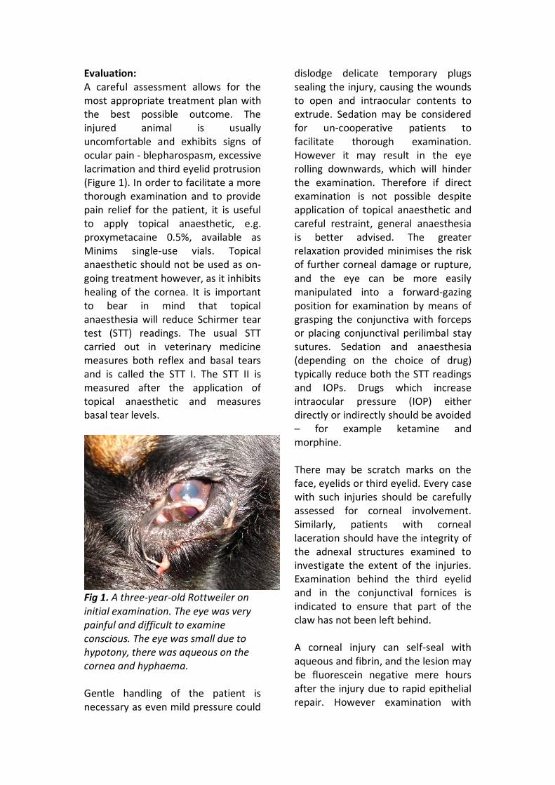
Evaluation: A careful assessment allows for the most appropriate treatment plan with the best possible outcome. The injured animal is usually uncomfortable and exhibits signs of ocular pain - blepharospasm, excessive lacrimation and third eyelid protrusion (Figure 1). In order to facilitate a more thorough examination and to provide pain relief for the patient, it is useful to apply topical anaesthetic, e.g. proxymetacaine 0.5%, available as Minims single-use vials. Topical anaesthetic should not be used as on-going treatment however, as it inhibits healing of the cornea. It is important to bear in mind that topical anaesthesia will reduce Schirmer tear test (STT) readings. The usual STT carried out in veterinary medicine measures both reflex and basal tears and is called the STT I. The STT II is measured after the application of topical anaesthetic and measures basal tear levels.
Fig 1. A three-year-old Rottweiler on initial examination. The eye was very painful and difficult to examine conscious. The eye was small due to hypotony, there was aqueous on the cornea and hyphaema. Gentle handling of the patient is necessary as even mild pressure could
dislodge delicate temporary plugs sealing the injury, causing the wounds to open and intraocular contents to extrude. Sedation may be considered for un-cooperative patients to facilitate thorough examination. However it may result in the eye rolling downwards, which will hinder the examination. Therefore if direct examination is not possible despite application of topical anaesthetic and careful restraint, general anaesthesia is better advised. The greater relaxation provided minimises the risk of further corneal damage or rupture, and the eye can be more easily manipulated into a forward-gazing position for examination by means of grasping the conjunctiva with forceps or placing conjunctival perilimbal stay sutures. Sedation and anaesthesia (depending on the choice of drug) typically reduce both the STT readings and IOPs. Drugs which increase intraocular pressure (IOP) either directly or indirectly should be avoided – for example ketamine and morphine. There may be scratch marks on the face, eyelids or third eyelid. Every case with such injuries should be carefully assessed for corneal involvement. Similarly, patients with corneal laceration should have the integrity of the adnexal structures examined to investigate the extent of the injuries. Examination behind the third eyelid and in the conjunctival fornices is indicated to ensure that part of the claw has not been left behind. A corneal injury can self-seal with aqueous and fibrin, and the lesion may be fluorescein negative mere hours after the injury due to rapid epithelial repair. However examination with
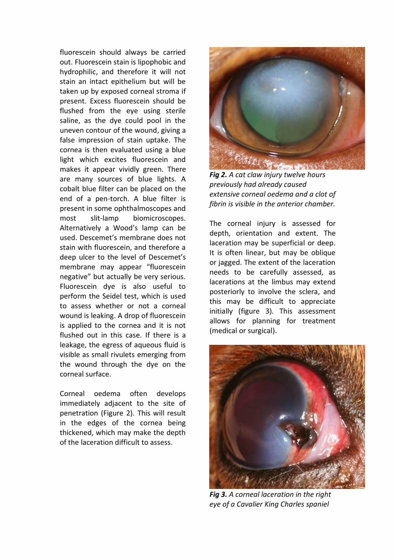
fluorescein should always be carried out. Fluorescein stain is lipophobic and hydrophilic, and therefore it will not stain an intact epithelium but will be taken up by exposed corneal stroma if present. Excess fluorescein should be flushed from the eye using sterile saline, as the dye could pool in the uneven contour of the wound, giving a false impression of stain uptake. The cornea is then evaluated using a blue light which excites fluorescein and makes it appear vividly green. There are many sources of blue lights. A cobalt blue filter can be placed on the end of a pen-torch. A blue filter is present in some ophthalmoscopes and most slit-lamp biomicroscopes. Alternatively a Wood’s lamp can be used. Descemet’s membrane does not stain with fluorescein, and therefore a deep ulcer to the level of Descemet’s membrane may appear “fluorescein negative” but actually be very serious. Fluorescein dye is also useful to perform the Seidel test, which is used to assess whether or not a corneal wound is leaking. A drop of fluorescein is applied to the cornea and it is not flushed out in this case. If there is a leakage, the egress of aqueous fluid is visible as small rivulets emerging from the wound through the dye on the corneal surface. Corneal oedema often develops immediately adjacent to the site of penetration (Figure 2). This will result in the edges of the cornea being thickened, which may make the depth of the laceration difficult to assess.
Fig 2. A cat claw injury twelve hours previously had already caused extensive corneal oedema and a clot of fibrin is visible in the anterior chamber. The corneal injury is assessed for depth, orientation and extent. The laceration may be superficial or deep. It is often linear, but may be oblique or jagged. The extent of the laceration needs to be carefully assessed, as lacerations at the limbus may extend posteriorly to involve the sclera, and this may be difficult to appreciate initially (figure 3). This assessment allows for planning for treatment (medical or surgical).
Fig 3. A corneal laceration in the right eye of a Cavalier King Charles spaniel
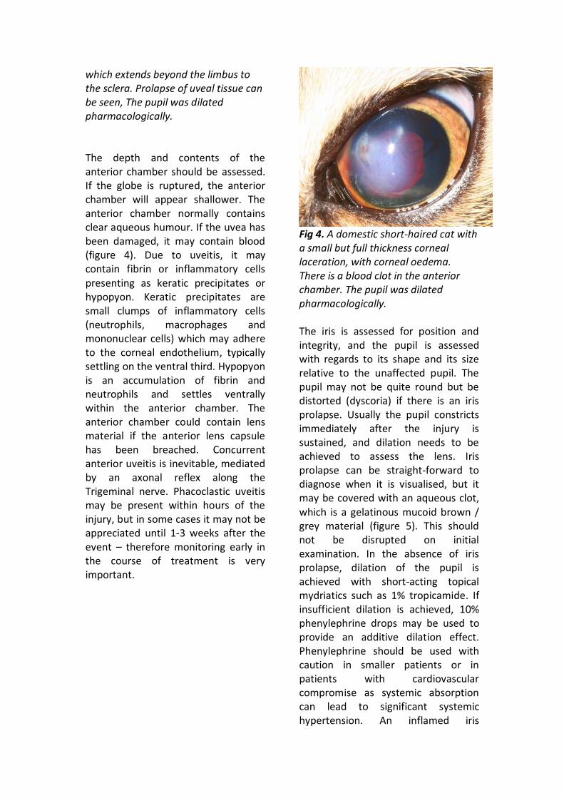
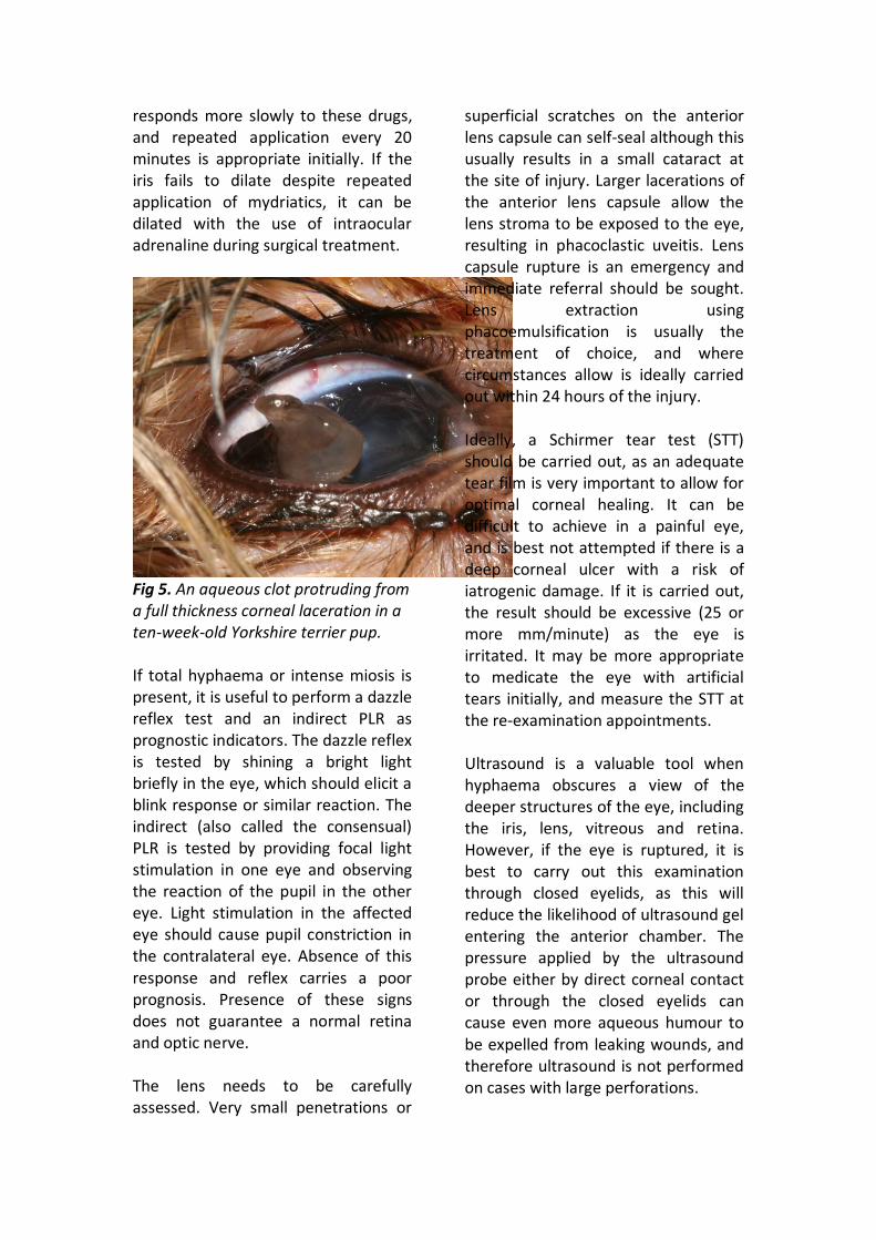
which extends beyond the limbus to the sclera. Prolapse of uveal tissue can be seen, The pupil was dilated pharmacologically. The depth and contents of the anterior chamber should be assessed. If the globe is ruptured, the anterior chamber will appear shallower. The anterior chamber normally contains clear aqueous humour. If the uvea has been damaged, it may contain blood (figure 4). Due to uveitis, it may contain fibrin or inflammatory cells presenting as keratic precipitates or hypopyon. Keratic precipitates are small clumps of inflammatory cells (neutrophils, macrophages and mononuclear cells) which may adhere to the corneal endothelium, typically settling on the ventral third. Hypopyon is an accumulation of fibrin and neutrophils and settles ventrally within the anterior chamber. The anterior chamber could contain lens material if the anterior lens capsule has been breached. Concurrent anterior uveitis is inevitable, mediated by an axonal reflex along the Trigeminal nerve. Phacoclastic uveitis may be present within hours of the injury, but in some cases it may not be appreciated until 1-3 weeks after the event – therefore monitoring early in the course of treatment is very important.
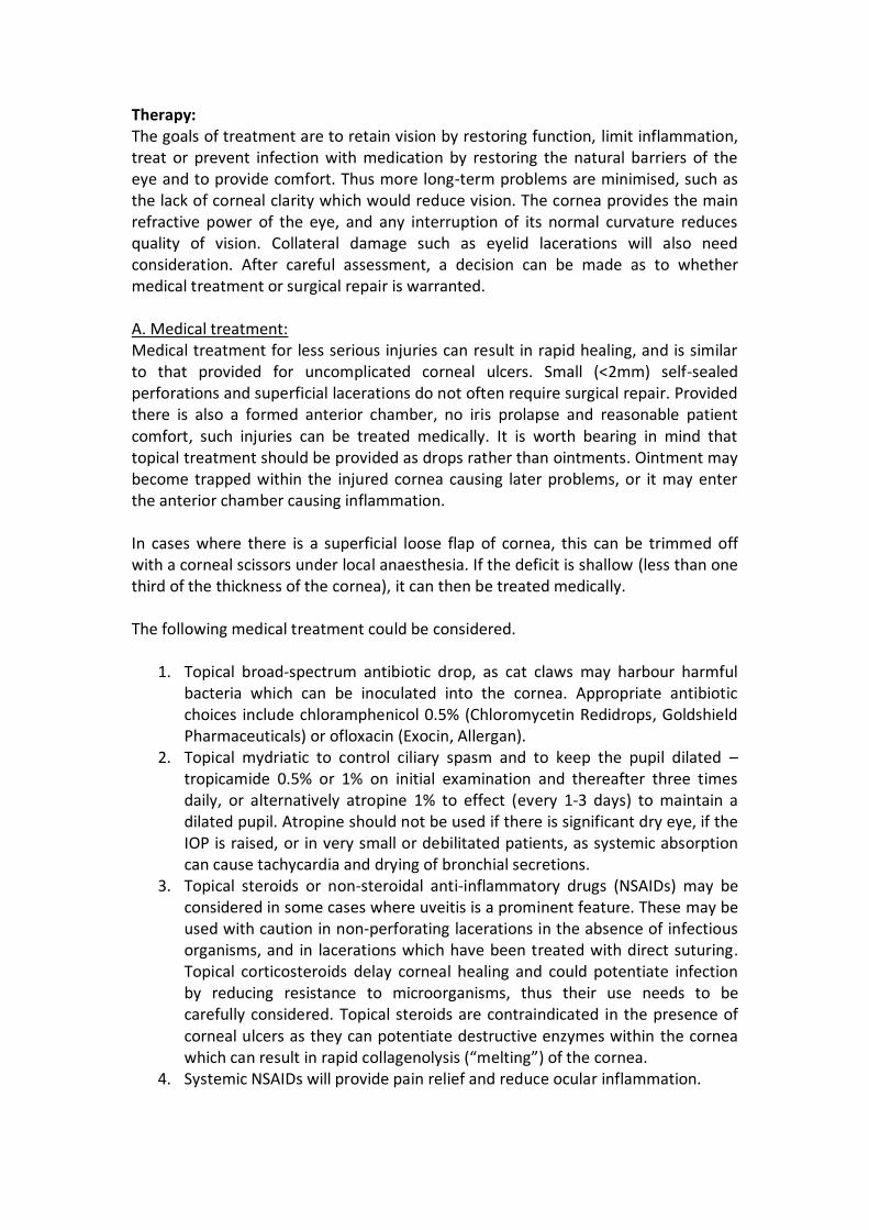
Fig 4. A domestic short-haired cat with a small but full thickness corneal laceration, with corneal oedema. There is a blood clot in the anterior chamber. The pupil was dilated pharmacologically. The iris is assessed for position and integrity, and the pupil is assessed with regards to its shape and its size relative to the unaffected pupil. The pupil may not be quite round but be distorted (dyscoria) if there is an iris prolapse. Usually the pupil constricts immediately after the injury is sustained, and dilation needs to be achieved to assess the lens. Iris prolapse can be straight-forward to diagnose when it is visualised, but it may be covered with an aqueous clot, which is a gelatinous mucoid brown / grey material (figure 5). This should not be disrupted on initial examination. In the absence of iris prolapse, dilation of the pupil is achieved with short-acting topical mydriatics such as 1% tropicamide. If insufficient dilation is achieved, 10% phenylephrine drops may be used to provide an additive dilation effect. Phenylephrine should be used with caution in smaller patients or in patients with cardiovascular compromise as systemic absorption can lead to significant systemic hypertension. An inflamed iris
responds more slowly to these drugs, and repeated application every 20 minutes is appropriate initially. If the iris fails to dilate despite repeated application of mydriatics, it can be dilated with the use of intraocular adrenaline during surgical treatment.
Fig 5. An aqueous clot protruding from a full thickness corneal laceration in a ten-week-old Yorkshire terrier pup. If total hyphaema or intense miosis is present, it is useful to perform a dazzle reflex test and an indirect PLR as prognostic indicators. The dazzle reflex is tested by shining a bright light briefly in the eye, which should elicit a blink response or similar reaction. The indirect (also called the consensual) PLR is tested by providing focal light stimulation in one eye and observing the reaction of the pupil in the other eye. Light stimulation in the affected eye should cause pupil constriction in the contralateral eye. Absence of this response and reflex carries a poor prognosis. Presence of these signs does not guarantee a normal retina and optic nerve. The lens needs to be carefully assessed. Very small penetrations or
superficial scratches on the anterior lens capsule can self-seal although this usually results in a small cataract at the site of injury. Larger lacerations of the anterior lens capsule allow the lens stroma to be exposed to the eye, resulting in phacoclastic uveitis. Lens capsule rupture is an emergency and immediate referral should be sought. Lens extraction using phacoemulsification is usually the treatment of choice, and where circumstances allow is ideally carried out within 24 hours of the injury. Ideally, a Schirmer tear test (STT) should be carried out, as an adequate tear film is very important to allow for optimal corneal healing. It can be difficult to achieve in a painful eye, and is best not attempted if there is a deep corneal ulcer with a risk of iatrogenic damage. If it is carried out, the result should be excessive (25 or more mm/minute) as the eye is irritated. It may be more appropriate to medicate the eye with artificial tears initially, and measure the STT at the re-examination appointments. Ultrasound is a valuable tool when hyphaema obscures a view of the deeper structures of the eye, including the iris, lens, vitreous and retina. However, if the eye is ruptured, it is best to carry out this examination through closed eyelids, as this will reduce the likelihood of ultrasound gel entering the anterior chamber. The pressure applied by the ultrasound probe either by direct corneal contact or through the closed eyelids can cause even more aqueous humour to be expelled from leaking wounds, and therefore ultrasound is not performed on cases with large perforations.
Therapy: The goals of treatment are to retain vision by restoring function, limit inflammation, treat or prevent infection with medication by restoring the natural barriers of the eye and to provide comfort. Thus more long-term problems are minimised, such as the lack of corneal clarity which would reduce vision. The cornea provides the main refractive power of the eye, and any interruption of its normal curvature reduces quality of vision. Collateral damage such as eyelid lacerations will also need consideration. After careful assessment, a decision can be made as to whether medical treatment or surgical repair is warranted. A. Medical treatment: Medical treatment for less serious injuries can result in rapid healing, and is similar to that provided for uncomplicated corneal ulcers. Small (<2mm) self-sealed perforations and superficial lacerations do not often require surgical repair. Provided there is also a formed anterior chamber, no iris prolapse and reasonable patient comfort, such injuries can be treated medically. It is worth bearing in mind that topical treatment should be provided as drops rather than ointments. Ointment may become trapped within the injured cornea causing later problems, or it may enter the anterior chamber causing inflammation. In cases where there is a superficial loose flap of cornea, this can be trimmed off with a corneal scissors under local anaesthesia. If the deficit is shallow (less than one third of the thickness of the cornea), it can then be treated medically.
The following medical treatment could be considered.
1. Topical broad-spectrum antibiotic drop, as cat claws may harbour harmful bacteria which can be inoculated into the cornea. Appropriate antibiotic choices include chloramphenicol 0.5% (Chloromycetin Redidrops, Goldshield Pharmaceuticals) or ofloxacin (Exocin, Allergan).
2. Topical mydriatic to control ciliary spasm and to keep the pupil dilated – tropicamide 0.5% or 1% on initial examination and thereafter three times daily, or alternatively atropine 1% to effect (every 1-3 days) to maintain a dilated pupil. Atropine should not be used if there is significant dry eye, if the IOP is raised, or in very small or debilitated patients, as systemic absorption can cause tachycardia and drying of bronchial secretions.
3. Topical steroids or non-steroidal anti-inflammatory drugs (NSAIDs) may be considered in some cases where uveitis is a prominent feature. These may be used with caution in non-perforating lacerations in the absence of infectious organisms, and in lacerations which have been treated with direct suturing. Topical corticosteroids delay corneal healing and could potentiate infection by reducing resistance to microorganisms, thus their use needs to be carefully considered. Topical steroids are contraindicated in the presence of corneal ulcers as they can potentiate destructive enzymes within the cornea which can result in rapid collagenolysis (“melting”) of the cornea.
4. Systemic NSAIDs will provide pain relief and reduce ocular inflammation.
5. Artificial tears such as hydroxypropyl methylcellulose (Hypromellose) (Isopto Plain, Alcon) lubricate the cornea, improve patient comfort and enhance healing.
6. A buster (Elizabethan) collar should be used to prevent further self-trauma. 7. Avoid topical local anaesthetic as a treatment, as it can inhibit corneal
healing. 8. The patient should be rested.
B. Surgical treatment: Surgical repair is indicated in the following circumstances:
1. Non-perforating corneal wound where the superficial edges are gaping 2. Perforating corneal wounds which are leaking (Seidel test positive) 3. Iris prolapse 4. Deep corneal ulcers i.e. greater or equal to one third corneal depth 5. Necrotic cornea at the edges of the laceration 6. Lens capsule rupture with release of lens material 7. Lens capsule rupture with phacoclastic uveitis 8. Cataract as a result of lens penetration
Preparation of the patient: General anaesthesia is induced with gentle patient handling. The patient is placed in lateral or dorsal recumbency, depending on the surgeon’s preference. The head is positioned so that the cornea is parallel to the operating table. The head is maintained in this position by the use of sandbags, rolled-up towels, or ideally a buster cushion / vacuum pillow. A lateral canthotomy may be planned in cases with deep-set or small eyes to increase surgical exposure, in which case an area on the lateral canthus should be clipped. Peri-ocular hair may also be gently clipped in certain cases, as it will retain ocular discharge post-operatively which can be irritating for the patient and it is difficult for the owner to keep clean. The eyelids are cleaned with 1:20 povidone iodine solution. The globe is irrigated using sterile saline. In the case of a perforated cornea, generally the use of disinfectants such as dilute povidone iodine in avoided, in case it enters the eye. The use of non-depolarising neuromuscular blocking agents can assist in achieving optimal surgical results e.g. vecuronium (Norcuron). It provides globe centralisation for easier visualisation of the affected cornea and thus improves access to the injured area. It relaxes the musculature around the eye which reduces the pressure on the globe allowing for easier manipulation and less risk of corneal rupture. A mechanical ventilator, peripheral nerve stimulator, monitoring equipment and anaesthetist familiar with the procedure are required. Corneal surgery is painful as the tissue contains a large number of nerve endings. Topical anaesthesia, at the start of and during the procedure, improves patient comfort and may allow for a slightly lighter plane of anaesthesia.
To achieve the best possible outcome for the patient, it is important to have access to adequate magnification and ophthalmic surgical instruments, and to have some experience with microsurgery before undertaking corneal surgery. Sometimes it will not have been possible to fully examine the lens completely before surgery due to corneal oedema, pupil constriction and turbid aqueous humour. Should a lens capsule rupture become apparent during surgery, it is better for the patient that phacoemulsification to remove the lens and implantation of an artificial lens is carried out at the same time, to minimise the effects of phacoclastic uveitis. Therefore prompt referral should be considered if the integrity of the lens is unknown. An example of the equipment useful for corneal surgery is outlined below. It does not include further equipment and instrumentation that would be required for phacoemulsification.
Equipment required:
Equipment Use
Magnification Some form of magnification is required in order to ensure precise tissue handling and accurate assessment and placement of sutures. Ideally an operating microscope is used. Head loupes are available with varying magnification, and at least 4X magnification is recommended.
Drapes A fenestrated drape, such as that used for neutering procedures, is appropriate for use around the eye. However the openings are usually large. To provide for optimal sterility, a disposable water-resistant drape with an adhesive plastic window is very useful in conjunction with the material drape. After placement on the eye, a scissors is used to incise the adhesive window in alignment with the eyelid aperture.
Eyelid speculum The incision made in the adhesive drape is folded back underneath the eyelids using eyelid speculum, which retracts the eyelids and enhances exposure of the cornea and conjunctiva when opened.
Tenotomy scissors For cutting sticky drape and larger suture material (e.g. 3/0 Vicryl)
Needle holders, e.g. Castroviejo
For the placement of stay sutures.
Toothed forceps e.g. Bishop-Harmon
For globe manipulation by grasping the conjunctiva.
Fine-tipped non-locking needle holder e.g. Barraquer
For suturing the cornea with small calibre needles.
Colibri forceps (0.12mm)
The delicate tips are useful for handling the cornea. They also have a tying platform, eliminating the need to change instrumentation (although tying forceps offer the advantage of causing less damage to fine suture material).
Tying forceps (pair) To grasp fine suture material when tying knots as an alternative to using the needle holders and Colibri forceps.
Vannus scissors For excision of damaged prolapsed iris and to cut fine suture material
Iris repositor / cyclodialysis spatula
To free adhesions of the iris to the corneal wound and to gently replace the iris in the anterior chamber.
Beaver blade No 64 or 69
May be required for debridement of the edge of a wound – these blades have rounded tips.
Beaver blade handle To hold the beaver blade.
Suture material 3/0-6/0 polyglactin 910 (Vicryl) for stay sutures 8/0 or 9/0 polyglactin 910 for corneal sutures
Viscoelastic e.g. sodium hyaluronate 1%
This agent fills the anterior chamber which reforms it, provides some rigidity to the cornea which makes suturing easier, separates tissue e.g. pushes the iris back, tamponades bleeding vessels and minimises synechiae.
Adrenaline Intraocular adrenaline will dilate the pupil, allowing for inspection of the anterior lens capsule, and will also reduce intraocular haemorrhage.
Cellulose spears Preferable to cotton or gauze swabs, as cotton fibres could enter the eye.
Balanced Salt Solution (BSS)
It is essential to keep the cornea and conjunctiva lubricated during the procedure with BSS or sterile saline. This can also be used to re-inflate the eye at the end of surgery.
Cannulas For corneal irrigation, viscoelastic injection and re-inflation of the anterior chamber at the end of surgery.
The most appropriate suture material for use in the cornea would be fine, monofilament and non-absorbable, for example 10/0 nylon. However the most practical choice in veterinary medicine is absorbable e.g. polyglactin 910 (Vicryl) which has good tensile strength and is does not cause a great degree of inflammation. 8/0 and 9/0 Vicryl are popular choices. Needles should be swaged on to minimise trauma while travelling through the tissue. The tip of the needle should be very sharp for penetration, and should be spatulated so that the needle can travel as atraumatically as possible through the corneal lamellae. The recommended needle curvature is ½ to ⅜ circle. Surgical techniques: Primary reconstructive surgical repair is possible in most cases of full or partial thickness corneal laceration as generally no tissue is missing. Therefore primary closure should be sufficient for a water-tight seal which heals uneventfully. As already outlined, surgical repair is not indicated for more superficial lacerations but is very useful for partial-thickness lacerations which have gaping edges. The aim is to accurately appose the edges of the wound, without excessive tension. Some tension needs to be exerted to prevent aqueous leakage, but should be appositional rather than compressive.
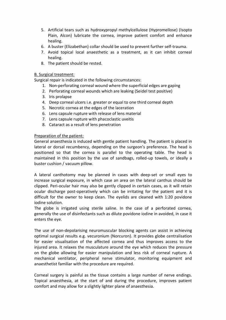
The surgeon must be seated and have arm-rests in order to maximise fine motor control for precision while performing fine surgery. The needle is grasped at the half-way point or slightly anterior. The needle is held perpendicular to the corneal surface. Using finger movement only, the needle holders are rotated so that the needle travels though the cornea following the curve of the needle such that it will exit perpendicular to the wound on the other side. Sutures should be placed at 70-90% depth of the cornea, as full-thickness sutures can provide a route for infectious organisms to enter the anterior chamber. The suture should exit 1-2 mm from the wound edge in order to avoid the oedematous wound margins. The most common pattern employed is the simple interrupted suture (figure 6). Simple continuous or double saw-tooth patterns may be used alternatively, depending on the preference of the surgeon. If the laceration is very large (>7mm), it may be useful to pre-place horizontal mattress sutures and close them once all are in place.
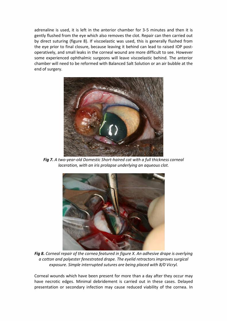
Fig 6. The Rottweiler featured in figure 1 the following morning. The eye was much more comfortable. Simple interrupted corneal sutures were used to repair the large corneal laceration. The pupil is not yet fully dilated despite application of mydriatics, due to uveitis. In perpendicular lacerations, sutures should be placed symmetrically. In other words, the corneal entry is at an equal distance from the laceration on both sides of the wound (1-2mm). For oblique laceration, the suture needs to be placed further from the wound on the side of the wound with the deeper laceration, and closer to the wound on the side of the more superficial wound, in order to create precise apposition. This causes the sutures to appear asymmetrical on the surface. If there is an aqueous clot present, it is gently debrided from the wound, bearing in mind that the iris might be prolapsed (figure 7). If it appears to be viable, replacement of the incarcerated iris is preferable to amputation. Replacement into the anterior chamber can be achieved using viscoelastic material or a cyclodialysis spatula. However, if the iris is devitalised or necrotic, or if the prolapse has been present for several days, it will be best to excise the damaged tissue. This can be achieved using sharp Vannus scissors. As the tissue is devitalised, no haemorrhage may occur. However, if haemorrhage does occur, it can be controlled with intracameral adrenaline 1:10,000 or may be tamponaded with viscoelastic. If
adrenaline is used, it is left in the anterior chamber for 3-5 minutes and then it is gently flushed from the eye which also removes the clot. Repair can then carried out by direct suturing (figure 8). If viscoelastic was used, this is generally flushed from the eye prior to final closure, because leaving it behind can lead to raised IOP post-operatively, and small leaks in the corneal wound are more difficult to see. However some experienced ophthalmic surgeons will leave viscoelastic behind. The anterior chamber will need to be reformed with Balanced Salt Solution or an air bubble at the end of surgery.
Fig 7. A two-year-old Domestic Short-haired cat with a full thickness corneal
laceration, with an iris prolapse underlying an aqueous clot.
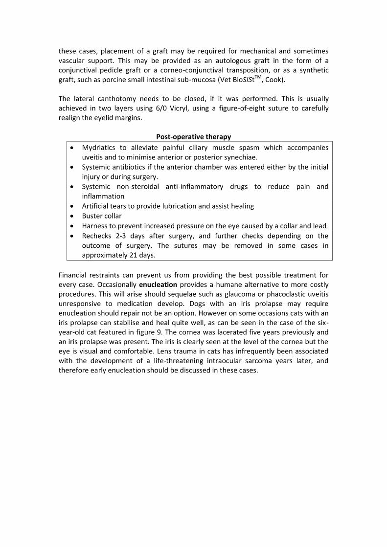
Fig 8. Corneal repair of the cornea featured in figure X. An adhesive drape is overlying
a cotton and polyester fenestrated drape. The eyelid retractors improves surgical exposure. Simple interrupted sutures are being placed with 8/0 Vicryl.
Corneal wounds which have been present for more than a day after they occur may have necrotic edges. Minimal debridement is carried out in these cases. Delayed presentation or secondary infection may cause reduced viability of the cornea. In
these cases, placement of a graft may be required for mechanical and sometimes vascular support. This may be provided as an autologous graft in the form of a conjunctival pedicle graft or a corneo-conjunctival transposition, or as a synthetic graft, such as porcine small intestinal sub-mucosa (Vet BioSIStTM, Cook). The lateral canthotomy needs to be closed, if it was performed. This is usually achieved in two layers using 6/0 Vicryl, using a figure-of-eight suture to carefully realign the eyelid margins.
Post-operative therapy
Mydriatics to alleviate painful ciliary muscle spasm which accompanies uveitis and to minimise anterior or posterior synechiae.
Systemic antibiotics if the anterior chamber was entered either by the initial injury or during surgery.
Systemic non-steroidal anti-inflammatory drugs to reduce pain and inflammation
Artificial tears to provide lubrication and assist healing
Buster collar
Harness to prevent increased pressure on the eye caused by a collar and lead
Rechecks 2-3 days after surgery, and further checks depending on the outcome of surgery. The sutures may be removed in some cases in approximately 21 days.
Financial restraints can prevent us from providing the best possible treatment for every case. Occasionally enucleation provides a humane alternative to more costly procedures. This will arise should sequelae such as glaucoma or phacoclastic uveitis unresponsive to medication develop. Dogs with an iris prolapse may require enucleation should repair not be an option. However on some occasions cats with an iris prolapse can stabilise and heal quite well, as can be seen in the case of the six-year-old cat featured in figure 9. The cornea was lacerated five years previously and an iris prolapse was present. The iris is clearly seen at the level of the cornea but the eye is visual and comfortable. Lens trauma in cats has infrequently been associated with the development of a life-threatening intraocular sarcoma years later, and therefore early enucleation should be discussed in these cases.
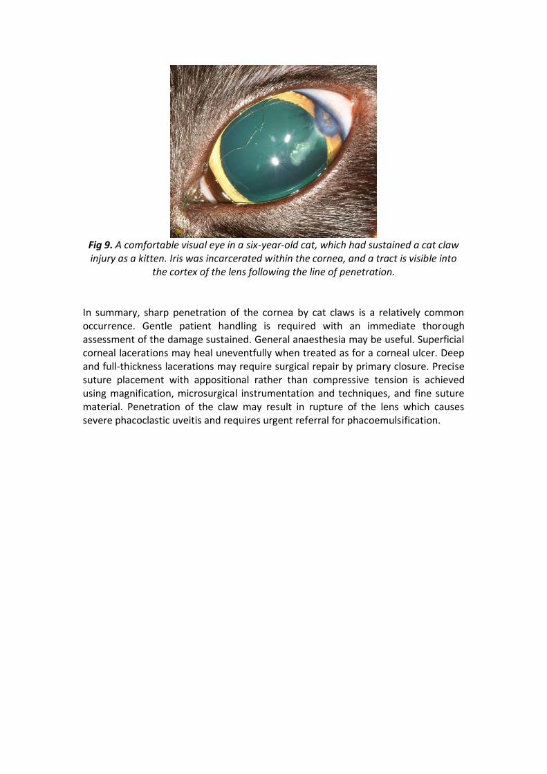
Fig 9. A comfortable visual eye in a six-year-old cat, which had sustained a cat claw injury as a kitten. Iris was incarcerated within the cornea, and a tract is visible into
the cortex of the lens following the line of penetration. In summary, sharp penetration of the cornea by cat claws is a relatively common occurrence. Gentle patient handling is required with an immediate thorough assessment of the damage sustained. General anaesthesia may be useful. Superficial corneal lacerations may heal uneventfully when treated as for a corneal ulcer. Deep and full-thickness lacerations may require surgical repair by primary closure. Precise suture placement with appositional rather than compressive tension is achieved using magnification, microsurgical instrumentation and techniques, and fine suture material. Penetration of the claw may result in rupture of the lens which causes severe phacoclastic uveitis and requires urgent referral for phacoemulsification.