Case Study: SCID/TREC/DBS/qPCR Monitoring - APHL · Case Study: SCID/TREC/DBS/qPCR. Monitoring....
64
Case Study: SCID/TREC/DBS/qPCR Monitoring April 25, 2016 APHL-CDC Carlos A. Saavedra-Matiz, MD Newborn Screening Program Wadsworth Center New York State Department of Health “Newborn screening is an extraordinarily successful example of the application of advances in medical genetics to personal and public health benefit.” Editorial , Nature Genetics, October 2010
Transcript of Case Study: SCID/TREC/DBS/qPCR Monitoring - APHL · Case Study: SCID/TREC/DBS/qPCR. Monitoring....

Case Study: SCID/TREC/DBS/qPCRMonitoring
April 25, 2016APHL-CDC
Carlos A. Saavedra-Matiz, MDNewborn Screening Program
Wadsworth CenterNew York State Department of Health
“Newborn screening is an extraordinarily successful example of the application of advances in medical genetics to personal and public health benefit.”
Editorial , Nature Genetics, October 2010

SCID: Severe Combined Immunodeficiency
• Heterogeneous group of genetic disorders
• Defects of cellular and humoral immune responses
• Absence or very low T-cell counts
• Incidence : 1 / 40,000-75,000 newborns
• Clinical symptoms: first months of life (median age at Dx 4-7 months)
• Multiple severe viral, bacterial and fungal infections after birth
• Often fatal within first year of life
• Survival rate is 94% when treated with HSCT by 3.5 months of age
• TREC (T-cell Receptor Excision Circles) are DNA by-products of T-cell maturation in the thymus
• Low or absent TRECs in DBS likely indicates T-cell deficiency


δRec-ψJα TREC Is Ideal
• Created from the sequential rearrangements of the TCR α/δlocus – 70% of thymocytes that express α/β TCR will form this specific TREC
• Signal joint region of this TREC is flanked by a conserved region– Allows for universal primer design that will always detect this TREC
• Occurs late in maturation– Likely to generate a functional and diverse T cell repertoire

Real Time PCR
• Reporter/Quencher
• 5’ Nuclease Activity
• Probe Cleavage
• Sequence Specific
• Multiplex Capability
Taqman

AbstractBACKGROUND: Dried blood spot (DBS) samples have been widely used in newborn screening (NBS) for the early identification of disease to facilitate the presymptomatic treatment of congenital diseases in newborns. As molecular genetics knowledge and technology progresses, there is an increased demand on NBS programs for molecular testing and a need to establish reliable, low-cost methods to perform those analyses. Here we report a flexible, cost-efficient, high-throughput DNA extraction method from DBS adaptable to small- and large-scale screening settings.METHODS: Genomic DNA (g.DNA) was extracted from single 3-mm diameter DBS by the sequential use of red cell lysis, detergent-alkaline, and acid-neutralizing buffers routinely used in whole blood and plant tissue DNA extractions. We performed PCR amplification of several genomic regions using standard PCR conditions and detection methods (agarose gel, melting-curve analysis, TaqMan-based assays). Amplicons were confirmed by BigDye® Terminator cycle sequencing and compared with reference sequences.RESULTS: High-quality g.DNA was extracted from hundreds of DBS, as proven by mutation detection of several human genes on multiple platforms. Manual and automated extraction protocols were validated. Quantification of g.DNA by Oligreen® fluorescent nucleic acid stain demonstrated a normal population distribution closely corresponding with white blood cell counts detected in newborn populations.CONCLUSIONS: High-quality, amplifiable g.DNA is extractable from DBSs. Our method is adaptable, reliable, and scalable to low- and high-throughput NBS at low cost ($0.10/sample). This method is routinely used for molecular testing in the New York State NBS program.
Clin Chem 59:7 (2013) Mar 25. [Epub ahead of print]doi:1373/clinchem.2013.205864 EditorialsNewborn Screening by Sequence and the Road Ahead.Sondheimer N.SourceDepartment of Pediatrics, University of Pennsylvania, and Section of Biochemical Genetics, Children’s Hospital Philadelphia, Philadelphia, PA.
Clin Chem 59:7 (2013) Mar 18. [Epub ahead of print] doi: 10.1373/clinchem.2012.198945 Cost-Effective and Scalable DNA Extraction Method from Dried Blood Spots.Saavedra-Matiz CA, Isabelle JT, Biski CK, Duva SJ, Sweeney ML, Parker AL, Young AJ, Diantonio LL, Krein LM, Nichols MJ, Caggana M.SourceNewborn Screening Program, Division of Genetics, Wadsworth Center, New York State Department of Health, Albany, NY.

SCID Screening Method
3.2mm punch ~1000 DNA extractions/day ~1200 RT-qPCR samples/day
Analysis

History of SCID Testing
• Automated assay developed and validated 12/2009-9/2010
• Submitted validation package to CLEP for approval on 9/08/2010
• CLEP and emergency regulation approved 9/27/2010
• SCID screening started 9/29/2010
• 1st “True SCID” baby detected 12/27/2010 (NICHD Support)
• Presumptive Positive (Borderline) category added 1/25/2011
• Commissioner of Health officially adds SCID to NSP panel 4/12/2011
Why Automate?
• Accommodate increased throughput
• Reduce repetitive stress injuries
• Address staffing shortages
• Increase reproducibility and consistency of results

Follow up of Infants ≥ 37 weeks Gestational Age with Positive NBS for SCID in NYS
Repeat NBS ASAP
≥ 200 TRECS/µL
•NBS calls PMD & specialist @ Specialty Treatment Center (STC) •Determine if child had cardiac surgery including thymectomy & if pre-thymectomy screen was negative
PMD contacts parent/guardian
•Child evaluated by PMD or specialist•Check CBC, lymphocyte subsets, mitogens•Repeat NBS card sent to Wadsworth Lab •Specialist interprets tests & determines management plan as needed
< 200 TRECS/µL
No further follow up
Presumptive Positive/Borderline*: 151-199 TRECS /µL
Referral: <150 TRECS /µL

Duplex Amplification PlotsTREC Amplification plot Rnase p amplification plot
ADA
ADA


Overall PPV = 18.7%Classic SCID PPV = 2.1%
J Clin Immunol. 2014 Mar 1. [Epub ahead of print]Newborn Screening for SCID in New York State: Experience from the First Two Years.Vogel BH1, Bonagura V, Weinberg GA, Ballow M, Isabelle J, Diantonio L, Parker A, Young A, Cunningham-Rundles C, Fong CT, Celestin J, Lehman H, Rubinstein A, Siegel S, Weiner L, Saavedra-Matiz C, Kay DM, Caggana M.


Race/ethnicity
Proportion screennegative infants
%
Proportion screenpositive infants
%
N infantsa Mean TREC copies/μl(95 % confidence
interval)a
White 48.4 29.6 31,315 1,885 (1,873–1,898)
Black 16.4 41.6 10,227 1,649 (1,628–1,669)
Hispanic 17.7 14.7 9,899 1,844 (1,821–1,866)
Asian 8.0 2.4 5,606 1,861 (1,833–1,888)
Other 9.5 11.3 6,512 1,819 (1,793–1,846)
aIncludes screen negative (not referred) and screen positive infants tested over a 3 month period

SEQUENCING FORWARD PRIMER SEQUENCING REVERSE PRIMER ASSAY FORWARD PRIMER ASSAY REVERSE PRIMER PROBE GAAGAAGAAGAAGGCTCTGTCTAGTGTGATAACATTTTGTTATCTTATTCATTGTCTTCATCCCTGAAA TACACTCTGCTCTCTCCTATCTCTGCTCTGAAAGGCAGAAAGAGGGCAGCCCTCTCCAAGGCAAAATG GGGCTCCTGTGGGGAACAGAGGGGTGCCTCTGTCAACAAAGGTGATGCCACATCCCTTTCAACCATGC TGACACCTCTGGTTTTTGTAAAGGTGCCCACTCCTGTG/CACGGTGATGCATAGGCACCTGCACCCCGT GCCTAAACCCTGCAGCTGGCACGGGCCCTGTCTGCTCTTCATTCACCGTTCTCACGAGTTGCAATAA


Mutation SNP % IINITIAL AVE QS AVE
c.999390 C 42.5% 1283 1409T 41.5% 117 595Y 8.0% 559 1220? 5.0% 474 702
c.999396 C 93.0% 714 1033T 0.8% 52 646Y 0.1% 58 527? 0.1% 82 227
DEL 0.3% 115 368
INS/DEL 9.0% 76 236POSS INS/DEL 11.0% 260 683
FAILURES 3.0% 15 12
QUESTIONABLES 9.5% 336 537
ALL 12 PLATES 1079 Samples 100.0% 665 984
rs76132819 (%) rs79211180 (%)Race C T Y C T Y
White 42.0 40.0 8.0 93.0 0.7 0.3
Black 30.0 54.0 9.0 92.0 2.0 2.0
Hispanic 54.0 32.0 9.0 96.0 0.0 1.0
Asian 85.0 3.0 4.0 95.0 0.0 0.0
Other 24.0 58.0 9.0 91.0 1.5 3.0

Liquid Handling System
Liquid Handling System is capable of 1200 DNA extractions per 8 hour day.


IN THE BEGINNING
Extract DNA Run real time assay for TRECs
Use RNaseP to determine quality of extraction Calculate number of TRECs per µl of blood >200 Trecs/ µl of blood sample is WAL ≤200 Trecs/ µl of blood retest in duplicate

IN THE BEGINNING
After retest: Average of 3 tests > 200 Trecs = WAL 2 retests are greater than 200, but average is less
than 200 = WAL ≤ 200 Trecs, infant is >37 weeks gestation age =
Referral ≤ 200 Trecs, infant is <37 weeks gestation age =
Rptpre As for a repeat specimen when infant has reached 37 weeks
gestational age

FIRST 3.5 MONTHS OF SCREENING
Screened 63,565 infants 100 Rptpre 127 Referrals 36/month .19% Referral rate

100 REPEAT PRE
88 individual infants 9 expired due to prematurity complications Received repeats on all others except 1 All went WAL ( some took multiple samples)
except 3 referrals All 3 diagnosed as No Disease

127 REFERRALS Received 85 (67%) repeats
47 >200 on repeat 38 ≤200 on repeat
73 (86%) closed as No Disease 3 DiGeorge syndrome 1 CHARGE syndrome 1 syndrome with T cell impairment 3 Idiopathic T cell lymphopenia 2 Secondary T cell lymphopenia 1 other

127 REFERRALS
4 expired with no diagnosis 2 lost to follow up 2 diagnosed with SCID
1 was born in May diagnosed with SCID Sent in sample for “confirmatory testing” No detectable TRECs
1 infant had 3 samples sent to NBS All 3 had no detectable TRECs
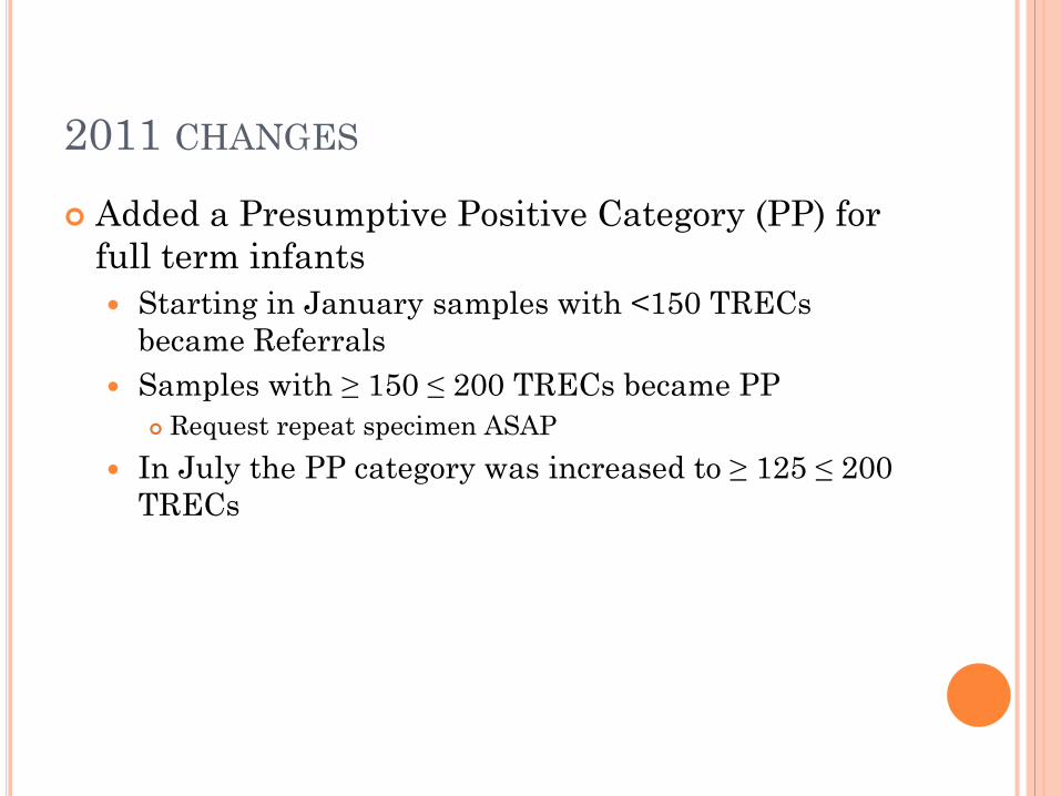
2011 CHANGES
Added a Presumptive Positive Category (PP) for full term infants Starting in January samples with <150 TRECs
became Referrals Samples with ≥ 150 ≤ 200 TRECs became PP
Request repeat specimen ASAP In July the PP category was increased to ≥ 125 ≤ 200
TRECs
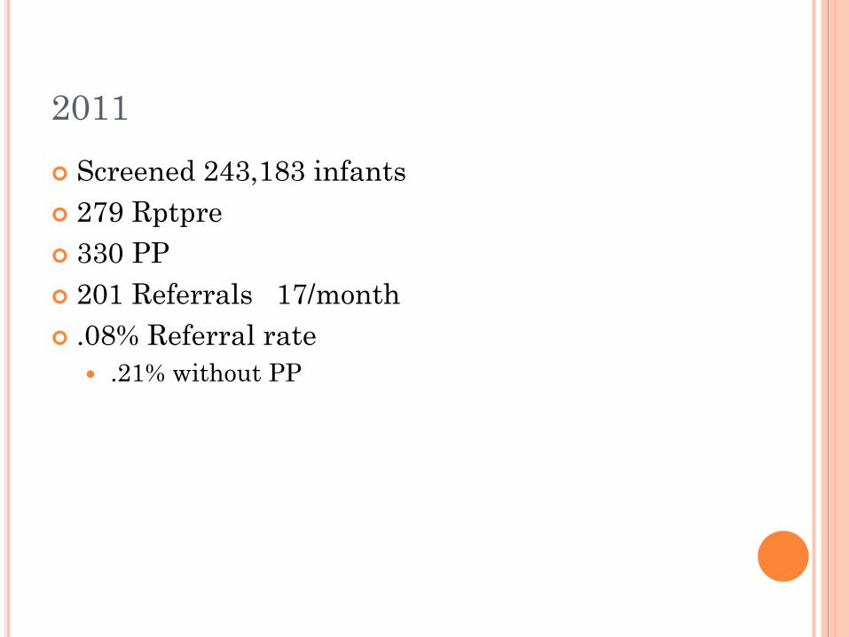
2011 Screened 243,183 infants 279 Rptpre 330 PP 201 Referrals 17/month .08% Referral rate
.21% without PP

2011 REPEAT PRE
279 Repeat Pre 254 individual infants 22 expired due to prematurity complications Received repeats on all others except 6 Most went WAL ( some took multiple samples)
10 samples became PP 1 expired 1 DiGeorge 1 Lost to follow up 7 No disease
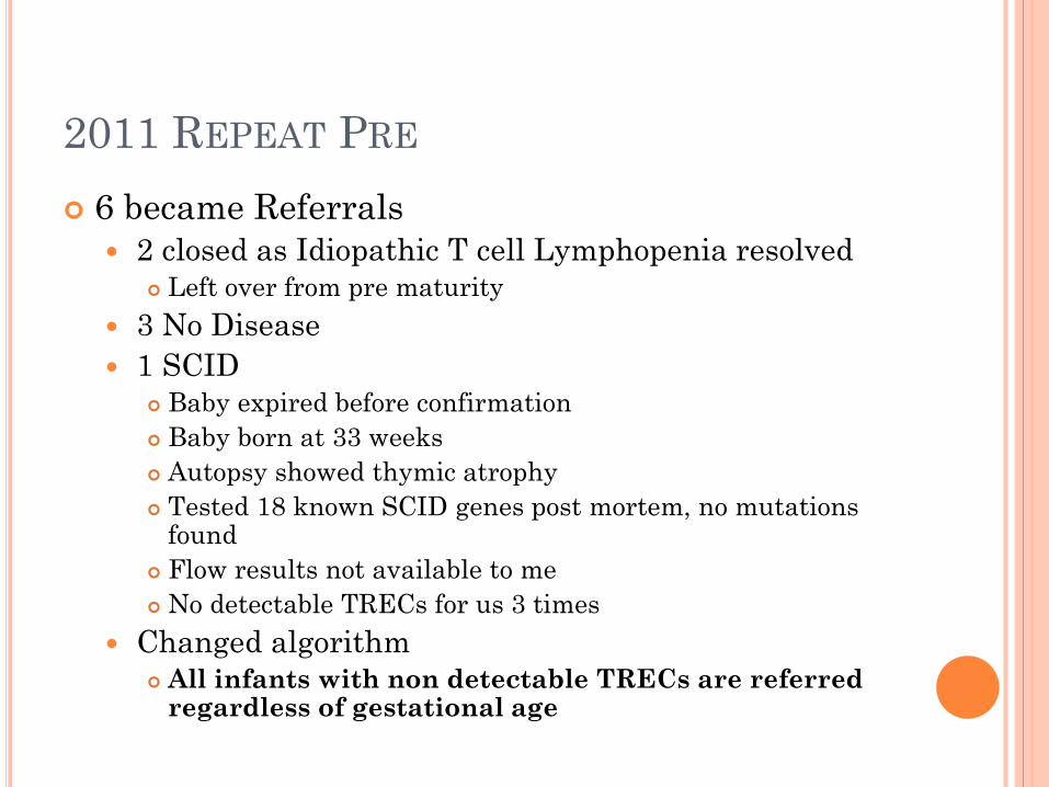
2011 REPEAT PRE
6 became Referrals 2 closed as Idiopathic T cell Lymphopenia resolved
Left over from pre maturity 3 No Disease 1 SCID
Baby expired before confirmation Baby born at 33 weeks Autopsy showed thymic atrophy Tested 18 known SCID genes post mortem, no mutations
found Flow results not available to me No detectable TRECs for us 3 times
Changed algorithm All infants with non detectable TRECs are referred
regardless of gestational age
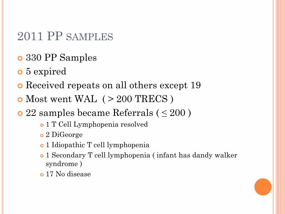
2011 PP SAMPLES
330 PP Samples 5 expired Received repeats on all others except 19 Most went WAL ( > 200 TRECS ) 22 samples became Referrals ( ≤ 200 )
1 T Cell Lymphopenia resolved 2 DiGeorge 1 Idiopathic T cell lymphopenia 1 Secondary T cell lymphopenia ( infant has dandy walker
syndrome ) 17 No disease

2011 REFERRALS
201 Referrals 133 No disease 5 B cell deficiency 7 Idiopathic T cell lymphopenia 8 Secondary T cell lymphopenia 4 Syndrome with T cell Impairment 16 expired 6 lost to follow up

2011 REFERRALS
1 Down Syndrome 10 DiGeorge 1 “other” 3 SCID
ADA SCID No detectable TRECs on 2 samples
IL2RG No detectable TRECs on 2 samples
Gene unknown No detectable TRECs on 3 samples This is the baby that was premature

249 Referrals 182 No Disease 5 SCID 1 Leaky SCID 15 Idiopathic T cell
Lymphopenia 8 Secondary T cell
Lymphopenia 2 Syndrome with T cell
impairment 10 DiGeorge 3 Down Syndrome 3 B cell Deficiency 3 “other” 9 expired 1 parental refusal 7 lost to follow up
233 Referrals 187 No Disease 5 SCID 1 Variant SCID 12 Idiopathic T cell
Lymphopenia 7 Secondary T cell
Lymphopenia 2 Syndrome with T cell
impairment 8 DiGeorge 1 Down Syndrome 4 B cell Deficiency 3 “other” 1 expired 1 parental refusal 5 lost to follow up
2012 Referrals 2013 Referrals

SNPS AND MOVING THE LAB
Extensive sequencing showed SNPs in the TREC probe region Sequenced 1079 Ref, PP and WAL samples 41% were homozygous for c.999390 and 8% were
heterozygous Learned we were moving the DAI Redesigned TREC probe New liquid handlers New Real Time Machines

1 YEAR OF SCREENING AT DAI WITH NEWPROBES/EQUIPMENT
March 2014 to March 2015 Screened 240,728 infants 277 Rptpre 182 PP 65 Referrals 5/month .03% Referral rate
.1% without PP

POST MOVE REPEAT PRE
277 Repeat Pre 240 individual infants 28 expired/no repeat 12 became PP 9 became Referrals
3 No Disease 3 Secondary T cell 1 DiGeorge 1 “other” 1 expired

POST MOVE PP 182 PP 11 expired/no repeat 18 became Referrals
9 No Disease 1 Variant SCID 1 Idiopathic T cell 3 Secondary T cell 2 DiGeorge 1 B cell Deficiency 1 “other”

POST MOVE REFERRALS
65 Referrals 26 expired/no repeat 25 No Disease 4 SCID 5 Idiopathic T cell Lymphopenia 2 Secondary T cell Lymphopenia 1 Syndrome with T cell impairment 9 DiGeorge 1 B cell Deficiency 1 “other” 7 expired 2 lost to follow up

# of infants screened
# of Referrals
# of PPs # of Rptpre
2011 243,183 201 330 279
2012 242,412 249 543 323
2013 239,410 233 554 320
Post Move 240,728 65 182 277

YEARLY AVERAGES
Pre Move
Post Move
Referrals 228 65PPs 475 182Rptpre 307 277Referral Rate
.09 .03
PP Rate .19 .07Rptpre Rate .13 .11

2011 2012 2013 Post moveNo Disease 133 (66%) 182 (73%) 187 (80%) 25 (38%)
SCID 3 (1.5%) 5 (2.5%) 5 (2.5%) 4 (6%)
Leaky SCID
0 1 0 0
Variant SCID
0 0 1 0
DiGeorge 10 10 8 9SecondaryT Cell
8 (4%) 8 (3%) 7 (3%) 2 (3%)
Idiopathic T Cell
7 (3.5%) 15 (6%) 12 (5%) 5 (3%)
Syndrome with T cell Impairment
4 2 2 1
B cell deficiency
5 3 4 1
Down Syndrome
1 3 1 0


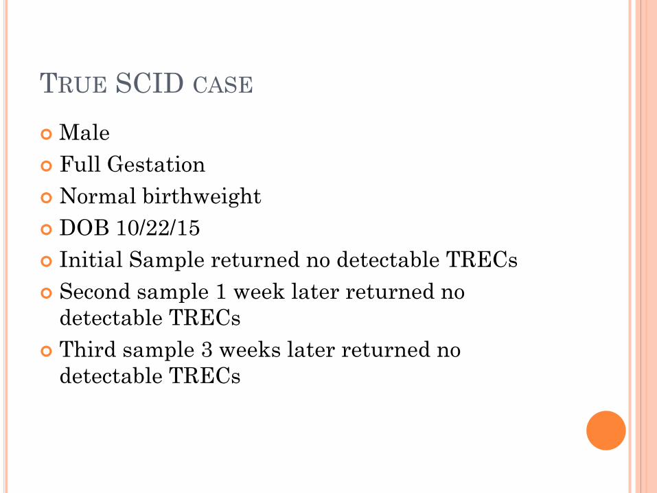
TRUE SCID CASE
Male Full Gestation Normal birthweight DOB 10/22/15 Initial Sample returned no detectable TRECs Second sample 1 week later returned no
detectable TRECs Third sample 3 weeks later returned no
detectable TRECs


TRUE SCID Male Full Gestation Normal birthweight DOB 5/17/15 Initial Sample returned no detectable TRECs Second sample 2 week later returned no
detectable TRECs Third sample 1 weeks later returned no
detectable TRECs
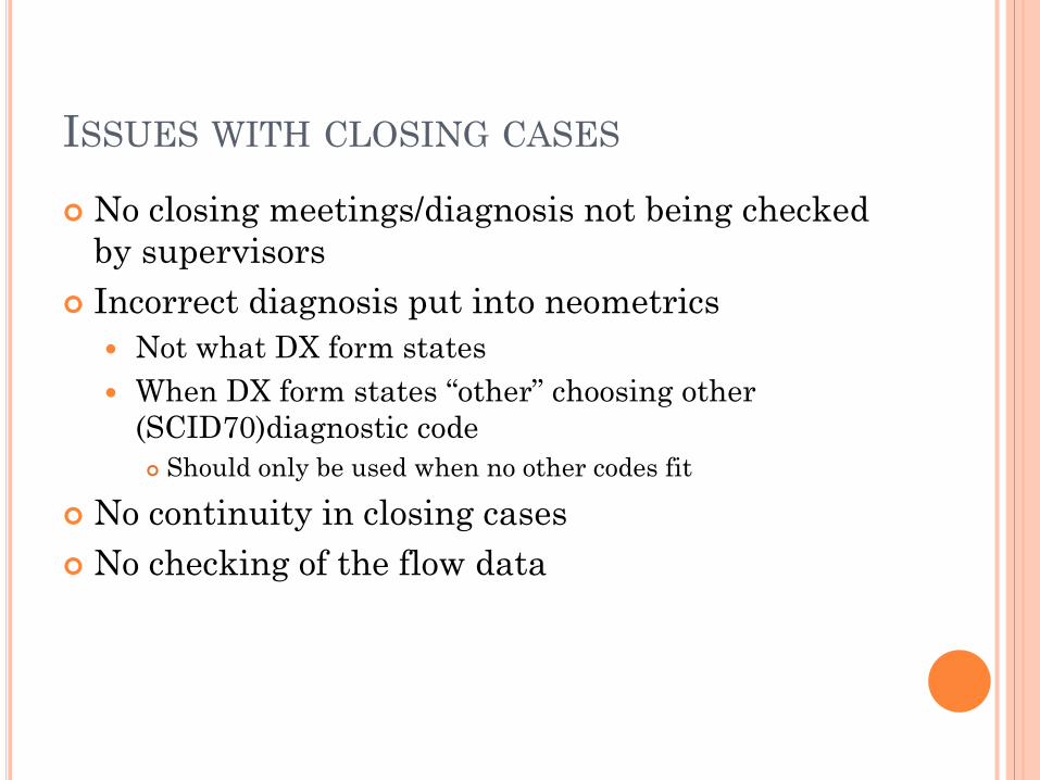
ISSUES WITH CLOSING CASES
No closing meetings/diagnosis not being checked by supervisors
Incorrect diagnosis put into neometrics Not what DX form states When DX form states “other” choosing other
(SCID70)diagnostic code Should only be used when no other codes fit
No continuity in closing cases No checking of the flow data

FLOW DATA SHOWS BORDERLINE T CELLSREASON UNKNOWN

SCID10 Disease, SCID, gene not specified
The baby has SCID and we don’t know what gene/mutation is the cause. Use this code if we don’t know if the baby was tested for mutations or if the baby was tested but we don’t know what mutations they have.
- SCID- SCID, gene unknown- SCID, genetics not done
SCID11 Disease, SCID, IL2RG (X-Linked)-related
Baby has SCID because of an IL2RG (interleukin 2 receptor gamma) mutation.
- Interleukin 2 receptor gamma deficiency/mutation
- IL2RG deficiency/mutation- IL2RG-SCID
SCID12 Disease, SCID, ADA-related Baby has SCID because of an ADA (adenosine deaminase) mutation.
- ADA deficiency/mutation- Adenosine deaminase
deficiency/mutation- ADA-SCID
SCID13 Disease, SCID, IL7Ra-related
Baby has SCID because of an IL7Ra (interleukin 7 receptor alpha) mutation.
- IL7Ra deficiency/mutation- Interleukin 7 receptor alpha
deficiency/mutation- IL7Ra-SCID
SCID14 Disease, JAK3-related Baby has SCID because of a JAK3 (Janus kinase 3) mutation.
- JAK3 deficiency/mutation- Janus kinase 3
deficiency/mutation- JAK3-SCID
SCID15 Disease, SCID, gene specified, not IL2RG, ADA, IL7Ra or JAK3
The baby has SCID and was tested for mutations in IL2RG, ADA, IL7Ra and JAK3. No mutations in any of these 4 genes (IL2RG, ADA, IL7Ra and JAK3) was found.
- Gene is specified but is not one that we have a diagnosis code for

SCID20 Disease, Leaky SCID, Omenn syndrome, gene not specified
Baby has Omenn syndrome, but we don’t know what mutation the baby has.
- Leaky SCID- Omenn syndrome - Absolute T cells (ABS T cells
(CD3)) are 300-1500, mitogens were tested and are abnormal.
SCID21 Disease, Leaky SCID, Omenn syndrome, gene specified
Baby has Omenn syndrome and the mutation causing the syndrome is known. Omenn syndrome is often due to mutations in:
- Leaky SCID- Omenn syndrome- Omenn syndrome with RAG1
deficiency/mutation- Omenn syndrome with RAG2
deficiency/mutation- Omenn syndrome with
DCLRE1C/Artemis deficiency/mutation
- Absolute T cells (ABS T cells (CD3)) are 300-1500, mitogens were done and are abnormal
SCID22 Disease, Non-SCID, Syndrome with T cell impairment
Infant has low T-cells and a syndrome other than DiGeorge (SCID23), Down’s (SCID24), CHARGE (SCID25), or Omenn (SCID20, SCID21) syndrome.
- VACTERL or VATER- Opitz syndrome/GBB- Trisomy 13/Patau syndrome- Trisomy 18/Edwards syndrome- Cartilage hair hypoplasia- Ring chromosome 17- 6p deletion syndrome- 17q duplication syndrome- Ataxia telangiectasia- Jacobsen syndrome- Multiple genetic defects/multiple
congenital anomalies/MCA/cytogenetic abnormalities (consult supervisor)

Absolute T cell count
>1500 300-1500 <300 ABS CD3; Absolute CD3;ABS CD3+; Absolute CD3+
Absolute T cell values are numbers, not percentages
The total number of T cells per cubic millimeter of blood (mm3), measured using flow cytometry.
Mitogens –phytohaemagglutinin test (“PHA”)
Varies by lab
Varies by lab Varies by lab
PHA Each baby needs at least 3 different mitogens tested. If 2 out of the 3 mitogen tests are in the abnormal range, the mitogens are considered to be abnormal.
Mitogen tests help determine whether the baby’s immune system is functional/can work properly.
Mitogens –pokeweed mitogen test (“PWM”)
Varies by lab
Varies by lab Varies by lab
PWM
Mitogens –Candida test (“CA”)
Varies by lab
Varies by lab Varies by lab
CA
Mitogens –tetanus toxin test (“TT”)
Varies by lab
Varies by lab Varies by lab
TT
Mitogens –concanavalin A test (“CONA”)
Varies by lab
Varies by lab Varies by lab
CON; CONA
If there are questions or something is confusing – ask a supervisor.

??? Doctor stated they believed baby to have SCID DX form sent in stating idiopathic T cell
lymphopenia Cases closed as secondary T cell lymphopenia
In April 2015 additional sample was sent in for testing because baby was a candidate for transplant and wanted TREC values
Sample had no detectable TRECs

In August began having case review meetings. Noticed DX form and neometrics had different
diagnosis. Noticed Dr initially thought SCID Because idiopathic was checked and flow was
borderline, decided to call and try to get additional information

Flow performed in March showed ABS T cells of 138, well within the SCID category.
Baby has confirmed Purine Nucleoside Phosphorylase (PNP) deficiency Metabolic disorder which can cause SCID Extremely rare, <50 cases known worldwide Doctor stated that it is pretty similar to ADA SCID Baby was preparing to undergo transplant
Case closed as SCID15 ( known gene ) Concern over >200 TREC value?

REFERRAL 868 DOB 5/19/14 Initial sample taken 6/21/14 Average of 49
TRECs ( 0,57,91 ) REF Repeat Specimen taken 6/2 UNS 6/9 received 2 sets of flow data with a DX of SCID
6/4 ABS T 74 6/5 ABS T 75

Repeat taken 6/23 Average of 167 TRECs RPTREF Trec values : 228,307,0,224,78
On 7/2 received additional flow data
6/9 ABS T 191 6/23 ABT T 428 Closed as SCID Last flow above SCID range, multiple TREC
values above 200

A.
B.
Figure 1. Simultaneous detection of TREC (blue), KREC (orange) and RNaseP (green) in a Triplex Assay from 348 DBS-derived normal controls (A) and 8 DBS-derived positive controls (B). RNaseP was used as an amplification control. All qPCR reactions were run on a QuantStudio 12K Flex system (Life Technologies). Taqpath Master Mix with Mustang Purple as the passive reference dye was utilized with our custom Taqman Probes: FAM-TREC-MGBNFQ, VIC-KREC-QSY and ABY2.0-RNaseP-QSY (Life Technologies).

ADA
Failure
XLA
DBSS

Gene HGNC Name
1 ADA ADENOSINE DEAMINASE
2 AK2 ADENYLATE KINASE 2
3 ATM ATM SERINE/THREONINE KINASE
4 BLNK B-CELL LINKER
5 BTK BRUTON AGAMMAGLOBULINEMIA TYROSINE KINASE
6 CD3D CD3d MOLECULE, DELTA (CD3-TCR complex)
7 CD3E CD3e MOLECULE, EPSILON (CD3-TCR complex)
8 CD3G CD3g MOLECULE, GAMMA (CD3-TCR complex)
9 CD247 CD247 MOLECULE
10 CD40LG CD40 LIGAND
11 PTPRC PROTEIN-TYROSINE PHOSPHATASE, RECEPTOR-TYPE, C
12 CHD7 CHROMODOMAIN HELICASE DNA BINDING PROTEIN 7
13 CORO1A CORONIN ACTIN BINDING PROTEIN 1A
14 DCLRE1C DNA CROSS LINK REPAIR 1C
15 DKC1 DYSKERATOSIS CONGENITA 1, DYSKERIN
16 DOCK2 DEDICATOR OF CYTOKINESIS 2
17 DOCK8 DEDICATOR OF CYTOKINESIS 8
18 FOXN1 FORKHEAD BOX N1
19 GATA2 GATA BINDING PROTEIN 2
20 IGHM IMMUNOGLOBULIN HEAVY CHAIN CONSTANT MU
21 IL2RG INTERLEUKIN 2 RECEPTOR, GAMMA
22 IL7R INTERLEUKIN 7 RECEPTOR
23 JAK3 JANUS KINASE 3
24 LIG4 LIGASE IV DNA ATP-DEPENDENT
25 MTHFD1 METHYLENETETRAHYDROFOLATE DEHYDROGENASE-1
26 MTR 5-METHYLTETRAHYDROFOLATE HOMOCYSTEINE METHYLTRANSFERASE
27 NHEJ1 NONHOMOLOGOUS END JOINING FACTOR 1
28 NBN NIBRIN
29 PNP PURINE NUCLEOSIDE PHOSPHORYLASE
30 PRKDC PROTEIN KINASE DNA ACTIVATED CATALYTIC POLYPEPTIDE
31 RAC2 RAS-RELATED C3 BOTULINUM TOXIN SUBSTRATE 2
32 RAG1 RECOMBINATION ACTIVATING GENE 1
33 RAG2 RECOMBINATION ACTIVATING GENE 2
34 RMRP RNA COMPONENT OF MITOCHONDRIAL RNA PROCESSING ENDORIBONUCLEASE 35 SLC46A1 SOLUTE CARRIER FAMILY 46 (folate transporter), MEMBER 1
36 STAT5B SIGNAL TRANSDUCER AND ACTIVATOR OF TRANSCRIPTION 5B
37 TBX1 T-BOX 1
38 WAS WISKOTT-ALDRICH SYNDROME
39 ZAP70 ZETA CHAIN (TCR) ASSOCIATED PROTEIN KINASE 70kDa