Getting The Certification Physiotherapy Treatment at Befit Clinic

Running Header: ANKLE FRACTURE
CHARLES UNIVERSITY IN PRAGUE
FACULTY OF PHYSICAL EDUCATION AND SPORTS
Departement of Physiotherapy
Case Study of a Physiotherapy Treatment of a patient with
Ankle fracture
Bachelor thesis
Prague, 2017
Thesis supervisor:
Mgr.Ilona kučerová
Elaborated by:
Salman Abdulmohsin
Alkhowildi
Clinical supervisor:
PhDr.Edwin Mahr, Ph.D

2 ANKLE FRACTURE

3 ANKLE FRACTURE
Dedication
I dedicate this bachelor thesis to my wonderful family in Saudi Arabia my
Father, My mother, my brothers and my beautiful twins sisters for their great support
and understanding from all views. I dedicate it to a very special lady Violetta for
changing my life and helping me to go throw my entire study. to my best friends,
Ahmad, Irina and Tanja for them support and being my second family Also to
Kafkova Marika for being not only a teacher but a mother in Prague. It is also
dedicated to the university staff who were cooperating with students in a nice manner
especially my professors for everything that they gave me during my study.

4 ANKLE FRACTURE
Abstract
Title:
A Case- Study of physiotherapy treatment of a patient who has experienced ankle
fracture in his right leg.
Goal:
The main objective of this thesis is the discuss all information related to ankle fracture
along with the rehabilitation plan to recover the ankle fracture, which will be
discussed in the theoretical and practical section of this thesis. The theoretical part
highlights the historical information, lower extremities anatomy, biomechanics and
kinesiology of the ankle joint, different injury types and the rehabilitation plan. The
clinical picture and the etiology of this fracture along with specific tests for ankle
fractures will also be discussed. The practical part presents a male patient case study
that recently experienced ankle fracture.
Methods:
The rehabilitation process of this patient majorly focused on the use of isometric
exercises, soft tissue techniques, sensorimotoric exercises and muscle stretching and
strengthening. It included a total of 8 therapy sessions where each session lasted for
30 minutes to 60 minutes along with two additional sessions where the initial and
final kinesiologic examinations were performed.
Results:
During these two weeks of rehabilitation, the patient demonstrated considerable
improvement in his ankle condition and had increased the active and passive range of
motion in his broken ankle. He also reported a decrease in the pain intensity and was
able to walk independently and smoothly.
Conclusion:
Based on the initial and final kinesiologic examination, it was evident that the patient
demonstrated a high level of satisfaction with the improvement in his ankle condition.
The rehabilitation therapies aided him in being fully active and independent.
Key words:
Ankle fracture, ankle joint, rehabilitation, kinesiology.

5 ANKLE FRACTURE
Declaration
I declare that this Bachelor Thesis is my own work written independently with
information and sources taken from literature which I have stated and based on
knowledge gained from the lectures during my academic studies in Fakulta Telesné
Výchovy a Sportu, Univerzita Karlova v Praze. Neither the thesis nor any other part
of it has already been submitted or presented for the obtainment of any other degree.
Prague, April 2017

6 ANKLE FRACTURE
Acknowledgments
I would like to thank first of all my family for all the support, the effort and the
understanding that they have shown to me through my studies and especially my
Parents my Father, for the great inspiration to me all these years, and my mother
which despite all the difficulties, they are standing still by my side in every step I take
also to my brother Ali for his support and care .
Also, I would like to thank PhDr. Edwin Mahr PhD. for the guidance, help, the
knowledge and the trust that he offered to me during my practice in the clinic and
gave me the chance to prove myself in practice. Additional thanks I will give to my
supervisor Mgr. Ilona kučerová
for her instructions and advices concerning my Bachelor thesis and for guiding
me through the entire process with her experience, patience and wisdom.
Also I will not forget to mention and give particular gratitude to Mgr. Helena
Vomáčková, Mgr. Michaela Stupková, Mgr. Kateřina Maršáková ,For them kindness,
help and support .
Likewise, I would like to thank my friends for their support and their help when I
needed it, and specifically Afro, also my classmates Murtada, Hussain, Ahmad and
whom have been there for me from the beginning of my “journey”.
My thanks also delivered to my great Saudi Arabian government for giving me
this opportunity to study abroad and gave me a scholarship and financial support .
Thank you all
Salman Alkhowildi Prague,

7 ANKLE FRACTURE
Contents
1. INTRODUCTION ............................................................................................................. 9
1.1 Historical Data and Literature review ....................................................................... 11
2. GENERAL PART ........................................................................................................... 14
2.1.1 Anatomy of the Ankle ............................................................................................ 14
2.1.2. Kinesiology of the Ankle Joint ........................................................................... 15
2.1.3 Biomechanics of the ankle joint .......................................................................... 16
2.2. Ankle Injuries ........................................................................................................... 18
2.2.1 Ankle Fractures ................................................................................................... 19
2.2.2 Ankle Sprain ......................................................................................................... 20
2.3 Diagnosis of ankle injuries .......................................................................................... 22
2.3.1 Medical Imaging .................................................................................................. 22
2.3.2 Ottawa Ankle and Midfoot Rules ........................................................................ 23
2.4 Management of Ankle Fractures .............................................................................. 25
2.4.1 Orthopaedic Management..................................................................................... 25
2.4.2 Interventions during Immobilization ................................................................... 25
2.4.3 Interventions following immobilization removal ............................................... 25
2.5 Etiology and Clinical Picture .................................................................................... 27
2.5.1 Etiology ............................................................................................................... 27
2.5.2 Clinical Picture .................................................................................................... 27
3. SPECIAL PART ........................................................................................................... 28
3.1 Methodology ............................................................................................................ 28
3.2 Anamnesis ................................................................................................................... 29
3.3 Initial kinesiologic examination ................................................................................ 32
Table 10: Muscle strength scores of different parts of the Lower Extremities (LRs) ............. 40
3.3.1. Conclusion ......................................................................................................... 44
3.4. Short- term and long-term rehabilitation plan .......................................................... 45
3.5. Therapy progress ...................................................................................................... 46
3.6 Final kinesiologic examination: ........................................................................................ 64
3.6.1. Conclusion ........................................................................................................... 76

8 ANKLE FRACTURE
3.7. Therapy effect evaluation and Prognosis...................................................................... 77
4. Conclusion ....................................................................................................................... 78
5. References ....................................................................................................................... 80
6. Supplement ...................................................................................................................... 87
6.1 List of Abbreviations ..................................................................................................... 87
6.2 List of Tables ................................................................................................................ 88
6.3. List of Figures ............................................................................................................ 89
6.4. Ethecal bored .............................................................................................................. 91
6.5. INFORMOVANÝ SOUHLAS ................................................................................... 92

9 ANKLE FRACTURE
1. INTRODUCTION
Ankle Fractures are the commonest injuries that are treated by most
orthopedic surgeons (Goost, et al., 2014). It has been the main subject of focus in
numerous articles and research studies that discussed ankle fractures, its
mechanism, classification of different types of fractures and their treatment
modalities. The ankle is one of the strongest mortise joint (also known as the
woodwork joints or the talocrural joint) in the body, which is the formation of a
hinge joint by the lower end of the tibia and the fibula that articulates with the talus
(American Academy of Orthopedic Surgeon, 2015). Ankle fractures occur when
one or both sides of an ankle are completely or partially broken due to twisting
injuries or falls or injuries experienced during play, or sports (DeAngelis,
Eskander, & French, 2007). Most ankle fractures were more frequently reported
among men under the age of 50 years while over the age of 50 years, most ankle
fractures have been reported in women (Moseley, Beckenkamp, Haas, Herbert, &
Lin, 2015).
The fracture type highly varies from simple fracture to complex fracture and
might or might involve one or all the three bones of the ankle joint. Irrespective of
the intervention methods, the primary goals of the orthopaedics are to restore the
normal anatomy of the ankle. The complication associated with both the operative
and non- operative management are the most important and primary consideration
in the decision- making process (Bugler, White, & Thordarson, 2011). For certain
types of fractures such as the undisplaced injuries, the most appropriate
management is the non- operative treatment as the operative management of such
injuries can expose the patients to unnecessary risks of operations/ surgeries and is
also deemed by experts as “over treatment” (Costigan, Thordarson, & Debnath,
2007).
In cases of ligamentous injuries or fractures of the ankle, ankles become
unstable due to its anatomical structure. Ankle fractures are treated with different

10 ANKLE FRACTURE
methods of internal fixation devices; however, the best possible implant as
reported by experts can be only determined based on the anatomy of the ankle
fracture (Lamontagne, Blachut, Broekhuyse, O'Brien, & Meek, 2002). However,
the ankle mobilization protocol during the post- operative period has been a heated
controversy. The outcomes of this type of fractures are of prime importance, as it is
important that the treatment of the ankle fracture not only benefit the patients in the
short- term but also in the long- run.
The treatment of the ankle fractures has its own challenges especially in cases
where the co- morbid conditions such as the neuropathic conditions, peripheral
vascular disease, and diabetes mellitus, which complicate the fracture condition as
well as the treatment process. These conditions can also majorly influence the
overall outcomes of the treatment process. Good results are expected only through
a thorough understanding of the injury mechanism, ankle anatomy, radiograph
injury and its adherence to the basic principles of management of the ankle fracture
(Appleton & McQueen, 2006).
At present, the conventional management approaches during the rehabilitation
ignore the impaired accessory movement roles that increase the recurrent injury
susceptibility. Hoch (2010) recommended the use of manual therapy in case of
restriction in physiological motion or range of accessory at the ankle is an
experience. However, research studies related to the manual therapy usage for the
talocrural joints have produced only controversial results. Studies have
demonstrated that single mobilization technique application to the talocrural joint
increases in loss of dorsiflexion (DF). Other manual therapy techniques such as
High- Velocity Low Amplitude (HVLA) thrust and Still technique were also
assessed and no change in the DF was reported (Greenman, 2003). The
combination of multiple treatment techniques has also demonstrated considerable
improvements in the DF post successive treatments.
Therefore, the restoration of the patient’s ankle anatomy and the ability of the
patient to walk normally and independently again majorly depend on the success of
the physiotherapy provided and on the therapeutic manipulation. (Evans & &
Lucas, 2010)

11 ANKLE FRACTURE
1.1 Historical Data and Literature review
Lindsjo (1985) was the first to introduce different classifications for the ankle
fracture. Following which, Bromer (1922) classified it as an external rotation leading
to abduction and adduction injuries. Percival Pott was the first to develop the
classification system for ankle fractures and described it in terms of the involved
malleoli number, thereby classifying the injuries into unimallelor, bimalleolar, and
trimalleolar (Pott, 1996). However, even though this system was easy to use when
used with better intraobserver reliability, yet it fails to distinguish between injuries
that are stable and unstable.
Later, Lauge Hansen’s classification system became the pioneering system,
which gave positive outcomes out of the conservative treatments provided. Later AO
group had put forward the Danis Weber classification, which was first developed by
Danis and then was modified by Weber (Weber, 1996). Today, there are two other
types of classification system that tries to aid the distinction between the stable and
unstable injuries: 1. The Weber classification system and 2. The Lange– Hansen
system. The former categorizes the fractures based on the position of the distal fibular
fracture with respect to the syndesmosis, while the later is a mechanistic classification
method, where it firstly describes the foot position at the time of injury and secondly
describes the ankle’s deforming force to provide more information on the stability.
The later also enables the physicians to make decisions for the kind of treatment that
needs to be given to the patient. However, The Danis Weber classification was
reported to be more beneficial or surgical treatments that the non- operative ones.
Joy (1947) later assessed the reduction obtained immediately after the post-
operative period, the reduction adequacy and correlated it with the result. Different
criterions were discussed and the Kristenson’s criterion was adopted. Leeds (1984)
assessed bimalleolar and trimalleolar fractures because of the supination external and
pronation external rotation injuries in which syndesmotic injuries were present. He
reported that the initial syndesmosis reduction and lateral malleolus fixing was ideal,
thereby influenced the syndesmotic stability for a longer period, and prevent late
arthritis. The syndesmosis reduction was important to achieve ankle stability in all

12 ANKLE FRACTURE
cases of pronation and supination external rotation injuries; however, it needs to be
combined with the lateral malleolus fixation (Joy, 1974).
On the other hand, Lindsjo (1985) used the open reduction and internal
fixation strategies to treat ankle fractures using the AO principle of internal fixation.
The authors reported that the most decisive factors were reduction adequacy, fracture
type, patient’s sex, and rigid fixation, exact reduction, subsequent full weight- bearing
walking, early post- operative exercises of the joint. However, this treatment led to
arthrosis development post- fixation suggesting improper reduction. Bauer (1985)
compared operative treatment with non- operative treatment and reported that the
operative treatment was favorable; however, in the long- run the arthrosis incidence
was common in the operative group than in the conservative group even if the
anatomical reduction as achieved (Bauer, 1985) .
Segal (1985) later compared the ability to perform weight bearing walking in
patients in both conservative and operative treatment. They reported that the patient
could immediately mobilize at the end of the 1st week while the other group could
mobilize by the end of the 5th
week upon stable fixation. The functional brace further
prevented rotational stress on the fractured ankle (Segal, 1985). Rowley (1986) when
compared all the treated ankle fractures at their institute with closed immobilization
and manipulation and those that were managed by surgical fixation found no
difference in the gait and range of motion of the fractured ankle. Baird (1987)
recommended open reduction and internal fixation of the lateral malleolus; however,
the deltoid ligament is not necessary for clinical recovery. Bostman (1989) achieved
medial malleolar fixation in their study using polylactide and polyglycolide and
reported that bioabsorbable screws for shoed bony union and fixation and its
reduction maintenance were comparable to that of the metallic implants, however, its
major advantage was that it did not require removal.
Finsen (1989) when compared the bone mineral concentration in surgically
fixed ankle fractures found that there was not much difference in between post-
traumatic osteopenia and their selected groups and was natural and maximum in the
initial seventy- six months post- trauma but it stabilized later and needed
improvement. Marti (1990) studied the malunited ankle fractures treated with
reconstructive osteotomies and reported that complete anatomical alignment
restoration of the joint should be attempted irrespective of the amount of time elapsed
and malunion. They further indicated that lateral or shortening rotation of the lateral

13 ANKLE FRACTURE
malleolus that resulted in the mortise widening and talus tilting was the commonest
cause of malunion. Winkler (1990) proved that the antiglide plate fixation in lateral
malleolus fixation in case of Weber fractures type B was bio- mechanically sound and
recommended its use in case of osteoporotic bones.
Carrage (1991) reported that fractures that recovered fast and the fixation that
was performed on emergency basis showed better outcomes that those that were
delayed. Cimino (1991) reported that open reduction and internal fixation in patients
allowed them to mobilize immediately after the patient was able to tolerate the partial
weight bearing along with crutches without any complications. Pritsch (1993)
reported that the pain post- arthroscopy was majorly due to distal tibiofibular joint
adhesion. Bucholz (1994) recommended the use of bio- absorbable implants so as to
prevent a repeat surgery from removing the metallic implants.
Schon (1995) suggested the need for neuroarthropathy presence in the
dislocation of the ankle fracture when performing open reduction and internal
fixation. They also further suggested that diabetes should not interfere with the
surgery or post- operative care; however, mobilization was recommended to prevent
neuroarthropathy. On the other hand, Cormack (1998) reported that the risk of
complications was 42% in case of an operative group who had diabetes where wound
infections were the commonest complication. Connolly (1998) later confirmed
Carrage (1991) findings stating that early internal fixation with restricted weight
bearing is a better option to prevent complications including swelling and hyperemia
around the ankle. Bibbo (2001) reported that presence of diabetes in patients who
have experienced ankle fractures increases their risks of developing infections, soft
tissue complications, and delayed the healing process. The delayed healing might also
result in impaired bone turnover and collagen syntheses.
Literature review of existing studies implies that there been a considerable
amount research conducted on ankle fracture and its treatment owing to which now
the ankle fractures can be treated and physicians and physiotherapists can aid the
patients in recovering their ankle anatomy and normal functionality of their lower
extremities.

14 ANKLE FRACTURE
2. GENERAL PART
2.1.1 Anatomy of the Ankle
The ankle joint is commonly known as the woodwork joint or the talocrural
joint is technically a hinge joint that is formed by the distal surfaces of tibia and fibula
articulating the talus (Dananberg, 2004; Magee, 2007). Usually, the movements that
take place in this joint are in the sagittal plane and comprises of dorsiflexion and
plantarflexion. The primary ankle fracture or injury mechanisms occur during the
movements that occur in the non- sagittal planes including the subtalar joint that
comprises eversion and inversion (Kisner, Colby, & Library, 2007).
The major muscles that span the ankle joint are plantarflexors (i.e., soleus and
gastrocnemius in the lower limb’s posterior compartment) and dorsiflexors (i.e., the
anterior tibialis that are present in the lower limbs’ anterior compartment) (Palastanga,
Field, & & Soames, 2006). The ligaments support the lateral and medial sides of the
distal tibiofibular joint and the ankle joints (Figure 1). On the medial side of the ankle,
the medial collateral ligament reinforces the ankle capsule stability. The medial
collateral ligament is also commonly known as deltoid, which is a strong ligament
reinforced by double layers. On the lateral side of the ankle, the lateral collateral
ligament protects the capsules that are formed by three different ligaments: anterior
talofibular, posterior talofibular and calcaneofibular. The anterior displacement
internal joint rotation and inversion are constrained by the anterior talofibular
ligament. The calcaneofibular ligament of the ankle controls the subtalar and ankle
joint eversion (Denegar, Hertel, & & Fonseca, 2002).
Lastly, the posterior talofibular ligaments then control the posterior joint
displacement. Since, this joint is tightly attached thus it might result in fracture of the
posterior malleoli avulsion upon application of the significant amount of force. The
joint at the distal tibiofibular is also known as the syndesmosis and the stability of this
joint id provided by the posterior and anterior inferior interosseous ligament and the
tibiofibular ligaments (Hubbard & Wikstrom, 2010).

15 ANKLE FRACTURE
2.1.2. Kinesiology of the Ankle Joint
The talocrural joint or the ankle joint is formed from the two malleoli and the
tibia that receives the talus trochlea. It comprises a strong ligament that is present on
the deltoid, medial side and a weak ligament on the lateral side. This weak ligament is
the one that is most injured. The movements that occur on the frontal and the coronal
plane are DF with 10ᵒ - 30ᵒ and PF with a range of motion in 45ᵒ - 50ᵒ (Figure 2). It is
also possible to provide circumduction (Anish, Kadakia, & M., 2014).

16 ANKLE FRACTURE
The physiological range of motion for EV is 15ᵒ to 30ᵒ and IN is 35ᵒ to 50ᵒ
(Figure 3). The ankle joint is primarily important for walking and has a major role in
the gait cycle, which is divided into the swing phase (which includes the 40% of the
gait cycle) and the stance phase (which includes the 60% of the cycle) for one leg
(Figure 4). The stance phase starts the moment the heel strikes the ground (i.e., the DF
of the ankle joint), following the loading response phase (i.e., flat foot), midstance
with the lower extremity support at this point followed by the terminal stance when
the heel takes off from the ground (i.e., the PF of the ankle joint). The final stage is
the pre- swing phase that needs both extremity support and this is the position where
the toes lift off the ground and give propulsory force.
The swing phase then starts when the toes lift off the ground and it ends up
striking the ground. This is done with the single support that it gets from the
ipsilateral lower extremity. At this point, the body weight gets transferred to the side
(Brookbush, 2011).
2.1.3 Biomechanics of the ankle joint
The biomechanics of both the ankle and the foot is very complex and
intricately connected. The foot is the most important mechanical part of the body as it
aids in walking and running with a steady and smooth pace. The weight of the whole
body, as well as the lower end of the foot, is carried by the ankle and thus affects the
foot orientation with the ground (Berme, 1985). The foot comprises 28 bones and the
movements of each of these bones are interdependent. Apart from being a structural

17 ANKLE FRACTURE
supportive platform that withstands repeated multiplied loads of the body weight, the
ankle is also capable of being adapted to varying and terrain speeds. The unique
anatomy of the foot allows it to act rigid as well as flexible enough as and when
required (Waugh, Blazevich, & Korff, 2012).
The ankle joint structure is formed by the distal surfaces of tibia and fibula
articulating the talus and the structure comprises a subtalar, talocrural and tibiofibular
structure (Figure 5). The talocrural joint is a modified hinge; a uniaxial and synovial
joint that is located in between the tibia’s medial malleolus wedged shaped talus and

18 ANKLE FRACTURE
the fibulas’ lateral malleolus (Berme, 1985). The hinge joint allows one- degree
freedom of movement in both DF and plantar flexion. However, due to the talus
plantarflexion shape, the DF needs to be described as a helical movement than a hinge
swing movement. The structure of the joint is very unstable and its stability highly
dependent on the articulation of the harmonic bone, anterior talofibular ligament,
inferior tibiofibular ligament, calcaneofibular ligament and posterior talofibular
ligament. The kinesiology part discussed above in section 2.1.2, has the details of the
role of the ankle in various steps involved in the gait cycle. During the DF and
plantarflexion, the relationship between the three joints is summarized in table 1.
Any kind of abnormal changes in the movement or structure of the ankle,
irrespective of the degree of change, has an obvious impact on promoting, cushioning
and ankle stability and edge of the foot. The change in the clinical correlation in the
biomechanical function has been discussed in a number of case studies. In the
Western Society, the footwear ranges from a soft moccasin to tough ski boot. The use
of these external materials can further result in the alteration of the physiological
biomechanics of the ankle function and the foot that eventually results in the
development of different pathological conditions (Berme, 1985).
2.2. Ankle Injuries
Ankle injuries are very common and have been most reported among the
young adults who are professionally and physically active (Fong, Hong, Chan, Yung,
& Chan, 2007). This population accounts for approximately 10% of the total visits to

19 ANKLE FRACTURE
the emergency departments of the healthcare services (Walker, 2014). The
commonest types of ankle injuries are ankle fracture and ankle sprain. For the
majority of the cases of ankle fractures and ankle sprains, the injury mechanisms
comprise a significant inversion force, which has been reported to most commonly
occurring during any sports activity like, running, rugby, soccer, etc (Martin,
Davenport, Paulseth, Wukich, & Godges, 2013 ).
2.2.1 Ankle Fractures
Ankle fractures have been reported to be very common among almost all age
groups and are commonly reported in older women (i.e., 16- 20 individuals per
10,000 individuals per year) and in young men (i.e., 13- 28 individuals per 10,000
individuals per year) (Giannini, et al., 2013; Robertson, Wood, Aitken, & Court
Brown, 2014). The annual incidence of ankle fractures overall has been reported to
occur in every five individual per 10,000 individuals (Scott, 2010; Singer,
McLauchlan, Robinson, & Christie, 1998). Most ankle fractures have been reported to
occur mostly during sports activity, which accounts for approximately 7% of the total
sports- related fractures (Court-Brown, Wood, & Aitken, 2008). As a whole, the
prevalence of ankle fractures has increased by 25% in the past 15 years i.e., 1986 to
2010, especially among the older population, where women who have osteoporosis
have been reported to be most susceptible (De Boer, Schepers, Panneman, Van Beeck,
& Van Lieshout, 2014). Osteoporosis, obesity, diabetes, and history of ankle injury
are some of the common risk factors that are associated with the fracture of the ankle.
Ankle fracture usually occurs as a result of excessive angular or rotational
force on the foot that is in relation to the limbs that leads to an excessive inversion
stress (Nightingale, Moseley, & Herbert, 2007). A fracture can be further classified
into the stable fracture and unstable fracture, which is determined based on the
whether or not the fragments were dislocated. If in case, the bone separation gap is
greater than 3mm then the fracture is said to be significant clinically. In such scenario,
the ankle injury will be treated as an ankle fracture (discussed in section 2.4), while
on the other hand, the insignificant fractures will be treated as an ankle sprain
(discussed in section 2.2.2). The fracture usually comprises of medial, posterior, or
lateral malleolus, where 2 or more malleoli are considered as a severe type of

20 ANKLE FRACTURE
fractures (Figure 6). In the severe cases, the ankle fractures usually include the
syndesmosis rupture between the distal fibula from the tibia (Stiell, et al., 1992).
2.2.2 Ankle Sprain
The ankle sprains are considered among the commonest type of
musculoskeletal injuries that occurs in the lower limb where most injuries being
reported to occur during exercise or sports activity (De Boer, Schepers, Panneman,
Van Beeck, & Van Lieshout, 2014). The incidence of ankle sprain has been reported
to occur in over 206 individuals per 100,000 individuals annually and is more
common among young adults (i.e., 700 per 100,000 in individuals who are between
15 to 19 years of age). Approximately 85% of the ankle sprains comprises of lateral
aspects of the ankle since most of it takes place during an inversion foot injury that
occurs in a certain degree of plantarflexion (Solveborn, 2014). Usually, anterior
talofibular is the first lateral ligament followed by the calcaneofibular that get affected
or injured during a lateral ankle sprain. However, the involvement of the posterior
talofibular ligament has been reported to be rare (Doherty, et al., 2014). The lateral
sprains are further classified into Grade I, Grade II and Grade III. Usually, the Grade
I ankle sprains does not demonstrate any ligament rupture, while the Grades II
demonstrate partial and Grade III demonstrates complete rupture (Martin, Davenport,
Paulseth, Wukich, & Godges, 2013 ; Lamb, Marsh, Hutton, Nakash, & Cooke, 2009).

21 ANKLE FRACTURE
However, the optimal mode of treating ankle sprains still remains unresolved.
Such injuries are usually treated using the conventional, non- surgical methods and
surgeries are considered only when the conventional treatment fails. The functional
treatment guidelines for treating ankle sprain emphasizes on early bearing with
support, using physical agents (such as electrotherapy and cryotherapy), using manual
therapy modalities (such as joint manipulation and mobilization), sports- related
activities and usual ankle exercises. In the case of severe ankle sprains, a short
immobilization period has been indicated below the knee cast (Lamb, Marsh, Hutton,
Nakash, & Cooke, 2009).
The functional treatment strategies usually include three different phases to
biologically heal the ligament injury. In the 1st phase, the PRICE protocol (i.e.,
Protection, Rest, Ice, Compression, Elevation) is followed in the initial 4- 5 days to
minimize the tissue inflammation and swelling. In the 2nd
phase, the sprained ankle is
immobilized using brace or tape to guard the ligament during the collagen
proliferation phase on the 5th
to 7th
day. Lastly, in the 3rd
phase, neuromuscular
training and weight bearing training are provided to promote the orientation of the
collagen fiber and also enable prevention of stiffness during the remodeling phase
(Martin, Davenport, Paulseth, Wukich, & Godges, 2013 ).

22 ANKLE FRACTURE
2.3 Diagnosis of ankle injuries
Differential diagnosis of an ankle sprain and fractures are difficult because
both exhibit similar mechanism of the injury that occurs because of a significant
amount of inversion force that affects the lateral aspects of the ankle. The clinical
presentation is also similar in acute injury and includes swelling, significant pain and
tenderness in the lateral malleolus (Scott, 2010). The most common and obvious
symptom upon a fracture is the inability to bear any kind of weight on the injured leg.
Although it is difficult to distinguish between ankle fracture and ankle sprains, yet it
is extremely important to know because the treatment process and management are
very different for both. In the case of ankle sprains, it is essential to promote early
mobilization and functional recovery while in the case of ankle fractures; early
mobilization is difficult, as fractures require a longer time to heal. However,
immobilization of the affected part for a longer period can result in ankle stiffness and
limitation of the increased activity; therefore it is important to avoid unnecessary
immobilization (Solveborn, 2014).
2.3.1 Medical Imaging
The gold standard for differentiating between an ankle fracture and an ankle
sprain usually includes diagnostic imaging, including magnetic resonance imaging
(MRI), radiography, ultrasonography, computed tomography (CT) scan, and
radionuclide bone scanning. Upon experiencing a musculoskeletal trauma, the initial
imaging that the patient is usually subjected to is the conventional radiography with
an aim to exclude anatomic abnormalities such as identifying or determining the
presence of fractures (Martin, Davenport, Paulseth, Wukich, & Godges, 2013 ). The
complex anatomy of the injured leg is then visualized using the computed tomography
and to assess any injuries in the soft tissues, the medical resonance imaging is used.
However, physicians perform radiological imaging based on the initial
physical examination and patient’s clinical history. The decision should consider that
the radiography use can increase the time of patient stay in the emergency department
and might expose the patient to unnecessary radiation and its various side- effects. It
also increases the expenditure of the health system (McKinnins, 2014). Recently, the
“Choosing Wisely” campaign has raised awareness among experts and practitioners
about the importance of avoiding unnecessary procedure and tests. Furthermore, the

23 ANKLE FRACTURE
number of patients subjected to medical imaging is essential to preserve resources and
improve patient safety. Moreover, the rules of clinical decision- making can be further
used to determine if the medical diagnostic imaging is needed in a patient presenting
signs and symptoms of an acute ankle injury (Volpp, Loewenstein, & Asch, 2012).
2.3.2 Ottawa Ankle and Midfoot Rules
The Ottawa Ankle and Midfoot rules are a set of rules that has been developed
to identify patients who have a low probability of midfoot and/ or ankle fracture and
doesn’t require radiography imaging, thereby aiding in the clinical decision- making
process. This set of rules were first introduced in the year 1992, which was later
refined in the year 1993 (Stiell, et al., A study to develop clinical decision rules for
the use of radiography in acute ankle injuries., 1992; Stiell, Greenberg, McKnight, &
al., 1993). This rule, since then, has been implemented and validated to use in
different settings and populations. The Ankle Rules indicates the need of radiography
when the pain is present in the malleolar zone and in the following scenarios: 1) bone
tenderness at the medial malleolus tip or at the posterior edge of the medial malleolus,
2) bone tenderness at the medial malleolus tip or at the posterior edge of the lateral
malleolus, and 3) unable to bear weight immediately and when in the emergency
department (Figure 8) (Spanos, Samdanis, Chytas, Beslikas, & Hatzokos, 2014;
Leeflang, 2014). The rules were developed to be applied in the adults; however,
theses can be used also in children above 5 years of age.

24 ANKLE FRACTURE
The accuracy and effectiveness of the diagnostic test can be measured by the
sensitivity of the test (i.e., the probability of the affected individuals exhibiting
positive test results) and specificity (i.e., the probability of the affected individuals
exhibiting negative test results). In order to minimize the number of the missed
fractures, the Ottawa Ankle and Midfoot Rules were calibrated to achieve high
sensitivity, therefore, the rules give high sensitivity when are implemented on patients
by practitioners in different hospital settings (Mallett, Halligan, Thompson, Collins, &
Altman, 2012). However, the heterogeneity of the estimates of the specificity further
prevented the pooling of earlier reviews.
Earlier reviews majorly focused on the accuracy of these rule when
implemented and examined its efficacy in the emergency departments by the doctors.
Nevertheless, the busy emergency departments require the participation of other
healthcare professionals as well on the triage of injured patients with midfoot and
ankle injuries (McClellan, Cramp, Powell, & Benger, 2010). Additionally, research
studies have reported that several orthopaedic conditions can be managed
appropriately in a primary care setting. Thus, these affected individuals can be triaged
and provided with the initial care in different community settings so as to avoid

25 ANKLE FRACTURE
unnecessary visits to the emergency departments (Marinos, Giannopoulos, Vlasis, &
al, 2008).
2.4 Management of Ankle Fractures
2.4.1 Orthopaedic Management
The goal of the orthopaedic management is the restoration of the anatomic
alignment of the fracture of the ankle that is usually achieved either by surgical or by
the non- surgical approach. However, evidence related to the beast approach is yet to
be determined, yet the uncomplicated and undisplaced fractures, in general, are
treated non- surgically in most cases by performing realigning the bone or closed
reduction, while the displaced ones are treated using the open reduction approach
using the internal surgical fixation using screws and plates (Bossuyt, Reitsma, Bruns,
& al, 2004). Post- closed reduction, the ankle is placed in a below- knee brace or cast
and is left immobilized for at least 4 – 6 weeks so that the bone fracture gets time to
heal. Fractures with syndesmosis rupture require diastasis screw that needs to be
removed post- 3 months of the surgery and patient is advised to restrict weight
bearing during this period (Donken, Al-Khateeb, Verhofstad, & van Laarhoven,
2012).
2.4.2 Interventions during Immobilization
The ankle fracture immobilization is achieved with a removable brace or a cast
and the patient is restricted to weight bearing on the injured leg for approximately 4- 6
weeks, depending on the stability of the fracture (Iyer, 2012). However, the most
common complication associated with restricted weight bearing and immobilizations
of the ankle treatment are stiffness and pain, which can result in limited activity.
Thus, to address such issues, patients are provided with removable braces to enable
them to practice exercises that allow a range of motion (Drakos & Murphy, 2014).
Research studies report that removable braces are very advantageous from both
functional and clinical aspects than casting.
2.4.3 Interventions following immobilization removal
Upon removal of immobilization for ankle fracture is done, patient
experienced residual pain, decreased muscle endurance and strength, ankle stiffness
and increased limitation of activity. Patients who are subjected to surgical fixation are

26 ANKLE FRACTURE
at higher risks of experiencing negative consequences of immobilization for a longer
period. During the removal of immobilization or participating in the rehabilitation
programs, that comprises exercise therapy, manual therapy and stretching post-
removal of immobilization, the interventions tend to act as brief advice to address
these consequences (Nightingale, Moseley, & Herbert, 2007).
Even though the impact of interventions during the immobilization phase is
known, yet, there very few studies that have proved the effectiveness of the
interventions post- removal of the immobilization (Donken, Al-Khateeb, Verhofstad,
& van Laarhoven, 2012). A systematic search conducted found only 5 relevant
articles that assessed interventions post- removal of immobilization and their
outcomes are inconclusive (Psatha, Wu, Gammie, & al., 2012). However, studies
proved the efficacy of the non- thermal shortwave diathermy in reducing swelling in
the ankle while 4 other studies reported that combination of manual therapy, thermal
shortwave diathermy, and passive stretch did not bring any advantage in the limitation
of the activity, pain, and range of motion of the ankle. On the other hand, upon
investigation, few other studies reported that they did not notice any difference on the
limitation of the activity between patients receiving individual physiotherapy exercise
program and the ones who received the standard care. However, the broad confidence
interval demonstrates that the clinically valuable effect cannot be ruled out.
Additionally, this particular study might have had underestimated the impacts of the
intervention due to the maximum uptake of physiotherapy in the control group. This
calls for the need for a well- designed trial to clarify the uncertainty (Nilsson, Jonsson,
Ekdahl, & Eneroth, 2009).
Furthermore, the intervention program uptake majorly depends upon the cost-
effectiveness and effectiveness of the given intervention by considering both the
consumers’ and healthcare perspectives. Only one study, so far, has evaluated the cost
of rehabilitation post- removal of immobilization. The outcomes f the study indicated
no difference in activity limitation in both the groups at the end of 4th
week of the
removal of immobilization and further concluded that the costs associated with
outpatient physiotherapy were considerably high post- 24 weeks of course where the
health care costs were 38.6% and the out- of- pocket costs were 41.7%. However, the
differences in the costs associated with the physiotherapists’ advice during the

27 ANKLE FRACTURE
removal of immobilization and the comprehensive physiotherapy rehabilitation
program are yet to be determined (Moseley, Herbert, Nightingale, & al., 2055).
Certain subgroups of patients with ankle fracture tend to be more benefitted
from the rehabilitation programs. Studies have revealed that the impact of the
interventions differs from one age group to another i.e., the effects were greater in the
younger age group than the age group that included patients above 40- years (Lin,
Moseley, Haas, Refshauge, & Herbert, 2008). The two probable groups that might
majorly benefit from the rehabilitation programs post removal of immobilization are
the older women and individuals with severe ankle fractures. Considering the
probable reasons of why this group of patients receives maximum benefit from the
rehabilitation program, it is important to further assess the outcomes by conducting an
adequately powered and well- designed study (Seiger, 2009).
2.5 Etiology and Clinical Picture
2.5.1 Etiology
One of the major and common causes of ankle fractures are low- energy falls.
Several research studies indicate that the percentage of ankle fractures reported is as a
result fall ranges from 38 % - 80 % (Court-Brown, Wood, & Aitken, 2008).
Following this, other most common causes include inversion injury that accounts for
31.5% of the ankle injuries, injury due to sports activity, which accounts for 10.2%,
falling down stairs accounting for 8%, falling from the height accounting for 4.5%
and injuries due to motor vehicle accidents accounting for 4.2% (Doherty, et al.,
2014).
2.5.2 Clinical Picture
When one of any three bones that forms the ankle joint breaks due to an
automobile accident or a weak fall or any other kind of trauma can result in ankle
fracture. Some of the common signs that indicate that one has broken his/ her ankle
are the initiation of severe pain immediately after the incident, swelling, tenderness
when touched, bruising, and inability to walk or put weight on the injured foot,
deformity in the dislocated or the fractured area. An ankle fracture can also result in
ligament injury or damage (Bostman, 1989).

28 ANKLE FRACTURE
3. SPECIAL PART
3.1 Methodology
This case study had taken place at a medical center named Centrum Lecby
Pohyboveho Aparatu Vysocany that is located at Sokolovska 810/ 304, PC 19061,
Prague.
My practice with this patient started from 16/01/2016 and continued until
21/01/2017.
This clinic is specialized in both orthopedics and providing rehabilitation to
the patients.
However, the care provided by the expert staff of this clinic offers a wide
range of therapies that varies from all types of orthopedics and surgery to Sport
injury/ General injury rehabilitation and also accommodates inpatients in special kind
of rooms comprising of maximum 2 beds, for post- surgery and out- patients with
ambulance services, which works and are available during the morning hours.
It provides a wide range of different therapeutical methods and techniques,
including hydrotherapy, electrotherapy, massage along with fully equipped fitness
room. The equipment used by the expert staff for rehabilitation therapies are latest in
the market.
The facility also provides all the means of therapies comprising:
electrotherapy (magnetotherapy, shockwave)
hydrotherapy (under water massage for whole body/ lower extremities/ upper
extremities, whirlpool, spa program )
gym (muscle strengthening machines, bicycles, sensorimotoric training
equipment, overballs, treadmills)
My supervisor for this study was PhDr. Edwin mahr PhD. and all the assessments
and therapeutical procedures necessary for the patient were provided under his
supervision and cooperation.
My patient was also informed prior to the initiation of his therapy that I will be
using his case as a case study for my Bachelor’s Degree Thesis and my work was
29 ANKLE FRACTURE
already approved by the Ethics Committee of the Faculty of Physical Education and
Sport at Charles University, Prague.
3.2 Anamnesis
Examined person: J.M (Male).
Date of birth: 1996.
Diagnosis: Ankle fracture (Right lower extremity).
Present state (Status presents):
Height: 1,82cm
Weight: 63kg
Body Mass Index: 19.0 (normal weight)
Today is my first contact with the patient i.e., 16\01\2017. He is on his second
day of his physiotherapy program. He did not complain of any dizziness or pain and
his face color appeared to be normal. Today, he was in a good mood as he
experienced a very little or negligible amount of pain in his ankle.
Mr. J.M needs to regain his ankle functioning as much as possible on his lower
extremity so that he can adjust himself back into his daily routine again.
Anamnesis/ History:
The patient experienced an ankle fracture on the 21/11/2016 and thus visited the
hospital named na Bulovce, on the same day for X- ray.
The X- ray report confirmed that he had an ankle fracture and thus had the surgery on
the day after i.e., 22/11/2016 where he stayed for 3 more days there.
He came at the Centrum Lecby Pohyboveho Aparatu Vysocany clinic (CLPA) on the
16/1/2017 for his first day of physiotherapy treatment, which was 8 weeks after his
surgery.
The injury took place when he was performing a back- flip while he was practicing
cheerleading when he landed up in the wrong way which resulted in a fracture in his
right ankle joint.

30 ANKLE FRACTURE
Immediately after landing in the wrong way, he experienced severe pain and was thus
immediately taken to the hospital.
Previous rehabilitation (RHB): There was no previous rehabilitation.
Personal anamnesis: Common childhood diseases
Family anamnesis: No diseases connected to this orthopedic state.
Hobbies-Activities of Daily Living: Rugby (which he stopped playing 3 years ago),
Cheerleader was his main activity now.
Social anamnesis: The patient is living on the first floor with his family. There is an
elevator but he prefers to walk up/down the stairs. He uses 10 stairs.
Occupation: The patient is a technician of medical equipment. He needs to drive for
many hours per day.
Allergic anamnesis: Flowers, celia
Operation anamnesis: None
Pharmaceutical anamnesis: None
Abuses: None
Diet: Gluten free diet
Statement from the patient’s medical documentation: None

31 ANKLE FRACTURE
Indication of rehabilitation:
Medical doctor’s indication of rehabilitation:
The patient needs to follow a specific RHB program under the standards after fracture
of ankle joint surgery.
Isometric exercises (that includes soleus, gastrocnemius, tibialis anterior and tibialis
posterior)
Exercises to increase Range of Motion (ROM) and strengthening the weak muscles
(soleus, gastrocnemius, tibialis anterior and tibialis posterior)
Differential diagnosis:
An ankle fracture can occur due to various reasons as discussed above. In this case,
the patient landed up in a wrong way while performing back flip during his
cheerleading exercise.
After ankle fracture surgery I expect there is:
Muscle weakness of lower extremity affected side
Overload muscles on the non- affected side
Shortness of the muscles on the affected side
Blockage of the joint on the affected side more than the non- affected side
Decrease of body balance
Loss of sensation in the affected area
Muscle tone imbalance
Decrease of Range of motion on the affected side
Hyper reflexes on affected side

32 ANKLE FRACTURE
3.3 Initial kinesiologic examination
Examination by observation of the patient (According to Kendall, 2005):
The examination of our patient was made in the standing position.
The type of breathing that he is using is abdominal (observation in the supine lying
position).
.
Anterior View:
Lateral rotation of foot right side
Edema on the right ankle joint
Right knee rotated outwards (patella)
Pelvis symmetrical both sides
Abdominal muscles higher on the left side than the right side
Left shoulder higher than the right one
Difference on the distance between the two arms and the intercostals (the
distance on the left hand is more than the right one)
Left clavicle is higher than the right one
Head shifted to the right side
Posterior View:
Lateral rotation of feet right side more than the left side
Edema on the right ankle joint (Achilles tendon not visible on the right side
due to edema after the surgery)
Hypotrophy of gastrocnemius muscle on the right side (affected side)
Knees symmetrical
Right scapula rotated outwards.

33 ANKLE FRACTURE
The space from the right scapula to the spine is smaller than the left scapula.
Left shoulder higher than the right one
Head shifted to the right side
Lateral View: (Left side)
Toes normal press and symmetrical
Knees symmetrical
Flat back posture
Both scapulas are visible
Shoulder protracted
Head in normal position
Lateral View: (Right side)
Lateral rotation of foot
Ankle joint swollen
Knees symmetrical
Scapula not visible from this side
Spine not visible from this side
Head in normal position
Observation of the scar and palpation of the area around the scar:
The patient has two scars (One is on the medial side of the ankle joint 6cm and the
other on the lateral side of the ankle joint 9cm) that have healed successfully.
The ankle joint was swollen; the skin around the scars was red in color and with
increased temperature on the ankle area.

34 ANKLE FRACTURE
Anthropometry Measurements:
Circumferences:
Right Left
Thigh:
( 15 cm above knee cap for the whole quadriceps )
41cm 42cm
( 10 cm above knee cap for vastus medialis ) 37cm 37cm
Knee cap: 35cm 35cm
Calf: 33cm 36cm
Ankle: 28cm 24 cm
Table 2: Circumference measurements for both lower extremities (LE’s)
Length of lower extremity in parts :
Right Left
Femur 39cm 39cm
Lower Leg 48cm 48cm
Foot 25cm 25cm
Table 3: Length of both the lower extremities in parts
Functional length:
Right Left
103cm 103cm
Table 4: Functional length measurement for both LE’s

35 ANKLE FRACTURE
Anatomical length:
Right Left
87 cm 87 cm
Table 5: Anatomical length measurement for both LE’s
ROM examination (According to Kendal):
Ankle Joint:
RIGHT LEFT
S : 45 - 0 - 10
F : 5 - 0 - 20
S : 60 - 0 - 10
F: 10 - 0 - 25
Table 6: ROM Examination of the ankle joint for both LE’s
Toes:
Right Left
Big toe:
(proximal phalanx)
S : 50 – 0 – 80 S : 70 – 0 - 80
Table 7: ROM examination in the toes for both Lower Extremities (LE’s)

36 ANKLE FRACTURE
Basic movement patterns (According to Janda, 2013):
Hip abduction:
During the examination of the hip abduction, the patient demonstrated the correct
pattern. He first contracted:
the gluteus medius and minimus
tensor fascia latae
quadrates lumborum.
Hip extension:
During the examination of the hip extension, the patient demonstrated a correct
pattern. He first contracted:
the gluteus maximus muscle
the hamstrings
the spinal extensors
the shoulder girdle muscles.
GAIT examination (According to Kendall, 2005):
We asked the patient to walk in the room from one door to the other so that we can
observe his gait movement.
During walking his walking we noticed that he was able to walk quite fluently and
quiet fast. Other factors that we noticed during his walk are listed below:
Heal strike wasn’t presented due to pain
Lateral rotation of the right ankle more than the left one
Asymmetry of shoulders (left shoulder higher than the right one)
Swinging his left hand more than the right one
Slightly limping his right lower extremity (affected side)
Putting more body weight during one leg stance on the left lower extremity
No rotation in trunk

37 ANKLE FRACTURE
Gait modification:
Walking on the heels: positive/ was painful for the patient but he could stand
on his heels
Walking on tip toes: positive/ was painful for the patient to walk but he could
stand on tip toes
Squats walking: positive/ was painful for the patient
Walking with eyes closed: negative/ he was able to perform it without pain, he
was stable
Walking backward or stair walking: He was able to walk backward but during
walking down the stairs he was experiencing pain and he has no problems
with going up the stair.
Trendelenburg test:
The patient has to stand on one of his lower extremity and the other leg with flexion
90ᵒ on hip and knee.
No pelvis drop on both side- stand.
Single leg stance:
Patient was asked to perform a simple stand on one leg.
Right leg: great balance but shaking a bit
Left leg: great balance without shaking better balance than the right one

38 ANKLE FRACTURE
Rhomberg test:
o ( I ) : standing with feet a parts and opened eyes
o ( II ) : feet together and opened eyes
o ( III ) : feet together and eyes closed
I = negative
II = negative
III = negative
Two Scale examination:
The physiological difference in loading is up to 5 kg, approximately 10% to 15% of
body- weight. Bigger difference indicates an imbalance.
Right Left
Total: 33kg 30kg
Table 1 Amount of weight patient could bear on both his lower extremities (LRs)
Muscle tone examination:
Lower extremity:
Right Left
Gluteus maximus: Normal tone Normal tone
Hip adductors:
(Pectineus, adductor magnus,
gracilis, adductor brevis, adductor
longus)
Normal tone Normal tone
Quadriceps femoris:
(Rectus femoris, vastus laterallis,
v.intermedius,
Normal tone Normal tone

39 ANKLE FRACTURE
v.mediallis)
Biceps femoris: Normal tone Normal tone
Semi- membranosus: Normal tone Normal tone
Semi- tendinosis: Normal tone Normal tone
Soleus: Hypotone Hypertone
Hamstrings: Hypotone Normal tone
Table 9: Muscle tone examination of different parts of the Lower Extremities.
Muscle strength test (According to Kendall, 2005):
The grading scale for this test is from 0 - 5 with:
0 = Zero (no muscle contraction)
1 = Trace (contraction felt but no movement)
2 = Poor (partial movement but in horizontal position)
3 = Fair (hold against gravity)
4 = Good (hold against moderate pressure)
5 = Normal (hold against strong pressure)
Lower extremity:
Right Left
Gluteus maximus: 5 5
Gluteus medius: 5 5
Gluteus minimus: 5 5
Lateral rotators of hip joint: 5 5
Hip adductors: 5 5
Tensor fasciae latae: 5 5
Sartorius: 5 5
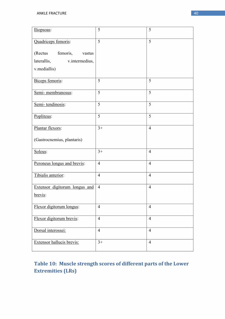
40 ANKLE FRACTURE
Iliopsoas: 5 5
Quadriceps femoris:
(Rectus femoris, vastus
laterallis, v.intermedius,
v.mediallis)
5 5
Biceps femoris: 5 5
Semi- membranosus: 5 5
Semi- tendinosis: 5 5
Popliteus: 5 5
Plantar flexors:
(Gastrocnemius, plantaris)
3+ 4
Soleus: 3+ 4
Peroneus longus and brevis: 4 4
Tibialis anterior: 4 4
Extensor digitorum longus and
brevis:
4 4
Flexor digitorum longus: 4 4
Flexor digitorum brevis: 4 4
Dorsal interossei: 4 4
Extensor hallucis brevis: 3+ 4
Table 10: Muscle strength scores of different parts of the Lower
Extremities (LRs)

41 ANKLE FRACTURE
Muscle length test (According to Janda, 2013):
The grading scale for this test is from 0 - 2 with:
0 = no muscle shortness
1 = moderate shortness
2 = marked shortness
Lower extremity:
Right Left
Iliopsoas: 0 0
Rectus femoris: 1 0
Tensor fascia latae: 1 1
Short hip adductors: 0 0
Gluteus maximus: 0 0
Minimus: 0 0
Medius: 0 0
Piriformis: 0 0
Hamstrings: 0 0
Tibialis anterior: 1 0
Soleus: 1 0
Gastrocnemius: 1 0
Table 11: Muscle length scores of different parts of the Lower Extremities (LRs)

42 ANKLE FRACTURE
Joint Play examination (According to Lewit, 2009):
When there is restriction present that means that the joint is blocked.
Joint name Right Left
Knee joint: Not restricted Not restricted
Patella: Present Not restricted
Head of fibula: Not restricted Not restricted
Talocrural: Present Not restricted
Subtalar joint:
Supination
Present Not restricted
Transverse tarsal joint:
(Chopart joint)
Present Not restricted
Tarsometatarsal joints:
(Lisfranc joints)
Present
Metatarsophalangeal joints:
(1st,2nd, 3rd, 4th, 5th)
Present Not restricted
Table 12: Assessment outcomes of the Joint Play Examination of different parts of the
lower extremities (LRs)
NEUROLOGICAL EXAMINATIONS:
Sensation in dermatomes:
The patient was in supine lying position with his eyes closed in order to make a more
accurate examination. We touch him with his hands on this thigh of the one lower
43 ANKLE FRACTURE
extremity and then on the other one. The patient was instructed to inform us if he
could feel the touch and if there was any difference in between both the lower
extremities.
Two types of examinations:
First, the patient was asked to say yes when he feels the touch in different
areas of his lower extremity.
Second, the patient was asked to say yes if he feels the touch on the same level
on both lower extremity
He had the same feeling on both sides; however, he reported less sensation
sometimes only around the scar otherwise patient said that he was unable to
understand if he feels any sensation or not around the scar on the lateral part.
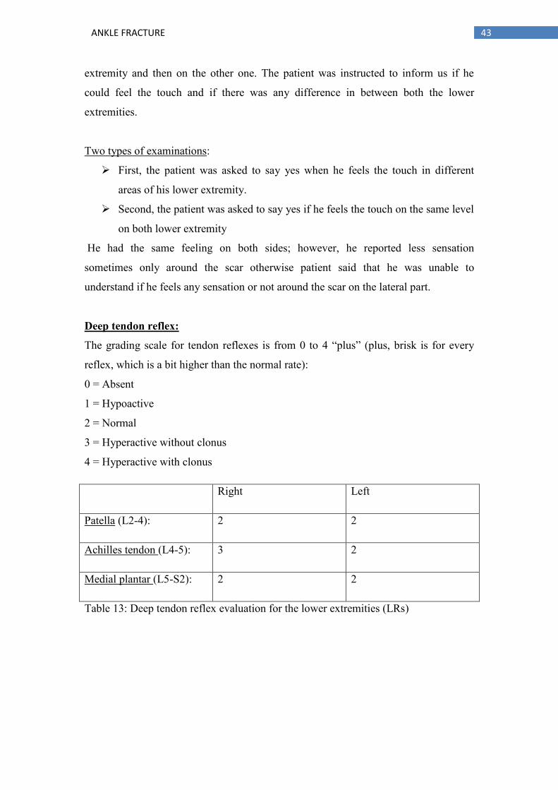
Deep tendon reflex:
The grading scale for tendon reflexes is from 0 to 4 “plus” (plus, brisk is for every
reflex, which is a bit higher than the normal rate):
0 = Absent
1 = Hypoactive
2 = Normal
3 = Hyperactive without clonus
4 = Hyperactive with clonus
Right Left
Patella (L2-4): 2 2
Achilles tendon (L4-5): 3 2
Medial plantar (L5-S2): 2 2
Table 13: Deep tendon reflex evaluation for the lower extremities (LRs)

44 ANKLE FRACTURE
Surface sensation (L4-S1):
L5 (from outside to inside): the patient had the same feeling on both sides
L4 (from inside to outside): the patient had the same feeling on both sides
S1 (in hamstrings) with bend knees: the patient had the same feeling on both sides
3.3.1. Conclusion
Conclusion of examination:
The patient visited our clinic right after recovering from his right ankle joint surgery.
He was in a good mood and was ready to cooperate immediately. Thus, we performed
few examinations on the patient in order to assess his current condition.
During anthropometry measurements, the right leg appeared to have less volume in
calf muscles and had a greater number around the ankle due to edema. The patient
also demonstrated less range of motion (ROM) in the direction to plantar flexion and
eversion on the right ankle joint. He also displayed less ROM in the direction to
plantar flexion of great toe on his right foot. He performed the basic movement
pattern examination correctly with no pathology present.
During the gait and gait modification on the affected side, there were no heel strikes,
lateral rotation of the right foot, and asymmetry of the shoulder. The patient was also
able to stand on heels and tip- toes but was unable to walk on it, walking on squat and
down stairs was also painful for him.
On the single leg stance examination, the patient performed with great balance but
was bit shaking on the affected side (i.e., the right side). During the muscle tone
examination, the soleus and hamstrings were observed to be hypotone on the right
side while the left side soleus and hamstrings were observed to be hypertonic and
therefore exhibited a normal tone.

45 ANKLE FRACTURE
Strength test examination demonstrated that the gastrocnemius, soleus and extensor
hallucis had fair levels, which are 3+ while very low pressure was observed on the
right side.
The length test examination demonstrated that the rectus femoris, TFL, tibialis
anterior, gastrocnemius, and soleus had moderately shortened on the right side, and
the left side TFL.
During the Joint play examination, the patella and the ankle joints were restricted to
the right side.
The neurological examination showed that he had sensation on his right limb and had
comparatively less sensation around the scar that was the lateral one. The deep tendon
reflex showed that the right limb Achilles tendon reflex was hyperactive without any
clonus.
We need to re- educate the patient about walking and his posture so as to help him
restore his functionality as it was prior to the injury. We also further need to continue
with the strengthening exercises and stretching exercises, especially on his shortened
muscles. Lastly, to achieve better results, the patient needs to perform his exercises
regularly and needs to follow all our instructions.
3.4. Short- term and long-term rehabilitation plan
Short term:
Our goal is to reduce patient’s edema
Scar care
Increase Range of Motion
Unblocked the blocked joints
Increase the proprioception
Re- education of walking
Decrease the instability

46 ANKLE FRACTURE
Long term:
Re- education of walking
Decrease pain
Restore ROM
Increase balance
Increase patient confident about his ankle
Goals of therapy:
Eliminate the pain
Decrease the edema
Increase of Rang of Motion
Increase the stability
Increase the sensation of the affected area
3.5. Therapy progress
Date: 16/01/2017
Status Present:
The patient is 8 weeks post- ankle fracture surgery. The scars have healed
successfully but there is swelling in ankle joint on the right side of the right lower
extremity, 1st therapy set.
Subjective:
The patient said that he still experiences pain in his ankle when he wakes up every
morning and also when he is stepping on it.

47 ANKLE FRACTURE
Objective:
The patient is in a good mood and is willing to cooperate, it is his first day of
treatment. Good communication, mentally normal, no assistive devices, left dominant
limb. The patient has pain on his right ankle joint due to surgery and he has a
limitation in the range of motion plus swelling.
Goals of today’s therapy unit:
Reduce pain/edema
Scar care treatment
Increase of sensation
Gait re-education
Increase range of motion
Strengthening of weak muscles
Stretching of shortened muscles
Increase balance
Procedure:
Soft tissue techniques: First by hands then by a soft ball for increasing the blood
circulation on the area, decreasing the pain and edema.
Scar therapy: Examination of the elasticity (elastic in all directions), massaging the
scar (Kippler’s fold, “S” shape, “D” shape, “C” shape, tapping, pressing on the scars
and on the sides).
Increase Sensation: By providing touch therapy around the scar area.
Gait re-education: Starting from the heel strike ending by the tip-toes. We also
corrected his posture during walking (Swinging in shoulders and hands, rotation of
trunk).
Mobilization techniques of the ankle joint and also the Chopart's joint and Lisfranc to
increase the range of motion in the ankle and Achilles tendon

48 ANKLE FRACTURE
Active exercises in the GYM in the standing position to strengthen the weak muscles
around the ankle joint (calf muscle).
PIR technique: To relax and stretch the shortened muscles (gastrocnemius, soleus)
SMT: For increasing the balance and proprioception of the patient
Group of sensorimotoric exercises: on soft pad, with wobble boards and difficulties
walking on a soft pad (forward, backward, and side walk both sides)
Walk on wobbles boards
Walk on the wobbles boards moving hands up and down
Walk on the wobbles boards holding a ball throw it up and catch it again
Walk on the wobbles boards and I throw him the ball, he catches it back and
throws it back to me.
Group of sensor motoric exercises: balance board-posturomed
Stand up with both legs on the board trying to balance moving forward and
sideways. 5 repetitions
Walking on board, standing in the middle of the board and then going down
again. 5 repetitions
Walking sideways, step with one leg up and then with the other staying on the
board for 3 seconds and again down with one leg and then the other. 5
repetitions
Results:
Pain reduced, restoring the ROM by opening the blockage in ankle joint using traction
technique.

49 ANKLE FRACTURE
Date: 17/01/2017
Status Present:
The patient is on his 2nd
therapy set, feels pain in his ankle joint but less than the day
before, no changes in ROM.
Subjective:
The patient said that he felt pain in the morning and could not move his right foot to
the direction of plantar flexion. He also pointed out that he could not move, not
because of the pain but because of the stiffness of the Achilles tendon.
Objective:
The patient is in a good mood and willing to cooperate, good communication,
mentally normal, no assistive devices, left dominant limb. The patient has pain on his
right ankle joint due to surgery and he has a limitation in the range of motion and
there is also some swelling.
Goals of today’s therapy unit:
Reduce pain
Scar therapy
Restore ROM
Increase balance
Stretched shorten muscles
Strengthening of weak muscles
Procedure:
Soft tissue techniques: First by hands and then by using soft ball for increasing the
blood circulation in the area, decreasing both pain and edema.
Scar therapy: Examination of elasticity (elastic in all directions), massaging the scar
(Kippler’s fold, “S” shape, “D” shape, “C” shape, tapping, pressing on the scars and
on the sides).
Increase Sensation: By providing touch therapy around the scar area.

50 ANKLE FRACTURE
Gait re- education: Starting from heel strike ending by the tip-toes. We also
corrected his posture during walking (Swinging in shoulders and hands, rotation of
trunk)
Mobilization techniques of the ankle joint also Chopart's joint and Lisfranc to increase
the range of motion in the ankle.
Active exercises in the GYM in the standing position to strengthen the weak muscles
around the ankle joint (calf muscle).
PIR technique: To relax and stretch the shortened muscles (gastrocnemius, soleus)
SMT : For increasing the balance and proprioception of the patient
Group of sensorimotoric exercises: on soft pad, with wobble boards and difficulties
walking on a soft pad (forward, backward, and side walk both sides)
Walk on wobbles boards
Walk on the wobbles boards moving hands up and down
Walk on the wobbles boards holding a ball throw it up and catch it again
Walk on the wobbles boards and we throw him the ball, he catches it and then
throws it back to us.
Group of sensor motoric exercises: balance board- posturomed
Stand up with both legs on the board trying to balance moving forward and
sideways. 5 repetitions
Walking on board, standing in the middle of the board and then going down
again. 5 repetitions
Walking sideways, step with one leg up and then with the other staying on the
board for 3 seconds and again down with one leg and then the other. 5
repetitions
For strengthening of the muscles: Exercise at the gym

51 ANKLE FRACTURE
Calf raise exercise: Patient stands in front of the mirror with hands support. He
practices calf raise on his tiptoes using his both lower extremities. 3 sets of 6
repetitions.
Results:
Decrease pain, reduce swelling
Date:18/1/2017
Status Present:
The patient is on the 3rd therapy set after ankle fracture surgery. He also has swelling
around the ankle joint
Subjective:
The patient said that he experiences pain in his ankle when he wakes up also when he
is stepping on it but the pain is less than the previous days.
Objective:
The patient is in a good mood and willing to cooperate, it is his 3rd day of treatment.
Good communication, mentally normal, no assistive devices, left dominant limb, no
glasses
Goals of today’s therapy unit:
Reduce pain
Scar care treatment
Increase of sensation
Gait re-education
Increase range of motion
Strengthening of weak muscles
Stretching of shortened muscles

52 ANKLE FRACTURE
Increase balance
Procedure:
Soft tissue techniques: First by hands then by a soft ball for increasing blood
circulation on the area, decreasing pain and edema.
Scar therapy: Examination of elasticity (elastic in all directions), massaging the scar
(Kippler’s fold, “S” shape , “D” shape, “C” shape, tapping, pressing on the scars and
on the sides).
Increase Sensation: By providing touch therapy around the scar area.
Gait re-education: Starting from heel strike ending by the tip-toes. We also corrected
his posture during walking (Swinging in shoulders and hands, rotation of trunk)
Mobilization techniques of Achilles tendon and the ankle joint also Chopart's joint
and Lisfranc to increase the range of motion in the ankle.
Active exercises in the GYM in standing position to strengthen the weak muscles
around the ankle joint (calf muscle).
PIR technique: To relax and stretch the shortened muscles (gastrocnemius, soleus,
hamstrings )
SMT: For increasing the balance and proprioception of the patient
Group of sensorimotoric exercises: on soft pad, with wobble boards and difficulties
walking on a soft pad (forward, backward, and side walk both sides)
Walk on wobbles boards
Walk on the wobbles boards moving hands up and down
Walk on the wobbles boards holding a ball throw it up and catch it again
Walk on the wobbles boards and we throw him the ball, he catches it and
throws it back to us .

53 ANKLE FRACTURE
Group of sensor motoric exercises: balance board-posturomed
Standing up with both legs on the board trying to balance moving forward and
sideways. 5 repetitions
Walking on board, standing in the middle of the board and then going down
again. 5 repetitions
Walking sideways, step with one leg up and then with the other staying on the
board for 3 seconds and again down with one leg and then the other. 5
repetitions
For strengthening of the muscles: Exercises at the gym
Calf raise exercise: Patient stands in front of the mirror with hands support. He
practices calf raise on his tiptoes using his both lower extremities.3 sets of 8
repetitions.
Results:
Pain reduced, reduced swelling, stiffness reduced in Achilles tendon
Date:19/1/2017
Status Present:
The patient is on the 4th
day of therapy set after ankle fracture surgery. There is also
some swelling around the ankle joint.
Subjective:
The patient said that he experiences pain in his ankle when he wakes up and he
couldn’t provide plantar flexion because of stiffness in Achilles tendon also when he
is stepping on it but the pain is less than before.
Objective:

54 ANKLE FRACTURE
The patient is in a good mood and willing to co- operate, it is his 4th
day of treatment.
Good communication, mentally normal, no assistive devices, left dominant limb, no
glasses
Goals of today’s therapy unit:
Scar care treatment
Increase of sensation
Gait re-education
Increase range of motion
Strengthening of weak muscles
Stretching of shortened muscles
Increase balance
Procedure:
Soft tissue techniques: First by hands then by using a soft ball for increasing the
blood circulation in the area so as to decrease pain and edema.
Scar therapy: Examination of elasticity (elastic in all directions), massaging the scar
(Kippler’s fold, “S” shape , “D” shape, “C” shape, tapping, pressing on the scars and
on the sides).
Increase Sensation: By providing touch therapy around the scar area.
Gait re-education: Starting from heel strike ending by the tip-toes. We also corrected
his posture during walking (Swinging in shoulders and hands, rotation of trunk)
Mobilization techniques of the ankle joint also Chopart's joint and Lisfranc to increase
the range of motion in the ankle and to Achilles tendon.
Active exercises in the GYM in a standing position to strengthen the weak muscles
around the ankle joint (calf muscle).
PIR technique: To relax and stretch the shortened muscles (gastrocnemius, soleus,
hamstrings)

55 ANKLE FRACTURE
PNF technique: To strength and relax the calf muscle, and to strength and relax
tibialis anterior 1st flexion and 1st extension diagonal lower extremities
SMT: For increasing the balance and proprioception of the patient
Group of sensorimotoric exercises: on soft pad, with wobble boards and difficulties
walking on a soft pad (forward, backward, and side walk both sides)
Walk on wobbles boards
Walk on the wobbles boards moving hands up and down
Walk on the wobbles boards holding a ball throw it up and catch it again
Walk on the wobbles boards and then I throw him the ball, which he had to
catch and throws it back to me.
Group of sensor motoric exercises: balance board-posturomed
Stand up with both legs on the board trying to balance moving forward and
sideways. 5 repetitions
Walking on board, standing in the middle of the board and then going down
again. 5 repetitions
Walking sideways, step with one leg up and then with the other staying on the
board for 3 seconds and again down with one leg and then the other. 5
repetitions
For strengthening of the muscles: Exercises at the gym
Calf raise exercise: Patient stands in front of the mirror with hands support. HeHe
practices calf raise on his tiptoes using his both lower extremities. 3 sets of 10
repetitions.
Results: reduced pain , reduced swelling , reduced stiffness in Achilles tendon ,
patient can walk on tip toes and on heels with small pain but the calf muscles on right
side still weak but stronger than before
Date: 20/1/2017

56 ANKLE FRACTURE
Status Present: patient is on the 5th
day of therapy set after ankle fracture surgery
also there is swelling around the ankle joint but the swelling is reduced now
Subjective: Patient said that he has a small pain in his ankle when he woke up and he
could provide plantar flexion but not the full range of motion because of stiffness in
Achilles tendon but the pain is less than all the days before.
Objective: The patient is in a good mood and willing to cooperate, it is his 5th
day of
treatment. Good communication, mentally normal, no assistive devices, left dominant
limb, no glasses
Goals of today’s therapy unit:
Scar care treatment
Increase of sensation
Gait re-education
Increase range of motion
Strengthening of weak muscles
Stretching of shortened muscles
Increase balance
Procedure:
Scar therapy: Examination of elasticity (elastic in all directions), massaging the scar
(Kippler’s fold, “S” shape, “D” shape, “C” shape, tapping, pressing on the scars and
on the sides).
Increase Sensation: By providing touch therapy around the scar area.
Gait re-education: Starting from heel strike ending by the tip-toes. We also corrected
his posture during walking (Swinging in shoulders and hands, rotation of trunk)
Mobilization techniques of Achilles tendon and the ankle joint also Chopart's joint
and lis franc to increase the range of motion in the ankle.
Active exercises in the GYM in a standing position to strengthen the weak muscles
around the ankle joint (calf muscle).

57 ANKLE FRACTURE
PIR technique: To relax and stretch the shortened muscles (gastrocnemius, soleus).
PNF technique: To strengthen and relax the calf muscle, and to strengthen and relax.
tibialis anterior 1st flexion and 1st extension diagonal lower extremities.
SMT: For increasing the balance and proprioception of the patient.
Group of sensorimotoric exercises: on soft pad, with wobble boards and difficulties
walking on a soft pad (forward, backward, and side walk both sides)
Walk on wobbles boards
Walk on the wobbles boards moving hands up and down
Walk on the wobbles boards holding a ball throw it up and catch it again
Walk on the wobbles boards and we throw him the ball, he catches it back and
throws it back to us.
Group of sensor motoric exercises: balance board-posturomed
Stand up with both legs on the board trying to balance moving forward and
sideways. 5 repetitions
Walking on board, standing in the middle of the board and then going down
again. 5 repetitions
Walking sideways, step with one leg up and then with the other staying on the
board for 3 seconds and again down with one leg and then the other. 5
repetitions
For strengthening of the muscles: Exercises at the gym
Calf raise exercise: Patient stands in front of the mirror with hands support. He
practices calf raise on his tiptoes using one lower extremity (his affected lower
extremities). 3 sets of 6 repetitions.
Results: Reduced pain, reduced swelling, reduced stiffness in Achilles tendon, patient
can walk on tip toes and on heels (no pain during performing) and the calf muscles on
right side stronger than before.

58 ANKLE FRACTURE
Date:23/1/2017
Status Present:
The patient is on the 6th
day of therapy set after ankle fracture surgery and there is still
swelling around the ankle joint but has reduced.
Subjective:
The patient said that he experienced small pain in his ankle when he woke up and he
could provide plantar flexion but not full range because of stiffness in Achilles tendon
but the pain is less than before.
Objective:
The patient is in a good mood and willing to cooperate, it is his 6th
day of treatment.
Good communication, mentally normal, no assistive devices, left dominant limb, no
glasses.
Goals of today’s therapy unit:
Scar care treatment
Increase of sensation
Gait re-education
Increase range of motion
Strengthening of weak muscles
Stretching of shortened muscles
Increase balance
Procedure:
Scar therapy: Examination of elasticity (elastic in all directions), massaging the scar
(Kippler’s fold, “S” shape, “D” shape, “C” shape, tapping, pressing on the scars and
on the sides).
Increase Sensation: By providing touch therapy around the scar area.

59 ANKLE FRACTURE
Gait re-education: Starting from heel strike ending by the tip- toes. We also
corrected his posture during walking (Swinging in shoulders and hands, rotation of
trunk ).
Mobilization techniques of the ankle joint also Chopart's joint and lis franc to increase
the range of motion in the ankle.
Active exercises in the GYM in a standing position to strengthen the weak muscles
around the ankle joint (calf muscle).
PIR technique: To relax and stretch the shortened muscles (gastrocnemius, soleus,
hamstrings).
PNF technique: To strength the calf muscle first extension, and to relax tibialis
anterior.
SMT: For increasing the balance and proprioception of the patient.
Group of sensorimotoric exercises: on soft pad, with wobble boards and difficulties
walking on a soft pad (forward, backward, and side walk both sides)
Walk on wobbles boards
Walk on the wobbles boards moving hands up and down
Walk on the wobbles boards holding a ball throw it up and catch it again
Walk on the wobbles boards and we throw him the ball, he catches it and
throws it back to us.
Group of sensorimotoric exercises: balance board- posturomed
Stand up with both legs on the board trying to balance moving forward and
sideways. 5 repetitions
Walking on board, standing in the middle of the board and then going down
again. 5 repetitions
Walking sideways, step with one leg up and then with the other staying on the
board for 3 seconds and again down with one leg and then the other. 5
repetitions
For strengthening of the muscles: Exercises at the gym

60 ANKLE FRACTURE
Calf raise exercise: Patient stands in front of the mirror with hands support.
He practices calf raise on his tiptoes using one lower extremity (his affected
lower extremity).3 sets of 7 repetitions. Walking on tip toes to strengthen the
calf muscles.
Results: Reduced pain, reduced swelling, reduced stiffness in Achilles tendon, the
patient can walk on tip toes and on heels (no pain during performing) but the calf
muscles on the right side are now stronger than before.
Date:24/1/2017
Status Present:
The patient is on the 7th
therapy set after ankle fracture surgery and there is still
swelling around the ankle joint but has reduced
Subjective:
The patient said that he has a small pain in his ankle when he woke up and he could
provide plantar flexion but not full range because of stiffness in Achilles tendon but
the pain is less than before.
Objective:
The patient is in a good mood and willing to cooperate, it is his first day of treatment.
Good communication, mentally normal, no assistive devices, left dominant limb, no
glasses.
Goals of today’s therapy unit:
Scar care treatment
Increase of sensation
Gait re-education
Increase range of motion

61 ANKLE FRACTURE
Strengthening of weak muscles
Stretching of shortened muscles
Increase balance
Procedure:
Scar therapy: Examination of elasticity (elastic in all directions), massaging the scar
(Kippler’s fold, “S” shape, “D” shape, “C” shape, tapping, pressing on the scars and
on the sides).
Increase Sensation: By providing touch therapy around the scar area.
Gait re-education: Starting from heel strike ending by the tip-toes. We also corrected
his posture during walking (Swinging in shoulders and hands, rotation of trunk)
Mobilization techniques of the ankle joint also Chopart's joint and lis franc to increase
the range of motion in the ankle.
Active exercises in the GYM in a standing position to strengthen the weak muscles
around the ankle joint (calf muscle).
PIR technique: To relax and stretch the shortened muscles (gastrocnemius, soleus,
hamstrings)
PNF technique: To strength the calf muscle first extension, and to relax tibialis
anterior
SMT: For increasing the balance and proprioception of the patient
Group of sensorimotoric exercises: on soft pad, with wobble boards and difficulties
walking on a soft pad (forward, backward, and side walk both sides)
Walk on wobbles boards
Walk on the wobbles boards moving hands up and down
Walk on the wobbles boards holding a ball throws it up and catch it again

62 ANKLE FRACTURE
Walk on the wobbles boards and we throw him the ball, he catches it back and
throws it back to us.
Group of sensor motoric exercises: balance board-posturomed
Stand up with both legs on the board trying to balance moving forward and
sideways. 7 repetitions
Walking on board, standing in the middle of the board and then going down
again. 7 repetitions
Walking sideways, step with one leg up and then with the other staying on the
board for 3 seconds and again down with one leg and then the other. 7
repetitions
For strengthening of the muscles: Exercises at the gym
Calf raise exercise: Patient stands in front of the mirror with hand support. He
practices calf raise on his tiptoes using one lower extremity (his affected lower
extremities). 3 sets of 8 repetitions. Walking on tip toes to strengthen the calf muscles.
Results: Reduced pain, reduced swelling, reduced stiffness in Achilles tendon, no
pain during walking and patient could perform walking in the squat (no pain during
performing) and the calf muscles on right side stronger than before.
Date:25/1/2017
Status Present:
The patient is on the 8th
day of therapy set after ankle fracture surgery and there is still
some swelling around the ankle joint but is reduced.
Subjective:
The patient said that he has no pain in his ankle when he woke up and he could
provide plantar flexion but not full range because of stiffness in Achilles tendon but
the pain is less than before.
Objective:
63 ANKLE FRACTURE
The patient is in a good mood and willing to cooperate, it is his 8th
day of treatment.
Good communication, mentally normal, no assistive devices, left dominant limb, no
glasses.
Goals of today’s therapy unit:
Scar care treatment
Increase of sensation
Gait re-education
Increase range of motion
Strengthening of weak muscles
Stretching of shortened muscles
Increase balance
Procedure:
Scar therapy: Examination of elasticity (elastic in all directions), massaging the scar
(Kippler’s fold, “S” shape, “D” shape, “C” shape, tapping, pressing on the scars and
on the sides).
Increase Sensation: By providing touch therapy around the scar area.
Gait re-education: Starting from heel strike ending by the tip-toes. We also corrected
his posture during walking (Swinging in shoulders and hands, rotation of trunk)
Mobilization techniques of the ankle joint also Chopart's joint and Lisfranc to increase
the range of motion in the ankle.
Active exercises in the GYM in a standing position to strengthen the weak muscles
around the ankle joint (calf muscle).
PIR technique: To relax and stretch the shortened muscles (gastrocnemius, soleus,
hamstrings ).
PNF technique: To strengthen the calf muscle first extension, and to relax tibialis
anterior.
SMT: For increasing the balance and proprioception of the patient.
Group of sensorimotoric exercises: on soft pad, with wobble boards and difficulties

64 ANKLE FRACTURE
walking on a soft pad (forward, backward, and side walk both sides)
Walk on wobbles boards
Walk on the wobbles boards moving hands ups and down
Walk on the wobbles boards holding a ball through it up and catch it again
Walk on the wobbles boards and we throw him the ball, he catches it back and
through it again to us.
Group of sensor motoric exercises: balance board- posturomed
Stand up with both legs on the board trying to balance moving forward and
sideways. 7 repetitions.
Walking on board, standing in the middle of the board and then going down
again. 7 repetitions.
Walking sideways, step with one leg up and then with the other staying on the
board for 3 seconds and again down with one leg and then the other. 7
repetitions.
For strengthening of the muscles: Exercises at the gym
Calf raise exercise: Patient stands in front of the mirror with hands support. He
practices calf raise on his tiptoes using one lower extremity (his affected lower
extremity). 3 sets of 10 repetitions. Walking on tip toes to strengthen the calf muscles.
Results: Reduced pain, reduced swelling, reduced stiffness in Achilles tendon, no
pain during walking and the calf muscle on right side was stronger than before
3.6 Final kinesiologic examination:
Examination by observation of the patient:
Anterior View:
Edema on the right ankle joint
Right knee rotated outwards (patella)
Pelvis symmetrical

65 ANKLE FRACTURE
Abdominal muscles higher on the left side than the right side
Left shoulder higher than the right one
Left clavicle is higher than the right one
Posterior View:
Same angle of feet rotation outward
Edema on the right ankle joint (Achilles tendon more visible on the right side
due to reducing edema resulting from therapy (after the surgery)
Hypotrophy of gastrocnemius muscle on the right side (affected side)
Knees symmetrical.
The space from the right scapula to the spine is smaller than the left scapula.
Left shoulder higher than the right one
Lateral View: (Left side)
Toes normal press and symmetrical
Knees symmetrical
Flat back posture
Both scapulas are visible
Shoulder protracted a bit
Head in normal position
Lateral View: (Right side)
Lateral rotation of foot
Ankle joint swollen
Knees symmetrical
Scapula not visible from this side
Spine not visible from this side
Head in normal position

66 ANKLE FRACTURE
Observation of the scar and palpation of the area around the scar:
Patient’s scars healed successfully. The ankle joint is less swollen; the skin around the
scars is pink in color and with slightly increased temperature on the ankle area.
Anthropometry Measurements:
Circumferences:
Right Left
Thigh:
( 15 cm above knee cap for the whole quadriceps )
45.5cm 46cm
( 10 cm above knee cap for vastus medialis ) 40cm 41,5cm
Knee cap: 35cm 35cm
Calf: 34cm 36cm
Ankle: 27cm 24 cm
Table 14: Final circumference measurements for both LE’s
Length of lower extremity in parts :
Right Left
Femur 39cm 39cm
Lower Leg 48cm 48cm
Foot 25cm 25cm
Table 15: Final length of the lower extremity in different parts

67 ANKLE FRACTURE
Functional length:
Right Left
103cm 103cm
Table 16: Final functional length measurement for both LE’s
Anatomical length:
Right Left
87 cm 87 cm
Table 17: Final anatomical length measurement for both LE’s
ROM examination (According to Kendal):
Ankle Joint:
RIGHT LIFT
S : 50 - 0 - 10
F : 5 - 0 – 20
S : 60 - 0 - 10
F: 10 - 0 - 25
Table 18: Final ROM examination in ankle joint for both LE’s
Toes:
Right Left
Big toe:
(proximal phalanx)
S : 50 – 0 - S : 70 – 0 -
Table 19: Final ROM examination in big toe for both LE’s

68 ANKLE FRACTURE
GAIT examination (According to Kendall, 2005):
We asked the patient to walk in the room so we can observe his gate
During walking:
Heal strike was present
Less lateral rotation in the right ankle
The symmetry of shoulders
swinging both hands
no limping
rotation in the trunk ( presented while walking by swinging upper extremities )
Gait modification:
Walking on heels: negative / he were able to perform it without pain.
Walking on tip toes: negative/ he was able to perform it without pain
Squats walking: negative / was able to perform.
Walking with eyes closed: negative /He was able to perform it without pain,
he was stable.
Walking backward or stair walking: negative / He was able to walk backward,
also up\down stairs ( upstairs no pain, downstairs less pain than before )
Trendelenburg test:
The patient has to stand on one lower extremity and the other leg with flexion 90ᵒ on
hip and knee.
No pelvis drop in both sides during standing.

69 ANKLE FRACTURE
Single leg stance:
Patient was asked to perform a simple stand on one leg.
Right leg: great balance without shaking.
Left leg: great balance without shaking.
Rhomberg test:
- ( I ) : standing with feet a parts and opened eyes
- ( II ) : feet together and opened eyes
- ( III ) : feet together and eyes closed
I = negative
II = negative
III = negative
Two Scale examination:
The physiological difference in loading is up to 5 kg, approximately 10 % to 15 % of
body weight. Bigger difference indicates an imbalance.
Right Left
Total: 33kg 30kg
Table 20: Physiological difference in loading weights in both lower extremities

70 ANKLE FRACTURE
Muscle tone examination:
Lower extremity:
Right Left
Gluteus maximus: Normal tone Normal tone
Hip adductors:
(Pectineus, adductor
magnus, gracilis, adductor
brevis, adductor longus)
Normal tone Normal tone
Quadriceps femoris:
(Rectus femoris, vastus
laterallis, v.intermedius,
v.mediallis)
Normal tone Normal tone
Biceps femoris: Normal tone Normal tone
Semimembranosus: Normal tone Normal tone
Semitendinosus: Normal tone Normal tone
Soleus: Normal tone Normal tone
Hamstrings: Normal tone Normal tone
Table 21: Final muscle tone test evaluation for both LE’s
Muscle strength test (According to Kendall, 2005):
The grading scale for this test is from 0 - 5 with:
0 = Zero (no muscle contraction)

71 ANKLE FRACTURE
1 = Trace (contraction felt but no movement)
2 = Poor (partial movement but in horizontal position)
3 = Fair (hold against gravity)
4 = Good (hold against moderate pressure)
5 = Normal (hold against strong pressure)
Lower extremity:
Right Left
Gluteus maximus: 5 5
Gluteus medius: 5 5
Gluteus minimus: 5 5
Lateral rotators of hip
joint:
5 5
Hip adductors: 5 5
Tensor fasciae latae: 5 5
Sartorius: 5 5
Iliopsoas: 5 5
Quadriceps femoris:
(Rectus femoris, vastus
laterallis, v.intermedius,
v.mediallis)
5 5
Biceps femoris: 5 5
Semimembranosus: 5 5
Semitendinosus: 5 5

72 ANKLE FRACTURE
Popliteus: 5 5
Plantar flexors:
(Gastrocnemius, plantaris)
4+ 5
Soleus: 4+ 5
Peroneus longus and
brevis:
4 5
Tibialis anterior: 4 5
Extensor digitorum longus
and brevis:
4 5
Flexor digitorum longus: 4 5
Flexor digitorum brevis: 4 4
Dorsal interossei: 4 4
Extensor hallucis brevis: 4 4
Table 22: Final muscle strength test evaluation for both LE’s

73 ANKLE FRACTURE
Muscle length test (According to Janda, 2013):
The grading scale for this test is from 0 - 2 with:
0 = no muscle shortness
1 = moderate shortness
2 = marked shortness
Lower extremity:
Right Left
Iliopsoas: 0 0
Rectus femoris: 0 0
Tensor fascia latae: 0 0
Short hip adductors: 0 0
Gluteus maximus: 0 0
Minimus: 0 0
Medius: 0 0
Piriformis: 0 0
Hamstrings: 0 0
Tibialis anterior: 0 0
Soleus: 1 0
Gastrocnemius: 1 0
Table 23: Final muscle length test evaluation for both LE’s

74 ANKLE FRACTURE
Joint Play examination (According to Lewit, 2009):
When there is restriction present that means that the joint is blocked.
Right Left
Knee joint: Not restricted Not restricted
Patella: Not Restricted Not restricted
Head of fibula: Not restricted Not restricted
Talocrural: Not Restricted Not restricted
Subtalar joint:
Supination
Not Restricted Not restricted
Transverse tarsal joint:
(Chopart joint)
Not Restricted Not restricted
Tarsometatarsal joints:
(Lisfranc joints)
Not Restricted Not Restricted
Metatarsophalangeal
joints:
(1st,2nd, 3rd, 4th, 5th)
Not Restricted Not Restricted
Table 24: Final joint play examination evaluation for L spine and LE’s
NEUROLOGICAL EXAMINATIONS:
Sensation in dermatomes:
The patient was in supine lying position with his eyes closed. Two types of
examinations were done:

75 ANKLE FRACTURE
First, he was asked to say yes when he feels the touch in different areas of his
lower extremity.
Second, patient was asked to say yes if he feels the touch on the same level on
both lower extremity
He had the same feeling on both sides but only around the scar, the sensation was
less. Sometimes, the patient was unable to understand if he feels the touch or not
around the scar lateral one but the patient mentioned that he feels better in sensation
than when he came for the first examination.
Deep tendon reflex:
The grading scale for tendon reflexes is from 0-4 “plus” (plus, brisk is for every
reflex, which is a bit higher than the normal rate):
0 = Absent
1 = Hypoactive
2 = Normal
3 = Hyperactive without clonus
4 = Hyperactive with clonus
Right Left
Patella (L2-4): 2 2
Achilles tendon (L4-5): 2 2
Medial plantar (L5-S2): 2 2
Table 25: Final deep tendon reflex evaluation for both LE’s
Surface sensation (L4-S1):
L5 (from outside to inside): the patient had the same feeling on both sides
L4 (from inside to outside): the patient had the same feeling on both sides
S1 (in hamstrings) with bend knees: the patient had the same feeling on both sides

76 ANKLE FRACTURE
3.6.1. Conclusion
Conclusion of Examination:
The patient after the ankle surgery had clearly restriction in range of motion,
the right ankle joint was swollen and painful, without sensation on the later part
around the scar. He also had blocked joints, shortened muscles on the affected side,
abnormal gait pattern. As soon as he visited our clinic we started the treatment and at
the end of the treatment, we performed our final examinations.
During the anthropometry measurements, the right leg still had less volume in
the calf muscles by 3 cm and greater number around the ankle due to edema but it was
better than the first day of his treatment. He experienced less ROM in the direction to
plantar flexion and eversion on the right ankle joint. He also had less ROM in the
direction to plantar flexion of great toe of the right foot.
Patient performed all the basic movement pattern correctly and no pathology
was present. During the gait plus gait modification on the affected side, during the
final treatment day, he was able to perform the heel strike and had a symmetrical
lateral rotation on both feet. In the end, there was no more asymmetry on the
shoulders.
The patient was able to stand and walk on his heels and tiptoes. Squatting and
climbing up and down the stairs was not painful anymore for him. The patient
performed well in the single leg stance test and showed great balance without shaking.
Muscle tone achieved normal strength test on gastrocnemius, soleus and extensor
hallucis, which had good level 4+. The length test on the gastrocnemius and soleus
were moderate and had shortened on the right side while the left side had no
shortness. The joint play was without any restriction on the joints.
During the Neurological examination, the sensation on the right limb had less
sensation around the scar on the lateral one but he experienced much more sensitivity
than the first day of treatment.

77 ANKLE FRACTURE
For better results, the patient needs to perform his exercises regularly and to
follow all of our instructions and recommendations.
3.7. Therapy effect evaluation and Prognosis
The patient provided feedback post the completion of the rehabilitation. In the
feedback, he described the importance and the need for strengthening, expert care and
the self- therapy, which he had to learn and follow during the therapy sessions in
order to avoid further damage to his lower extremities and especially his injured ankle
or provoking a new injury.
The Self- therapy was introduced to him by the doctor just prior to his last
check- up session. The self- therapy included all the exercises he performed during
the rehabilitation treatment sessions.
The patient was recommended to continue practicing the exercises he learned
during the program and also follow the rehabilitation plan in order to maintain and
achieve better ROM.
He needs to continue practicing his exercises in the ankle fracture program
pattern as it was executed during the rehabilitation therapies. It would be wise to
continue the sensomotoric exercises and practicing the gait movement and
mobilization techniques to restore his previous ankle condition. In the case of
swelling, massaging the scar would help him in getting some relief.
His ROM of the ankle joint had considerably increased since his first visit to the
rehabilitation sessions as described by the patient himself.
However, the major concern yet remained for the patient, which was the
increase in the ROM of the ankle joint during the rehabilitation process. Therefore,
we measured it by using a goniometer at the beginning of every session. The final
assessments of various parameters highlighted the effectiveness of the rehabilitation
therapy, which was provided for each and every session.
Additionally, the comparison of the values of the initial and final kinesiologic
examination further demonstrates considerable improvement in the muscular

78 ANKLE FRACTURE
condition, gait cycle, posture and the joint play upon examining the injured ankle
joint.
The final assessments indicate an overall improvement in the ROM and joint
mobility of the fractured ankle joint. His prognosis appeared to be very good and his
ankle joint had fully recovered from the fracture without provoking any further issues.
4. Conclusion
I straight away choose my patient because he was in the beginning of his
rehabilitation treatment plan at the clinic I worked in and thus it gave me an
opportunity to observe the progress of the treatment and the improvement in his
condition right from Day 1 and work with him closely and in an effective manner.
When the patient first visited our clinic, he complained about pain in his injured ankle
area and restricted range of motion; however, he was not in a very bad condition.
The restriction in the ROM was clearly visible in all the motions of the affected
ankle joint, especially in the DF and PF. This issue was very critical and was
preventing him from performing simple daily activities so he was a bit furious.
Moreover, he was unhappy about the fact that he had become dependent on others.
Thus, the rehabilitation treatment plan was designed based on his issues and with the
goal of improving his gait, ROM, reducing his pain and swelling of the affected area.
The place where I worked, i.e., Centrum Léčby Pohybového Aparátu Vysočany,
Prague, routinely and frequently provided similar kind of therapies (like the one that
was provided to this patient by us) to patients’ who come up with similar issues. The
rehabilitation program planned by me with the help of my supervisor was based on
the patient’s ROM restriction, swelling, and pain. I chose to focus on these factors
majorly so as to treat the ROM, and reduce the pain and swelling in the patient and
restore his ankle condition and gait cycle back to the previous state before the injury.
The issues that he was facing post- surgery were common in these kinds of surgeries.

79 ANKLE FRACTURE
Before initiation of the rehabilitation session, I discussed my treatment protocol
with the patient as well as my supervisor to ensure all issues are considered and
patient’s needs were taken care of. This helped in fastening the recovery process,
which further helped both of us in different ways i.e., the patient, could recover faster
and start working and practice his daily activities independently and I could finish my
thesis and practice.
I also applied some muscle relaxation techniques on him in order to release the
hypertonus and tension n the muscles, which was found during the initial
examination.
The outcomes of the initial and final kinesiologic examinations when compared
showed positive and significant improvements along with increased ROM in the
injured ankle joint. The restricted joint play was also restored with the help of the
exercises and his ability to perform the ankle movements independently and the
improvement was significant. These were considered to be the most significant
parameters in this case, as from the initiation of the treatment protocol, the goal was
to reduce swelling and pain and increase his ROM and improve his gait in order to
help him to be more independent. Improvements in his health condition were
expected; however, the actual improvement was seen as a result of the efficiency of
the therapies provided to him, the chronological timing of each session and patient’s
cooperation and efforts to recover faster.
Too much of muscle strength or too much pressure on the ankle on the operated
ankle was non- questionable; however, the patients’ main interest was to recover as
soon as possible so that he can join back at his work, perform his tasks independently,
recover from the pain and swelling and start cheerleading again. Since the patient had
a strong will power and showed positive attidute towards the therapies and put
exceptional efforts to perform all tasks with excellence, we also made sure that he is
able to work on his ROM and ankle movement all by himself.
For future reference, at the end of the sessions, I instructed him to continue
practicing the exercises taught to him during the treatment sessions so as to continue
improve his gait and restore back his functionality. It was noticed that the patient was
already motivated to continue self- therapy and thus required no counseling from us
and he promised to stay as active as possible.

80 ANKLE FRACTURE
After considering the total two weeks therapy, I can conclude that the treatment
plan that I had made for this patient had successfully helped him in recovering from
his fracture and regain his functionality and body posture. During the whole
rehabilitation process, the patient appeared to be very active, enthusiastic and
optimistic about performing new exercises and also cooperated well with us by
following all our instructions without any issues.
5. References
American Academy of Orthopedic Surgeon. (2015). Fractures. Retrieved March 13,
2017
Anish, R., Kadakia, K., & M., M. (2014). Rehabilitation After Acute Ruptures.
Retrieved from http://books.google.com.sa/books?id=6OXb-
Appleton, P., & McQueen, M. C.-B. (2006). The fibula nail for treatment of ankle
fractures in elderly and high risk patients. Tech Foot Ankle Surg, 204- 208.
Baird, R. (1987). Fractures of the distal part of the fibula with associated disruption of
the deltoid ligament, treatment without repair of the deltoid ligament. JBJS,
1346.
Bauer, M. (1985). Malleolar fractures: nonoperative versus operative treatment. A
controlled study. CORR, 199.
Berme, N. (1985). Biomechanincs in normal and pathological human articulating
joints. Dordrecht: Martinus Nijhoff.
Bibbo, C. (2001). Complications of ankle fractures in diabetic patients. OCNA, 113-
117.
Bossuyt, P., Reitsma, J., Bruns, D., & al, e. (2004). Towards complete and accurate
reporting of studies of diagnostic accuracy: the STARD initiative. Clin Chem,
21 (1): 4- 10.
Bostman, O. (1989). Ankle fractures treated using biodegradable internal fixation.
CORR, 195.

81 ANKLE FRACTURE
Brookbush, B. (2011). Kinesiology of the Ankle. Retrieved from
http://brentbrookbush.com/kinesiology-ofthe-ankle
Broomer, A. (1996). Rockwood and greens fractures in adult, 4th ed.
Bucholz, R. (1994). Fixation with bioabsorbable screws for the treatment of fractures
of the ankle. JBJS, 319.
Bugler, K., White, D., & Thordarson, B. (2011). Foot & Ankle: FOcus on Ankle
Fractures. Bones & Joint.
Carrage, E. (1991). Early complications in the operative treatment of ankle fractures.
JBJS, 79.
Cimino, W. (1991). Early mobilization of the ankle fractures after open reduction and
internal fixation. CORR, 2201.
Connolly, J. (1998). Limb threatening neuropathic complications from ankle fractures
in patients with diabetes. CORR, 212.
Costigan, W., Thordarson, D., & Debnath, U. (2007). Operative management of ankle
fractures in patients with diabetes mellitus. Foot Ankle Int, 32- 7.
Court-Brown, C., Wood, A., & Aitken, S. (2008). The epidemiology of acute sports-
related fractures in adults. Injury, 39 (12): 1365- 1372.
Dananberg, H. (2004). Manipulation of the ankle as a method of treatment for ankle
and foot pain. Journal of the American Podiatric Medical Association, 395-
399.
De Boer, A., Schepers, T., Panneman, M., Van Beeck, E., & Van Lieshout, E. (2014).
Health care consumption and costs due to foot and ankle injuries in the
Netherlands, 1986-2010. BMC Musculoskelet Disord, 15: 128.
DeAngelis, N., Eskander, M., & French, B. (2007). Does medial tenderness predict
deep deltoid ligament incompetence in supination-external rotation type ankle
fractures? J Orthop Trauma, 244- 7.

82 ANKLE FRACTURE
Denegar, C. R., Hertel, J., & & Fonseca, J. (2002). The effect of lateral ankle sprain
on dorsiflexion range of motion, posterior talar glide, and joint laxity. The
Journal of Orthopaedic and Sports Physical Therapy, 166- 176.
Doherty, C., Delahunt, E., Caulfield, B., Hertel, J., Ryan, J., & Bleakley, C. (2014).
The incidence and prevalence of ankle sprain injury: a systematic review and
meta-analysis of prospective epidemiological studies. Sports Med, 44 (1): 123-
40.
Donken, C., Al-Khateeb, H., Verhofstad, M., & van Laarhoven, C. (2012). Surgical
versus conservative interventions for treating ankle fractures in adults.
COchrane Database Syst Rev, 8- 10.
Drakos, M., & Murphy, C. (2014). Bracing versus casting in ankle fractures. Phys
Sportsmed, 42 (4): 60- 70.
Evans, D., & & Lucas, N. (2010). What is 'manipulation'? A reappraisal. . Manual
Therapy, 286- 291.
Finsen, V. (1989). Osteopenia after ankle fractures. CORR, 261.
Fong, D., Hong, Y., Chan, L., Yung, P., & Chan, K. (2007). A systematic review on
ankle injury and ankle sprain in sports. Sports Med, 37 (1): 73- 94.
Giannini, S., Chiarello, E., Persiani, V., Luciani, D., Cadossi, M., & Tedesco, G.
(2013). Ankle fractures in elderly patients. Aging Clin Exp Res., 77- 79.
Goost, H., Wimmer, M., Barg, A., Kabir, K., Valderrabano, V., & Burger, C. (2014).
Fractures of the ankle joint: investigation and treatment options. . Dtsch
Arztebl, 111(21), 377- 388.
Greenman, P. (2003). Principles of manual medicine (3rd ed.). Philadelphia: ippincott
Williams & Wilkins.
Hoch, M., & McKeon, P. (2010). The effectiveness of mobilisation with movement at
improving dorsiflexion after ankle sprain. J Sport Rehabil, 19: 226- 32.

83 ANKLE FRACTURE
Hubbard, T., & Wikstrom, E. (2010). Ankle sprain: pathophysiology, predisposing
factors, and management strategies. Open Access Journal of Sports Medicine,
115- 122.
Iyer, K. (2012). Trauma and Management in Orthopedics. Springer.
Joy, G. (1974). Precise evaluation of the reduction of severe ankle fractures. JBJS,
979.
Kisner, C., Colby, L., & Library, R. (2007). Therapeutic exercise: foundations and
techniques. Davis.
Lamb, S., Marsh, J., Hutton, J., Nakash, R., & Cooke, M. (2009). Mechanical
supports for acute, severe ankle sprain: a pragmatic, multicentre, randomised
controlled trial. Lancet, 575- 81.
Lamontagne, J., Blachut, P., Broekhuyse, H., O'Brien, P., & Meek, R. (2002).
Surgical treatment of a displaced lateral malleolus fracture: the antiglide
technique versus lateral plate fixation. J Orthop Trauma, 498- 502.
Leeds, H. (1984). Instability of the distal tibiofibular syndesmosis after bimalleolar
and trimalleolar fractures. JBJS, 90.
Leeflang, M. (2014). Systematic reviews and meta-analyses of diagnostic test
accuracy. Clin Microbiol, 20 (2): 105- 113.
Lin, C., Moseley, A., Haas, M., Refshauge, K., & Herbert, R. (2008). Manual therapy
in addition to physiotherapy does not improve clinical or economic outcomes
after ankle fracture. J Rehabil Med, 40 (6): 433- 439.
Lindsjo, U. (1985). Classification of ankle fractures: the Lauge Hansen or AO
System. CORR, 12-1 5.
Magee, D. (2007). Orthopedic physical assessment (5th ed.). ST. Louis: Mo:
Saunders Elsevier.
Mallett, S., Halligan, S., Thompson, M., Collins, G., & Altman, D. (2012).
Interpreting diagnostic accuracy studies for patient care. BMJ.
84 ANKLE FRACTURE
Marinos, G., Giannopoulos, A., Vlasis, K., & al, e. (2008). Primary care in the
management of common orthopaedic problems. Quality in Primary Care, 16
(5): 345- 9.
Marti, R. (1990). Malunited ankle fractures. JBJS, 709].
Martin, R., Davenport, T., Paulseth, S., Wukich, D., & Godges, J. (2013 ). Ankle
stability and movement coordination impairments: ankle ligament sprains. J
orthop Sports Phys The, 43(9): 1- 40.
McClellan, C., Cramp, F., Powell, J., & Benger, J. (2010). Extended scope
physiotherapists in the emergency department: a literature review. Phys Ther
Rev, 106- 11.
McCormack, R. (1998). Ankle fractures in diabetics. JBJS, 689.
McKinnins, L. (2014). Fundaments of muskeletal imaging. In: McKinnis LN, ed. 4 ed.
Philadelphia: F.A. Davis Company.
Moseley, A., Beckenkamp, P., Haas, M., Herbert, R., & Lin, C. (2015). Rehabilitation
After Immobilization for Ankle Fracture: The EXACT Randomized Clinical
Trial. JAMA, 314(13), 1375- 1385.
Moseley, A., Herbert, R., Nightingale, E., & al., e. (2055). Passive stretching does not
enhance outcomes in patients with plantarflexion contracture after cast
immobilization for ankle fracture: a randomized controlled trial. Arch Phys
Med Rehabil, 86 (6)L 1118- 26.
Nightingale, E., Moseley, A., & Herbert, R. (2007). Passive dorsiflexion flexibility
after cast immobilization for ankle fracture. CLin Orthop Relat Res, 456: 65-
69.
Nilsson, G., Jonsson, K., Ekdahl, C., & Eneroth, M. (2009). Effects of a training
program after surgically treated ankle fracture: a prospective randomised
controlled trial. BMC Musculoskelet Disord, 118.
Palastanga, N., Field, D., & & Soames, R. (2006). Anatomy and human movement :
structure and function (5th ed.). New York: Butterworth Heinmann/ Elsevier.

85 ANKLE FRACTURE
Pott, P. (1996). Rockwood and Greens fractures in adults, fourth edition.
Pritsch, M. (1993). Adhesions of Distal tibiofibular syndesmosis. A cause for chronic
ankle pain after fracture. CORR, 220.
Psatha, M., Wu, Z., Gammie, F., & al., e. (2012). A longitudinal MRI study of muscle
atrophy during lower leg immobilization following ankle fracture. Magn
Reson Imaging, 35 (2): 686- 695.
Robertson, G., Wood, A., Aitken, S., & Court Brown, C. (2014). Epidemiology,
management, and outcome of sport-related ankle fractures in a standard UK
population. Foot Ankle Int., 32 (11): 1143- 52.
Rowley, D. (1986). A prospective trial comparing operative and manipulative
treatment of ankle fractures. JBJS, 610.
Schon, L. (1995). The management of neuroarthropathic fracture dislocations in
diabetic patient. . OCNA, 375.
Scott, A. (2010). Diagnosis and treatment of ankle fractures. Radiol Technol, 457- 75.
Segal, D. (1985). Functional bracing and rehabilitation of ankle fractures. CORR, 29.
Seiger, C. (2009). Effect of pulsed shortwave diathermy and joint mobilization on
range of motion, pain, and edema of surgically repaired hypomobile ankles.
ProQuest Dissertations & Thesis.
Singer, B., McLauchlan, G., Robinson, C., & Christie, J. (1998). Epidemiology of
fractures in 15 000 adults: the influence of age and gender. J Bone Joint Surg
Br., 80 (2): 243- 248.
Solveborn, S. (2014). Emergency orthopedics: a manual on acute conditions of the
locomotor system. Solverborn: Springer.
Spanos, I., Samdanis, V., Chytas, A., Beslikas, T., & Hatzokos, I. (2014).
Implementation of the Ottawa Ankle Rules by resident orthopaedic aurgeons
in an emergency department. CLinical Research on Foot & Ankle, 2 (!): 127-
129.

86 ANKLE FRACTURE
Stiell, I., Greenberg, G., McKnight, R., & al., e. (1993). Decision rules for the use of
radiography in acute ankle injuries: Refinement and prospective validation.
JAMA, 269 (9):1127-32.
Stiell, I., Greenberg, G., McKnight, R., Nair, R., McDowell, I., & Worthington, J.
(1992). A study to develop clinical decision rules for the use of radiography in
acute ankle injuries. Ann Emerg Med, 21 (4): 384- 90.
Volpp, K., Loewenstein, G., & Asch, D. (2012). Choosing wisely: low-value services,
utilization, and patient cost sharing. JAMA, 308 (16): 1635- 1636.
Walker, J. (2014). Assessment and management of patients with ankle injuries. Nurs
Stand, 28 (50): 52- 9.
Waugh, A., Blazevich, j., & Korff, T. (2012). Age-related changes in mechanical
properties of the Acjilles tendon. J Anat. , 220(2): 144- 155.
Weber, R. (1996). Rockwood and Greens fractures in adults, fourth edition.
Winkler, B. (1990). The dorsal antiglide plate in the treatment of Danis Weber type B
fractures of the distal fibula. CORR, 204.
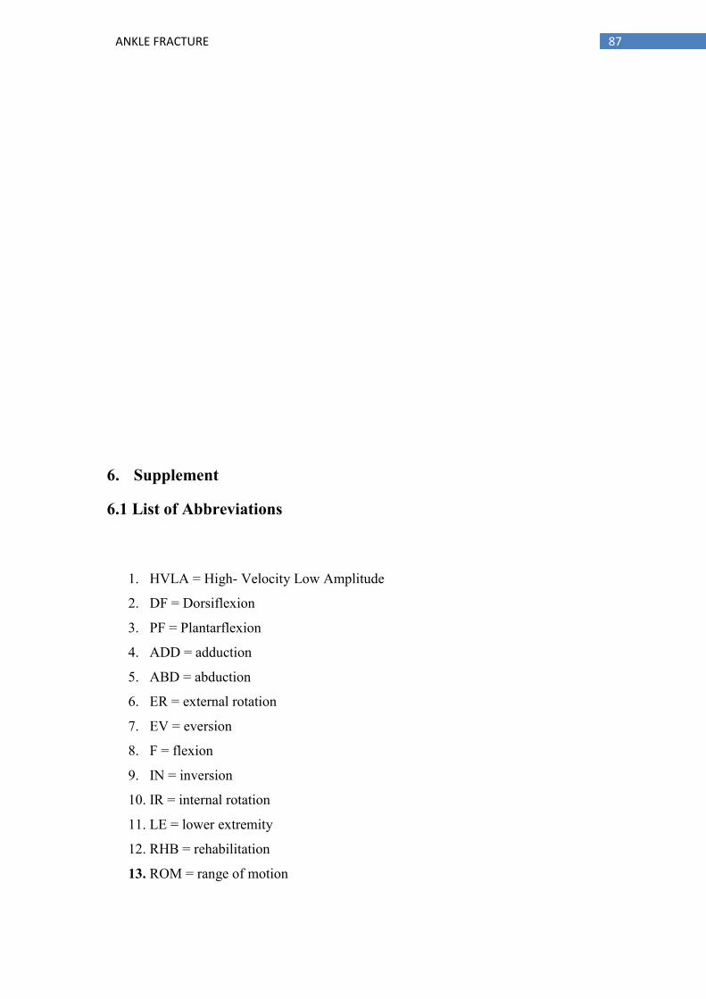
87 ANKLE FRACTURE
6. Supplement
6.1 List of Abbreviations
1. HVLA = High- Velocity Low Amplitude
2. DF = Dorsiflexion
3. PF = Plantarflexion
4. ADD = adduction
5. ABD = abduction
6. ER = external rotation
7. EV = eversion
8. F = flexion
9. IN = inversion
10. IR = internal rotation
11. LE = lower extremity
12. RHB = rehabilitation
13. ROM = range of motion

88 ANKLE FRACTURE
6.2 List of Tables
Table 1 The component movements o dorsiflexion and plantarflexion
Table 2 Circumference measurements for both lower extremities (LE’s)
Table 3 Length of both the lower extremities in parts
Table 4 Functional length measurement for both LE’s
Table 5 Anatomical length measurement for both LE’s
Table 6 ROM Examination of the ankle joint for both LE’s
Table 7 ROM examination in the toes for both Lower Extremities (LE’s)
Table 8 Amount of weight patient could bear on both his lower extremities (LRs)
Table 9 Muscle tone examination of different parts of the Lower Extremities
Table 10 Muscle strength scores of different parts of the Lower Extremities (LRs)
Table 11 Muscle length scores of different parts of the Lower Extremities (LRs)
Table 12 Assessment outcomes of the Joint Play Examination of different parts of the
lower extremities (LRs)
Table 13 Deep tendon reflex evaluation for the lower extremities (LRs)
Table 14 Final Circumference measurements of both LEs
Table 15 Final length of the lower extremity in different parts

89 ANKLE FRACTURE
Table 16 Final functional length measurement for both LE’s
Table 17 Final anatomical length measurement for both LE’s
Table 18 Final ROM examination in ankle joint for both LE’s
Table 19 Final ROM examination in big toe for both LE’s
Table 20 Physiological difference in loading weights in both lower extremities
Table 21 Final muscle tone test evaluation for both LE’s
Table 22 Final muscle strength test evaluation for both LE’s
Table 23 Final muscle length test evaluation for both LE’s
Table 24 Final joint play examination evaluation for L spine and LE’s
Table 25 Final deep tendon reflex evaluation for both LE’s
6.3. List of Figures
Figure 1 The Ankle Joint
Figure 2 DF and PF movements of the ankle joint
Figure 3 Inversion and Eversion movements of the Ankle Joint
Figure 4 Presentation of Gait Cycle in the Ankle Joint
Figure 5 Ankle Structure in the Latera and Anterior View
Figure 6 Ankle Fracture
Figure 7 Lateral Ankle Sprains
Figure 8 Ottawa Ankle and Midfoot Rules

90 ANKLE FRACTURE

91 ANKLE FRACTURE
6.4. Ethecal bored

92 ANKLE FRACTURE
6.5. INFORMOVANÝ SOUHLAS
Vážená paní, vážený pane,
v souladu se Všeobecnou deklarací lidských práv, zákonem č. 101/2000 Sb., o
ochraně osobních údajů a o změně některých zákonů, ve znění pozdějších předpisů,
Helsinskou deklarací, přijatou 18. Světovým zdravotnickým shromážděním v roce
1964 ve znění pozdějších změn (Fortaleza, Brazílie, 2013) a dalšími obecně
závaznými právními předpisy Vás žádám o souhlas s prezentováním a uveřejněním
výsledků vyšetření a průběhu terapie prováděné v rámci praxe na1
………………….………………………., kde Vás příslušně kvalifikovaná osoba
seznámila s Vaším vyšetřením a následnou terapií. Výsledky Vašeho vyšetření a
průběh Vaší terapie bude publikován v rámci bakalářské práce
na UK FTVS, s názvem2 …………………
Získané údaje, fotodokumentace, průběh a výsledky terapie budou uveřejněny
v bakalářské práci v anonymizované podobě. Osobní data nebudou uvedena a budou
uchována v anonymní podobě. V maximální možné míře zabezpečím, aby získaná
data nebyla zneužita.
Jméno a příjmení řešitele ........................................................................
Podpis:........................
Jméno a příjmení osoby, která provedla poučení3..................................
Podpis:........................
Prohlašuji a svým níže uvedeným vlastnoručním podpisem potvrzuji, že dobrovolně
souhlasím s prezentováním a uveřejněním výsledků vyšetření a průběhu terapie ve
výše uvedené bakalářské práci, a že mi osoba, která provedla poučení, osobně vše
podrobně vysvětlila, a že jsem měl(a) možnost si řádně a v dostatečném čase zvážit
všechny relevantní informace, zeptat se na vše podstatné a že jsem dostal(a) jasné a
srozumitelné odpovědi na své dotazy. Byl(a) jsem poučen(a) o právu odmítnout
prezentování a uveřejnění výsledků vyšetření a průběhu terapie v bakalářské práci
nebo svůj souhlas kdykoli odvolat bez represí, a to písemně zasláním Etické komisi
UK FTVS, která bude následně informovat řešitele.
Místo, datum ....................
Jméno a příjmení pacienta .............................................. Podpis pacienta:
..............................
Jméno a příjmení zákonného zástupce4 .........................................…..
Vztah zákonného zástupce k pacientovi .................................... Podpis:
.............................

93 ANKLE FRACTURE