Reendodontic treatment ( possibilities apexlocators in endodontic therapy)
International Journal of Dental Health Concerns (2015), 1, 1-4
1
INTRODUCTION
“Failure is only the opportunity to begin again, only this time more wisely.”
The aims of traditional endodontic treatment are to elimination of bacteria from the root canal system and to the establishment of effective barriers against root recontamination.[1] Presence of residual bacteria or re-infection in a previously disinfected canal or secondary infection[2] is frequently related to failure of conventional endodontic treatment. Other factors responsible are presence of unresolved cystic lesion, extra-radicular infections, systemic reactions to a foreign body due to apical extrusion of endodontic material, and endogenous cholesterol crystal accumulation in the apical tissues. Thus, success of the treatment relies on different factors and can be verified through clinical and radiographic evaluations during the follow-up period.[2-4] This article aims to report a case of apicoectomy indicated due to failure of an endodontically treated tooth.
CASE REPORT
A 14-year-old girl reported with the chief complaint of discoloration in upper front tooth with a history of trauma to this region due to fall, 3 years back, followed by root canal treatment. 6 months later, there was recurrent swelling and pus discharge associated with the discolored tooth. Examination revealed black discoloration of the clinical crown of maxillary right central incisor (MRCI) with an amalgam restoration on the palatal aspect, Grade I mobility and tenderness on percussion. There was an active discharging sinus on the labial aspect of MRCI, in the attached gingiva (Figure 1).
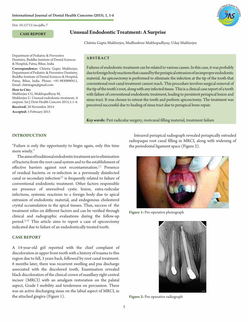
Intraoral periapical radiograph revealed periapically extruded radiopaque root canal filling in MRCI, along with widening of the periodontal ligament space (Figure 2).
CASE REPORT Unusual Endodontic Treatment: A Surprise
Chitrita Gupta Mukherjee, Madhushree Mukhopadhyay, Uday Mukherjee
Department of Pediatric & Preventive Dentistry, Buddha Institute of Dental Sciences & Hospital, Patna, Bihar, IndiaCorrespondence: Chitrita Gupta Mukherjee, Department of Pediatric & Preventive Dentistry, Buddha Institute of Dental Sciences & Hospital, Patna, Bihar, India. Phone: +91-9830890411, Email: [email protected] How to Cite:Mukherjee CG, Mukhopadhyay M, Mukherjee U. Unusual endodontic treatment: A surprise. Int J Dent Health Concern 2015;1:1-4.Received: 28 November 2014Accepted: 5 February 2015
ABSTRACT
Failures of endodontic treatment can be related to various causes. In this case, it was probably due to foreign body reactions that caused by the periapical extrusion of an improper endodontic material. An apicoectomy is performed to eliminate the infection at the tip of the tooth that conventional root canal treatment cannot reach. This procedure involves surgical removal of the tip of the tooth’s root, along with any infected tissue. This is a clinical case report of a tooth with failure of conventional endodontic treatment, leading to persistent periapical lesion and sinus tract. It was chosen to retreat the tooth and perform apicoectomy. The treatment was perceived successful due to healing of sinus tract due to periapical bone repair.
Key words: Peri radicular surgery, rootcanal filling material, treatment failure
Figure 1: Pre-operative photograph
Figure 2: Pre-operative radiograph
Doi: 10.15713/ins.ijdhc.7
Improper obturation material… Mukherjee, et al.. I J D H C
2
Consent was obtained from the parents for re-randomized controlled trials of MRCI, and if periapical infection remained uncontrolled, endodontic surgery might be required later on.
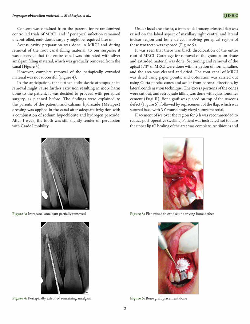
Access cavity preparation was done in MRCI and during removal of the root canal filling material, to our surprise; it was observed that the entire canal was obturated with silver amalgam filling material, which was gradually removed from the canal (Figure 3).
However, complete removal of the periapically extruded material was not successful (Figure 4).
In the anticipation, that further enthusiastic attempts at its removal might cause further extrusion resulting in more harm done to the patient, it was decided to proceed with periapical surgery, as planned before. The findings were explained to the parents of the patient, and calcium hydroxide (Metapex) dressing was applied in the canal after adequate irrigation with a combination of sodium hypochlorite and hydrogen peroxide. After 1-week, the tooth was still slightly tender on percussion with Grade I mobility.
Under local anesthesia, a trapezoidal mucoperiosteal flap was raised on the labial aspect of maxillary right central and lateral incisor region and bony defect involving periapical region of these two teeth was exposed (Figure 5).
It was seen that there was black discoloration of the entire root of MRCI. Curettage for removal of the granulation tissue and extruded material was done. Sectioning and removal of the apical 1/3rd of MRCI were done with irrigation of normal saline, and the area was cleaned and dried. The root canal of MRCI was dried using paper points, and obturation was carried out using Gutta-percha cones and sealer from coronal direction, by lateral condensation technique. The excess portions of the cones were cut out, and retrograde filling was done with glass ionomer cement (Fugi II). Bone graft was placed on top of the osseous defect (Figure 6), followed by replacement of the flap, which was sutured back with 3-0 round body vicryl suture material.
Placement of ice over the region for 3 h was recommended to reduce post-operative swelling. Patient was instructed not to raise the upper lip till healing of the area was complete. Antibiotics and
Figure 3: Intracanal amalgam partially removed
Figure 4: Periapically extruded remaining amalgam Figure 6: Bone graft placement done
Figure 5: Flap raised to expose underlying bone defect
Improper obturation material… Mukherjee, et al.. I J D H C
3
analgesics were prescribed, and instructions were given about soft diet and regular rinsing of the mouth with chlorhexidine solution.
The patient reported for review after 2 weeks, and it was noticed that healing of the sinus tract had occurred, along with a reduction in the tooth mobility.
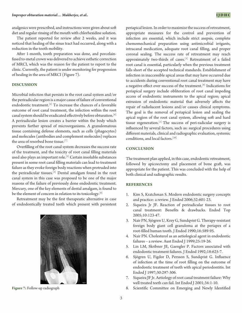
After 1-month, tooth preparation was done, and porcelain-fused to-metal crown was delivered to achieve esthetic correction of MRCI, which was the reason for the patient to report to the clinic. Currently, the patient is under monitoring for progression of healing in the area of MRCI (Figure 7).
DISCUSSION
Microbial infection that persists in the root canal system and/or the periradicular region is a major cause of failure of conventional endodontic treatment.[5] To increase the chances of a favorable outcome of root canal treatment, the infection within the root canal system should be eradicated effectively before obturation.[6] A periradicular lesion creates a barrier within the body which prevents further spread of microorganisms. A granulomatous tissue containing defense elements, such as cells (phagocytes) and molecules (antibodies and complement molecules) replaces the area of resorbed bone tissue.[7]
Overfilling of the root canal system decreases the success rate of the treatment, and the toxicity of root canal filling materials used also plays an important role.[5] Certain insoluble substances present in some root canal filling materials can lead to treatment failure as they evoke foreign body reactions when protruded into the periradicular tissues.[3] Dental amalgam found in the root canal system in this case was proposed to be one of the major reasons of the failure of previously done endodontic treatment. Mercury, one of the key elements of dental amalgam, is found to be the element of concern in relation to its toxicology.[8]
Retreatment may be the first therapeutic alternative in case of endodontically treated teeth which present with persistent
periapical lesion. In order to maximize the success of retreatment, appropriate measures for the control and prevention of infection are essential, which include strict asepsis, complete chemomechanical preparation using antimicrobial irrigants, intracanal medication, adequate root canal filling, and proper coronal sealing. The success rate of retreatment may reach approximately two-thirds of cases.[5] Retreatment of a failed root canal is essential, particularly when the previous treatment falls short of the accepted technical standards. Establishment of infection in inaccessible apical areas that may have occurred due to accidents during conventional root canal treatment may have a negative effect over success of the treatment.[9] Indications for periapical surgery include obliteration of root canal impeding access of endodontic instruments to the apical region; apical extrusion of endodontic material that adversely affects the repair of radiolucent lesions and/or causes clinical symptoms. The goal is the removal of periapical lesion and sealing the apical region of the root canal system, allowing soft and hard tissue regeneration.[1] The success of peri-radicular surgery is influenced by several factors, such as: surgical procedures using different materials, clinical and radiographic evaluation, systemic conditions, and local factors.[10]
CONCLUSION
The treatment plan applied, in this case, endodontic retreatment, followed by apicoectomy and placement of bone graft, was appropriate for the patient. This was concluded with the help of both clinical and radiographic results.
REFERENCES
1. Kim S, Kratchman S. Modern endodontic surgery concepts and practice: a review. J Endod 2006;32:601-23.
2. Siqueira Jr JF. Reaction of periradicular tissues to root canal treatment: Benefits & drawbacks. Endod Top 2005;10:123-47.
3. Nair PN, Sjögren U, Krey G, Sundqvist G. Therapy-resistant foreign body giant cell granuloma at the periapex of a root-filled human tooth. J Endod 1990;16:589-95.
4. Nair PN. Cholesterol as an aetiological agent in endodontic failures – a review. Aust Endod J 1999;25:19-26.
5. Lin LM, Skribner JE, Gaengler P. Factors associated with endodontic treatment failures. J Endod 1992;18:625-7.
6. Sjögren U, Figdor D, Persson S, Sundqvist G. Influence of infection at the time of root filling on the outcome of endodontic treatment of teeth with apical periodontitis. Int Endod J 1997;30:297-306.
7. Siqueira JF Jr. Aetiology of root canal treatment failure: Why well treated teeth can fail. Int Endod J 2001;34:1-10.
8. Scientific Committee on Emerging and Newly Identified Figure 7: Follow-up radiograph
Improper obturation material… Mukherjee, et al.. I J D H C
4
Health Risks (SCENIHR). The safety of dental amalgam and alternate dental restoration materials for patients and users; May 2008.
9. Wu MK, Dummer PM, Wesselink PR. Consequences of and strategies to deal with residual post-treatment root canal
infection. Int Endod J 2006;39:343-56.10. Tsesis I, Faivishevsky V, Kfir A, Rosen E. Outcome of
surgical endodontic treatment performed by a modern technique: a meta-analysis of literature. J Endod 2009;35:1505-11.