
Case Report Spontaneous and bilateral necrosis of the ... · J Toxicol Pathol 2015; 28: 121–124...
4
J Toxicol Pathol 2015; 28: 121–124 Case Report Spontaneous and bilateral necrosis of the femoral head in a young experimental beagle dog Ryosuke Kobayashi 1* , Tetsuro Kurotaki 1 , Naoaki Yamada 1 , Shino Kumabe 1 , Takuya Doi 1 , Yumi Wako 1 , and Minoru Tsuchitani 1 1 Pathology Department, LSI Medience Corporation, 14-1 Sunayama, Kamisu-shi, Ibaraki 314-0255, Japan Abstract: This report describes the pathological characterizations of a rare case of necrosis of the femoral head that was spontaneous, bilateral, avascular and nontraumatic. A 14-month-old beagle dog was presented with pain in the hind limbs. At necropsy, the articular surface in the bilateral femoral head was markedly irregular. There were no gross abnormalities other than in the hip joints. Micro- scopically, a wide range of trabecular bone necrosis localized in the subchondral area was observed in both femoral heads. In the right femoral head, fibrosis and proliferative vessels were noted in the subchondral area. The articular cartilage was thickened irregularly, but there was no evidence of cartilage necrosis. The bone marrow adjacent to the affected area showed severe depression. In the me- taphysis, atrophic bone marrow, but not bone necrosis, was observed. This was a rare case of spontaneous necrosis of the femoral head in an experimental beagle dog. (DOI: 10.1293/tox.2014-0060; J Toxicol Pathol 2015; 28: 121–124) Key words: avascular osteonecrosis, dog, Legg-Calvé-Perthes disease, necrosis of the femoral head, spontaneous lesion In toxicological studies, beagle dogs are generally used as a non-rodent animal species, and there is an abundant of background data for them. However, there is no report of spontaneous osteonecrosis among these data 1-3 . Osteonecro- sis generally occurs under avascular conditions due to bone fracture, acute inflammatory diseases or infarction in do- mestic animals. Microscopically, bone necrosis is character- ized by cell death and loss of osteocytes from their lacunae, although scattered empty lacunae do not justify a diagnosis of necrosis, as they could possibly represent a normal turn- over or artifact of the preparation 4, 5 . It is known that the femoral head is especially predisposed to hypoxia among the systemic bones because the region is served by the end- arteries and has poor collateral circulation 4 . Necrosis of the femoral head (NFH) occurs sporadi- cally in dogs mainly due to bone fracture. Meanwhile, NFH particularly that referred to as Legg-Calvé-Perthes disease (LCPD), which is speculated to be a genetic disorder with an autosomal recessive inheritance, is known to occur be- fore the growth plate closes in small breeds and young dogs. It is thought that NFH in non-small breeds of dogs should be discriminated from LCPD in terms of the difference in pathogenesis 4 . We encountered a case of LCPD-like NFH that was spontaneous, bilateral and nontraumatic in a young experimental beagle dog. This report deals with rare case of spontaneous NFH and its pathological characteristics in this laboratory beagle dog. A 14-month-old female beagle dog was imported from Beijing Marshall Biotechnology Co., Ltd. (Beijing). The dog was housed in an animal room maintained at 22 ± 3°C and a humidity of 55 ± 20% and was given 300 g/day DS-A (Oriental Yeast Co., Ltd., Tokyo, Japan) with free access to water. As an experimental procedure, cerebrospinal fluid was collected once from the neck. Two days thereafter, a gait abnormality and pain were noted in the right hind limb. No drugs were administered to the dog. Since the dog did not show any improvement in the abnormality of the hind limbs over the course of one month, it was euthanized under pentobarbital sodium anesthesia and necropsied. The body weight of the dog before necropsy was 6.1 kg. All proce- dures were conducted with permission of the Institutional Animal Care and Use Committee of the facility, which was fully accredited by AAALAC International. At necropsy, the bilateral femurs including the knee joint, coxal bone and humeral bone were fixed in 10% neutral-buffered formalin. These tissues were sliced and decalcified with 10% EDTA at 37°C for several weeks, embedded in paraffin, cut at 4 μm and stained with hematoxylin and eosin (HE), Prussian blue staining and Schmorl’s method. Grossly, no lesions associated with collection of cere- brospinal fluid were observed in the cervical spinal cord. In the hip joints, mobility decreased slightly but joint flu- id did not increase. The articular surfaces of both femoral Received: 9 December 2014, Accepted: 5 February 2015 Published online in J-STAGE: 1 March 2015 *Corresponding author: R Kobayashi (e-mail: [email protected]) ©2015 The Japanese Society of Toxicologic Pathology This is an open-access article distributed under the terms of the Cre- ative Commons Attribution Non-Commercial No Derivatives (by-nc- nd) License <http://creativecommons.org/licenses/by-nc-nd/3.0/>.
Transcript of Case Report Spontaneous and bilateral necrosis of the ... · J Toxicol Pathol 2015; 28: 121–124...

J Toxicol Pathol 2015; 28: 121–124
Case Report
Spontaneous and bilateral necrosis of the femoral head in a young experimental beagle dog
Ryosuke Kobayashi1*, Tetsuro Kurotaki1, Naoaki Yamada1, Shino Kumabe1, Takuya Doi1, Yumi Wako1, and Minoru Tsuchitani1
1Pathology Department, LSI Medience Corporation, 14-1 Sunayama, Kamisu-shi, Ibaraki 314-0255, Japan
Abstract: This report describes the pathological characterizations of a rare case of necrosis of the femoral head that was spontaneous, bilateral, avascular and nontraumatic. A 14-month-old beagle dog was presented with pain in the hind limbs. At necropsy, the articular surface in the bilateral femoral head was markedly irregular. There were no gross abnormalities other than in the hip joints. Micro-scopically, a wide range of trabecular bone necrosis localized in the subchondral area was observed in both femoral heads. In the right femoral head, fibrosis and proliferative vessels were noted in the subchondral area. The articular cartilage was thickened irregularly, but there was no evidence of cartilage necrosis. The bone marrow adjacent to the affected area showed severe depression. In the me-taphysis, atrophic bone marrow, but not bone necrosis, was observed. This was a rare case of spontaneous necrosis of the femoral head in an experimental beagle dog. (DOI: 10.1293/tox.2014-0060; J Toxicol Pathol 2015; 28: 121–124)
Key words: avascular osteonecrosis, dog, Legg-Calvé-Perthes disease, necrosis of the femoral head, spontaneous lesion
In toxicological studies, beagle dogs are generally used as a non-rodent animal species, and there is an abundant of background data for them. However, there is no report of spontaneous osteonecrosis among these data1-3. Osteonecro-sis generally occurs under avascular conditions due to bone fracture, acute inflammatory diseases or infarction in do-mestic animals. Microscopically, bone necrosis is character-ized by cell death and loss of osteocytes from their lacunae, although scattered empty lacunae do not justify a diagnosis of necrosis, as they could possibly represent a normal turn-over or artifact of the preparation4, 5. It is known that the femoral head is especially predisposed to hypoxia among the systemic bones because the region is served by the end-arteries and has poor collateral circulation4.
Necrosis of the femoral head (NFH) occurs sporadi-cally in dogs mainly due to bone fracture. Meanwhile, NFH particularly that referred to as Legg-Calvé-Perthes disease (LCPD), which is speculated to be a genetic disorder with an autosomal recessive inheritance, is known to occur be-fore the growth plate closes in small breeds and young dogs. It is thought that NFH in non-small breeds of dogs should be discriminated from LCPD in terms of the difference in
pathogenesis4. We encountered a case of LCPD-like NFH that was spontaneous, bilateral and nontraumatic in a young experimental beagle dog. This report deals with rare case of spontaneous NFH and its pathological characteristics in this laboratory beagle dog.
A 14-month-old female beagle dog was imported from Beijing Marshall Biotechnology Co., Ltd. (Beijing). The dog was housed in an animal room maintained at 22 ± 3°C and a humidity of 55 ± 20% and was given 300 g/day DS-A (Oriental Yeast Co., Ltd., Tokyo, Japan) with free access to water. As an experimental procedure, cerebrospinal fluid was collected once from the neck. Two days thereafter, a gait abnormality and pain were noted in the right hind limb. No drugs were administered to the dog. Since the dog did not show any improvement in the abnormality of the hind limbs over the course of one month, it was euthanized under pentobarbital sodium anesthesia and necropsied. The body weight of the dog before necropsy was 6.1 kg. All proce-dures were conducted with permission of the Institutional Animal Care and Use Committee of the facility, which was fully accredited by AAALAC International. At necropsy, the bilateral femurs including the knee joint, coxal bone and humeral bone were fixed in 10% neutral-buffered formalin. These tissues were sliced and decalcified with 10% EDTA at 37°C for several weeks, embedded in paraffin, cut at 4 μm and stained with hematoxylin and eosin (HE), Prussian blue staining and Schmorl’s method.
Grossly, no lesions associated with collection of cere-brospinal fluid were observed in the cervical spinal cord. In the hip joints, mobility decreased slightly but joint flu-id did not increase. The articular surfaces of both femoral
Received: 9 December 2014, Accepted: 5 February 2015Published online in J-STAGE: 1 March 2015*Corresponding author: R Kobayashi (e-mail: [email protected])©2015 The Japanese Society of Toxicologic PathologyThis is an open-access article distributed under the terms of the Cre-ative Commons Attribution Non-Commercial No Derivatives (by-nc-nd) License <http://creativecommons.org/licenses/by-nc-nd/3.0/>.

Necrosis of the Femoral Head in a Beagle Dog122
heads appeared irregular and roughened and showed a se-verely thickened synovium and joint capsule (Fig. 1). On the longitudinal cut surface of both femoral heads, articular cartilage was severely thickened and showed ulceration and subchondral cracks (Fig. 2). The physis of the femurs were closed bilaterally. In the bilateral acetabulum, brown dis-coloration was seen at the dorsal aspect, which was consid-ered a weight-bearing area. There was no gross abnormality in other systemic bones, cartilages, or joints. The bilateral femoral muscles showed atrophy.
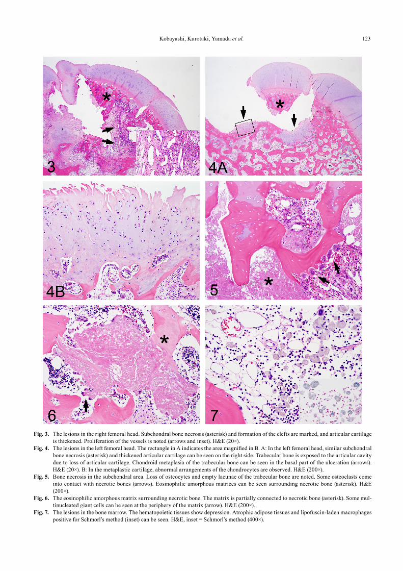
Histopathologically, a broad range of necrosis of the trabecular bone with limited inflammatory changes was ob-served beneath the irregularly thickened articular cartilage in the subchondral area of the proximal femur (Figs. 3, 4A). The thickened cartilages contained an abundant hypocellu-lar cartilage matrix without necrosis. Synovial hyperplasia around the hip joint was severe. A portion of the articular cartilage and necrotic bone was detached from the intact trabecular bone, which formed clefts or ulcerations. Focal chondroid metaplasia of the trabecular bone was noted in the basal part of the ulceration (Fig. 4B). Fibrosis and prolif-eration of the vessels were seen in the subchondral areas of the right femoral head (Fig. 3). The bone necrosis was char-acterized by loss of osteocytes with empty lacunae of the poorly stained bone matrix. Osteoclasts were rare around the bone matrix (Fig. 5). Regenerating new bone formation (creeping substitution) were not seen in any area around the necrotic bone. In addition, eosinophilic amorphous matrices were scattered around the necrotic bone (Figs. 5, 6). The matrix was partially connected to the necrotic bone and in contact with some osteoclasts. Thus it was considered that the eosinophilic matrices were debris of necrotic bone. In the bone marrow adjacent to the area of bone necrosis, there was severe depression with atrophic adipose tissue and mild fibrosis. Some lipofuscin-laden macrophages, which were positive for Schmorl’s method and negative for Prussian blue staining were also observed (Fig. 7). Thrombus for-mation indicative of local circulatory disturbance was not observed in any of the blood vessels of the affected area. On the other hand, there were degenerative changes in the bone marrow of the metaphysis of the femur, but no necrotic trabecular bone was recognized.
In the bilateral acetabulum, ulcerative articular carti-lage and severe depression of the bone marrow tissues were observed accompanied by brown discoloration. In other ar-eas of the articular surface, thickening of the cartilage and synovial hyperplasia were evident. Bone necrosis and eo-sinophilic amorphous matrices as seen in the femoral head were not observed in the subchondral or epiphyseal area. On the other hand, there were no lesions in the knee joints or humeral bone.
We diagnosed this case as bilateral NFH. Although there have been experimental studies using canine models of NFH, spontaneous NFH has not yet been reported in lab-oratory beagle dogs6-8. As for the distribution of the lesions, bone necrosis was localized in the subchondral area of the femoral heads in our case. Generally, NFH is histologically
characterized by subchondral bone necrosis in the femoral head, and the histopathological changes in our case were comparable to those reported as typical lesions of avascu-lar and nontraumatic osteonecrosis5. Meanwhile, the lesions of the cartilage, bone marrow and synovium in the femoral heads and acetabula were thought to be secondary and reac-tive lesions, as seen in spontaneous LCPD9. In the systemic joints other than the femoral joint in our case, there were no chondral lesions suggesting primary disorder of cartilage. Thus it was considered that the chondral lesions observed in our case were secondary physical injuries subsequent to ab-normal conformity in the hip joint. These reactive changes and severe deformation of the femoral heads suggested that the disease was progressive and occurred before becoming clinically apparent.
Commonly, avascular osteonecrosis in dogs results from trauma, intramedullary neoplasms, or thrombosis5. Sporadic NFH in dogs is generally caused by bone frac-tures4. However, these lesions were not confirmed in either femur in our case. Meanwhile, bone necrosis was clearly localized to the subchondral area of the femoral head, and
Fig. 1. Macroscopic changes in the femoral heads. The spherical shape of the bilateral femoral head is completely collapsed, and the articular surface shows marked irregularity.
Fig. 2. The longitudinal cut surface of the left femoral head (A). B: age-matched control at the same magnification. The articu-lar cartilage thickened irregularly. Cracks in the subchondral area (black arrow head) or ulceration (white arrow) form.
Kobayashi, Kurotaki, Yamada et al. 123
Fig. 3. The lesions in the right femoral head. Subchondral bone necrosis (asterisk) and formation of the clefts are marked, and articular cartilage is thickened. Proliferation of the vessels is noted (arrows and inset). H&E (20×).
Fig. 4. The lesions in the left femoral head. The rectangle in A indicates the area magnified in B. A: In the left femoral head, similar subchondral bone necrosis (asterisk) and thickened articular cartilage can be seen on the right side. Trabecular bone is exposed to the articular cavity due to loss of articular cartilage. Chondroid metaplasia of the trabecular bone can be seen in the basal part of the ulceration (arrows). H&E (20×). B: In the metaplastic cartilage, abnormal arrangements of the chondrocytes are observed. H&E (200×).
Fig. 5. Bone necrosis in the subchondral area. Loss of osteocytes and empty lacunae of the trabecular bone are noted. Some osteoclasts come into contact with necrotic bones (arrows). Eosinophilic amorphous matrices can be seen surrounding necrotic bone (asterisk). H&E (200×).
Fig. 6. The eosinophilic amorphous matrix surrounding necrotic bone. The matrix is partially connected to necrotic bone (asterisk). Some mul-tinucleated giant cells can be seen at the periphery of the matrix (arrow). H&E (200×).
Fig. 7. The lesions in the bone marrow. The hematopoietic tissues show depression. Atrophic adipose tissues and lipofuscin-laden macrophages positive for Schmorl’s method (inset) can be seen. H&E, inset = Schmorl’s method (400×).

Necrosis of the Femoral Head in a Beagle Dog124
there were proliferative vessels in the right femoral head. In-flammatory reactions were limited. Therefore, we presumed that the dog had latent abnormal blood circulation in the bilateral proximal femurs. Evidence has been reported for complex causes of LCPD, including abnormal epiphyseal circulation10 and increasing intra-articular pressure caused by effusion from synovitis11 or intracapsular tamponade12, 13. It is thought that a variety of developmental and anatomical factors are included in the pathogenesis of LCPD. We as-sume that a vascular anomaly similar to LCPD is associated with the cause of NFH in our case, although the etiology remained unclear. The difference in the lesions between the right and left femoral head in our case might be attributed to the extent of ischemia or the degree of repairing reactions. Sporadic occurrence of the disease in the animal group ex-cluded the effects of genetic factors.
In the dog, hip dysplasia needs to be considered for the differential diagnosis. Hip dysplasia takes chronic and mild course with occasional microfracture, but there is no wide range of bone necrosis14, 15. Additionally, our case did not clinically show subluxation or increased joint laxity, and acetabula were not shallow at necropsy. In conclusion, the experimental beagle dog was diagnosed with avascular, spontaneous and sporadic NFH.
Acknowledgments: We would like to thank Ms. Chie Shiroumaru for her special technical supports, and Mr. Ste-phen Filiatrault and Ms. Kanae Tamatsukuri for language editing of the manuscript.
References
1. Hottendorf GH, and Hirth RS. Lesions of spontaneous sub-clinical disease in Beagle dogs. Vet Pathol. 11: 240–258. 1974. [Medline] [CrossRef]
2. Kobayashi K, Hirouchi Y, Iwata H, Yamakawa S, Mikami S, Yamamoto S, Hashiguchi J, and Enomoto M. Historical control data of spontaneous lesions in beagle dogs. J Toxi-col Pathol. 7: 329–343. 1994. [CrossRef]
3. Sato J, Doi T, Wako Y, Hamamura M, Kanno T, Tsuchitani M, and Narama I. Histopathology of incidental findings in beagles used in toxicity studies. J Toxicol Pathol. 25: 103–
134. 2012. [Medline] [CrossRef] 4. Thompson K. Osteonecrosis. In: Pathology of Domestic
Animals 5th ed. MG Maxie (ed). Elsevier, Philadelphia. Vol. 1. 88-92. 2007.
5. Carlson CS, and Weisbrode SE. Aseptic necrosis of bone. In: Pathologic Basis of Veterinary Disease, 5th ed. JF Zach-ary, and MD Mcgavin (eds). Elsevier, Missouri. 953-955. 2012.
6. Takaoka K, Yoshioka T, Hosoya T, Ono K, and Takase T. The repair process in experimentally induced avascular ne-crosis of the femoral head in dogs. Arch Orthop Trauma Surg. 99: 109–115. 1981. [Medline] [CrossRef]
7. Alpaslan AM, Aksoy MC, and Yazici M. Interruption of the blood supply of femoral head: an experimental study on the pathogenesis of Legg-Calve-Perthes Disease. Arch Orthop Trauma Surg. 127: 485–491. 2007. [Medline] [CrossRef]
8. Shen F, Yan ZQ, Guo CA, Shi HC, Gu YS, Zeng MS, Lu XY, and Liu J. Prediction of traumatic avascular necrosis of the femoral head by single photon emission computer-ized tomography and computerized tomography: an experi-mental study in dogs. Chin J Traumatol. 14: 227–232. 2011. [Medline]
9. Mickelson MR, McCurnin DM, Awbrey BJ, Maynard JA, and Martin RK. Legg-Calvé-Perthes disease in dogs: a comparison to human Legg-Calvé-Perthes disease. Clin Orthop Relat Res.: 287–300. 1981. [Medline]
10. Fujikawa K. Comparative vascular anatomy of the hip of the miniature dog and of the normal-size mongrel. Kurume Med J. 38: 159–165. 1991. [Medline] [CrossRef]
11. Warren DV, and Dingwall JS. Legg-Perthes disease in the dog—a review. Can Vet J. 13: 135–137. 1972. [Medline]
12. Singleton WB, and Jones EL. The experimental induction of subclinical Perthes’ disease in the puppy following ar-throtomy and intracapsular tamponade. J Comp Pathol. 89: 57–71. 1979. [Medline] [CrossRef]
13. Kemp HB. Perthes’ disease: the influence of intracapsular tamponade on the circulation in the hip joint of the dog. Clin Orthop Relat Res.: 105–114. 1981. [Medline]
14. Fries CL, and Remedios AM. The pathogenesis and di-agnosis of canine hip dysplasia: a review. Can Vet J. 36: 494–502. 1995. [Medline]
15. Ginja MM, Silvestre AM, Gonzalo-Orden JM, and Ferreira AJ. Diagnosis, genetic control and preventive management of canine hip dysplasia: a review. Vet J. 184: 269–276. 2010. [Medline] [CrossRef]


![BilateralSpontaneousPneumothorax, Pneumomediastinum ... · as pneumothorax [2]. The ratio of simultaneous bilateral spontaneous pneumothorax is approximately 1.3% among all cases](https://static.fdocuments.in/doc/165x107/5cc1981888c993110a8c6f12/bilateralspontaneouspneumothorax-pneumomediastinum-as-pneumothorax-2.jpg)
