Case Report Postcoital Internal Carotid Artery Dissection...
4
Hindawi Publishing Corporation Case Reports in Neurological Medicine Volume 2013, Article ID 403647, 3 pages http://dx.doi.org/10.1155/2013/403647 Case Report Postcoital Internal Carotid Artery Dissection Presenting as Isolated Painful Horner Syndrome: A Case Report Eren Gozke, Hilal Tastekin Toz, PJnar Kahraman Koytak, and Funda Alparslan Department of Neurology, FSM Teaching and Research Hospital, 34752 Istanbul, Turkey Correspondence should be addressed to Eren Gozke; [email protected] Received 11 January 2013; Accepted 31 January 2013 Academic Editors: P. Berlit, J. L. Gonz´ alez-Guti´ errez, J. C. Kattah, and Y. Wakabayashi Copyright © 2013 Eren Gozke et al. is is an open access article distributed under the Creative Commons Attribution License, which permits unrestricted use, distribution, and reproduction in any medium, provided the original work is properly cited. Postcoital artery dissection is a rare condition. Here we report a 40-year-old male patient with painful Horner syndrome related to postcoital internal carotid artery (ICA) dissection. In neurologic examination of the patient, semiptosis, enophthalmus, and myosis were observed on the leſt side. ere were no carotid bruits. On T1-weighted and fat-suppressed cranial MRI, hyperintensity consistent with intramural hematoma was observed within cervical and temporal petrous segments of leſt ICA. On cervical and cranial MRA, marked decrease in the calibration of C1 and C2 segments of the leſt ICA was remarkable. e patient was diagnosed as leſt ICA dissection and anticoagulant therapy was initiated. A prominent improvement was noted in clinical findings during two months of followup period. 1. Introduction Carotid artery dissection is characterized by the formation of mural hematoma as a result of rupture of tunica intima with an incidence of 2.5–3/100.000. It can develop sponta- neously or secondary to trauma. Although postcoital artery dissections are rarely reported in the literature, any incident of postcoital carotid artery dissection in patients presented with isolated Horner syndrome has not been encountered so far [1, 2]. A case with postcoital internal carotid artery (ICA) dissection in a patient referred with painful Horner syndrome is presented. 2. Case Report A 40-year-old male patient was referred to our outpatient clinic with complaints of rapid onset of pain localized on behind his leſt eye during intercourse and subsequent development of ptosis of his leſt eyelid. Physical examination did not reveal any evidence of abnormality. In neurologic examination, semiptosis, enophthalmus, and myosis were observed on the leſt side. ere was no sweating abnormality on his face. Ophthalmologic evaluation revealed normal visual acuity and intact fundi, bilaterally. ere were no carotid bruits. He was smoking a pack per day for 20 years, without any history of alcohol and any known substance and drug abuse. Cranial magnetic resonance imaging (MRI) and craniocervical magnetic resonance angiography (MRA) were performed on the patient with diagnosis of leſt Horner syndrome. On diffusion MRI, any acute ischemic lesion was not detected. On T1-weighted and fat-suppressed cranial MRI, hyperintensity consistent with intramural hematoma was observed within cervical and temporal petrous segments of leſt ICA (Figures 1(a) and 1(b)). On cervical and cranial MRA, marked decrease in the calibration of C1, and C2 segments of the leſt ICA was remarkable (Figure 1(c)). Any abnormal contrast enhancement in cerebral parenchyma was not observed aſter administration of contrast material. Any abnormality in routine laboratory test and markers of vasculitis was not detected. e patient was diagnosed as leſt ICA dissection, and anticoagulant therapy was initiated. First intravenous heparin was given, and then warfarin was started. A prominent improvement was noted in clinical findings during two months of followup period. 3. Discussion Horner syndrome manifests itself with signs of ptosis, myosis, enophthalmus, and facial anhidrosis. It is characterized
Transcript of Case Report Postcoital Internal Carotid Artery Dissection...
Hindawi Publishing CorporationCase Reports in Neurological MedicineVolume 2013, Article ID 403647, 3 pageshttp://dx.doi.org/10.1155/2013/403647
Case ReportPostcoital Internal Carotid Artery Dissection Presenting asIsolated Painful Horner Syndrome: A Case Report
Eren Gozke, Hilal Tastekin Toz, PJnar Kahraman Koytak, and Funda Alparslan
Department of Neurology, FSM Teaching and Research Hospital, 34752 Istanbul, Turkey
Correspondence should be addressed to Eren Gozke; [email protected]
Received 11 January 2013; Accepted 31 January 2013
Academic Editors: P. Berlit, J. L. Gonzalez-Gutierrez, J. C. Kattah, and Y. Wakabayashi
Copyright © 2013 Eren Gozke et al. This is an open access article distributed under the Creative Commons Attribution License,which permits unrestricted use, distribution, and reproduction in any medium, provided the original work is properly cited.
Postcoital artery dissection is a rare condition. Here we report a 40-year-old male patient with painful Horner syndrome relatedto postcoital internal carotid artery (ICA) dissection. In neurologic examination of the patient, semiptosis, enophthalmus, andmyosis were observed on the left side.There were no carotid bruits. On T1-weighted and fat-suppressed cranial MRI, hyperintensityconsistent with intramural hematoma was observed within cervical and temporal petrous segments of left ICA. On cervical andcranial MRA, marked decrease in the calibration of C1 and C2 segments of the left ICA was remarkable. The patient was diagnosedas left ICA dissection and anticoagulant therapy was initiated. A prominent improvement was noted in clinical findings during twomonths of followup period.
1. Introduction
Carotid artery dissection is characterized by the formationof mural hematoma as a result of rupture of tunica intimawith an incidence of 2.5–3/100.000. It can develop sponta-neously or secondary to trauma. Although postcoital arterydissections are rarely reported in the literature, any incidentof postcoital carotid artery dissection in patients presentedwith isolated Horner syndrome has not been encountered sofar [1, 2]. A case with postcoital internal carotid artery (ICA)dissection in a patient referredwith painfulHorner syndromeis presented.
2. Case Report
A 40-year-old male patient was referred to our outpatientclinic with complaints of rapid onset of pain localizedon behind his left eye during intercourse and subsequentdevelopment of ptosis of his left eyelid. Physical examinationdid not reveal any evidence of abnormality. In neurologicexamination, semiptosis, enophthalmus, and myosis wereobserved on the left side. There was no sweating abnormalityon his face. Ophthalmologic evaluation revealed normalvisual acuity and intact fundi, bilaterally. There were nocarotid bruits. He was smoking a pack per day for 20 years,
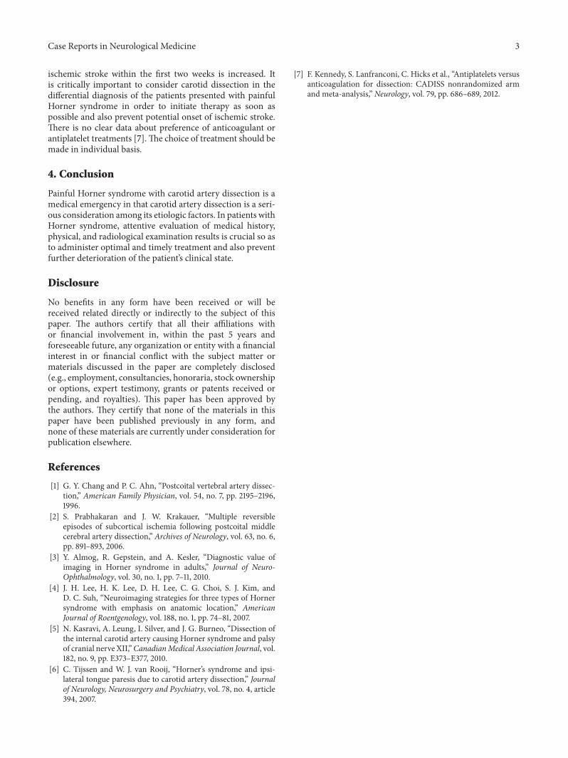
without any history of alcohol and any known substanceand drug abuse. Cranial magnetic resonance imaging (MRI)and craniocervical magnetic resonance angiography (MRA)were performed on the patient with diagnosis of left Hornersyndrome. On diffusion MRI, any acute ischemic lesion wasnot detected. On T1-weighted and fat-suppressed cranialMRI, hyperintensity consistent with intramural hematomawas observed within cervical and temporal petrous segmentsof left ICA (Figures 1(a) and 1(b)). On cervical and cranialMRA, marked decrease in the calibration of C1, and C2segments of the left ICA was remarkable (Figure 1(c)). Anyabnormal contrast enhancement in cerebral parenchymawas not observed after administration of contrast material.Any abnormality in routine laboratory test and markers ofvasculitis was not detected. The patient was diagnosed as leftICA dissection, and anticoagulant therapy was initiated. Firstintravenous heparinwas given, and thenwarfarinwas started.A prominent improvement was noted in clinical findingsduring two months of followup period.
3. Discussion
Horner syndromemanifests itself with signs of ptosis,myosis,enophthalmus, and facial anhidrosis. It is characterized

2 Case Reports in Neurological Medicine
(a) (b)
(c)
Figure 1: (a) Hyperintense area surrounding the left ICA on T1-weighted axial cranial MRI (white arrow). (b) Intramural hematoma on theleft ICA on T1-weighted, fat-suppressed axial cranial MRI (white arrow). (c) Marked decrease in blood flow within cervical and temporalpetrous segment of the left ICA on cervical MRA (white arrow).
by interruption of oculosympathetic pathway at any pointbetween hypothalamus and the eye. Denervations of pupil-lary dilator and Muller muscle result in myosis and ptosis,respectively. Ciliospinal reflex is lost. Facial flushing can beseen. Since sympathetic fibers responsible for sudomotorinnervation diverge from oculosympathetic pathway beforesuperior cervical ganglion, anhidrosis is not seen in post-ganglionic lesions [3]. Horner syndrome can be classified inthree groups according to the location of the lesion. Centraltype can occur in lesions of hypothalamus, brain stem,and medulla spinalis which affect descendant sympatheticpathways. Most frequently seen types are lateral medullarysyndrome (Wallenberg syndrome) and syringomyelia. Otherconcomitant neurologic findings might suggest this type.Preganglionic type results from root avulsions in medulla
spinalis, brachial plexus lesions, traumas, pulmonary, andmediastinal tumors (Pancoast syndrome: pulmonary apicaltumors, and concomitant shoulder-armpain). Postganglionictype is related to internal carotid artery diseases, basis craniilesions, cavernous sinus diseases, superior orbital fissure, ororbital apex diseases [4].
Although patients with spontaneous ICA dissectionsfrequently present with clinical manifestations of stroke, only20% of the patients seek medical care with local complaintsof head and neck pain, pulsatile tinnitus, and symptoms ofisolated Horner syndrome. In the literature, in 91% of thecases with Horner syndrome developed secondary to ICAdissection, ipsilateral pains on neck and also headache havebeen reported. More rarely lower cranial nerve palsies canbe seen [5, 6]. In untreated cases, the risk of early onset of
Case Reports in Neurological Medicine 3
ischemic stroke within the first two weeks is increased. Itis critically important to consider carotid dissection in thedifferential diagnosis of the patients presented with painfulHorner syndrome in order to initiate therapy as soon aspossible and also prevent potential onset of ischemic stroke.There is no clear data about preference of anticoagulant orantiplatelet treatments [7].The choice of treatment should bemade in individual basis.
4. Conclusion
Painful Horner syndrome with carotid artery dissection is amedical emergency in that carotid artery dissection is a seri-ous consideration among its etiologic factors. In patients withHorner syndrome, attentive evaluation of medical history,physical, and radiological examination results is crucial so asto administer optimal and timely treatment and also preventfurther deterioration of the patient’s clinical state.
Disclosure
No benefits in any form have been received or will bereceived related directly or indirectly to the subject of thispaper. The authors certify that all their affiliations withor financial involvement in, within the past 5 years andforeseeable future, any organization or entity with a financialinterest in or financial conflict with the subject matter ormaterials discussed in the paper are completely disclosed(e.g., employment, consultancies, honoraria, stock ownershipor options, expert testimony, grants or patents received orpending, and royalties). This paper has been approved bythe authors. They certify that none of the materials in thispaper have been published previously in any form, andnone of these materials are currently under consideration forpublication elsewhere.
References
[1] G. Y. Chang and P. C. Ahn, “Postcoital vertebral artery dissec-tion,” American Family Physician, vol. 54, no. 7, pp. 2195–2196,1996.
[2] S. Prabhakaran and J. W. Krakauer, “Multiple reversibleepisodes of subcortical ischemia following postcoital middlecerebral artery dissection,” Archives of Neurology, vol. 63, no. 6,pp. 891–893, 2006.
[3] Y. Almog, R. Gepstein, and A. Kesler, “Diagnostic value ofimaging in Horner syndrome in adults,” Journal of Neuro-Ophthalmology, vol. 30, no. 1, pp. 7–11, 2010.
[4] J. H. Lee, H. K. Lee, D. H. Lee, C. G. Choi, S. J. Kim, andD. C. Suh, “Neuroimaging strategies for three types of Hornersyndrome with emphasis on anatomic location,” AmericanJournal of Roentgenology, vol. 188, no. 1, pp. 74–81, 2007.
[5] N. Kasravi, A. Leung, I. Silver, and J. G. Burneo, “Dissection ofthe internal carotid artery causing Horner syndrome and palsyof cranial nerve XII,”CanadianMedical Association Journal, vol.182, no. 9, pp. E373–E377, 2010.
[6] C. Tijssen and W. J. van Rooij, “Horner’s syndrome and ipsi-lateral tongue paresis due to carotid artery dissection,” Journalof Neurology, Neurosurgery and Psychiatry, vol. 78, no. 4, article394, 2007.
[7] F. Kennedy, S. Lanfranconi, C. Hicks et al., “Antiplatelets versusanticoagulation for dissection: CADISS nonrandomized armand meta-analysis,” Neurology, vol. 79, pp. 686–689, 2012.

Submit your manuscripts athttp://www.hindawi.com
Stem CellsInternational
Hindawi Publishing Corporationhttp://www.hindawi.com Volume 2014
Hindawi Publishing Corporationhttp://www.hindawi.com Volume 2014
MEDIATORSINFLAMMATION
of
Hindawi Publishing Corporationhttp://www.hindawi.com Volume 2014
Behavioural Neurology
EndocrinologyInternational Journal of
Hindawi Publishing Corporationhttp://www.hindawi.com Volume 2014
Hindawi Publishing Corporationhttp://www.hindawi.com Volume 2014
Disease Markers
Hindawi Publishing Corporationhttp://www.hindawi.com Volume 2014
BioMed Research International
OncologyJournal of
Hindawi Publishing Corporationhttp://www.hindawi.com Volume 2014
Hindawi Publishing Corporationhttp://www.hindawi.com Volume 2014
Oxidative Medicine and Cellular Longevity
Hindawi Publishing Corporationhttp://www.hindawi.com Volume 2014
PPAR Research
The Scientific World JournalHindawi Publishing Corporation http://www.hindawi.com Volume 2014
Immunology ResearchHindawi Publishing Corporationhttp://www.hindawi.com Volume 2014
Journal of
ObesityJournal of
Hindawi Publishing Corporationhttp://www.hindawi.com Volume 2014
Hindawi Publishing Corporationhttp://www.hindawi.com Volume 2014
Computational and Mathematical Methods in Medicine
OphthalmologyJournal of
Hindawi Publishing Corporationhttp://www.hindawi.com Volume 2014
Diabetes ResearchJournal of
Hindawi Publishing Corporationhttp://www.hindawi.com Volume 2014
Hindawi Publishing Corporationhttp://www.hindawi.com Volume 2014
Research and TreatmentAIDS
Hindawi Publishing Corporationhttp://www.hindawi.com Volume 2014
Gastroenterology Research and Practice
Hindawi Publishing Corporationhttp://www.hindawi.com Volume 2014
Parkinson’s Disease
Evidence-Based Complementary and Alternative Medicine
Volume 2014Hindawi Publishing Corporationhttp://www.hindawi.com