Case Report Oncocytic Sialolipoma of the Submandibular Glande-ceo.org/upload/pdf/ceo-7-149.pdf ·...
4
149 Copyright © 2014 by Korean Society of Otorhinolaryngology-Head and Neck Surgery. This is an open-access article distributed under the terms of the Creative Commons Attribution Non-Commercial License (http://creativecommons.org/licenses/by-nc/3.0) which permits unrestricted non-commercial use, distribution, and reproduction in any medium, provided the original work is properly cited. Oncocytic Sialolipoma of the Submandibular Gland Dongbin Ahn 1 · Tae In Park 2 · Junesik Park 1 · Sung Jae Heo 1 Departments of 1 Otolaryngology-Head and Neck Surgery and 2 Pathology, Kyungpook National University Hospital, Kyungpook National University School of Medicine, Daegu, Korea Clinical and Experimental Otorhinolaryngology Vol. 7, No. 2: 149-152, June 2014 http://dx.doi.org/10.3342/ceo.2014.7.2.149 Case Report INTRODUCTION Although benign soft-tissue tumors represent 2%–5% of all sal- ivary gland neoplasms [1], lipoma rarely occurs in the salivary glands, accounting for less than 0.5% of all parotid gland tu- mors. Lipomas are classified as simple lipoma, fibrolipoma, an- giolipoma, spindle cell lipoma, and pleomorphic lipoma. Sialoli- poma, a newly reported variant of salivary gland lipoma, is a well-circumscribed salivary gland tumor composed of mature adipocytes and glandular tissue. Since the first seven cases were reported in 2001 by Nagao et al. [2], 23 additional cases of si- alolipoma have been reported to date [3]. We report a case of oncocytic sialolipoma, a new pathological subtype of sialolipo- ma described by Pusiol et al. [4]. This case is only the second re- ported case of oncocytic sialolipoma and the fourth case of sub- mandibular gland sialolipoma. CASE REPORT A previous healthy 43-year-old woman was seen for evaluation of swelling in the right submandibular region. The patient first noticed the mass 2 months before visiting the hospital. She had no pain, trismus, or discomfort during eating, but complained of cosmetic problems. The mass was very soft and movable within the submandibular region; no tenderness was reported during physical examination. Ultrasonography (USG) showed that the mass was of a rela- tively heterogeneous, hypoechoic nature, with ill-defined mar- gins compared to typical submandibular glands. The tumor was located in the superficial and deep portions of the normal sub- mandibular gland and extended externally to the gland (Fig. 1A). A computerized tomography (CT) image revealed a fatty mass with an irregular margin. Multiple soft tissue density was seen inside the mass in the right parapharyngeal space and sub- mandibular region (Fig. 1B). Because the mass was located at the superior and medial aspect of the submandibular gland, the residual gland was displaced inferolaterally, creating the cosmet- ic problem about which the patient complained. As we had no previous experience with sialolipoma, our clinical impression of the mass was that it was a simple lipoma of the submandibular gland. Tumorectomy with preservation of submandibular gland was Sialolipoma, a rare tumor of the salivary gland, is a recently described variant of salivary gland lipoma. Oncocytic sialoli- poma was first described by Pusiol et al. in 2009. We report the case of an oncocytic sialolipoma of the submandibular gland in a 43-year-old female. Excision of the tumor was performed with preservation of the submandibular gland. The tu- mor had a thin, fibrous capsule and consisted of abundant adipose tissue, an oncocytic nodule, and scattered normal glan- dular structures surrounded by adipose tissue. Four cases of sialolipoma of the submandibular gland, including the present case, were reviewed. All 4 tumors were developed on the right submandibular glands, with a composition of adipose tissue as high as that of sialolipoma of the parotid gland; in contrast to previous reports, three cases were in females. As newly described tumor type, care should be taken to distinguish oncocytic sialolipoma from other salivary gland neoplasms such as simple lipoma, pleomorphic adenoma, or oncocytoma. Keywords. Sialolipoma, Oncocyte, Submandibular gland, Lipoadenoma • Received August 16, 2010 Revised April 4, 2011 Accepted April 24, 2011 • Corresponding author: Sung Jae Heo Department of Otolaryngology-Head and Neck Surgery, Kyungpook National University Hospital, 130 Dongdeok-ro, Jung-gu, Daegu 700-721, Korea Tel: +82-53-420-5783, Fax: +82-53-423-4524 E-mail: [email protected] pISSN 1976-8710 eISSN 2005-0720
Transcript of Case Report Oncocytic Sialolipoma of the Submandibular Glande-ceo.org/upload/pdf/ceo-7-149.pdf ·...

149
Copyright © 2014 by Korean Society of Otorhinolaryngology-Head and Neck Surgery.This is an open-access article distributed under the terms of the Creative Commons Attribution Non-Commercial License (http://creativecommons.org/licenses/by-nc/3.0) which permits unrestricted non-commercial use, distribution, and reproduction in any medium, provided the original work is properly cited.
Oncocytic Sialolipoma of the Submandibular Gland
Dongbin Ahn1·Tae In Park2·Junesik Park1·Sung Jae Heo1
Departments of 1Otolaryngology-Head and Neck Surgery and 2Pathology, Kyungpook National University Hospital, Kyungpook National University School of Medicine, Daegu, Korea
Clinical and Experimental Otorhinolaryngology Vol. 7, No. 2: 149-152, June 2014 http://dx.doi.org/10.3342/ceo.2014.7.2.149
Case Report
INTRODUCTION
Although benign soft-tissue tumors represent 2%–5% of all sal-ivary gland neoplasms [1], lipoma rarely occurs in the salivary glands, accounting for less than 0.5% of all parotid gland tu-mors. Lipomas are classified as simple lipoma, fibrolipoma, an-giolipoma, spindle cell lipoma, and pleomorphic lipoma. Sialoli-poma, a newly reported variant of salivary gland lipoma, is a well-circumscribed salivary gland tumor composed of mature adipocytes and glandular tissue. Since the first seven cases were reported in 2001 by Nagao et al. [2], 23 additional cases of si-alolipoma have been reported to date [3]. We report a case of oncocytic sialolipoma, a new pathological subtype of sialolipo-ma described by Pusiol et al. [4]. This case is only the second re-ported case of oncocytic sialolipoma and the fourth case of sub-mandibular gland sialolipoma.
CASE REPORT
A previous healthy 43-year-old woman was seen for evaluation of swelling in the right submandibular region. The patient first noticed the mass 2 months before visiting the hospital. She had no pain, trismus, or discomfort during eating, but complained of cosmetic problems. The mass was very soft and movable within the submandibular region; no tenderness was reported during physical examination. Ultrasonography (USG) showed that the mass was of a rela-tively heterogeneous, hypoechoic nature, with ill-defined mar-gins compared to typical submandibular glands. The tumor was located in the superficial and deep portions of the normal sub-mandibular gland and extended externally to the gland (Fig. 1A). A computerized tomography (CT) image revealed a fatty mass with an irregular margin. Multiple soft tissue density was seen inside the mass in the right parapharyngeal space and sub-mandibular region (Fig. 1B). Because the mass was located at the superior and medial aspect of the submandibular gland, the residual gland was displaced inferolaterally, creating the cosmet-ic problem about which the patient complained. As we had no previous experience with sialolipoma, our clinical impression of the mass was that it was a simple lipoma of the submandibular gland. Tumorectomy with preservation of submandibular gland was
Sialolipoma, a rare tumor of the salivary gland, is a recently described variant of salivary gland lipoma. Oncocytic sialoli-poma was first described by Pusiol et al. in 2009. We report the case of an oncocytic sialolipoma of the submandibular gland in a 43-year-old female. Excision of the tumor was performed with preservation of the submandibular gland. The tu-mor had a thin, fibrous capsule and consisted of abundant adipose tissue, an oncocytic nodule, and scattered normal glan-dular structures surrounded by adipose tissue. Four cases of sialolipoma of the submandibular gland, including the present case, were reviewed. All 4 tumors were developed on the right submandibular glands, with a composition of adipose tissue as high as that of sialolipoma of the parotid gland; in contrast to previous reports, three cases were in females. As newly described tumor type, care should be taken to distinguish oncocytic sialolipoma from other salivary gland neoplasms such as simple lipoma, pleomorphic adenoma, or oncocytoma.
Keywords. Sialolipoma, Oncocyte, Submandibular gland, Lipoadenoma
• Received August 16, 2010 Revised April 4, 2011 Accepted April 24, 2011
• Corresponding author: Sung Jae Heo Department of Otolaryngology-Head and Neck Surgery, Kyungpook National University Hospital, 130 Dongdeok-ro, Jung-gu, Daegu 700-721, Korea Tel: +82-53-420-5783, Fax: +82-53-423-4524 E-mail: [email protected]
pISSN 1976-8710 eISSN 2005-0720

150 Clinical and Experimental Otorhinolaryngology Vol. 7, No. 2: 149-152, June 2014
performed under general anesthesia via submandibular incision. A marginal mandibular branch of the facial nerve was identified and preserved. Because the tumor was well-encapsulated, it could be easily dissected from the main submadibular gland. The tumor measured 4 cm in its largest dimension and had a fatty consistency like a simple lipoma. Gross examination revealed
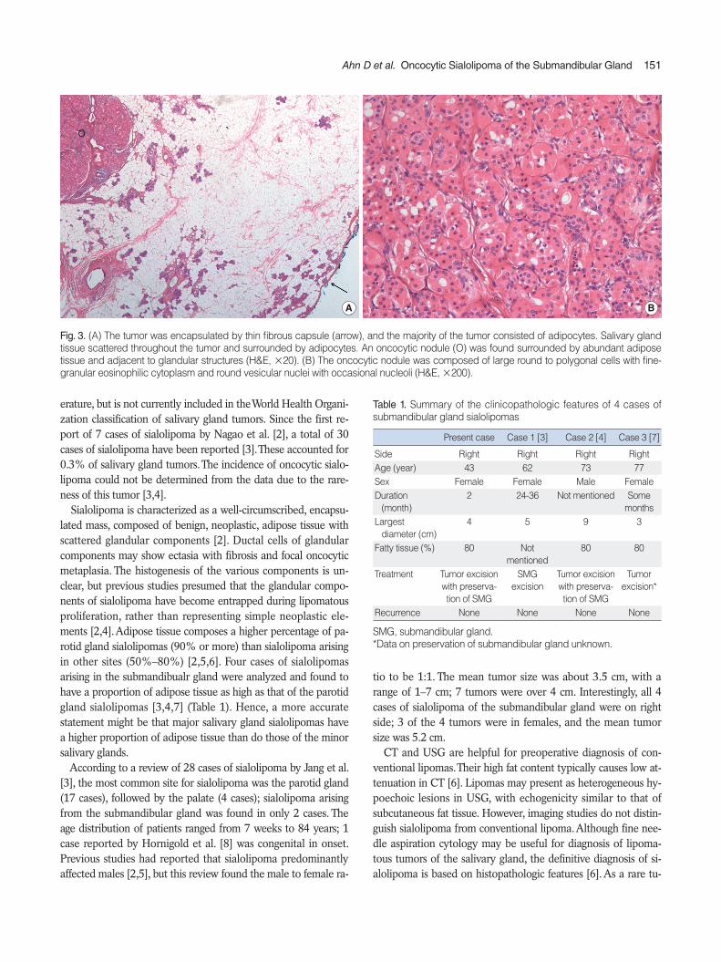
that the tumor was well-circumscribed, soft, yellowish, and had a well-demarcated light pink-colored nodular component sur-rounded by fatty tissue. Ill-defined brown lesions were scattered around the nodule; histological examination identified these as an oncocytic nodule and glandular tissue (Fig. 2). Microscopic examination showed that the tumor was encapsu-lated by a thin fibrous capsule; the majority of the tumor consist-ed of adipocytes, which had no histological differences from sim-ple lipoma. Salivary gland tissues within the tumor were sparse. Those present were surrounded by adipocytes; hence, theses gland tissues were completely isolated from the tumor capsule. An oncocytic nodule was found surrounded by adipose tissue and mainly located at the periphery of the tumor, adjacent to ductal structures (Fig. 3A). The nodule, measuring about 1 cm across, was composed of large round to polygonal cells with fine granular, eosinophilic cytoplasm, and round vesicular nuclei with occasional nucleoli (Fig. 3B).
DISCUSSION
Lipomatous tumors admixed with salivary gland tissue have been studied with interest for the last decade. New tumor types, including sialolipoma, lipoadenoma, oncocytic lipoadenoma and oncocytic lipoadenoma with sebaceous differentiation have been described [4]. Oncocytic sialolipoma of the salivary gland is a unique benign tumor that has recently been described in the lit-
A B
SMG
MH
HG
Fig. 1. (A) Ultrasonography showing that the mass (arrows) was of a heterogeneous, hypoechoic nature with ill-defined margins compared to typical submandibular gland (SMG) tissue; the mass was located throughout the superficial and deep portions of normal submandibular gland tissue (MH, mylohyoid muscle; HG, hyoglossus muscle). (B) The computed tomography image revealed a fatty mass with irregular mar-gins and multiple soft tissue density was seen inside the mass in the right parapharyngeal space and submandbular region.
Fig. 2. Gross pathology. The tumor was well-circumscribed, soft, yel-lowish and had a well-demarcated light-pink colored nodular com-ponent (arrow heads) surrounded by fat tissue and ill-defined brown lesions (arrows) scattered peripherally in the tumor; histological ex-amination identified these structures as an oncocytic nodule and glandular tissue.
Ahn D et al. Oncocytic Sialolipoma of the Submandibular Gland 151
erature, but is not currently included in the World Health Organi-zation classification of salivary gland tumors. Since the first re-port of 7 cases of sialolipoma by Nagao et al. [2], a total of 30 cases of sialolipoma have been reported [3]. These accounted for 0.3% of salivary gland tumors. The incidence of oncocytic sialo-lipoma could not be determined from the data due to the rare-ness of this tumor [3,4]. Sialolipoma is characterized as a well-circumscribed, encapsu-lated mass, composed of benign, neoplastic, adipose tissue with scattered glandular components [2]. Ductal cells of glandular components may show ectasia with fibrosis and focal oncocytic metaplasia. The histogenesis of the various components is un-clear, but previous studies presumed that the glandular compo-nents of sialolipoma have become entrapped during lipomatous proliferation, rather than representing simple neoplastic ele-ments [2,4]. Adipose tissue composes a higher percentage of pa-rotid gland sialolipomas (90% or more) than sialolipoma arising in other sites (50%–80%) [2,5,6]. Four cases of sialolipomas arising in the submandibualr gland were analyzed and found to have a proportion of adipose tissue as high as that of the parotid gland sialolipomas [3,4,7] (Table 1). Hence, a more accurate statement might be that major salivary gland sialolipomas have a higher proportion of adipose tissue than do those of the minor salivary glands. According to a review of 28 cases of sialolipoma by Jang et al. [3], the most common site for sialolipoma was the parotid gland (17 cases), followed by the palate (4 cases); sialolipoma arising from the submandibular gland was found in only 2 cases. The age distribution of patients ranged from 7 weeks to 84 years; 1 case reported by Hornigold et al. [8] was congenital in onset. Previous studies had reported that sialolipoma predominantly affected males [2,5], but this review found the male to female ra-
tio to be 1:1. The mean tumor size was about 3.5 cm, with a range of 1–7 cm; 7 tumors were over 4 cm. Interestingly, all 4 cases of sialolipoma of the submandibular gland were on right side; 3 of the 4 tumors were in females, and the mean tumor size was 5.2 cm. CT and USG are helpful for preoperative diagnosis of con-ventional lipomas. Their high fat content typically causes low at-tenuation in CT [6]. Lipomas may present as heterogeneous hy-poechoic lesions in USG, with echogenicity similar to that of subcutaneous fat tissue. However, imaging studies do not distin-guish sialolipoma from conventional lipoma. Although fine nee-dle aspiration cytology may be useful for diagnosis of lipoma-tous tumors of the salivary gland, the definitive diagnosis of si-alolipoma is based on histopathologic features [6]. As a rare tu-
A B
O
Fig. 3. (A) The tumor was encapsulated by thin fibrous capsule (arrow), and the majority of the tumor consisted of adipocytes. Salivary gland tissue scattered throughout the tumor and surrounded by adipocytes. An oncocytic nodule (O) was found surrounded by abundant adipose tissue and adjacent to glandular structures (H&E, ×20). (B) The oncocytic nodule was composed of large round to polygonal cells with fine-granular eosinophilic cytoplasm and round vesicular nuclei with occasional nucleoli (H&E, ×200).
Table 1. Summary of the clinicopathologic features of 4 cases of submandibular gland sialolipomas
Present case Case 1 [3] Case 2 [4] Case 3 [7]
Side Right Right Right RightAge (year) 43 62 73 77Sex Female Female Male FemaleDuration (month)
2 24-36 Not mentioned Some months
Largest diameter (cm)
4 5 9 3
Fatty tissue (%) 80 Notmentioned
80 80
Treatment Tumor excision with preserva-tion of SMG
SMGexcision
Tumor excision with preserva-tion of SMG
Tumorexcision*
Recurrence None None None None
SMG, submandibular gland.*Data on preservation of submandibular gland unknown.

152 Clinical and Experimental Otorhinolaryngology Vol. 7, No. 2: 149-152, June 2014
mor, oncocytic sialolipoma of the salivary gland can have differ-ential diagnoses, primarily other tumors with a large adipose tis-sue component. Sialolipoma is a proliferation of mature adipose tissue, sur-rounded by a thin fibrous capsule. It is characterized by islands of epithelial salivary glandular components that consist of ductal, acinar, basal, and myoepithelial cells that are enclosed in mature adipose tissue [2]. Lipoadenoma is characterized by an admix-ture of mature adipose tissue and branching epithelial tubules that lack myoepithelial cells. Immunohistochemical examination reveals no acini [1,4]. The presence of a fibrous capsule can easily distinguish sialolipoma from lipomatosis; the presence of normal salivary gland with duct dialatation and fibrosis precludes the possibility of a pleomorphic adenoma with extensive adipose content [2,9]. If a tumor has oncocytic nodules as in our case, it should be differentiated from oncocytoma, which has some ma-lignant potential. Distinguishing between these 2 tumor types is not difficult because oncocytic nodules of sialolipoma are sur-rounded by abundant adipocytes adjacent to normal glandular structures. To our knowledge, 5 cases of oncocytic lipoadenomas have been previously reported in the literature [1]. Our case in only the second report of oncocytic sialolipoma. In the first report, oncocytic sialolipoma was characterized as sialolipoma with on-cocytic adenoma in the tumor. The oncocytic micronodules have been considered to be the result of oncocytic metaplasia and hy-perplasia of ducts. In our case, an oncocytic micronodule was found adjacent to ductal structures and isolated from fatty tissue, just as reported by Pusiol et al. [4]. This arrangement suggests that the oncocytic nodule originated from ductal cells, which be-came separated from the ducts by proliferation of adipose tissue [4]. Treatment for sialolipoma is complete surgical excision of the tumor. Because the tumor in our case was well-encapsulated and the initial impression was that of a simple lipoma, the treatment of choice was tumorectomy with preservation of the submandib-ular gland. Superficial parotidectomy has been performed in most cases of parotid gland sialolipomas. No recurrences have been reported in any of the 30 cases of sialolipoma [3,5,6]. In conclusion, sialolipoma is a very rare, but distinct salivary
gland neoplasm. It is found predominantly in parotid glands, but could develop in any salivary gland. Many variations of sialoli-poma such as oncocytic change and sebaceous differentiation are possible. As a newly described tumor type, care should be taken to distinguish from other salivary gland neoplasms such as sim-ple lipoma, pleomorphic adenoma, or oncocytoma.
CONFLICT OF INTEREST
No potential conflict of interest relevant to this article was re-ported.
REFERENCES
1. Ilie M, Hofman V, Pedeutour F, Attias R, Santini J, Hofman P. Onco-cytic lipoadenoma of the parotid gland: immunohistochemical and cytogenetic analysis. Pathol Res Pract. 2010 Jan;206(1):66-72.
2. Nagao T, Sugano I, Ishida Y, Asoh A, Munakata S, Yamazaki K, et al. Sialolipoma: a report of seven cases of a new variant of salivary gland lipoma. Histopathology. 2001 Jan; 38(1):30-6.
3. Jang YW, Kim SG, Pai H, Park JW, Lee YC, Rotaru H. Sialolipoma: case report and review of 27 cases. Oral Maxillofac Surg. 2009 Jun; 13(2):109-13.
4. Pusiol T, Franceschetti I, Scialpi M, Piscioli I. Oncocytic sialolipoma of the submandibular gland with sebaceous differentiation: a new pathological entity. Indian J Pathol Microbiol. 2009 Jul-Sep;52(3): 379-82.
5. Ramer N, Lumerman HS, Ramer Y. Sialolipoma: report of two cases and review of the literature. Oral Surg Oral Med Oral Pathol Oral Radiol Endod. 2007 Dec;104(6):809-13.
6. Yum DJ, Kang JH, Kim YJ, Kim SW. Sialolipoma of the parotid gland. Korean J Otorhinolaryngol-Head Neck Surg. 2008 Apr; 51(4): 371-5.
7. Parente P, Longobardi G, Bigotti G. Hamartomatous sialolipoma of the submandibular gland: case report. Br J Oral Maxillofac Surg. 2008 Oct;46(7):599-600.
8. Hornigold R, Morgan PR, Pearce A, Gleeson MJ. Congenital sialoli-poma of the parotid gland first reported case and review of the liter-ature. Int J Pediatr Otorhinolaryngol. 2005 Mar;69(3):429-34.
9. Lin YJ, Lin LM, Chen YK, Shen YH, Hsue SS, Wang WC, et al. Sialo-lipoma of the floor of the mouth: a case report. Kaohsiung J Med Sci. 2004 Aug;20(8):410-4.