CASE REPORT - KoreaMed · Clinic, Universal Aksaray Hospital, Istanbul, Turkey Reconstruction of...
5
Copyright © 2013, the Korean Surgical Society J Korean Surg Soc 2013;84:61-65 http://dx.doi.org/10.4174/jkss.2013.84.1.61 CASE REPORT JKSS Journal of the Korean Surgical Society pISSN 2233-7903ㆍeISSN 2093-0488 Received April 6, 2012, Revised July 25, 2012, Accepted August 5, 2012 Correspondence to: Yeliz Emine Ersoy Department of General Surgery, Bezmialem Vakif University Faculty of Medicine, Estonsehir KV-28/1, Samlar Koyu, Tatarcik Mevkii, Bahcesehir Yani, Basaksehir 34306, Istanbul, Turkey Tel: +90-532-6102713, Fax: +90-212-6318918, E-mail: [email protected] This study was presented as a poster in a national surgical congress (17. Ulusal Cerrahi Kongresi, May 26-29, 2010, Ankara, Turkey). cc Journal of the Korean Surgical Society is an Open Access Journal. All articles are distributed under the terms of the Creative Commons Attribution Non-Commercial License (http://creativecommons.org/licenses/by-nc/3.0/) which permits unrestricted non-commercial use, distribution, and reproduction in any medium, provided the original work is properly cited. Repair of a postappendectomy massive ventral hernia using tissue expanders Yeliz Emine Ersoy, Fatih Celebi 1 , Fazilet Erozgen 2 , Selma Sonmez Ergun 3 , Murat Akaydin 4 , Rafet Kaplan 2 Department of General Surgery, Bezmialem Vakif University Faculty of Medicine, Istanbul, 1 2nd General Surgery Clinic, Bagcilar Training and Research Hospital, Istanbul, 2 1st General Surgery Clinic, Haseki Training and Research Hospital, Istanbul, 3 Department of Plastic and Reconstructive Surgery, Bezmialem Vakif University Faculty of Medicine, Istanbul, 4 General Surgery Clinic, Universal Aksaray Hospital, Istanbul, Turkey Reconstruction of large abdominal wall defects is a challenging problem. Various reconstructive techniques have been de- scribed in the surgical literature each with its advantages and disadvantages. In this report we describe our experience in treating a patient with large abdominal wall defect by staged abdominal wall reconstruction utilizing prosthetic mesh in con- junction with tissue expanders. A 41-year-old male presented with abdominal pain. Exploratory laparotomy showed perfo- rated appendicitis with intraabdominal abscess of 1,500 mL. Postoperatively, he developed intraperitoneal sepsis. To prevent abdominal compartment syndrome, he was reoperated and left with “open abdomen”. After several open abdomen lavages, his abdominal wall defect was allowed to granulate. After epithelization of the defect, the abdominal wall was reconstructed using prosthetic mesh and tissue expanders. The tissue expansion process was well tolerated. We suggest that the use of tis- sue expanders provides reliable and well-vascularized soft-tissue coverage in abdominal wall reconstruction. Key Words: Abdominal hernia, Tissue expander, Reconstruction INTRODUCTION Advances in surgical intensive care have enabled pa- tients with severe abdominal pathologies to survive. En bloc tumor resections, previous surgeries, intraabdominal catastrophes, necrotizing fascial infections, or traumatic abdominal injuries cause abdominal wall defects in these kind of patients [1,2]. The aims of reconstruction in such defects are full restoration of abdominal wall function, in- cluding muscular support, prevention of visceral eventra- tion, and adequate soft tissue coverage [3]. Wide undermining of the skin and subcutaneous tissue usually permits primary wound closure. In a minority of patients, lateral migration of the rectus abdominis muscles from contraction of the flank muscles, and concomitant visceral sac protrusion, result in loss of abdominal domain
-
Upload
trankhuong -
Category
Documents
-
view
214 -
download
0
Transcript of CASE REPORT - KoreaMed · Clinic, Universal Aksaray Hospital, Istanbul, Turkey Reconstruction of...

Copyright © 2013, the Korean Surgical Society
J Korean Surg Soc 2013;84:61-65http://dx.doi.org/10.4174/jkss.2013.84.1.61
CASE REPORT
JKSSJournal of the Korean Surgical Society
pISSN 2233-7903ㆍeISSN 2093-0488
Received April 6, 2012, Revised July 25, 2012, Accepted August 5, 2012
Correspondence to: Yeliz Emine ErsoyDepartment of General Surgery, Bezmialem Vakif University Faculty of Medicine, Estonsehir KV-28/1, Samlar Koyu, Tatarcik Mevkii, Bahcesehir Yani, Basaksehir 34306, Istanbul, TurkeyTel: +90-532-6102713, Fax: +90-212-6318918, E-mail: [email protected]
This study was presented as a poster in a national surgical congress (17. Ulusal Cerrahi Kongresi, May 26-29, 2010, Ankara, Turkey).
cc Journal of the Korean Surgical Society is an Open Access Journal. All articles are distributed under the terms of the Creative Commons Attribution Non-Commercial License (http://creativecommons.org/licenses/by-nc/3.0/) which permits unrestricted non-commercial use, distribution, and reproduction in any medium, provided the original work is properly cited.
Repair of a postappendectomy massive ventral hernia using tissue expanders
Yeliz Emine Ersoy, Fatih Celebi1, Fazilet Erozgen2, Selma Sonmez Ergun3, Murat Akaydin4, Rafet Kaplan2
Department of General Surgery, Bezmialem Vakif University Faculty of Medicine, Istanbul, 12nd General Surgery Clinic, Bagcilar Training and Research Hospital, Istanbul, 21st General Surgery Clinic, Haseki Training and Research Hospital, Istanbul, 3Department of Plastic and Reconstructive Surgery, Bezmialem Vakif University Faculty of Medicine, Istanbul, 4General Surgery Clinic, Universal Aksaray Hospital, Istanbul, Turkey
Reconstruction of large abdominal wall defects is a challenging problem. Various reconstructive techniques have been de-scribed in the surgical literature each with its advantages and disadvantages. In this report we describe our experience in treating a patient with large abdominal wall defect by staged abdominal wall reconstruction utilizing prosthetic mesh in con-junction with tissue expanders. A 41-year-old male presented with abdominal pain. Exploratory laparotomy showed perfo-rated appendicitis with intraabdominal abscess of 1,500 mL. Postoperatively, he developed intraperitoneal sepsis. To prevent abdominal compartment syndrome, he was reoperated and left with “open abdomen”. After several open abdomen lavages, his abdominal wall defect was allowed to granulate. After epithelization of the defect, the abdominal wall was reconstructed using prosthetic mesh and tissue expanders. The tissue expansion process was well tolerated. We suggest that the use of tis-sue expanders provides reliable and well-vascularized soft-tissue coverage in abdominal wall reconstruction.
Key Words: Abdominal hernia, Tissue expander, Reconstruction
INTRODUCTION
Advances in surgical intensive care have enabled pa-tients with severe abdominal pathologies to survive. En bloc tumor resections, previous surgeries, intraabdominal catastrophes, necrotizing fascial infections, or traumatic abdominal injuries cause abdominal wall defects in these kind of patients [1,2]. The aims of reconstruction in such
defects are full restoration of abdominal wall function, in-cluding muscular support, prevention of visceral eventra-tion, and adequate soft tissue coverage [3].
Wide undermining of the skin and subcutaneous tissue usually permits primary wound closure. In a minority of patients, lateral migration of the rectus abdominis muscles from contraction of the flank muscles, and concomitant visceral sac protrusion, result in loss of abdominal domain
Yeliz Emine Ersoy, et al.
62 thesurgery.or.kr
that make this approach impossible [2]. Generally, two methods are used for the treatment of abdominal wall de-fects that are not amenable to tensionless approximation of the natural tissues. The first method is to bridge the de-fect with the patients’ own tissue, synthetic products, or a composite material; and the second one is to reappro-ximate the natural tissue after utilizing relaxing incisions or preoperative measures like tissue expansion or pro-gressive pneumoperitoneum [3].
Synthetic meshes, autologous tissue flaps, mostly my-ocutaneous flaps such as tensor fasciae latae, rectus femo-ris, rectus abdominis, and latissimus dorsi, with or with-out incorporation of synthetic meshes, pedicled omentum flaps, some biological materials like human acellular der-mal matrix, were introduced in hernia repair and abdomi-nal wall reconstruction [4-6]. Abdominal wall re-construction with prosthetic mesh can be safe and effec-tive if well-vascularized soft-tissue coverage is provided.
Tissue expanders have been used in this clinical sit-uation to provide soft-tissue coverage and to restore ab-dominal domain by increasing both the size and the vascu-larity of the donor tissue. Tissue expansion produces a strong, vascularized capsule around the expanders. Our patient’s vascularized capsule, combined with the existing rectus sheath, was used to reconstruct abdominal wall de-fects [7]. Tissue expanders placed in the subcutaneous space, abdominal wall intramuscular spaces (between the internal oblique and transverse abdominis muscles), inter-muscular sites (between the external and internal oblique muscles), and finally intraabdominally, have been re-ported as a method to increase the size of potential skin or muscle flaps before closure and also for stable coverage of prosthetic material [3,8].
Simplicity, safety and efficacy should be the main con-siderations in the management. In this report we describe our experience in treating a patient with large abdominal wall defect by staged abdominal wall reconstruction uti-lizing prosthetic mesh in conjunction with tissue expan-ders.
CASE REPORT
A 41-year-old male patient presented to our emergency department with abdominal pain. Exploratory laparoto-my performed through a subumbilical incision showed perforated appendicitis with intraabdominal abscess of 1,500 mL. In addition, there was a large hemangioma occu-pying 80% of the liver. After surgery he developed intra-peritoneal sepsis and in order to prevent abdominal com-partment syndrome, he was reoperated and left with “open abdomen”. Several open abdomen lavages were performed and his abdominal wall defect was allowed to granulate.
After epithelization of the defect approximately ten months later (Fig. 1), reconstruction of the abdominal wall was planned. Skin incisions were made 2 cm lateral to the defective area. Two rectangular tissue expanders (LS 82 2000, Laboratoires Sebbin, Boissy-l'Aillerie, France) of 2,000 mL volume (230 × 130 × 70 mm) were placed in the subcutaneous pockets created just above the anterior rec-tus sheath and adjacent to the abdominal wound bi-laterally to reconstruct the defect of 23 × 37 cm in size. The ports of the tissue expanders were placed on the thoracic cage region.
Each of the tissue expanders were inflated with 200 mL of saline at the time of the initial operation. Over the next several weeks, the expanders were gradually inflated to a total volume of 2,350 mL each over a period of four months (Fig. 2). After four months, the patient returned to the op-erating room for definitive repair of the defect. The ex-panders were removed (Fig. 3) and since the fascial defect was too large to close primarily, utilizing prosthetic mesh (expanded polytetrafluoroethylene; GORE-TEX soft tis-sue patch) (Fig. 4) in conjunction with tissue expanders, the abdominal wall defect was reconstructed. Expanded skin flaps were advanced toward the midline and closed in layers without tension. A more acceptable midline scar was then obtained. Suction drains were placed bilaterally and removed three days later.
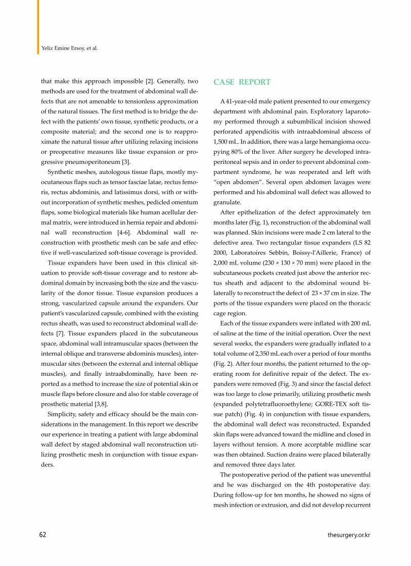
The postoperative period of the patient was uneventful and he was discharged on the 4th postoperative day. During follow-up for ten months, he showed no signs of mesh infection or extrusion, and did not develop recurrent
Postappendectomy ventral hernia repair using tissue expanders
thesurgery.or.kr 63
Fig. 1. Preoperative appearance of patient (before tissue expan-sion).
Fig. 2. Appearance after inflation of tissue expanders to total volume of 2,350 mL each.
Fig. 3. Appearance after removal of expanders and granulation tissue over intestines.
Fig. 4. Appearance after prosthetic mesh placement.
Fig. 5. Appearance of patients two months later.
hernia, ulceration or enteric fistula. The tissue expansion process was well tolerated by the patient (Fig. 5).
DISCUSSION
Treatment of large ventral hernias is difficult, partly due to a lack of available abdominal wall, and owing to the dif-ficulty in finding sufficient space for the abdominal con-tents after reposition. The abdominal wall is a multi-layered structure composed of several different tissues. The myofascial layer is the major supportive part of the ab-dominal wall, which is important for abdominal contents
Yeliz Emine Ersoy, et al.
64 thesurgery.or.kr
protection and dynamic function [4].There is no consensus about which technique or materi-
al is suitable for a certain kind of defect. Selection should be made mainly on the basis of the size, location and depth of the defect, wound bed preparedness and the medical status of the patient [1,4]. When assessing the patient ini-tially, a complete history, physical and laboratory workup are essential with the involvement of multiple specialties, including general surgery, internal medicine, nutrition, pulmonology, and infectious disease [1]. Although multi-ple strategies are advocated, even for different situations, it requires careful planning to determine the optimal choice in each case [2].
The goals of abdominal reconstruction are; restoration of function and integrity of the musculofascial abdominal wall, prevention of visceral eventration, and provision of dynamic muscle support [1]. There are three types of ab-dominal wall defects. Type I involves only the loss of skin and can be corrected with primary suture; type II is a my-ofascial defect with intact skin coverage for reconstruction of which a myofascial substitute is needed; and type III is a myofascial defect without skin coverage that needs au-tologous tissues and synthetic or biological mesh [4]. Our patient is included in type III and was repaired by pros-thetic mesh and tissue expanders.
Ideally, the abdominal wall is reconstructed by mobiliz-ing local tissue and using existing fascia. However, pri-mary closure performed under tension results in tissue is-chemia, wound dehiscence, herniation, pulmonary com-plications secondary to increased intraabdominal pres-sure. Reduced diaphragmatic excursion is associated with high morbidity and mortality rates. The use of prosthetic mesh in conjunction with tissue-expanded skin provides a durable abdominal closure and is technically simpler than flap closure methods.
With minimal donor site morbidity relative to most musculofascial techniques, tissue expansion provides well-vascularized skin and soft tissue over the prosthetic mesh, allows excision of unsightly scars and skin grafts and obtains excellent color and texture match [2]. Subcuta-neously placed tissue expanders have been successfully used in surgical situations with the primary intent of ex-panding skin to achieve wound closure, including adult
patients who had loss of the abdominal wall from either traumatic or infectious events and in pediatric patients with abdominal wall defects [8]. Subcutaneous placement under healthy skin provides sufficient skin and subcutis to be approximated and allows uncomplicated placement of expanders and ample recruitment of soft-tissue coverage, with prosthetic mesh being used to close fascial defects [3]. But, the relatively superficial location of the expander can cause thinning of the skin and result in extrusion that ne-cessitates early removal and placement of a new expander at a later date [8]. Subcutaneous tissue expansion does not result in any alteration to the structure of the Musculofascia labdominal wall but can serve as an adjunct to bridge these defects [3].
Subfascial placement of expanders permits expansion of muscle and fascial layers to provide full-thickness au-tologous abdominal wall reconstruction, but at the cost of slightly more difficult dissection and subsequent removal. Placing expanders in the plane between the transverse and internal oblique muscles is risky because this area contains the nervous and arterial supplies for these two muscles and the rectus [3]. As a prevention, the vertical incision made in the posterior layer of the internal oblique aponeu-rosis should be minimal in length and performed with caution [8]. From an anatomic point of view, the plane be-tween the external oblique and internal oblique muscles seems to be the most convenient to use, but, it is a time-consuming, expensive form of therapy, as the process of gradual expansion takes several weeks resulting in lim-ited enlargement of the structures needed [3].
De Ugarte et al. [8] presented a novel approach to using tissue expanders placed within the intramuscular layers of the abdominal wall of a patient with a giant omphalocele, allowing creation of an outer layer composed of skin and internal and external oblique muscles. Intraabdominal placement of tissue expanders offers the advantage of cre-ating “tissue flaps” that include all layers of the abdominal wall, including skin, muscle, and peritoneum. On the oth-er hand, expansion of the abdominal cavity can increase intraabdominal pressure, impair respiratory function, cause visceral ischemia and increase the risk of displace-ment of viscera [8].
The early use of tissue expanders to increase the amount

Postappendectomy ventral hernia repair using tissue expanders
thesurgery.or.kr 65
of tissue available for coverage would be limited by the presence of intestinal contamination, stomas, and drains [9]. Before starting filling of the expander, a period of wound healing is usually awaited for three weeks to pre-vent expander exclusion. Tissue expanders can become in-fected or exposed during the course of expansion with a related complication rate of less than 15% . The predicted surface area gain after completion of tissue expansion of-ten falls short of the clinical requirements [10]. Due to the reversible phenomena of expanded skin to retract after re-moval of the expanders, overexpansion (10% to 20% over implant capacity) is necessary. Instead of resorting to seri-al expansions, overexpansion techniques help in reaching the desired dimensions and avoiding a shortage of donor site for expansion, and eventually reconstruction can be completed in one-stage [10]. It appears safe without risk of implant failure at least to 5 to 10 times the vendors’ stated maximum volume.
As a conclusion, the ideal reconstruction of abdominal wall should provide tension-free repair and dynamic muscle support if possible. Tissue expanders can be used as an additional therapeutic modality in the treatment of complex abdominal wall defects not amenable to delayed primary closure. The location for placement of a tissue ex-pander depends on the medical condition and specific re-constructive needs of the patient. This method has pro-vided reliable and durable abdominal wall closure with adequate soft-tissue coverage of prosthetic mesh as well as partially restoring abdominal domain. It also minimizes the potential mesh related complications, and is aestheti-cally superior to that achieved with skin graft and muscle flap techniques. When the size, place, and depth of the ex-pander is chosen carefully, and the expanding process and antibiotic prophylaxis is managed properly, this staged method of abdominal wall reconstruction is safe and sim-ple regarding the literature and our experience. Although the expansion process takes longer and, in some countries, the use of expanders has some medical and socioecono-
mical problems (cost, insurance coverage problems, etc) it allows maximization of patient healing and provides good functional and aesthetic results as in our patient.
CONFLICTS OF INTEREST
No potential conflict of interest relevant to this article was reported.
REFERENCES
1. Rohrich RJ, Lowe JB, Hackney FL, Bowman JL, Hobar PC. An algorithm for abdominal wall reconstruction. Plast Reconstr Surg 2000;105:202-16.
2. Carlson GW, Elwood E, Losken A, Galloway JR. The role of tissue expansion in abdominal wall reconstruction. Ann Plast Surg 2000;44:147-53.
3. Van Geffen HJ, Simmermacher RK. Incisional hernia re-pair: abdominoplasty, tissue expansion, and methods of augmentation. World J Surg 2005;29:1080-5.
4. Tang R, Gu Y, Gong DQ, Qian YL. Immediate repair of ma-jor abdominal wall defect after extensive tumor excision in patients with abdominal wall neoplasm: a retrospective re-view of 27 cases [corrected]. Ann Surg Oncol 2009;16: 2895-907.
5. Pless TK, Pless JE. Giant ventral hernias and their repair. A 10 year follow up study. Scand J Plast Reconstr Surg Hand Surg 1993;27:311-5.
6. Kim Z, Kim YJ. Components separation technique for large abdominal wall defect. J Korean Surg Soc 2011; 80(Suppl 1):S63-6.
7. Bax NM, van der Zee DC, Pull ter Gunne AJ, Rovekamp MH. Treatment of giant omphalocele by enlargement of the abdominal cavity with a tissue expander. J Pediatr Surg 1993;28:1181-4.
8. De Ugarte DA, Asch MJ, Hedrick MH, Atkinson JB. The use of tissue expanders in the closure of a giant omphalocele. J Pediatr Surg 2004;39:613-5.
9. Livingston DH, Sharma PK, Glantz AI. Tissue expanders for abdominal wall reconstruction following severe trau-ma: technical note and case reports. J Trauma 1992;32:82-6.
10. Hallock GG. Safety of clinical overinflation of tissue expanders. Plast Reconstr Surg 1995;96:153-7.