Case Report - Hindawi Publishing Corporationdownloads.hindawi.com/journals/cric/2012/471759.pdf ·...
4
Hindawi Publishing Corporation Case Reports in Cardiology Volume 2012, Article ID 471759, 3 pages doi:10.1155/2012/471759 Case Report ALCAPA Presents in an Adult with Exercise Intolerance but Preserved Cardiac Function Yan Liu and Beth W. Miller Department of Internal Medicine, University of Texas Southwestern Medical School Residency Programs at Austin, University Medical Center Brackenridge, 601 E 15th Street, Austin, TX 78701, USA Correspondence should be addressed to Beth W. Miller, [email protected] Received 26 June 2012; Accepted 12 August 2012 Academic Editors: K.-R. Chiou, C. Firschke, M. R. Movahed, K. Nikus, and C. S. Snyder Copyright © 2012 Y. Liu and B. W. Miller. This is an open access article distributed under the Creative Commons Attribution License, which permits unrestricted use, distribution, and reproduction in any medium, provided the original work is properly cited. Anomalous origin of the left coronary artery from the pulmonary artery (ALCAPA) is a rare congenital anomaly that usually manifests as severe left-sided heart failure and mitral valve insufficiency during the first one to two months of life. The majority of these cases die in infancy if not corrected early upon presentation. Adulthood presentation is rare and most of the untreated patients who reach adulthood present with left ventricular dysfunction, severe mitral regurgitation, and sometimes myocardial infarction. Here we report a case of a 20-year-old woman with a history of exercise intolerance since childhood that was misinterpreted as asthma until a 2D-Echo revealed ALCAPA with RCA collaterals to the left anterior descending artery, preserved LV ejection fraction, and absence of apparent mitral valve abnormality. One month after the ALCAPA diagnosis, she successfully underwent surgical reconstruction of left main and pulmonary artery without any major complications. She had normal left ventricular function without apparent ischemic cardiac symptoms eighteen months after procedure. 1. Introduction Anomalous origin of the left coronary artery from the pulmonary artery (ALCAPA) is a rare congenital anomaly occurring in 1 of 300,000 live births [1, 2] and accounts for 0.4 percent of congenital cardiac abnormalities. ALCAPA was first reported in 1885 by Brooks [1] and its first clinical description in conjunction with autopsy findings was described by Bland et al. in 1933 [3]. Therefore the condition is also known as Bland-White-Garland-syndrome. The ALCAPA anomaly may result from abnormal septa- tion of the conotruncus into the aorta and pulmonary artery, or from persistence of the pulmonary buds together with involution of the aortic buds that eventually form the coro- nary arteries. ALCAPA is usually an isolated cardiac anomaly that does not present prenatally because of the favorable fetal physiology that includes equivalent pressures in the main pulmonary artery and aorta secondary to a nonrestrictive patent ductus arteriosus, and the relatively equivalent oxygen concentrations due to parallel circulations. This results in normal myocardial perfusion and, therefore, no stimulus for collateral vessel formation between the right and left coronary artery systems is present. Shortly after birth, as the circulation becomes one in series, pulmonary artery pressure and resistance decrease, as does oxygen content of pul- monary blood flow. This causes a drop in antegrade flow and oxygen content of the anomalous left coronary artery, leading to myocardial ischemia. Furthermore, collateral circulation between the right and left coronary systems ensues and the left coronary artery flow reverses and enters the pulmonary trunk due to the low pulmonary arterial pressure, causing coronary steal phenomena. Consequently, fixed ischemia or myocardial infarction occurs. Eighty-five percent of all cases of ALCAPA present within the first two months of life. The typical clinical course involves severe left-sided heart failure and significant mitral valve insufficiency secondary to papillary muscle ischemia, permanent fibrosis, or left ventricular dilatation. The late presentation is extremely rare and usually with significant cardiac function compromise. 2. Case Presentation A 20-year-old woman presents with chronic dyspnea on exertion and exercise intolerance that was attributed to and
Transcript of Case Report - Hindawi Publishing Corporationdownloads.hindawi.com/journals/cric/2012/471759.pdf ·...
Hindawi Publishing CorporationCase Reports in CardiologyVolume 2012, Article ID 471759, 3 pagesdoi:10.1155/2012/471759
Case Report
ALCAPA Presents in an Adult with Exercise Intolerance butPreserved Cardiac Function
Yan Liu and Beth W. Miller
Department of Internal Medicine, University of Texas Southwestern Medical School Residency Programs at Austin,University Medical Center Brackenridge, 601 E 15th Street, Austin, TX 78701, USA
Correspondence should be addressed to Beth W. Miller, [email protected]
Received 26 June 2012; Accepted 12 August 2012
Academic Editors: K.-R. Chiou, C. Firschke, M. R. Movahed, K. Nikus, and C. S. Snyder
Copyright © 2012 Y. Liu and B. W. Miller. This is an open access article distributed under the Creative Commons AttributionLicense, which permits unrestricted use, distribution, and reproduction in any medium, provided the original work is properlycited.
Anomalous origin of the left coronary artery from the pulmonary artery (ALCAPA) is a rare congenital anomaly that usuallymanifests as severe left-sided heart failure and mitral valve insufficiency during the first one to two months of life. The majorityof these cases die in infancy if not corrected early upon presentation. Adulthood presentation is rare and most of the untreatedpatients who reach adulthood present with left ventricular dysfunction, severe mitral regurgitation, and sometimes myocardialinfarction. Here we report a case of a 20-year-old woman with a history of exercise intolerance since childhood that wasmisinterpreted as asthma until a 2D-Echo revealed ALCAPA with RCA collaterals to the left anterior descending artery, preservedLV ejection fraction, and absence of apparent mitral valve abnormality. One month after the ALCAPA diagnosis, she successfullyunderwent surgical reconstruction of left main and pulmonary artery without any major complications. She had normal leftventricular function without apparent ischemic cardiac symptoms eighteen months after procedure.
1. Introduction
Anomalous origin of the left coronary artery from thepulmonary artery (ALCAPA) is a rare congenital anomalyoccurring in 1 of 300,000 live births [1, 2] and accountsfor 0.4 percent of congenital cardiac abnormalities. ALCAPAwas first reported in 1885 by Brooks [1] and its firstclinical description in conjunction with autopsy findings wasdescribed by Bland et al. in 1933 [3]. Therefore the conditionis also known as Bland-White-Garland-syndrome.
The ALCAPA anomaly may result from abnormal septa-tion of the conotruncus into the aorta and pulmonary artery,or from persistence of the pulmonary buds together withinvolution of the aortic buds that eventually form the coro-nary arteries. ALCAPA is usually an isolated cardiac anomalythat does not present prenatally because of the favorable fetalphysiology that includes equivalent pressures in the mainpulmonary artery and aorta secondary to a nonrestrictivepatent ductus arteriosus, and the relatively equivalent oxygenconcentrations due to parallel circulations. This results innormal myocardial perfusion and, therefore, no stimulusfor collateral vessel formation between the right and left
coronary artery systems is present. Shortly after birth, as thecirculation becomes one in series, pulmonary artery pressureand resistance decrease, as does oxygen content of pul-monary blood flow. This causes a drop in antegrade flow andoxygen content of the anomalous left coronary artery, leadingto myocardial ischemia. Furthermore, collateral circulationbetween the right and left coronary systems ensues and theleft coronary artery flow reverses and enters the pulmonarytrunk due to the low pulmonary arterial pressure, causingcoronary steal phenomena. Consequently, fixed ischemia ormyocardial infarction occurs. Eighty-five percent of all casesof ALCAPA present within the first two months of life.The typical clinical course involves severe left-sided heartfailure and significant mitral valve insufficiency secondaryto papillary muscle ischemia, permanent fibrosis, or leftventricular dilatation. The late presentation is extremely rareand usually with significant cardiac function compromise.
2. Case Presentation
A 20-year-old woman presents with chronic dyspnea onexertion and exercise intolerance that was attributed to and
2 Case Reports in Cardiology
RCA
LP
LM
LAD
L 164W 213
(a)
RCA
LP
LM
LAD
L 165W 194
(b)
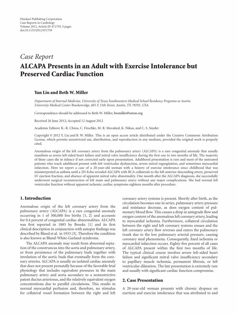
Figure 1: Coronary angiogram showing ALCAPA with left-to-right shunt. Retrograde filling was seen through collaterals arising from theenlarged right coronary artery (RCA), which gives rise to collaterals to a large left anterior descending artery (LAD) and left main artery(LMA) originated from the left main pulmonary (LP) trunk. The LAD and LMA are filling the pulmonary artery through a pronouncedleft-to-right shunt.
treated as presumed exercise-induced asthma since child-hood. She experienced worsening left-sided chest heavinessand was subsequently referred to our institute. She has nocoronary risk factors and no family history of prematurecoronary artery disease or congenital heart condition. Phys-ical examination was normal except a subtle continuousmurmur at apex and lower right sternal border. ECG showedsinus rhythm without ST or T wave changes (SupplementalFigure 1 available at doi:10.1155/2012/471759). Chest X-ray showed no cardiomegaly (Supplemental Figure 2). Serialcardiac enzymes were negative. In the light of cardiacmurmur, an echocardiogram was obtained and revealedanomalous coronary arteries with a large left coronaryartery to main pulmonary artery fistula. There was mildleft ventricular enlargement with preserved left ventricularcontractile function and an ejection fraction of 65%. Theappearance of the left ventricle and left atrium was consistentwith a systemic to pulmonary vascular shunt with increasedstroke volumes in the left heart. There were no structuralabnormalities of the aortic, mitral, tricuspid, or pulmonicvalves, and there was very mild mitral regurgitation byDoppler. A subsequent cardiac catheterization confirmedthe diagnosis of ALCAPA with retrograde filling throughcollaterals arising from an enlarged right coronary artery(12 mm) (Figure 1), pronounced left-to-right shunting fromthe left main coronary artery into the left main pulmonaryartery trunk; and the right coronary artery gives rise tocollaterals to a large left anterior descending artery whichhas ectasia in its proximal segment into a smaller circumflexartery. The patient underwent surgical treatment by thecreation of a pulmonary artery tube graft from the aorta tothe left coronary artery and reconstruction of the main pul-monary artery with a bovine pericardial patch. She had nomajor complications or ischemic symptoms 18 months afteroperation. A follow-up echocardiogram at that time showednormalized stroke volume and left atrial and ventricular size
secondary to reversed left-to-right shunt, preserved LV EF at55%, and absence of valvular abnormalities.
3. Discussion
As a rare congenital heart condition, ALCAPA even morerarely has a late presentation in adulthood as few of thesepatients survive past childhood without surgical repair [4].Among the reported adult cases [5–10], most of them, ifnot all, presented with evidence of irreversible impairment ofcardiac function such as severe dilated cardiomyopathy, sud-den cardiac death, acute myocardial infarction, malignantarrhythmias secondary to myocardial scar tissue, impairedLV contractile function, and development of significantmitral regurgitation. In this case, although she has anadulthood presentation, her presentation with a preservedLV contractile function without apparent structural orfunctional cardiac impairment may have prevented her froma life-threatening event later in life. While it is known thatthe sufficient collateral blood supply from the right coronaryartery could help some patients with ALCAPA pass throughchildhood with relatively minor symptoms, such as dyspnea,chest pain, and exercise intolerance, these symptoms areoften misinterpreted, (as it was initially the case for thepresented patient), as exercise-induced asthma. The keyfor an early diagnosis before the occurrence of permanentcardiac damage is a careful cardiac exam and detailedphysical examination, maintaining a high clinical suspicionwhen children have exercise intolerance. Early employmentof first line diagnostic modality echocardiogram is essential,as ECG in this case is usually unrevealing.
In infants, most of the patients with surgically correctedALCAPA show normalization of both ventricular functionand mitral valve insufficiency [11, 12]. Estimated long-termsurvival at 20 years was recently shown to be 94.8% [12].Although there is no large clinical trial study on surgically
Case Reports in Cardiology 3
corrected adult ALCAPA, the prognosis is usually determinedby the extent of irreversible left ventricular dysfunction andpresence of myocardial scar tissue. This patient had almostcomplete reversal of previous left atrial and ventriculardilation with maintained normal LV contractile function18 months after surgical repair, which is largely attributedto the absence of irreversible LV dysfunction or myocardialinfarction when the diagnosis was made.
In conclusion, even as a rare scenario of a rare disorder,diagnosis of adulthood ALCAPA should be considerednot only in adult patients presenting with clear evidenceof ischemic heart disease, left ventricular dysfunction, orarrhythmias, but also more importantly in patients withminor symptoms of exercise intolerance or dyspnea thatcould be easily misinterpreted or missed without high clinicalsuspicion and detailed physical and diagnostic evaluation, asearly diagnosis and timely surgical treatment usually resultsin an excellent prognosis.
References
[1] H. S. J. Brooks, “Two cases of an abnormal coronary arteryof the heart, arising from the pulmonary artery, with someremarks upon the effect of this anomaly in producing cirsoiddilatation of the vessels,” Journal of Anatomy and Physiology,vol. 20, pp. 26–29, 1885.
[2] S. Menahem and A. W. Venables, “Anomalous left coronaryartery from the pulmonary artery: a 15 year sample,” BritishHeart Journal, vol. 58, no. 4, pp. 378–384, 1987.
[3] E. F. Bland, P. D. White, and J. Garland, “Congenital anomaliesof the coronary arteries: report of an unusual case associatedwith cardiac hypertrophy,” American Heart Journal, vol. 8, no.6, pp. 787–801, 1933.
[4] T. P. Singh, M. F. Di Carli, N. M. Sullivan, M. F. Leonen, and W.R. Morrow, “Myocardial flow reserve in long-term survivorsof repair of anomalous left coronary artery from pulmonaryartery,” Journal of the American College of Cardiology, vol. 31,no. 2, pp. 437–443, 1998.
[5] T. Kristensen, K. F. Kofoed, S. Helqvist, M. Helvind, andL. Søndergaard, “Anomalous origin of the left coronaryartery from the pulmonary artery (ALCAPA) presenting withventricular fibrillation in an adult: a case report,” Journal ofCardiothoracic Surgery, vol. 3, no. 1, article 33, 2008.
[6] A. Facciorusso, P. Lanna, C. Vigna et al., “Anomalous originof the left coronary artery from the pulmonary artery inan elderly patient, football player in youth,” Journal ofCardiovascular Medicine, vol. 9, no. 10, pp. 1066–1069, 2008.
[7] A. Separham and P. Aliakbarzadeh, “Anomalous left coronaryartery from the pulmonary artery presenting with abortedsudden death in an octogenarian: a case report,” Journal ofMedical Case Reports, vol. 6, article 12, 2012.
[8] M. Das, P. Mahindrakar, D. Das, S. K. Behera, S. R. Chowd-hury, and B. Bandyopadhyay, “Anomalous left coronary arteryfrom pulmonary artery with mitral stenosis,” Annals ofThoracic Surgery, vol. 92, no. 2, pp. 735–737, 2011.
[9] P. Bajona, D. Maselli, R. Dore, and G. Minzioni, “Anomalousorigin of the left main artery from the pulmonary artery:adult presentation with systemic collateral supply and giantright coronary artery aneurysm,” The Journal of Thoracic andCardiovascular Surgery, vol. 134, no. 2, pp. 518–520, 2007.
[10] A. C. Aykan, M. Yildiz, G. Kahveci, and M. Ozkan, “Two adultcases of anomalous left coronary artery from the pulmonary
artery,” Turk Kardiyoloji Dernegi Arsivi, vol. 40, pp. 48–51,2012.
[11] Y. Isomatsu, Y. Imai, T. Shin’oka, M. Aoki, and Y. Iwata, “Sur-gical intervention for anomalous origin of the left coronaryartery from the pulmonary artery: the Tokyo experience,” TheJournal of Thoracic and Cardiovascular Surgery, vol. 121, no. 4,pp. 792–797, 2001.
[12] R. Lange, M. Vogt, J. Horer et al., “Long-term results of repairof anomalous origin of the left coronary artery from thepulmonary artery,” The Annals of Thoracic Surgery, vol. 83, no.4, pp. 1463–1471, 2007.

Submit your manuscripts athttp://www.hindawi.com
Stem CellsInternational
Hindawi Publishing Corporationhttp://www.hindawi.com Volume 2014
Hindawi Publishing Corporationhttp://www.hindawi.com Volume 2014
MEDIATORSINFLAMMATION
of
Hindawi Publishing Corporationhttp://www.hindawi.com Volume 2014
Behavioural Neurology
EndocrinologyInternational Journal of
Hindawi Publishing Corporationhttp://www.hindawi.com Volume 2014
Hindawi Publishing Corporationhttp://www.hindawi.com Volume 2014
Disease Markers
Hindawi Publishing Corporationhttp://www.hindawi.com Volume 2014
BioMed Research International
OncologyJournal of
Hindawi Publishing Corporationhttp://www.hindawi.com Volume 2014
Hindawi Publishing Corporationhttp://www.hindawi.com Volume 2014
Oxidative Medicine and Cellular Longevity
Hindawi Publishing Corporationhttp://www.hindawi.com Volume 2014
PPAR Research
The Scientific World JournalHindawi Publishing Corporation http://www.hindawi.com Volume 2014
Immunology ResearchHindawi Publishing Corporationhttp://www.hindawi.com Volume 2014
Journal of
ObesityJournal of
Hindawi Publishing Corporationhttp://www.hindawi.com Volume 2014
Hindawi Publishing Corporationhttp://www.hindawi.com Volume 2014
Computational and Mathematical Methods in Medicine
OphthalmologyJournal of
Hindawi Publishing Corporationhttp://www.hindawi.com Volume 2014
Diabetes ResearchJournal of
Hindawi Publishing Corporationhttp://www.hindawi.com Volume 2014
Hindawi Publishing Corporationhttp://www.hindawi.com Volume 2014
Research and TreatmentAIDS
Hindawi Publishing Corporationhttp://www.hindawi.com Volume 2014
Gastroenterology Research and Practice
Hindawi Publishing Corporationhttp://www.hindawi.com Volume 2014
Parkinson’s Disease
Evidence-Based Complementary and Alternative Medicine
Volume 2014Hindawi Publishing Corporationhttp://www.hindawi.com