
Case Report Extranodal marginal zone lymphoma of … · Case Report Extranodal marginal zone...
6
Int J Clin Exp Pathol 2013;6(5):951-956 www.ijcep.com /ISSN:1936-2625/IJCEP1303035 Case Report Extranodal marginal zone lymphoma of mucosa-associated lymphoid tissue (MALT lymphoma) of the ileum in a 35-year-old Japanese woman Tadashi Terada Departments of Pathology, Shizuoka City Shimizu Hospital, Shizuoka, Japan Received March 15, 2013; Accepted April 3, 2013; Epub April 15, 2013; Published April 30, 2013 Abstract: MALT lymphoma of the ileum is extremely rare: only several cases have been reported. A 34-year-old woman presented abdominal pain and melena. Colorectal and small intestinal endoscopes revealed multiple tu- mors and ulcers of the entire ileum. Biopsy was taken. Histologically, the biopsy consisted of 6 tissue specimens taken from the various sites of the ileum. All the tissue specimens showed infiltration of small atypical cells resem- bling centrocyte-like cells (CLC). Immunoblastic cells were scattered, though the number was scant. Monocytoid, plasma cell differentiation, and germinal centers were seen. Lymphoepithelial lesions (LEL) were scattered. Some small atypical lymphocyte were destructive the vessels and stromal tissues. Giemsa and Gram stains demonstrated no Helicobacter pylori and any bacteria. Immunohistochemically, the atypical small lymphocytes were positive for vimentin, but negative for various kinds of cytokeratins (CKs), EMA, CEA and CA19-9. The CK highlighted the LEL. They were positive for CD45, and B-cell markers (CD20, CD79a, CD10, CD23, bcl-2). CD138-positive plasma cells were seen in large number. CD68-positive macrophages were scattered. CD30- and CD15-positive immunoblastic cells were scattered. Most of the lymphoid cells were negative for T-cell markers (CD3, CD4, CD5, CD45RO, and CD43) and negative for NK cell markers (CD56 and CD57). The lymphoid cells were positive for κ-chain but negative for λ-chain; thus the light chain restriction was seen. TdT and cyclin D1 were negative. P53 was positive and Ki-67 labeling index was 67%. The lymphoid cells were negative for neuroendocrine markers (NCAM, NSE, chromogranin, and synaptophysin). The pathological diagnosis was MALT lymphoma of the ileum. Post-biopsy imaging techniques including CT, MRI, PET endoscope and gallium scintigraphy identified no tumors and no lymphadenopathy in the body except the ileum. The stomach was free from MALT lymphoma. She was treated by low dose chemotherapy and strictly followed up. Keywords: Ileum, MALT lymphoma, histopathology, immunohistochemistry Introduction Extranodal marginal zone lymphoma of muco- sa-associated lymphoid tissue (MALT lympho- ma) is defined as an extranodal lymphoma composed of morphologically heterogenous small B-cells including marginal (centrocyte- like) cells, cell resembling monocytoid cells, small lymphocytes, and scattered immuno- blasts and centroblast-like cells [1, 2]. This enti- ty was first described by Isaacson and White in 1983 [2]. There is a plasma cell differentiation in a proportion of cases. The infiltrate is in the marginal zone of reactive B-cell follicles and extends into the interfollicular region. In epithe- lial tissues, the neoplastic cells typically infil- trate the epithelium forming lymphoepithelial lesions. MALT lymphoma most commonly involves gas- trointestinal (GI) tract (50%), followed in order by salivary glands, lung (14%), head and neck (15%), ocular adnexa (12%), skin (11%), thyroid (4%) and breast (4%). In the GI tract, the major- ity of MALT lymphoma occurs in the stomach [3-14], where Helicobacter Pylori (HP) are regarded as the causative agent [1]. Elimination of HP commonly cures the gastric MALT lymphoma. Primary gastrointestinal lymphoma comprises 10-15% of all non-Hodgkin lymphomas and
Transcript of Case Report Extranodal marginal zone lymphoma of … · Case Report Extranodal marginal zone...

Int J Clin Exp Pathol 2013;6(5):951-956 www.ijcep.com /ISSN:1936-2625/IJCEP1303035
Case ReportExtranodal marginal zone lymphoma of mucosa-associated lymphoid tissue (MALT lymphoma) of the ileum in a 35-year-old Japanese woman
Tadashi Terada
Departments of Pathology, Shizuoka City Shimizu Hospital, Shizuoka, Japan
Received March 15, 2013; Accepted April 3, 2013; Epub April 15, 2013; Published April 30, 2013
Abstract: MALT lymphoma of the ileum is extremely rare: only several cases have been reported. A 34-year-old woman presented abdominal pain and melena. Colorectal and small intestinal endoscopes revealed multiple tu-mors and ulcers of the entire ileum. Biopsy was taken. Histologically, the biopsy consisted of 6 tissue specimens taken from the various sites of the ileum. All the tissue specimens showed infiltration of small atypical cells resem-bling centrocyte-like cells (CLC). Immunoblastic cells were scattered, though the number was scant. Monocytoid, plasma cell differentiation, and germinal centers were seen. Lymphoepithelial lesions (LEL) were scattered. Some small atypical lymphocyte were destructive the vessels and stromal tissues. Giemsa and Gram stains demonstrated no Helicobacter pylori and any bacteria. Immunohistochemically, the atypical small lymphocytes were positive for vimentin, but negative for various kinds of cytokeratins (CKs), EMA, CEA and CA19-9. The CK highlighted the LEL. They were positive for CD45, and B-cell markers (CD20, CD79a, CD10, CD23, bcl-2). CD138-positive plasma cells were seen in large number. CD68-positive macrophages were scattered. CD30- and CD15-positive immunoblastic cells were scattered. Most of the lymphoid cells were negative for T-cell markers (CD3, CD4, CD5, CD45RO, and CD43) and negative for NK cell markers (CD56 and CD57). The lymphoid cells were positive for κ-chain but negative for λ-chain; thus the light chain restriction was seen. TdT and cyclin D1 were negative. P53 was positive and Ki-67 labeling index was 67%. The lymphoid cells were negative for neuroendocrine markers (NCAM, NSE, chromogranin, and synaptophysin). The pathological diagnosis was MALT lymphoma of the ileum. Post-biopsy imaging techniques including CT, MRI, PET endoscope and gallium scintigraphy identified no tumors and no lymphadenopathy in the body except the ileum. The stomach was free from MALT lymphoma. She was treated by low dose chemotherapy and strictly followed up.
Keywords: Ileum, MALT lymphoma, histopathology, immunohistochemistry
Introduction
Extranodal marginal zone lymphoma of muco-sa-associated lymphoid tissue (MALT lympho-ma) is defined as an extranodal lymphoma composed of morphologically heterogenous small B-cells including marginal (centrocyte-like) cells, cell resembling monocytoid cells, small lymphocytes, and scattered immuno-blasts and centroblast-like cells [1, 2]. This enti-ty was first described by Isaacson and White in 1983 [2]. There is a plasma cell differentiation in a proportion of cases. The infiltrate is in the marginal zone of reactive B-cell follicles and extends into the interfollicular region. In epithe-lial tissues, the neoplastic cells typically infil-
trate the epithelium forming lymphoepithelial lesions.
MALT lymphoma most commonly involves gas-trointestinal (GI) tract (50%), followed in order by salivary glands, lung (14%), head and neck (15%), ocular adnexa (12%), skin (11%), thyroid (4%) and breast (4%). In the GI tract, the major-ity of MALT lymphoma occurs in the stomach [3-14], where Helicobacter Pylori (HP) are regarded as the causative agent [1]. Elimination of HP commonly cures the gastric MALT lymphoma.
Primary gastrointestinal lymphoma comprises 10-15% of all non-Hodgkin lymphomas and
Ileal MALT lymphoma
952 Int J Clin Exp Pathol 2013;6(5):951-956
encompasses 30-40% of the total extranodal lymphomas. Approximately 60-75% of cases occur in the stomach, followed by colon, cecum, jejunum, ileum, and rectum [3-14]. Lymphoid neoplasms may consist of mature B, T and less commonly extranodal NK/T cells. Of these, the two most frequently encountered histologic subtypes are MALT lymphoma, diffuse large B cell lymphoma (DLBCL). Enteropathy-associated T cell lymphoma, type I in particular, usually arises in a background of celiac dis-ease. T cell gene rearrangement confirms clon-ality. NK/T cell neoplasms are invariably asso-ciated with Epstein-Barr virus infection.
Primary ileal MALT lymphoma is extremely rare; only several cases have been reported in the literature [15-19]. Herein reported is a case of ileal MALT lymphoma occurred in a young woman.
Case report
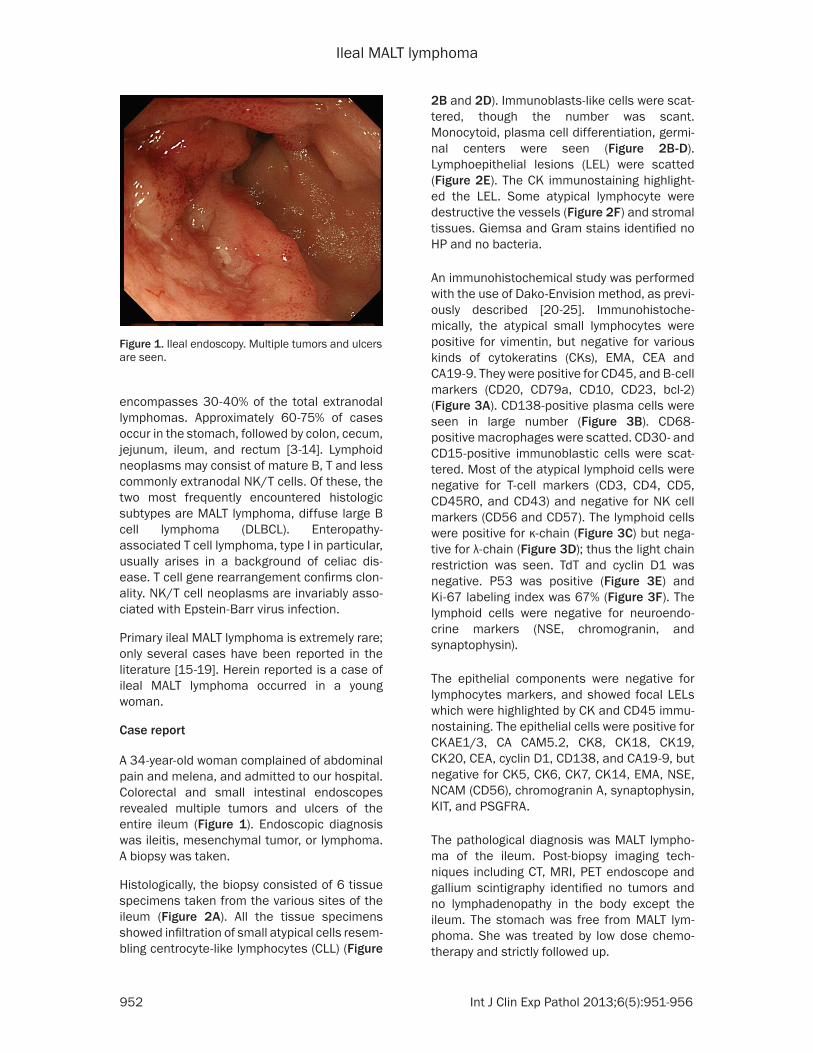
A 34-year-old woman complained of abdominal pain and melena, and admitted to our hospital. Colorectal and small intestinal endoscopes revealed multiple tumors and ulcers of the entire ileum (Figure 1). Endoscopic diagnosis was ileitis, mesenchymal tumor, or lymphoma. A biopsy was taken.
Histologically, the biopsy consisted of 6 tissue specimens taken from the various sites of the ileum (Figure 2A). All the tissue specimens showed infiltration of small atypical cells resem-bling centrocyte-like lymphocytes (CLL) (Figure
2B and 2D). Immunoblasts-like cells were scat-tered, though the number was scant. Monocytoid, plasma cell differentiation, germi-nal centers were seen (Figure 2B-D). Lymphoepithelial lesions (LEL) were scatted (Figure 2E). The CK immunostaining highlight-ed the LEL. Some atypical lymphocyte were destructive the vessels (Figure 2F) and stromal tissues. Giemsa and Gram stains identified no HP and no bacteria.
An immunohistochemical study was performed with the use of Dako-Envision method, as previ-ously described [20-25]. Immunohistoche- mically, the atypical small lymphocytes were positive for vimentin, but negative for various kinds of cytokeratins (CKs), EMA, CEA and CA19-9. They were positive for CD45, and B-cell markers (CD20, CD79a, CD10, CD23, bcl-2) (Figure 3A). CD138-positive plasma cells were seen in large number (Figure 3B). CD68-positive macrophages were scatted. CD30- and CD15-positive immunoblastic cells were scat-tered. Most of the atypical lymphoid cells were negative for T-cell markers (CD3, CD4, CD5, CD45RO, and CD43) and negative for NK cell markers (CD56 and CD57). The lymphoid cells were positive for κ-chain (Figure 3C) but nega-tive for λ-chain (Figure 3D); thus the light chain restriction was seen. TdT and cyclin D1 was negative. P53 was positive (Figure 3E) and Ki-67 labeling index was 67% (Figure 3F). The lymphoid cells were negative for neuroendo-crine markers (NSE, chromogranin, and synaptophysin).
The epithelial components were negative for lymphocytes markers, and showed focal LELs which were highlighted by CK and CD45 immu-nostaining. The epithelial cells were positive for CKAE1/3, CA CAM5.2, CK8, CK18, CK19, CK20, CEA, cyclin D1, CD138, and CA19-9, but negative for CK5, CK6, CK7, CK14, EMA, NSE, NCAM (CD56), chromogranin A, synaptophysin, KIT, and PSGFRA.
The pathological diagnosis was MALT lympho-ma of the ileum. Post-biopsy imaging tech-niques including CT, MRI, PET endoscope and gallium scintigraphy identified no tumors and no lymphadenopathy in the body except the ileum. The stomach was free from MALT lym-phoma. She was treated by low dose chemo-therapy and strictly followed up.
Figure 1. Ileal endoscopy. Multiple tumors and ulcers are seen.

Ileal MALT lymphoma
953 Int J Clin Exp Pathol 2013;6(5):951-956
Discussion
The author reported a very rare case of MALT lymphoma of the ileum. In general, MALT lym-phoma is almost always seen in the stomach, where HP infection is very prevalent. HP is thought to the etiology of MALT lymphoma in addition to gastric carcinoma. The lesion (MALT lymphoma) was once called reactive lymphoid hyperplasia and pseudolymphoma, but recent-ly it is called as MALT lymphoma since the paper of Isaacson and Wright in 1983 [2]. Gastric MALT lymphoma usually cures by eradi-cation of HP. In the present case, Giemsa and Gram stains revealed no HP. The present case did not examined HP culture. In other case reports of ileal MALT lymphoma did not show HP [15-19]. Thus, the etiology of ileal MALT lym-phoma is unknown. It may be true low grade malignancy developed by genetic alterations.
The diagnosis of the present study was MALT lymphoma of the ileum. Most of the GI lympho-ma except for the stomach is diffuse large B-cell lymphoma, followed by follicular lympho-ma and mantle cell lymphoma and T-cell neo-plasm is very rare [3-14]. The present study showed tumorous proliferation of small atypical lymphocytes. They resembled centrocyte-like
lymphocytes and monocytoid cells. Germinal centers were scattered and plasma cell differ-entiation, as evidenced by CD138 immunos-taining, was broad. In addition, apparent LELs were seen. The tumor was seen to be destruc-tive vessels and stromal cells, but no angiocen-tric lymphoma (T-cells) features were seen. These histological features are highly sugges-tive or confirmative of MALT lymphoma. However, the involvement site is not stomach; the author investigated an immunohistochemi-cal study.
Immunohistochemistry revealed the tumor was composed largely of B-cells, though a small number of T-cells were seen. Plasma cells were confirmed by CE138 immunostaining. CD30-, CD15-positive immunoblastic cells were scat-tered in a very small number. The LEL was high-lighted by immunostaining of CK and B cell markers. There were germinal centers not stained completely by bcl-2. Importantly, the present tumor was positive κ-light chain and negative for λ-light chain; thus the so called light chain restriction was seen. This phenome-non implies the monoclonality of the lesion and strongly suggest the lesion is true neoplasm. The p53 was positive, suggesting p53 gene mutations and malignant potentials of the pres-
Figure 2. Histological findings. A. Very low power view of the one specimen of the ileal biopsy. Severe proliferation of atypical small lymphocytes with destruction of normal architectures is seen. HE, x20. B. Medium size view. Proliferation of small atypical lymphocytes is seen. HE, x200. C. High power view. Proliferation of small atypical lymphocytes is seen. Monocytoid cells, centrocytes, immunoblastic cells and plasm cells are also seen. HE, x400. D. Lymphoepithelial lesions. The glands (center) are infiltrated by atypical lymphocytes. HE, x200. E. The angiodestructive features are seen. HE, x200.

Ileal MALT lymphoma
954 Int J Clin Exp Pathol 2013;6(5):951-956
ent lesion. Ki-67 labeling index (67%) was very high, suggesting that the cell proliferation is severe and the lesion is malignant. TdT was negative, suggesting that the present tumor is not precursor lymphoma. These immunohisto-chemical data, together with histological find-ings, confirms that the present ileal lesion is MALT lymphoma.
The differential diagnosis includes low-grade small B-cell neoplasms including small lympho-cytic lymphoma/CLL, lymphoplasmacytic lym-phoma, follicular lymphoma, and mantle cell lymphoma, and plasmacytoma [26-30]. The present case is not small lymphocytic lympho-ma, in which the lymphoid proliferation is monotonous and no LEL, plasma cell differen-tiation, monocytoid B-cells, CLC, or germinal centers are present. The present case is differ-ent from follicular lymphoma, in which the lym-phoid proliferation is monotonous and no other above mentioned MALT features were seen. In the diagnosis of follicular lymphoma, the bcl-2 and bcl-6 immunostaining are mandatory; in follicular lymphoma, the follicles were strongly stained with bcl-2 and bcl-6. In the present case, the nodular aggregate of lymphocytes are not follicles but germinal centers, and they were only faintly stained by bcl-2 immunostain-
ing. Histologically and immunohistochemically, the present tumor is entirely different from fol-licular lymphoma. The present case is not man-tle cell lymphoma, in which the lymphoma cells have characteristic nuclear groove seen in cells of mantle zone and are positive for cyclin D1. In the present tumor, no such nuclear features were seen, and cyclin D1 was negative. The present case is apparently not plasmacytoma or myeloma.
In conclusion, the author reported an extremely rare case of MALT lymphoma of the ileum in a young Japanese woman.
Declaration
The author has no conflict of interest.
Address correspondence to: Dr. Tadashi Terada, Department of Pathology, Shizuoka City Shimizu Hospital, Miyakami 1231, Shimizu-Ku, Shizuoka 424-8636, Japan. Phone: +81-54-336-1111; Fax: +81-54-334-1173; E-mail: [email protected]
References
[1] Isaacson PG, Chott A, Nakamura S, Muller-Her-melink HK, Harris NL, Swerdlow SH. Extranodal marginal zone lymphoma of mucosa-associat-
Figure 3. Immunohistochemical findings of the ileal MALT lymphoma. A. The atypical lymphocytes are strongly and diffusely positive for CD20. X200. B. The plasma cell differentiation cells are strongly positive for CD138. X200. C. The atypical lymphocytes are strongly positive for κ-light chain. x200. D. In contrast, the atypical lymphocytes are almost negative for λ-light chain. x200. E. p53 is expressed in the atypical lymphocytes. X200. F. The Ki-67 labeling is very high. X200.
Ileal MALT lymphoma
955 Int J Clin Exp Pathol 2013;6(5):951-956
ed lymphoid tissue (MALT lymphoma). In: Swerdlow SH, Campo E, Harris NL, Jaffe ES, Pileri SA, Stein H, Thiele J, Vardiman JW, eds. WHO classification of tumours of haematopoi-etic and lymphoid tissues. Lyon: IARC, 2008; pp: 214-217.
[2] Isaacson P, Wright DH. Malignant lymphoma of mucosa-associated lymphoid tissue: a distinc-tive type of B-cell lymphoma. Cancer 1983; 52: 1410-1416.
[3] Terada T. Gastrointestinal malignant lympho-ma: a pathologic study of 37 cases in a single Japanese institution. Am J Blood Res 2012; 2: 194-200.
[4] Bautista-Quach MA, Ake CD, Chen M, Wang J. Gastrointestinal lymphomas: Morphology, im-munophenotype and molecular features. J Gastrointest Oncol 2012; 3: 209-225.
[5] Terada T. One patient with double lymphomas: simultaneous gastric MALT lymphoma and ile-al diffuse large B-cell lymphoma. Int J Clin Exp Pathol 2012; 5: 260-263.
[6] Terada T. Histopathologic study of the rectum in 1,464 consecutive rectal specimens in a single Japanese hospital: II. malignant lesions. Int J Clin Exp Pathol 2013; 6: 385-394.
[7] Terada T. A clinicopathologic study of esopha-geal 860 benign and malignant lesions in 910 cases of consecutive esophageal biopsies. Int J Clin Exp Pathol 2013; 6: 191-198.
[8] Terada T. Malignant tumors of the small intes-tine: a histopathologic study of 41 cases among 1,312 consecutive specimens of small intestine. Int J Clin Exp Pathol 2012; 5: 203-209.
[9] Terada T. Esophageal cancers: a clinicopatho-logic and immunohistochemical study of 223 cases. Gastroenterol Res 2009; 2: 148-151.
[10] Terada T. An immunohistochemical study of primary signet-ring cell carcinoma of the stom-ach and colorectum: II. Expresssion of MUC1, MUC2, MUC5AC, and MUC6 in normal mucosa and in 42 cases. Int J Clin Exp Pathol 2013; 6: 613-21.
[11] Terada T. An immunohistochemical study of primary signet-ring cell carcinoma of the stom-ach and colorectum: I. Cytokeratin profile in 42 cases. Int J Clin Exp Pathol 2013; 6: 703-10.
[12] Terada T. An immunohistochemical study of primary signet-ring cell carcinoma of the stom-ach and colorectum: III. Expressions of EMA, CEA, CA19-9, CDX-2, p53, Ki-67 antigen, TTF-1, vimentin, and p63 in normal mucosa and in 42 cases. Int J Clin Exp Pathol 2013; 6: 630-8.
[13] Terada T. Histopathologic study of the stomach using computer database in 10,000 consecu-tive gastric specimens: II. Malignant lesions. Int J Clin Exp Pathol 2013; in press.
[14] Terada T. Histopathologic study using comput-er database of the stomach in 10,000 consec-utive gastric specimens: I. Benign conditions. Int J Clin Exp Pathol 2013; in press.
[15] Makino Y, Suzuki H, Nishizawa T, Kameyama K, Hisamatsu T, Imaeda H, Mukai M, Hibi T. Il-eal mucosa-zssociated lymphoid tissue (MALT) lymphoma with a large-cell component that regressed spontaneously. Gut Liver 2010; 4: 117-121.
[16] Ohashi S, Yazumi S, Watanabe N, Matsumoto S, Fukui T, Nishio A, Chiba T. Education and im-aging. Gastrointestinal: MALT lymphoma of the terminal ileum. J Gastroenterol Hepatol 2006; 21: 1495.
[17] Saito T, Toyoda H, Yamaguchi M, Nakamura T, Nakamura S, Mukai K, Fuke H, Wakita Y, Iwata M, Adachi Y, Shiku H. Ileocolonic lymphomas: a series of 16 cases. Endoscopy 2005; 37: 466-469.
[18] Matsumoto Y, Matsumoto T, Nakamura S, Ka-wasaki A, Aso M, Aoyagi K, Sadoshima S, On-oyama K, Fujishima M. Primary ileal plasmacy-toma arising in mixed low- and high-grade B-cell lymphoma of mucosa-associated lym-phoid tissue type. Abdom Imaging 2000; 25: 139-141.
[19] Hasegawa N, Tsuboi Y, Kato K, Yamada K, Morita K, Kuroiwa M, Ito H, Matsushima T, Ono K, Oshiro M. Endoscopic diagnosis of ileocecal mucosa-associated lymphoid tissue lympho-ma. Gastrointest Endosc 1999; 50: 115-117.
[20] Terada T, Kawaguchi M, Furukawa K, Sekido Y, Osamura Y. Minute mixed ductal-endocrine carcinoma of the pancreas with predominant intraductal growth. Pathol Int 2002; 52: 740-746.
[21] Terada T, Takeuchi T, Taniguchi M. Hepatobili-ary cystadenocarcinoma with cystadenoma el-ements of the gall bladder in an old man. Pathol Int 2003; 53: 790-795.
[22] Terada T, Tanigichi M. Intraductal oncocytic papillary neoplasm of the liver. Pathol Int 2004; 54: 116-123.
[23] Terada T, Kawaguchi M. Primary clear cell ad-enocarcinoma of the peritoneum. Tohoku J Exp Med 2005; 206: 271-275.
[24] Terada T. Extramuscular subcutaneous fibroli-poma containing foci of striated muscle cells: a hitherto unreported condition. Int J Clin Exp Pathol 2013; 6: 113-115.
[25] Terada T. Vascular leiomyoma of the lung aris-ing from pulmonary artery. Int J Clin Exp Pathol 2013; 6: 97-99.
[26] Muller-Hermlink HK, Montserrat E, Catovsky D, Campo E, Harris NL, Stein H. Chronic lympho-cytic leukemia/small lymphocytic lymphoma. In: Swerdlow SH, Campo E, Harris NL, Jaffe ES,

Ileal MALT lymphoma
956 Int J Clin Exp Pathol 2013;6(5):951-956
Pileri SA, Stein H, Thiele J, Vardiman JW, eds. WHO classification of tumours of haematopoi-etic and lymphoid tissues. IARC: Lyon, 2008; pp: 180-182.
[27] Harris NL, Nathwani BN, Swerdlow SH, de Jong D, Jaffe ES, Yoshino T, Ott G, Spagnoto D. Fol-licular lymphoma. In: Swerdlow SH, Campo E, Harris NL, Jaffe ES, Pileri SA, Stein H, Thiele J, Vardiman JW, eds. WHO classification of tu-mours of haematopoietic and lymphoid tis-sues. Lyon: IARC, 2008; pp: 220-226.
[28] Swerdlow SH, Campo E, Seto M, Muller-Herm-link HK. Mantle cell lymphoma. In: Swerdlow SH, Campo E, Harris NL, Jaffe ES, Pileri SA, Stein H, Thiele J, Vardiman JW, eds. WHO clas-sification of tumours of haematopoietic and lymphoid tissues. Lyon: IARC, 2008; pp: 229-232.
[29] McKenna RW, Kyle RA, Kuehl WM, Grogan TM, Harris NL, Coupland RW. Extraosseous plas-macytoma. In: Swerdlow SH, Campo E, Harris NL, Jaffe ES, Pileri SA, Stein H, Thiele J, Vard-iman JW. WHO Classification of tumours of haematopietic and lymphoid tissues. Lyon: IARC, 2008; pp: 208-209.
[30] McKenna RW, Kyle RA, Kuehl WM, Grogan TM, Harris NL, Coupland RW. Plasma cell myeloma. In: Swerdlow SH, Campo E, Harris NL, Jaffe ES, Pileri SA, Stein H, Thiele J, Vardiman JW. WHO Classification of tumours of haematopietic and lymphoid tissues. Lyon: IARC, 2008; pp: 202-208.


