Case Report Extensive Post-Partum Spontaneous Coronary Artery · Gestational diabetes was the only...
4
Central Journal of Cardiology & Clinical Research Cite this article: Sarfaraz M, Alhajiri A, Hasan SR (2019) Extensive Post-Partum Spontaneous Coronary Artery Dissection: Is Medical Therapy Safe? An Expe- rience from Middle Eastern District General Hospital. J Cardiol Clin Res 7(1): 1146. *Corresponding author Moaziz Sarfaraz, Department of Emergency, Fujairah Hospital, Fujairah, UAE Submitted: 25 October 2018 Accepted: 14 January 2019 Published: 17 January 2019 Copyright © 2019 Sarfaraz et al. OPEN ACCESS Keywords • Acute coronary syndrome (ACS); Spontaneous coronary artery dissection (SCAD); Coronary artery bypass graft (CABG); Percutaneous coronary intervention (PCI); Coronary angiography; CT angiography; Intervascular imaging; Left anterior descending artery (LAD) Case Report Extensive Post-Partum Spontaneous Coronary Artery Dissection: Is Medical Therapy Safe? An Experience from Middle Eastern District General Hospital Moaziz Sarfaraz 1 *, Abdulla Alhajiri 2 , and Syeda Rana Hasan 3 1 Department of Emergency, Fujairah Hospital, UAE 2 Department of Cardiology, Fujairah Hospital, UAE 3 Ministry of Health, Fujairah, UAE Abstract Extensive coronary artery dissection is an uncommon presentation of acute coronary syndrome (ACS). The etiology is unknown. Hemodynamic modification, hormonal transformations, revision in autoimmune status, peri and post-partum stress in females has been postulated as the possible factors. High index of suspicion plays a major role in the diagnosis and hence urgent intervention can be requisite. Early diagnosis followed by meticulous measures can lead to a successful outcome. Management options can range from conservative therapy to Coronary Artery Bypass Graft (CABG). Medical treatment is reserved for uncomplicated cases. We have the opportunity to present a unique case of extensive Spontaneous Coronary Artery Dissection (SCAD) which was treated initially conservatively then escalated to CABG. INTRODUCTION Spontaneous dissection of the coronary artery is a very unique form of ACS. It may present as an unstable angina and even sudden cardiac death. SCAD is a recognised cause of myocardial infarction. Its reported incidence ranges from 0.1- 1.1% [1,2] . It affects females of middle age in 26-38% of cases, especially in late pregnancy, peripartum or postpartum periods [1-3]. The condition is found to have a 10 year major adverse cardiac episodes rate of 47.4% [3,4] . The most frequent location of dissection is Left anterior descending (LAD) . However, right coronary artery, left main coronary artery and left circumflex arteries could also be involved. There is <1% chances of multivessel disease [2,5-8] . The factual etiopathological mechanisms are obscure [2,6] . Coronary angiogram is the initial diagnostic modality which should be carried out in acute phase in order to reach a definitive diagnosis [2,3] . Different diagnostic modalities can be used, such as CT coronary angiography which is another useful technique for diagnosis in some suspected cases. By virtue of its rare occurrence, no treatment guidelines are available and sudden cardiac death is a common presentation [2,6] . CASE PRESENTATION A 40 year old North African lady presented to our hospital’s accident and emergency department following severe left sided chest pain for 20 minutes. Her pain was sharp in nature and radiated to the left arm. It was associated with shortness of breath, nausea and diaphoresis. She did not have any other cardiac symptoms. Patient’s symptoms started 10 days ago following a lower Caesarean section procedure under general anaesthesia and the delivery of her first two healthy twins. Gestational diabetes was the only significant past medical history in her case. Family history was also unsubstantial. On arrival to the ER: her body temperature was 36.6 O C, blood pressure was 129/102 mmHg, pulse rate 82 beats per minutes, respiratory rate 12 breaths per minutes and her oxygen saturation was 98% on room air. Cardiovascular examination revealed normal jugular venous pressure, normal carotid pulses,
Transcript of Case Report Extensive Post-Partum Spontaneous Coronary Artery · Gestational diabetes was the only...

CentralBringing Excellence in Open Access
Journal of Cardiology & Clinical Research
Cite this article: Sarfaraz M, Alhajiri A, Hasan SR (2019) Extensive Post-Partum Spontaneous Coronary Artery Dissection: Is Medical Therapy Safe? An Expe-rience from Middle Eastern District General Hospital. J Cardiol Clin Res 7(1): 1146.
*Corresponding author
Moaziz Sarfaraz, Department of Emergency, Fujairah Hospital, Fujairah, UAE
Submitted: 25 October 2018
Accepted: 14 January 2019
Published: 17 January 2019
Copyright© 2019 Sarfaraz et al.
OPEN ACCESS
Keywords•Acute coronary syndrome (ACS); Spontaneous
coronary artery dissection (SCAD); Coronary artery bypass graft (CABG); Percutaneous coronary intervention (PCI); Coronary angiography; CT angiography; Intervascular imaging; Left anterior descending artery (LAD)
Case Report
Extensive Post-Partum Spontaneous Coronary Artery Dissection: Is Medical Therapy Safe? An Experience from Middle Eastern District General HospitalMoaziz Sarfaraz1*, Abdulla Alhajiri2, and Syeda Rana Hasan3
1Department of Emergency, Fujairah Hospital, UAE2Department of Cardiology, Fujairah Hospital, UAE3Ministry of Health, Fujairah, UAE
Abstract
Extensive coronary artery dissection is an uncommon presentation of acute coronary syndrome (ACS). The etiology is unknown. Hemodynamic modification, hormonal transformations, revision in autoimmune status, peri and post-partum stress in females has been postulated as the possible factors. High index of suspicion plays a major role in the diagnosis and hence urgent intervention can be requisite. Early diagnosis followed by meticulous measures can lead to a successful outcome. Management options can range from conservative therapy to Coronary Artery Bypass Graft (CABG). Medical treatment is reserved for uncomplicated cases. We have the opportunity to present a unique case of extensive Spontaneous Coronary Artery Dissection (SCAD) which was treated initially conservatively then escalated to CABG.
INTRODUCTIONSpontaneous dissection of the coronary artery is a very
unique form of ACS. It may present as an unstable angina and even sudden cardiac death. SCAD is a recognised cause of myocardial infarction. Its reported incidence ranges from 0.1-1.1% [1,2]. It affects females of middle age in 26-38% of cases, especially in late pregnancy, peripartum or postpartum periods [1-3]. The condition is found to have a 10 year major adverse cardiac episodes rate of 47.4% [3,4]. The most frequent location of dissection is Left anterior descending (LAD) . However, right coronary artery, left main coronary artery and left circumflex arteries could also be involved. There is <1% chances of multivessel disease [2,5-8]. The factual etiopathological mechanisms are obscure [2,6]. Coronary angiogram is the initial diagnostic modality which should be carried out in acute phase in order to reach a definitive diagnosis [2,3]. Different diagnostic modalities can be used, such as CT coronary angiography which is another useful technique for diagnosis in some suspected cases. By virtue of its rare occurrence, no treatment guidelines
are available and sudden cardiac death is a common presentation [2,6].
CASE PRESENTATIONA 40 year old North African lady presented to our hospital’s
accident and emergency department following severe left sided chest pain for 20 minutes. Her pain was sharp in nature and radiated to the left arm. It was associated with shortness of breath, nausea and diaphoresis. She did not have any other cardiac symptoms. Patient’s symptoms started 10 days ago following a lower Caesarean section procedure under general anaesthesia and the delivery of her first two healthy twins. Gestational diabetes was the only significant past medical history in her case. Family history was also unsubstantial.
On arrival to the ER: her body temperature was 36.6 OC, blood pressure was 129/102 mmHg, pulse rate 82 beats per minutes, respiratory rate 12 breaths per minutes and her oxygen saturation was 98% on room air. Cardiovascular examination revealed normal jugular venous pressure, normal carotid pulses,
CentralBringing Excellence in Open Access
Sarfaraz et al. (2019)
2/4J Cardiol Clin Res 7(1): 1146 (2019)
normal first and second heart sounds. No added sounds or murmurs were appreciated at that moment. Peripheral pulses were observed regular. Her chest examination revealed good air entry bilaterally without any crepitations or wheeze. A lower abdominal Caesarean section surgical scar was evident without any abdominal mass and or organomegally. There was no evidence of aortic dissection.
ECG revealed sinus rhythm with 90 beats/minute. 2 mm ST segment elevations in lateral leads. Blood workup, including full blood count, initial troponin, D-Dimer and electrolytes, was all normal. Chest radiography failed to show any acute disease process. Echocardiography affirmed hypokinetic septum, anterior wall and antero-septal walls. LVEF was 35% with normal pulmonary pressure. No valvular dysfunction was observed. There was a mild global pericardial effusion. In view of the above findings, the patient was treated for Acute ST elevation MI. She received oxygen, morphine, IV nitrates, Aspirin 300mg and clopidogrel 600mg. Her symptoms stabilized by then. She was taken immediately to our hospital catheterization laboratory for Primary Percutaneous Intervention (PPCI).
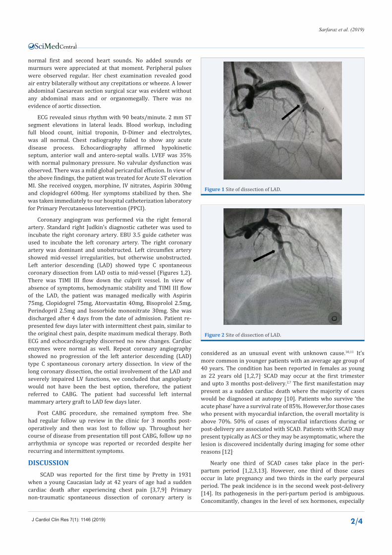
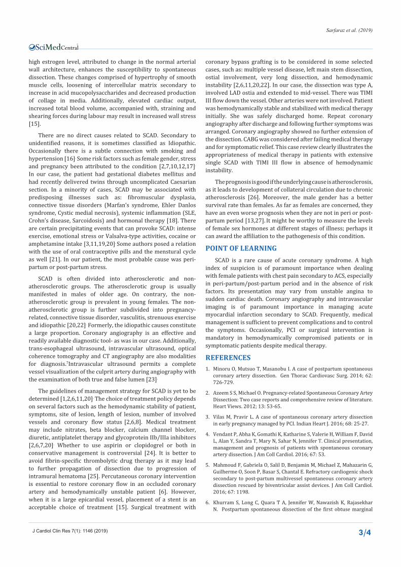
Coronary angiogram was performed via the right femoral artery. Standard right Judkin’s diagnostic catheter was used to incubate the right coronary artery. EBU 3.5 guide catheter was used to incubate the left coronary artery. The right coronary artery was dominant and unobstructed. Left circumflex artery showed mid-vessel irregularities, but otherwise unobstructed. Left anterior descending (LAD) showed type C spontaneous coronary dissection from LAD ostia to mid-vessel (Figures 1,2). There was TIMI III flow down the culprit vessel. In view of absence of symptoms, hemodynamic stability and TIMI III flow of the LAD, the patient was managed medically with Aspirin 75mg, Clopidogrel 75mg, Atorvastatin 40mg, Bisoprolol 2.5mg, Perindopril 2.5mg and Isosorbide mononitrate 30mg. She was discharged after 4 days from the date of admission. Patient re-presented few days later with intermittent chest pain, similar to the original chest pain, despite maximum medical therapy. Both ECG and echocardiography discerned no new changes. Cardiac enzymes were normal as well. Repeat coronary angiography showed no progression of the left anterior descending (LAD) type C spontaneous coronary artery dissection. In view of the long coronary dissection, the ostial involvement of the LAD and severely impaired LV functions, we concluded that angioplasty would not have been the best option, therefore, the patient referred to CABG. The patient had successful left internal mammary artery graft to LAD few days later.
Post CABG procedure, she remained symptom free. She had regular follow up review in the clinic for 3 months post-operatively and then was lost to follow up. Throughout her course of disease from presentation till post CABG, follow up no arrhythmia or syncope was reported or recorded despite her recurring and intermittent symptoms.
DISCUSSION SCAD was reported for the first time by Pretty in 1931
when a young Caucasian lady at 42 years of age had a sudden cardiac death after experiencing chest pain [3,7,9]. Primary non-traumatic spontaneous dissection of coronary artery is
considered as an unusual event with unknown cause.10,11 It’s more common in younger patients with an average age group of 40 years. The condition has been reported in females as young as 22 years old [1,2,7]. SCAD may occur at the first trimester and upto 3 months post-delivery.2,7 The first manifestation may present as a sudden cardiac death where the majority of cases would be diagnosed at autopsy [10]. Patients who survive ‘the acute phase’ have a survival rate of 85%. However,for those cases who present with myocardial infarction, the overall mortality is above 70%. 50% of cases of myocardial infarctions during or post-delivery are associated with SCAD. Patients with SCAD may present typically as ACS or they may be asymptomatic, where the lesion is discovered incidentally during imaging for some other reasons [12].
Nearly one third of SCAD cases take place in the peri-partum period [1,2,3,13]. However, one third of those cases occur in late pregnancy and two thirds in the early perpeural period. The peak incidence is in the second week post-delivery [14]. Its pathogenesis in the peri-partum period is ambiguous. Concomitantly, changes in the level of sex hormones, especially
Figure 1 Site of dissection of LAD.
Figure 2 Site of dissection of LAD.
CentralBringing Excellence in Open Access
Sarfaraz et al. (2019)
3/4J Cardiol Clin Res 7(1): 1146 (2019)
high estrogen level, attributed to change in the normal arterial wall architecture, enhances the susceptibility to spontaneous dissection. These changes comprised of hypertrophy of smooth muscle cells, loosening of intercellular matrix secondary to increase in acid mucopolysaccharides and decreased production of collage in media. Additionally, elevated cardiac output, increased total blood volume, accompanied with, straining and shearing forces during labour may result in increased wall stress [15].
There are no direct causes related to SCAD. Secondary to unidentified reasons, it is sometimes classified as Idiopathic. Occasionally there is a subtle connection with smoking and hypertension [16]. Some risk factors such as female gender, stress and pregnancy been attributed to the condition [2,7,10,12,17]. In our case, the patient had gestational diabetes mellitus and had recently delivered twins through uncomplicated Caesarian section. In a minority of cases, SCAD may be associated with predisposing illnesses such as: fibromuscular dysplasia, connective tissue disorders (Marfan’s syndrome, Ehler Danlos syndrome, Cystic medial necrosis), systemic inflammation (SLE, Crohn’s disease, Sarcoidosis) and hormonal therapy [18]. There are certain precipitating events that can provoke SCAD: intense exercise, emotional stress or Valsalva-type activities, cocaine or amphetamine intake [3,11,19,20]. Some authors posed a relation with the use of oral contraceptive pills and the menstural cycle as well [21]. In our patient, the most probable cause was peri-partum or post-partum stress.
SCAD is often divided into atherosclerotic and non-atherosclerotic groups. The atherosclerotic group is usually manifested in males of older age. On contrary, the non-atherosclerotic group is prevalent in young females. The non-atherosclerotic group is further subdivided into pregnancy-related, connective tissue disorder, vasculitis, strenuous exercise and idiopathic [20,22]. Formerly, the idiopathic causes constitute a large proportion. Coronary angiography is an effective and readily available diagnostic tool- as was in our case. Additionally, trans-esophageal ultrasound, intravascular ultrasound, optical coherence tomography and CT angiography are also modalities for diagnosis.7Intravascular ultrasound permits a complete vessel visualization of the culprit artery during angiography with the examination of both true and false lumen [23].
The guidelines of management strategy for SCAD is yet to be determined [1,2,6,11,20]. The choice of treatment policy depends on several factors such as the hemodynamic stability of patient, symptoms, site of lesion, length of lesion, number of involved vessels and coronary flow status [2,6,8]. Medical treatment may include nitrates, beta blocker, calcium channel blocker, diuretic, antiplatelet therapy and glycoprotein IIb/IIIa inhibitors [2,6,7,20]. Whether to use aspirin or clopidogrel or both in conservative management is controversial [24]. It is better to avoid fibrin-specific thrombolytic drug therapy as it may lead to further propagation of dissection due to progression of intramural hematoma [25]. Percutaneous coronary intervention is essential to restore coronary flow in an occluded coronary artery and hemodynamically unstable patient [6]. However, when it is a large epicardial vessel, placement of a stent is an acceptable choice of treatment [15]. Surgical treatment with
coronary bypass grafting is to be considered in some selected cases, such as: multiple vessel disease, left main stem dissection, ostial involvement, very long dissection, and hemodynamic instability [2,6,11,20,22]. In our case, the dissection was type A, involved LAD ostia and extended to mid-vessel. There was TIMI III flow down the vessel. Other arteries were not involved. Patient was hemodynamically stable and stabilized with medical therapy initially. She was safely discharged home. Repeat coronary angiography after discharge and following further symptoms was arranged. Coronary angiography showed no further extension of the dissection. CABG was considered after failing medical therapy and for symptomatic relief. This case review clearly illustrates the appropriateness of medical therapy in patients with extensive single SCAD with TIMI III flow in absence of hemodynamic instability.
The prognosis is good if the underlying cause is atherosclerosis, as it leads to development of collateral circulation due to chronic atherosclerosis [26]. Moreover, the male gender has a better survival rate than females. As far as females are concerned, they have an even worse prognosis when they are not in peri or post-partum period [13,27]. It might be worthy to measure the levels of female sex hormones at different stages of illness; perhaps it can award the affiliation to the pathogenesis of this condition.
POINT OF LEARNINGSCAD is a rare cause of acute coronary syndrome. A high
index of suspicion is of paramount importance when dealing with female patients with chest pain secondary to ACS, especially in peri-partum/post-partum period and in the absence of risk factors. Its presentation may vary from unstable angina to sudden cardiac death. Coronary angiography and intravascular imaging is of paramount importance in managing acute myocardial infarction secondary to SCAD. Frequently, medical management is sufficient to prevent complications and to control the symptoms. Occasionally, PCI or surgical intervention is mandatory in hemodynamically compromised patients or in symptomatic patients despite medical therapy.
REFERENCES1. Minoru O, Mutsuo T, Masanobu I. A case of postpartum spontaneous
coronary artery dissection. Gen Thorac Cardiovasc Surg. 2014; 62: 726-729.
2. Azeem S S, Michael O. Pregnancy-related Spontaneous Coronary Artey Dissection: Two case reports and comprehensive review of literature. Heart Views. 2012; 13: 53-65.
3. Vilas M, Pravir L. A case of spontaneous coronary artery dissection in early pregnancy managed by PCI. Indian Heart J. 2016; 68: 25-27.
4. Vendant P, Abha K, Gomathi K, Katharine S, Valerie H, William F, David L, Alan Y, Sandra T, Mary N, Sahar N, Jennifer T. Clinical presentation, management and prognosis of patients with spontaneous coronary artery dissection. J Am Coll Cardiol. 2016; 67: 53.
5. Mahmoud F, Gabriela O, Salil D, Benjamin M, Michael Z, Mahazarin G, Guilherme O, Soon P, Basar S, Chantal E. Refractory cardiogenic shock secondary to post-partum multivessel spontaneous coronary artery dissection rescued by biventricular assist devices. J Am Coll Cardiol. 2016; 67: 1198.
6. Khurram S, Long C, Quara T A, Jennifer W, Nawazish K, Rajasekhar N. Postpartum spontaneous dissection of the first obtuse marginal
CentralBringing Excellence in Open Access
Sarfaraz et al. (2019)
4/4J Cardiol Clin Res 7(1): 1146 (2019)
Sarfaraz M, Alhajiri A, Hasan SR (2019) Extensive Post-Partum Spontaneous Coronary Artery Dissection: Is Medical Therapy Safe? An Experience from Middle Eastern District General Hospital. J Cardiol Clin Res 7(1): 1146.
Cite this article
branch of the left circumflex coronary artery causing acute coronary syndrome: a case report and literature review. J Med case rep. 2013; 7: 82.
7. Jatinder S P, Lawrence J, William A M. Spontaneous coronary artery dissection as a cause of sudden cardiac death in the peripartum period. BMJ Case Reports. 2010.
8. Marysia S T, Sharonne N H, Sridevi R P, Robert D S, Amir L, Ryan J L, et al. Clinical features, management and prognosis of spontaneous coronary artery dissection. Circulation. 2012; 126: 579-588.
9. Pretty H C. Dissecting aneurysm of coronary artery in a woman aged 42. BMJ. 1931; 1: 667.
10. Branden V D, Bruggeling W A, Corbeij H M. Spontaneous coronary artery dissection in the postpartum period. Neth Heart J. 2008; 16: 412-414.
11. Massimo D M, Daniela C, Daniela F. Spontaneous coronary artery dissection in a young woman resolved with conservative strategy. A case report. Journal of Cardiology Cases. 2016; 14: 59-61.
12. Liang J J, Skalski J H, Mankad R. Spontaneous coronary artery dissection: is there a metabolic association. Perfusion. 2013; 28: 457-458.
13. De Maio S J Jr, Kinsella S H, Silverman M E. Clinical course and longterm prognosis of spontaneous coronary artery dissection. Am J Cardiol. 1989; 64: 471-474.
14. Koul A K, Hollander G, Moskovits N, Frankel R, Herrera L, Shani J. Coronary artery dissection during pregnancy and the postpartum period: two cases and revision of the literature. Catheter CardiovascInterv. 2001; 52: 88-94.
15. Tanis W, Stell P R, Pijlman A H, Kirkels J H, Peters R H J, Dc Man F H. Spontaneous coronary artery dissection: current insights and therapy. Neth Heart J. 2008; 16: 344-349.
16. Dhawan R, Singh G, Fesniak H. Spontaneous coronary artery dissection: the clinical spectrum. Angiology 2002; 53: 89-93.
17. Tsuyoshi N, Atsushi T, Yuichi O, Fukuda S, Takashi A. Spontaneous
coronary artery dissection in patients with acute coronary syndrome. J Am Coll Cardiol. 2013; 61.
18. Fernando A, Teresa B. Spontaneous coronary artery dissection. Novel diagnostic insights from large series of patients. Circ Cardiovasc Interv. 2014; 7: 638-641.
19. Henkin S, Negrotto S M, Tweet M S. Spontaneous coronary artery dissection and its association with heritable connective tissue disorders. Heart Jun. 2016; 102: 876-881.
20. Amelia Y, Jacqqueline S. Spontaneous coronary artery dissection-Areview. Cardiovasc Diagn Ther. 2015; 5: 37-48.
21. Slight R, Behranwala AA, Nzewi O, Sivaprakasam R, Brackenbury E, Mankand P. Spontaneous coronary artery dissection: a report of two cases occurring during mensturation. NZ Med J. 2003; 116: 44-47.
22. Saranya B, Parichart J, Mahek S, Prasit P, Harish RSR. Spontaneous coronary artey dissection following a preeclampsia pregnancy. Int J Cardiol. 2014; 173: 3-4.
23. Manuel P, Jorge S, Vera L, Jamie D, Miguel M, Nieves G, Pilar J, Javier E, Camino B, Rosana H, Carlos M, Fernando A. Combined use of OCT and IVUS in spontaneous coronary artery dissection. JACC: Cardiovascular Imaging. 2013; 6: 830-832.
24. Maeder M, Ammann P, Angehrn W, Rickli H. Idiopathic spontaneous coronary artery dissection: incidence, diagnosis and treatment. Int J Card. 2005; 101: 363-369.
25. Buys E M, Suttorp M J, Morshuis W J, Plokker H W. Extension of a spontaneous coronary artery dissection due to thrombolytic therapy. Cathet Cardiovasc Diagn. 1994; 33: 157-160.
26. Celik S K, Saggan A, Altintig A, Yuksel M, Akin M, Kultursay H. Primary spontaneous coronary artery dissection in atherosclerotic patient. Report of nine cases with review of the pertinent literature. Eur J Cardiothorac Surg. 2001; 20: 573-576.
27. Kamineni R, Sadhu S, Alpert J S. Spontaneous coronary artery dissection: report of two cases and a 50 years review of the literature. Cardiol Rev. 2002; 10: 279-284.