Case Report Combined Awake Craniotomy with Endoscopic...
4
Hindawi Publishing Corporation Case Reports in Medicine Volume 2013, Article ID 401359, 3 pages http://dx.doi.org/10.1155/2013/401359 Case Report Combined Awake Craniotomy with Endoscopic Port Surgery for Resection of a Deep-Seated Temporal Lobe Glioma: A Case Report Lance Bodily, 1 Arlan H. Mintz, 2 and Johnathan Engh 1 1 Department of Neurological Surgery, University of Pittsburgh School of Medicine, UPMC Presbyterian, Suite B-400, 200 Lothrop Street, Pittsburgh, PA 15213, USA 2 Capital Insititute for Neurosciences, Two Capital Way, Suite 456, Pennington, NJ 08534, USA Correspondence should be addressed to Johnathan Engh; [email protected] Received 24 December 2012; Accepted 13 March 2013 Academic Editor: Aaron S. Dumont Copyright © 2013 Lance Bodily et al. is is an open access article distributed under the Creative Commons Attribution License, which permits unrestricted use, distribution, and reproduction in any medium, provided the original work is properly cited. e authors describe the combination of awake craniotomy and minimally invasive endoscopic port surgery to resect a high-grade glioma located near eloquent structures of the temporal lobe. Combined minimally invasive techniques such as these may facilitate deep tumor resection within eloquent regions of the brain, allowing minimum white matter dissection. Technical aspects of this procedure, a case outcome involving this technique, and the direction of further investigations for the utility of these techniques are discussed. 1. Introduction Awake craniotomy with cortical mapping has served as the gold standard for functional preservation in brain tumor surgery since its introduction over half a century ago [1]. For the resection of tumors near the motor cortex or language pathway areas, this technique can help maximize the volume of tumor removal, while minimizing the risk of iatrogenic injury [2–4]. For deep-seated brain tumors, resection using a min- imally invasive cylindrical retractor has the potential to minimize damage to the surrounding white matter incurred as a result of the surgical channel into the tumor. is approach, initially championed by Kelly [5–7], has been modified into an even less invasive endoscopic port surgery in more recent years. Endoscopic port surgery (EPS) has been described for the removal of both intraventricular and intraparenchymal tumors [8–11]. In this paper, the authors describe the combination of awake craniotomy with electrophysiological brain mapping and EPS to resect a glioblastoma of the dominant temporal lobe. e case is an example of a deep-seated, eloquent tumor for which the combination of the two techniques seemed intuitive in order to facilitate a favorable outcome. 2. Case Report 2.1. Patient Presentation. A 63-year-old, right-handed man presented with a 6-month history of gait deterioration, requiring a walker. For the 3-4 weeks prior to presentation, he had also experienced a new onset of memory difficulties and subtle trouble with word finding, as well as mild headaches and declining vision. Of note, the patient had a history of spinal cerebellar atrophy (SCA). e patient’s neurologic exam was remarkable for mild dysmetria, dysdiadochokine- sia, and a positive Romberg sign, in addition to subtle fluent dysphasia and memory dysfunction. An MRI scan revealed a leſt temporal lobe mass mea- suring 3.5 cm × 2 cm abutting the temporal horn of the lateral ventricle (Figure 1). Notable T2 signal prolongation was observed, extending from the ventricle to more anterior and superior portions of the temporal lobe. Mass effect was present, with mild effacement of the ventricle and minimal midline shiſt. Radiological signs secondary to the SCA were also observed, including cerebellar atrophy with enlargement of the foramina of Luschka bilaterally, as well as a fourth ventricular enlargement. A craniotomy for tumor resection was recommended to the patient in order to achieve a tissue diagnosis, alleviate
Transcript of Case Report Combined Awake Craniotomy with Endoscopic...
Hindawi Publishing CorporationCase Reports in MedicineVolume 2013, Article ID 401359, 3 pageshttp://dx.doi.org/10.1155/2013/401359
Case ReportCombined Awake Craniotomy with Endoscopic PortSurgery for Resection of a Deep-Seated Temporal Lobe Glioma:A Case Report
Lance Bodily,1 Arlan H. Mintz,2 and Johnathan Engh1
1 Department of Neurological Surgery, University of Pittsburgh School of Medicine, UPMC Presbyterian, Suite B-400,200 Lothrop Street, Pittsburgh, PA 15213, USA
2Capital Insititute for Neurosciences, Two Capital Way, Suite 456, Pennington, NJ 08534, USA
Correspondence should be addressed to Johnathan Engh; [email protected]
Received 24 December 2012; Accepted 13 March 2013
Academic Editor: Aaron S. Dumont
Copyright © 2013 Lance Bodily et al. This is an open access article distributed under the Creative Commons Attribution License,which permits unrestricted use, distribution, and reproduction in any medium, provided the original work is properly cited.
The authors describe the combination of awake craniotomy and minimally invasive endoscopic port surgery to resect a high-gradeglioma located near eloquent structures of the temporal lobe. Combined minimally invasive techniques such as these may facilitatedeep tumor resection within eloquent regions of the brain, allowing minimum white matter dissection. Technical aspects of thisprocedure, a case outcome involving this technique, and the direction of further investigations for the utility of these techniquesare discussed.
1. Introduction
Awake craniotomy with cortical mapping has served as thegold standard for functional preservation in brain tumorsurgery since its introduction over half a century ago [1]. Forthe resection of tumors near the motor cortex or languagepathway areas, this technique can help maximize the volumeof tumor removal, while minimizing the risk of iatrogenicinjury [2–4].
For deep-seated brain tumors, resection using a min-imally invasive cylindrical retractor has the potential tominimize damage to the surrounding white matter incurredas a result of the surgical channel into the tumor. Thisapproach, initially championed by Kelly [5–7], has beenmodified into an even less invasive endoscopic port surgeryin more recent years. Endoscopic port surgery (EPS) hasbeen described for the removal of both intraventricular andintraparenchymal tumors [8–11].
In this paper, the authors describe the combination ofawake craniotomy with electrophysiological brain mappingand EPS to resect a glioblastoma of the dominant temporallobe.The case is an example of a deep-seated, eloquent tumorfor which the combination of the two techniques seemedintuitive in order to facilitate a favorable outcome.
2. Case Report
2.1. Patient Presentation. A 63-year-old, right-handed manpresented with a 6-month history of gait deterioration,requiring a walker. For the 3-4 weeks prior to presentation, hehad also experienced a new onset of memory difficulties andsubtle trouble with word finding, as well as mild headachesand declining vision. Of note, the patient had a historyof spinal cerebellar atrophy (SCA). The patient’s neurologicexam was remarkable for mild dysmetria, dysdiadochokine-sia, and a positive Romberg sign, in addition to subtle fluentdysphasia and memory dysfunction.
An MRI scan revealed a left temporal lobe mass mea-suring 3.5 cm × 2 cm abutting the temporal horn of thelateral ventricle (Figure 1). Notable T2 signal prolongationwas observed, extending from the ventricle to more anteriorand superior portions of the temporal lobe. Mass effect waspresent, with mild effacement of the ventricle and minimalmidline shift. Radiological signs secondary to the SCA werealso observed, including cerebellar atrophy with enlargementof the foramina of Luschka bilaterally, as well as a fourthventricular enlargement.
A craniotomy for tumor resection was recommended tothe patient in order to achieve a tissue diagnosis, alleviate
2 Case Reports in Medicine
(a) (b) (c)
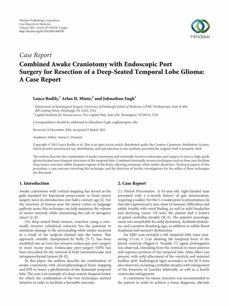
Figure 1: Preoperative and postoperative images. (a) Preresection T1 axial MRI with contrast showing a temporal lesion with an estimated5.0 cubic centimeter volume. (b) Immediate postresection T1 axial MRI with contrast showing a slight amount of residual enhancementat the anteromedial margin of the resection cavity. (Yellow arrow indicates residual tumor area not present in (c)). (c) The correspondingnoncontrast enhanced T1 MRI image taken at the same time demonstrates that the posteromedial and lateral hyperintensities are in factblood products and postoperative change rather than gross residual tumor. A 93.3% volumetric resection was achieved.
(a) (b)
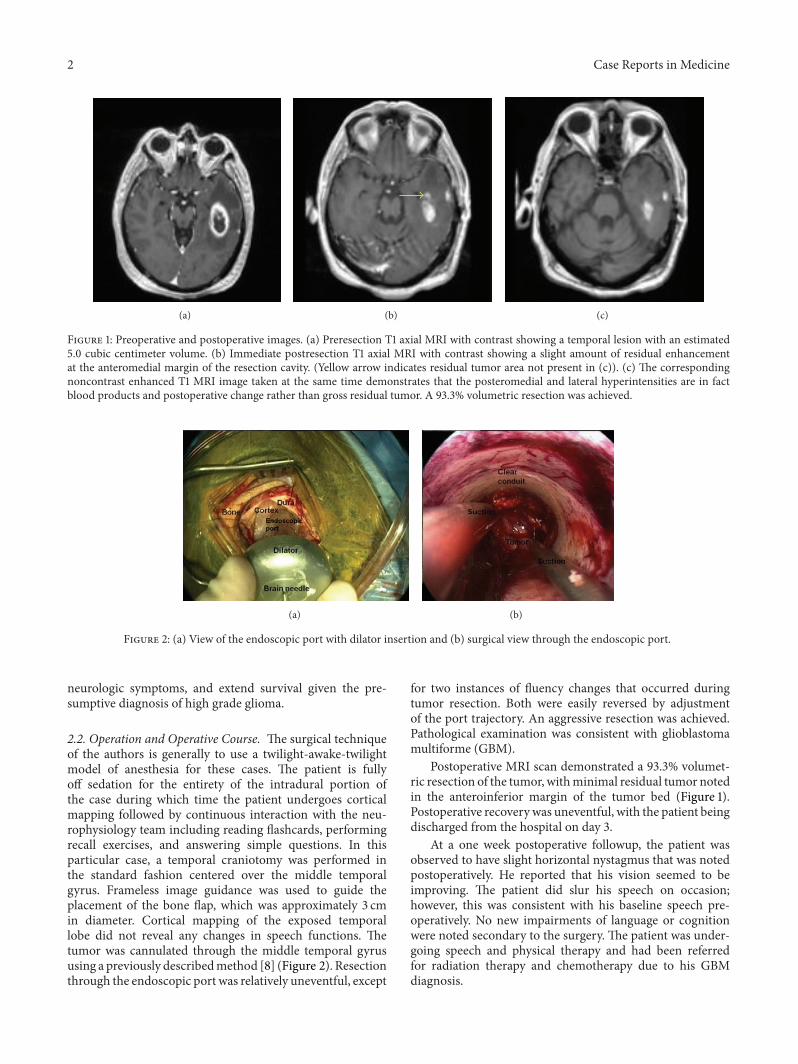
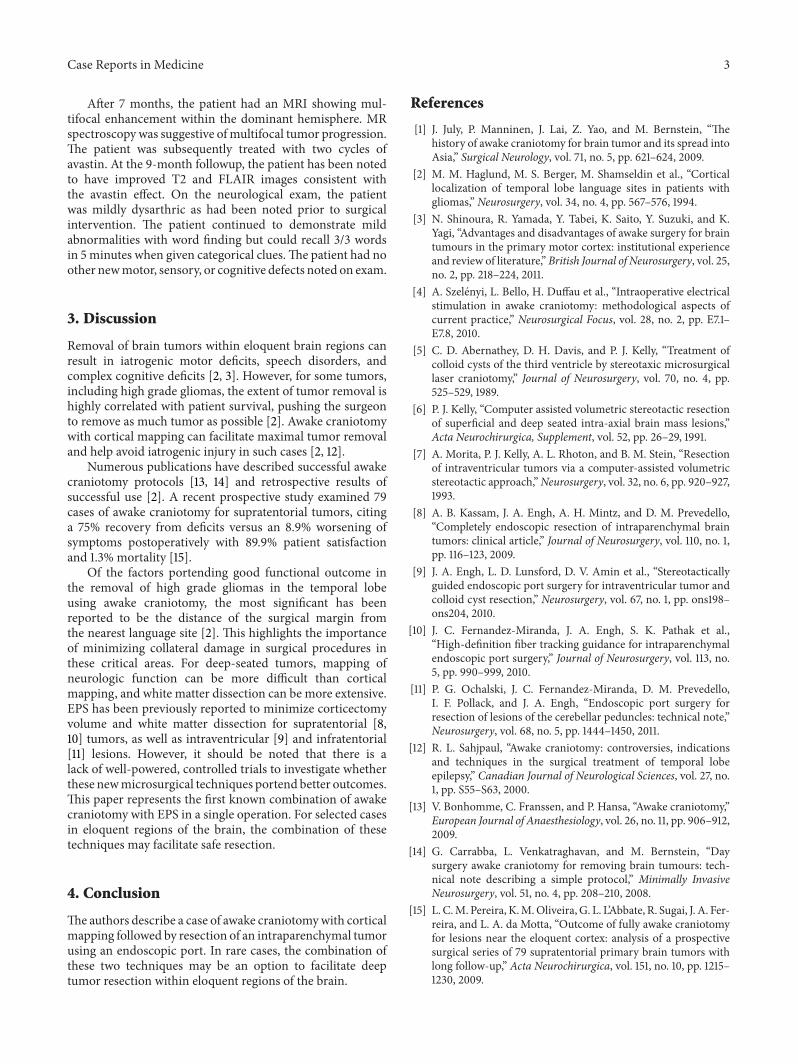
Figure 2: (a) View of the endoscopic port with dilator insertion and (b) surgical view through the endoscopic port.
neurologic symptoms, and extend survival given the pre-sumptive diagnosis of high grade glioma.
2.2. Operation and Operative Course. The surgical techniqueof the authors is generally to use a twilight-awake-twilightmodel of anesthesia for these cases. The patient is fullyoff sedation for the entirety of the intradural portion ofthe case during which time the patient undergoes corticalmapping followed by continuous interaction with the neu-rophysiology team including reading flashcards, performingrecall exercises, and answering simple questions. In thisparticular case, a temporal craniotomy was performed inthe standard fashion centered over the middle temporalgyrus. Frameless image guidance was used to guide theplacement of the bone flap, which was approximately 3 cmin diameter. Cortical mapping of the exposed temporallobe did not reveal any changes in speech functions. Thetumor was cannulated through the middle temporal gyrususing a previously describedmethod [8] (Figure 2). Resectionthrough the endoscopic port was relatively uneventful, except
for two instances of fluency changes that occurred duringtumor resection. Both were easily reversed by adjustmentof the port trajectory. An aggressive resection was achieved.Pathological examination was consistent with glioblastomamultiforme (GBM).
Postoperative MRI scan demonstrated a 93.3% volumet-ric resection of the tumor, withminimal residual tumor notedin the anteroinferior margin of the tumor bed (Figure 1).Postoperative recoverywas uneventful, with the patient beingdischarged from the hospital on day 3.
At a one week postoperative followup, the patient wasobserved to have slight horizontal nystagmus that was notedpostoperatively. He reported that his vision seemed to beimproving. The patient did slur his speech on occasion;however, this was consistent with his baseline speech pre-operatively. No new impairments of language or cognitionwere noted secondary to the surgery. The patient was under-going speech and physical therapy and had been referredfor radiation therapy and chemotherapy due to his GBMdiagnosis.
Case Reports in Medicine 3
After 7 months, the patient had an MRI showing mul-tifocal enhancement within the dominant hemisphere. MRspectroscopy was suggestive ofmultifocal tumor progression.The patient was subsequently treated with two cycles ofavastin. At the 9-month followup, the patient has been notedto have improved T2 and FLAIR images consistent withthe avastin effect. On the neurological exam, the patientwas mildly dysarthric as had been noted prior to surgicalintervention. The patient continued to demonstrate mildabnormalities with word finding but could recall 3/3 wordsin 5 minutes when given categorical clues.The patient had noother newmotor, sensory, or cognitive defects noted on exam.
3. Discussion
Removal of brain tumors within eloquent brain regions canresult in iatrogenic motor deficits, speech disorders, andcomplex cognitive deficits [2, 3]. However, for some tumors,including high grade gliomas, the extent of tumor removal ishighly correlated with patient survival, pushing the surgeonto remove as much tumor as possible [2]. Awake craniotomywith cortical mapping can facilitate maximal tumor removaland help avoid iatrogenic injury in such cases [2, 12].
Numerous publications have described successful awakecraniotomy protocols [13, 14] and retrospective results ofsuccessful use [2]. A recent prospective study examined 79cases of awake craniotomy for supratentorial tumors, citinga 75% recovery from deficits versus an 8.9% worsening ofsymptoms postoperatively with 89.9% patient satisfactionand 1.3% mortality [15].
Of the factors portending good functional outcome inthe removal of high grade gliomas in the temporal lobeusing awake craniotomy, the most significant has beenreported to be the distance of the surgical margin fromthe nearest language site [2]. This highlights the importanceof minimizing collateral damage in surgical procedures inthese critical areas. For deep-seated tumors, mapping ofneurologic function can be more difficult than corticalmapping, and white matter dissection can be more extensive.EPS has been previously reported to minimize corticectomyvolume and white matter dissection for supratentorial [8,10] tumors, as well as intraventricular [9] and infratentorial[11] lesions. However, it should be noted that there is alack of well-powered, controlled trials to investigate whetherthese newmicrosurgical techniques portend better outcomes.This paper represents the first known combination of awakecraniotomy with EPS in a single operation. For selected casesin eloquent regions of the brain, the combination of thesetechniques may facilitate safe resection.
4. Conclusion
Theauthors describe a case of awake craniotomywith corticalmapping followed by resection of an intraparenchymal tumorusing an endoscopic port. In rare cases, the combination ofthese two techniques may be an option to facilitate deeptumor resection within eloquent regions of the brain.
References
[1] J. July, P. Manninen, J. Lai, Z. Yao, and M. Bernstein, “Thehistory of awake craniotomy for brain tumor and its spread intoAsia,” Surgical Neurology, vol. 71, no. 5, pp. 621–624, 2009.
[2] M. M. Haglund, M. S. Berger, M. Shamseldin et al., “Corticallocalization of temporal lobe language sites in patients withgliomas,” Neurosurgery, vol. 34, no. 4, pp. 567–576, 1994.
[3] N. Shinoura, R. Yamada, Y. Tabei, K. Saito, Y. Suzuki, and K.Yagi, “Advantages and disadvantages of awake surgery for braintumours in the primary motor cortex: institutional experienceand review of literature,” British Journal of Neurosurgery, vol. 25,no. 2, pp. 218–224, 2011.
[4] A. Szelenyi, L. Bello, H. Duffau et al., “Intraoperative electricalstimulation in awake craniotomy: methodological aspects ofcurrent practice,” Neurosurgical Focus, vol. 28, no. 2, pp. E7.1–E7.8, 2010.
[5] C. D. Abernathey, D. H. Davis, and P. J. Kelly, “Treatment ofcolloid cysts of the third ventricle by stereotaxic microsurgicallaser craniotomy,” Journal of Neurosurgery, vol. 70, no. 4, pp.525–529, 1989.
[6] P. J. Kelly, “Computer assisted volumetric stereotactic resectionof superficial and deep seated intra-axial brain mass lesions,”Acta Neurochirurgica, Supplement, vol. 52, pp. 26–29, 1991.
[7] A. Morita, P. J. Kelly, A. L. Rhoton, and B. M. Stein, “Resectionof intraventricular tumors via a computer-assisted volumetricstereotactic approach,”Neurosurgery, vol. 32, no. 6, pp. 920–927,1993.
[8] A. B. Kassam, J. A. Engh, A. H. Mintz, and D. M. Prevedello,“Completely endoscopic resection of intraparenchymal braintumors: clinical article,” Journal of Neurosurgery, vol. 110, no. 1,pp. 116–123, 2009.
[9] J. A. Engh, L. D. Lunsford, D. V. Amin et al., “Stereotacticallyguided endoscopic port surgery for intraventricular tumor andcolloid cyst resection,” Neurosurgery, vol. 67, no. 1, pp. ons198–ons204, 2010.
[10] J. C. Fernandez-Miranda, J. A. Engh, S. K. Pathak et al.,“High-definition fiber tracking guidance for intraparenchymalendoscopic port surgery,” Journal of Neurosurgery, vol. 113, no.5, pp. 990–999, 2010.
[11] P. G. Ochalski, J. C. Fernandez-Miranda, D. M. Prevedello,I. F. Pollack, and J. A. Engh, “Endoscopic port surgery forresection of lesions of the cerebellar peduncles: technical note,”Neurosurgery, vol. 68, no. 5, pp. 1444–1450, 2011.
[12] R. L. Sahjpaul, “Awake craniotomy: controversies, indicationsand techniques in the surgical treatment of temporal lobeepilepsy,” Canadian Journal of Neurological Sciences, vol. 27, no.1, pp. S55–S63, 2000.
[13] V. Bonhomme, C. Franssen, and P. Hansa, “Awake craniotomy,”European Journal of Anaesthesiology, vol. 26, no. 11, pp. 906–912,2009.
[14] G. Carrabba, L. Venkatraghavan, and M. Bernstein, “Daysurgery awake craniotomy for removing brain tumours: tech-nical note describing a simple protocol,” Minimally InvasiveNeurosurgery, vol. 51, no. 4, pp. 208–210, 2008.
[15] L. C.M. Pereira, K.M.Oliveira, G. L. L’Abbate, R. Sugai, J. A. Fer-reira, and L. A. da Motta, “Outcome of fully awake craniotomyfor lesions near the eloquent cortex: analysis of a prospectivesurgical series of 79 supratentorial primary brain tumors withlong follow-up,” Acta Neurochirurgica, vol. 151, no. 10, pp. 1215–1230, 2009.

Submit your manuscripts athttp://www.hindawi.com
Stem CellsInternational
Hindawi Publishing Corporationhttp://www.hindawi.com Volume 2014
Hindawi Publishing Corporationhttp://www.hindawi.com Volume 2014
MEDIATORSINFLAMMATION
of
Hindawi Publishing Corporationhttp://www.hindawi.com Volume 2014
Behavioural Neurology
EndocrinologyInternational Journal of
Hindawi Publishing Corporationhttp://www.hindawi.com Volume 2014
Hindawi Publishing Corporationhttp://www.hindawi.com Volume 2014
Disease Markers
Hindawi Publishing Corporationhttp://www.hindawi.com Volume 2014
BioMed Research International
OncologyJournal of
Hindawi Publishing Corporationhttp://www.hindawi.com Volume 2014
Hindawi Publishing Corporationhttp://www.hindawi.com Volume 2014
Oxidative Medicine and Cellular Longevity
Hindawi Publishing Corporationhttp://www.hindawi.com Volume 2014
PPAR Research
The Scientific World JournalHindawi Publishing Corporation http://www.hindawi.com Volume 2014
Immunology ResearchHindawi Publishing Corporationhttp://www.hindawi.com Volume 2014
Journal of
ObesityJournal of
Hindawi Publishing Corporationhttp://www.hindawi.com Volume 2014
Hindawi Publishing Corporationhttp://www.hindawi.com Volume 2014
Computational and Mathematical Methods in Medicine
OphthalmologyJournal of
Hindawi Publishing Corporationhttp://www.hindawi.com Volume 2014
Diabetes ResearchJournal of
Hindawi Publishing Corporationhttp://www.hindawi.com Volume 2014
Hindawi Publishing Corporationhttp://www.hindawi.com Volume 2014
Research and TreatmentAIDS
Hindawi Publishing Corporationhttp://www.hindawi.com Volume 2014
Gastroenterology Research and Practice
Hindawi Publishing Corporationhttp://www.hindawi.com Volume 2014
Parkinson’s Disease
Evidence-Based Complementary and Alternative Medicine
Volume 2014Hindawi Publishing Corporationhttp://www.hindawi.com