Case Report An 80-Year-Old Man with Inferior Wall ...
5
1052 J Med Assoc Thai Vol. 99 No. 9 2016 Case Report J Med Assoc Thai 2016; 99 (9): 1052-6 Full text. e-Journal: http://www.jmatonline.com Correspondence to: Krittayaphong R, Division of Cardiology, Department of Medicine Faculty of Medicine Siriraj Hospital, Mahidol University, 2 Wanglang Road, Bangkoknoi, Bangkok 10700, Thailand. Phone: +66-2-4196104, Fax: +66-2-4127412 E-mail: [email protected] An 80-Year-Old Man with Inferior Wall Myocardial Infarction and Cardiogenic Shock: Case Report Korakoth Towashiraporn MD*, Decho Jakrapanichakul MD**, Wanchai Wongkornrat MD***, Rungroj Krittayaphong MD** * Her Majesty Cardiac Center, Siriraj Hospital, Mahidol University, Bangkok, Thailand ** Division of Cardiology, Department of Medicine, Faculty of Medicine Siriraj Hospital, Mahidol University, Bangkok, Thailand *** Division of Cardiothoracic Surgery, Department of Surgery, Faculty of Medicine Siriraj Hospital, Mahidol University, Bangkok, Thailand We report a case of ruptured head of posteromedial papillary muscle and prolapse of anterior mitral valve leaflet that caused severe mitral regurgitation (MR) and cardiogenic shock as a complication of inferior wall myocardial infarction in an 80-year-old man. Emergency coronary angiogram revealed thrombotic occlusion at proximal right coronary artery. Transesophageal echocardiogram revealed ruptured head of posteromedial papillary muscle and prolapse of anterior mitral valve leaflet causing severe MR. After percutaneous coronary intervention with bare metal stent, the patient underwent mitral valve repair with saphenous vein graft to the posterolateral branch of the right coronary artery. The patient was discharged from hospital on day 14 of admission. Keywords: Complication of inferior wall myocardial infarction, Ruptured head of papillary muscle Ruptured papillary muscle resulting in acute severe mitral regurgitation (MR) is infrequent, and often fatal mechanical complication of acute myocardial infarction (1-4) . In acute severe MR, regurgitant volume in a noncompliant left atrium results in a marked increase in left atrial pressure (5) . This increase in pressure progresses into the pulmonary capillaries (6) , causing acute pulmonary edema, decreased cardiac output, abrupt hemodynamic instability, and cardiogenic shock (7) , all of which results in multi-organ failure and death. Two papillary muscles (posteromedial and anterolateral) that are attached to the mitral valve leaflets via the chordae tendinae act to preserve mitral valvular competence (1,8) . The anterolateral papillary muscle is supplied by both the first diagonal branch, which originates from left anterior descending artery (LAD), and the obtuse marginal branch, which originates from the left circumflex artery (LCX) (9) . The posteromedial papillary muscle is supplied by only the right coronary artery (RCA) (1) . Rupture of the posteromedial papillary muscle is more frequently seen than rupture of the anterolateral papillary muscle, due to its single blood supply system (6,7) . Patients with ruptured papillary muscle typically present with inferior ST-segment elevated myocardial infarction (STEMI) and cardiogenic shock (2) . On physical examination, if the left atrium (LA) and left ventricle (LV) become close to a common chamber due to acute severe MR, any observed murmur may shorten or disappear. There are no obvious physical signs or findings in acute severe MR, such as apical displacement. Echocardiography is a useful imaging modality in diagnosis of ruptured papillary muscle, with a diagnostic sensitivity of 65 to 85% by transthoracic echocardiogram (TTE) (8) , and improved sensitivity to 95 to 100% by transesophageal echocardiogram (TEE) (10) . Direct visualization of the rupture is necessary for definitive diagnosis (1) . TTE may reveal normal LV size, different from chronic severe MR (5,6) and regional wall motion abnormalities of ventricular function due to myocardial ischemia (5) with good overall LV systolic function. From a study by Nishimura et al (11) , mean right ventricular systolic pressure (RVSP) was 55 mmHg and mean pulmonary capillary wedge pressure (PCWP) was 21 mmHg. Conservative treatment was found to be associated with poor clinical outcomes (2,12) . Medical treatment is only a bridging therapy to surgical correction. Gold et al (13) reported a 50% mortality rate within 24 hours in patients who did not undergo
Transcript of Case Report An 80-Year-Old Man with Inferior Wall ...

1052 J Med Assoc Thai Vol. 99 No. 9 2016
Case Report
J Med Assoc Thai 2016; 99 (9): 1052-6Full text. e-Journal: http://www.jmatonline.com
Correspondence to:Krittayaphong R, Division of Cardiology, Department of Medicine Faculty of Medicine Siriraj Hospital, Mahidol University, 2 Wanglang Road, Bangkoknoi, Bangkok 10700, Thailand.Phone: +66-2-4196104, Fax: +66-2-4127412E-mail: [email protected]
An 80-Year-Old Man with Inferior Wall Myocardial Infarction and Cardiogenic Shock: Case Report
Korakoth Towashiraporn MD*, Decho Jakrapanichakul MD**, Wanchai Wongkornrat MD***, Rungroj Krittayaphong MD**
* Her Majesty Cardiac Center, Siriraj Hospital, Mahidol University, Bangkok, Thailand** Division of Cardiology, Department of Medicine, Faculty of Medicine Siriraj Hospital, Mahidol University, Bangkok, Thailand
*** Division of Cardiothoracic Surgery, Department of Surgery, Faculty of Medicine Siriraj Hospital, Mahidol University, Bangkok, Thailand
We report a case of ruptured head of posteromedial papillary muscle and prolapse of anterior mitral valve leaflet that caused severe mitral regurgitation (MR) and cardiogenic shock as a complication of inferior wall myocardial infarction in an 80-year-old man. Emergency coronary angiogram revealed thrombotic occlusion at proximal right coronary artery. Transesophageal echocardiogram revealed ruptured head of posteromedial papillary muscle and prolapse of anterior mitral valve leaflet causing severe MR. After percutaneous coronary intervention with bare metal stent, the patient underwent mitral valve repair with saphenous vein graft to the posterolateral branch of the right coronary artery. The patient was discharged from hospital on day 14 of admission.
Keywords: Complication of inferior wall myocardial infarction, Ruptured head of papillary muscle
Ruptured papillary muscle resulting in acute severe mitral regurgitation (MR) is infrequent, and often fatal mechanical complication of acute myocardial infarction(1-4). In acute severe MR, regurgitant volume in a noncompliant left atrium results in a marked increase in left atrial pressure(5). This increase in pressure progresses into the pulmonary capillaries(6), causing acute pulmonary edema, decreased cardiac output, abrupt hemodynamic instability, and cardiogenic shock(7), all of which results in multi-organ failure and death. Two papillary muscles (posteromedial and anterolateral) that are attached to the mitral valve leaflets via the chordae tendinae act to preserve mitral valvular competence(1,8). The anterolateral papillary muscle is supplied by both the first diagonal branch, which originates from left anterior descending artery (LAD), and the obtuse marginal branch, which originates from the left circumflex artery (LCX)(9). The posteromedial papillary muscle is supplied by only the right coronary artery (RCA)(1). Rupture of the posteromedial papillary muscle is more frequently seen than rupture of the anterolateral papillary muscle, due to its single blood supply
system(6,7). Patients with ruptured papillary muscle typically present with inferior ST-segment elevated myocardial infarction (STEMI) and cardiogenic shock(2). On physical examination, if the left atrium (LA) and left ventricle (LV) become close to a common chamber due to acute severe MR, any observed murmur may shorten or disappear. There are no obvious physical signs or findings in acute severe MR, such as apical displacement. Echocardiography is a useful imaging modality in diagnosis of ruptured papillary muscle, with a diagnostic sensitivity of 65 to 85% by transthoracic echocardiogram (TTE)(8), and improved sensitivity to 95 to 100% by transesophageal echocardiogram (TEE)(10). Direct visualization of the rupture is necessary for definitive diagnosis(1). TTE may reveal normal LV size, different from chronic severe MR(5,6) and regional wall motion abnormalities of ventricular function due to myocardial ischemia(5) with good overall LV systolic function. From a study by Nishimura et al(11), mean right ventricular systolic pressure (RVSP) was 55 mmHg and mean pulmonary capillary wedge pressure (PCWP) was 21 mmHg. Conservative treatment was found to be associated with poor clinical outcomes(2,12). Medical treatment is only a bridging therapy to surgical correction. Gold et al(13) reported a 50% mortality rate within 24 hours in patients who did not undergo
J Med Assoc Thai Vol. 99 No. 9 2016 1053
surgery. Intra-aortic balloon pump (IABP) was reported to be useful for stabilizing patients by reducing after load and improving cardiac output(13). Emergency surgery (i.e., mitral valve surgery with or without coronary artery bypass grafting [CABG]) remains the treatment of choice for ruptured papillary muscle(14). Although surgery is the treatment of choice, the morbidity and mortality rates of surgical treatment remain high. Lorusso et al(14) reported a 30-day mortality rate of 26.9% and Kishon et al(15) reported a 7-year postoperative survival rate of 64%.
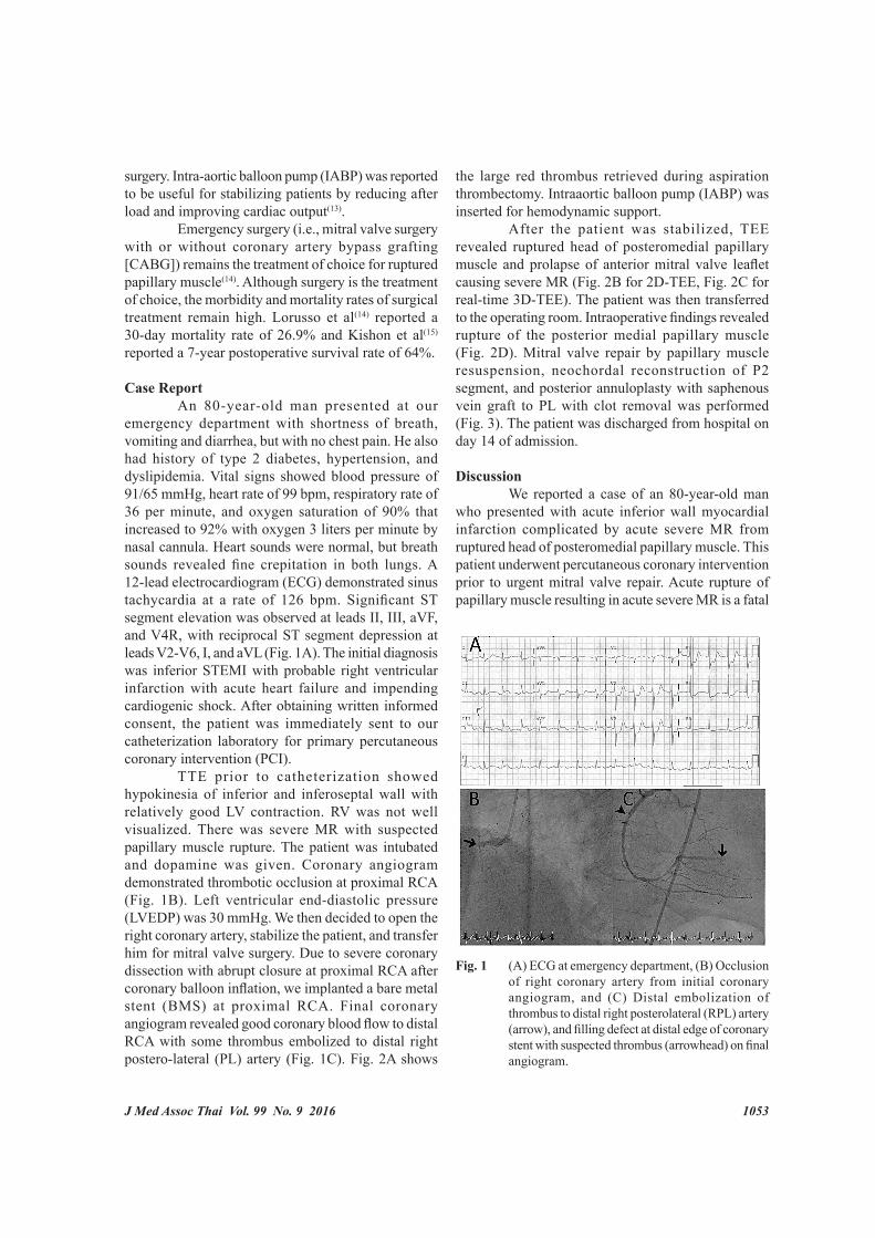
Case Report An 80-year-old man presented at our emergency department with shortness of breath, vomiting and diarrhea, but with no chest pain. He also had history of type 2 diabetes, hypertension, and dyslipidemia. Vital signs showed blood pressure of 91/65 mmHg, heart rate of 99 bpm, respiratory rate of 36 per minute, and oxygen saturation of 90% that increased to 92% with oxygen 3 liters per minute by nasal cannula. Heart sounds were normal, but breath sounds revealed fine crepitation in both lungs. A 12-lead electrocardiogram (ECG) demonstrated sinus tachycardia at a rate of 126 bpm. Significant ST segment elevation was observed at leads II, III, aVF, and V4R, with reciprocal ST segment depression at leads V2-V6, I, and aVL (Fig. 1A). The initial diagnosis was inferior STEMI with probable right ventricular infarction with acute heart failure and impending cardiogenic shock. After obtaining written informed consent, the patient was immediately sent to our catheterization laboratory for primary percutaneous coronary intervention (PCI). TTE prior to catheterization showed hypokinesia of inferior and inferoseptal wall with relatively good LV contraction. RV was not well visualized. There was severe MR with suspected papillary muscle rupture. The patient was intubated and dopamine was given. Coronary angiogram demonstrated thrombotic occlusion at proximal RCA (Fig. 1B). Left ventricular end-diastolic pressure (LVEDP) was 30 mmHg. We then decided to open the right coronary artery, stabilize the patient, and transfer him for mitral valve surgery. Due to severe coronary dissection with abrupt closure at proximal RCA after coronary balloon inflation, we implanted a bare metal stent (BMS) at proximal RCA. Final coronary angiogram revealed good coronary blood flow to distal RCA with some thrombus embolized to distal right postero-lateral (PL) artery (Fig. 1C). Fig. 2A shows
the large red thrombus retrieved during aspiration thrombectomy. Intraaortic balloon pump (IABP) was inserted for hemodynamic support. After the patient was stabilized, TEE revealed ruptured head of posteromedial papillary muscle and prolapse of anterior mitral valve leaflet causing severe MR (Fig. 2B for 2D-TEE, Fig. 2C for real-time 3D-TEE). The patient was then transferred to the operating room. Intraoperative findings revealed rupture of the posterior medial papillary muscle (Fig. 2D). Mitral valve repair by papillary muscle resuspension, neochordal reconstruction of P2 segment, and posterior annuloplasty with saphenous vein graft to PL with clot removal was performed (Fig. 3). The patient was discharged from hospital on day 14 of admission.
Discussion We reported a case of an 80-year-old man who presented with acute inferior wall myocardial infarction complicated by acute severe MR from ruptured head of posteromedial papillary muscle. This patient underwent percutaneous coronary intervention prior to urgent mitral valve repair. Acute rupture of papillary muscle resulting in acute severe MR is a fatal
Fig. 1 (A) ECG at emergency department, (B) Occlusion of right coronary artery from initial coronary angiogram, and (C) Distal embolization of thrombus to distal right posterolateral (RPL) artery (arrow), and filling defect at distal edge of coronary stent with suspected thrombus (arrowhead) on final angiogram.

1054 J Med Assoc Thai Vol. 99 No. 9 2016
mechanical complication of myocardial infarction. In untreated patients, the mortality rate was reported to be 80%(2). Rupture of the posteromedial papillary muscle was found more frequently (in about 75% of
cases) than rupture of the anterolateral papillary muscle(9). The posteromedial papillary muscle is supplied by only the RCA(1); whereas, the anterolateral papillary muscle is supplied by both the LAD and the LCX(9). Direct visualization of the rupture by echocardiogram is necessary for definitive diagnosis. TTE is often useful, but Kishon et al reported that the ruptured papillary muscle could only be identified in 45% of patients(16). As a result, TEE is necessary for diagnosing ruptured papillary muscle in some patients. Emergency surgical correction is the treatment of choice for ruptured papillary muscle(14). Repair of the mitral apparatus rather than valve replacement was found to be associated with better long-term and short-term survival(17,18). A previous report(18) showed a survival rate at 4 years of 89% in patients with mitral valve repair and of 59% in patients with mitral valve replacement. However, mitral valve repair is difficult and sometimes not possible in high-risk patients(19). Recent myocardial infarction and emergency procedure were associated with operative mortality rate in combined CABG concomitant with mitral valve surgery(20). Therefore, percutaneous coronary intervention of the infarct-related artery to salvage myocardial ischemia, with immediate subsequent mitral valve repair may be the best treatment strategy in selected patient.
Conclusion A high index of clinical suspicion and a skilled heart team are essential for the management of acute severe MR with inferior STEMI. Definite surgical treatment should not be delayed.
What is already known on this topic? Myocardial infarction complicated by rupture of the posteromedial papillary muscle is an almost always fatal condition. Emergency surgery is the treatment of choice. However, even with surgery, morbidity and mortality rates are still high.
What this study adds? PCI of infarct-related artery to salvage myocardial ischemia with immediate surgical correction of mitral valve regurgitation may result in reduced morbidity and mortality rate in patients with acute severe MR-complicated myocardial infarction.
Potential conflicts of interest None.
Fig. 2 (A) Large red thrombus retrieved from right coronary artery, (B) Ruptured posteromedial papillary muscle (white arrow) and prolapse of anterior mitral valve leaflet (white arrowhead), eccentric jet of severe mitral regurgitation (black arrow) on color Doppler of 2D TEE, (C) Ruptured posteromedial papillary muscle (arrow and arrowhead) from left ventricle (LV) view and from left atrium (LA) view of 3D TEE, and (D) Ruptured posteromedial papillary muscle (arrowhead) with LA retraction (arrow) in the operative field.
Fig. 3 MV repair by papillary muscle resuspension, neochordal reconstruction of P2 and posterior annuloplasty with saphenous vein graft to PL with clot removal.

J Med Assoc Thai Vol. 99 No. 9 2016 1055
References1. Fradley MG, Picard MH. Rupture of the
posteromedial papillary muscle leading to partial flail of the anterior mitral leaflet. Circulation 2011; 123: 1044-5.
2. Thompson CR, Buller CE, Sleeper LA, Antonelli TA, Webb JG, Jaber WA, et al. Cardiogenic shock due to acute severe mitral regurgitation complicating acute myocardial infarction: a report from the SHOCK Trial Registry. SHould we use emergently revascularize Occluded Coronaries in cardiogenic shocK? J Am Coll Cardiol 2000; 36: 1104-9.
3. Birnbaum Y, Chamoun AJ, Conti VR, Uretsky BF. Mitral regurgitation following acute myocardial infarction. Coron Artery Dis 2002; 13: 337-44.
4. Hillis GS, Moller JE, Pellikka PA, Bell MR, Casaclang-Verzosa GC, Oh JK. Prognostic significance of echocardiographically defined mitral regurgitation early after acute myocardial infarction. Am Heart J 2005; 150: 1268-75.
5. Mokadam NA, Stout KK, Verrier ED. Management of acute regurgitation in left-sided cardiac valves. Tex Heart Inst J 2011; 38: 9-19.
6. Jeresaty RM. Left ventricular function in acute non-ischaemic mitral regurgitation. Eur Heart J 1991; 12 (Suppl B): 19-21.
7. Jain SK, Larsen TR, Darda S, Saba S, David S. A forgotten devil; Rupture of mitral valve papillary muscle. Am J Case Rep 2013; 14: 38-42.
8. Czarnecki A, Thakrar A, Fang T, Lytwyn M, Ahmadie R, Pascoe E, et al. Acute severe mitral regurgitation: consideration of papillary muscle architecture. Cardiovasc Ultrasound 2008; 6: 5.
9. Jayawardena S, Renteria AS, Burzyantseva O, Lokesh G, Thelusmond L. Anterolateral papillary muscle rupture caused by myocardial infarction: A case report. Cases J 2008; 1: 172.
10. Hozumi T, Yoshikawa J, Yoshida K, Yamaura Y, Akasaka T, Shakudo M. Direct visualization of ruptured chordae tendineae by transesophageal two-dimensional echocardiography. J Am Coll Cardiol 1990; 16: 1315-9.
11. Nishimura RA, Schaff HV, Shub C, Gersh BJ,
Edwards WD, Tajik AJ. Papillary muscle rupture complicating acute myocardial infarction: analysis of 17 patients. Am J Cardiol 1983; 51: 373-7.
12. Sanders RJ, Neubuerger KT, Ravin A. Rupture of papillary muscles: occurrence of rupture of the posterior muscle in posterior myocardial infarction. Dis Chest 1957; 31: 316-23.
13. Gold HK, Leinbach RC, Sanders CA, Buckley MJ, Mundth ED, Austen WG. Intraaortic balloon pumping for ventricular septal defect or mitral regurgitation complicating acute myocardial infarction. Circulation 1973; 47: 1191-6.
14. Lorusso R, Gelsomino S, De Cicco G, Beghi C, Russo C, De Bonis M, et al. Mitral valve surgery in emergency for severe acute regurgitation: analysis of postoperative results from a multicentre study. Eur J Cardiothorac Surg 2008; 33: 573-82.
15. Kishon Y, Oh JK, Schaff HV, Mullany CJ, Tajik AJ, Gersh BJ. Mitral valve operation in postinfarction rupture of a papillary muscle: immediate results and long-term follow-up of 22 patients. Mayo Clin Proc 1992; 67: 1023-30.
16. Kishon Y, Iqbal A, Oh JK, Gersh BJ, Freeman WK, Seward JB, et al. Evolution of echocardiographic modalities in detection of postmyocardial infarction ventricular septal defect and papillary muscle rupture: study of 62 patients. Am Heart J 1993; 126: 667-75.
17. Kay GL, Kay JH, Zubiate P, Yokoyama T, Mendez M. Mitral valve repair for mitral regurgitation secondary to coronary artery disease. Circulation 1986; 74: I88-I98.
18. David TE, Ho WC. The effect of preservation of chordae tendineae on mitral valve replacement for postinfarction mitral regurgitation. Circulation 1986; 74: I116-I120.
19. David TE. Chordal preservation in mitral valve replacement. Operat Tech Thorac Cardiovasc Surg 1998; 3: 130-3.
20. Edwards FH, Peterson ED, Coombs LP, DeLong ER, Jamieson WR, Shroyer ALW, et al. Prediction of operative mortality after valve replacement surgery. J Am Coll Cardiol 2001; 37: 885-92.

1056 J Med Assoc Thai Vol. 99 No. 9 2016
ผูปวยชายที่มีอาการกลามเนื้อหัวใจดานลางตายเฉียบพลัน รวมกับภาวะช็อคจากหัวใจ
กรกฏ โตวชิราภรณ, เดโช จักราพานิชกุล, วันชัย วงศกรรัตน, รุงโรจน กฤตยพงษ
ผูปวยชายอายุ 80 ป มารับการรักษาดวยอาการกลามเน้ือหัวใจดานลางตายเฉียบพลัน รวมกับมีภาวะช็อคจากหัวใจ การตรวจคล่ืนสะทอนหวัใจผานหลอดอาหาร พบวามกีารฉกีขาดของ posteromedial papillary muscle ซึง่ทาํใหเกดิการลิน้หวัใจไมตรัลร่ัวแบบรุนแรงอยางเฉียบพลัน ผูปวยไดรับการสวนหัวใจอยางเรงดวน พบวามีการอุดก้ันของหลอดเลือดหัวใจดานขวาจากลิ่มเลือด ผูปวยจึงไดรับการถางขยายหลอดเลือดหัวใจดานขวาดวยขดลวดชนิดไมเคลือบยา และไดรับการผาตัดซอมแซมลิ้นหัวใจไมตรัล รวมกับผาตัดทําทางเบ่ียงเสนเลือดดานขวา ผูปวยสามารถกลับบานไดในวันท่ี 14 ของการรักษา