Case Report Alveolar rhabdomyosarcoma of … sinuses with metastasis to breast in a middle-aged...
6
Int J Clin Exp Pathol 2015;8(11):15316-15321 www.ijcep.com /ISSN:1936-2625/IJCEP0015989 Case Report Alveolar rhabdomyosarcoma of nasopharynx and paranasal sinuses with metastasis to breast in a middle-aged woman: a case report and literature review Hongmei Liu 1* , Wei Zhao 1* , Meijuan Huang 1,2 , Xiaojuan Zhou 1,2 , Youling Gong 1,2 , You Lu 1,2 1 Department of Thoracic Oncology, Cancer Center, West China Hospital, Medical School, Sichuan University, Chengdu, China; 2 State Key Laboratory of Biotherapy, West China Hospital, Medical School, Sichuan University, Chengdu, China. * Equal contributors. Received September 11, 2015; Accepted October 22, 2015; Epub November 1, 2015; Published November 15, 2015 Abstract: Alveolar rhabdomyosarcoma (ARMS) is a common soft tissue tumor in children which can rarely metas- tasize to the breast in adults. Here we report the rare case of a 42-year-old Asian woman, who was diagnosed with ARMS of the nasopharynx and paranasal sinuses, and got a complete remission (CR) after surgery and chemora- diotherapy. Then the patient relapsed in the unilateral breast seventeen months later. Histology and immunohis- tochemistry of the primary sites and the breast lesions, combined with FISH, have been performed to confirm the diagnosis of metastatic alveolar rhabdomyosarcoma. With a rational therapeutic regimen of surgery, chemotherapy and radiotherapy, the patient has got a complete remission again. Keywords: Breast metastasis, alveolar rhabdomyosarcoma, nasopharynx and paranasal sinuses Introduction Breast involvement from distant foci is seldom. The incidence of breast metastasis in malig- nancies has been reported accounting for only 0.5-2% [1, 2]. The most common metastatic diseases of the breast are malignant melano- mas, lymphoma and leukemia [1]. Rhab- domyosarcoma (RMS) is a very common soft tissue sarcoma in the pediatric age group, which most often occurs in the head and neck areas, the extremities and the urogenital organs [3]. Here we report a case of breast metastasis of alveolar RMS in a 42-year-old woman. Breast examination, imaging study, histopathological features combined gene test- ing are used to identify the diagnosis. To the best of our knowledge, it is the first case described that alveolar rhabdomyosarcoma of the nasopharynx and paranasal sinuses metas- tasize to the breast in middle-aged woman. Case report A 42-year-old woman was admitted to our Ear Nose Throat (ENT) department for a year histo- ry of nasal obstruction and recurrent epistaxis. The nasal endoscopic examination revealed the presence of a 10×10 mm mass in the naso- pharynx. And the head computed tomography (CT) scan found a destructive soft-tissue mass in the right ethmoid sinus and frontal sinus (Figure 1). There was also a palpable subman- dibular lymph node on the right side through neck examination. Endoscopic complete excision of the mass in the nasopharynx and paranasal sinuses was done for surgical treatment, and the right sub- mandibular lymphadenectomy was conducted at the same time. Histologic examination sho- wed sheets of small round cells arranged in an alveolar pattern. Immunohistochemical exami- nation of the mass and lymph node presented positive reaction for desmin, myogenin and CD- 56 (Figure 2), but negative reaction for EBER- ISH, PCK, EMA, synaptophysin and CD-99. Fluorescence in situ hybridization (FISH) analy- sis revealed the FOXO1 (FKHR) mutation. Investigations after the surgery included a full blood count, biochemical tests and PET-CT.
Transcript of Case Report Alveolar rhabdomyosarcoma of … sinuses with metastasis to breast in a middle-aged...

Int J Clin Exp Pathol 2015;8(11):15316-15321www.ijcep.com /ISSN:1936-2625/IJCEP0015989
Case ReportAlveolar rhabdomyosarcoma of nasopharynx and paranasal sinuses with metastasis to breast in a middle-aged woman: a case report and literature review
Hongmei Liu1*, Wei Zhao1*, Meijuan Huang1,2, Xiaojuan Zhou1,2, Youling Gong1,2, You Lu1,2
1Department of Thoracic Oncology, Cancer Center, West China Hospital, Medical School, Sichuan University, Chengdu, China; 2State Key Laboratory of Biotherapy, West China Hospital, Medical School, Sichuan University, Chengdu, China. *Equal contributors.
Received September 11, 2015; Accepted October 22, 2015; Epub November 1, 2015; Published November 15, 2015
Abstract: Alveolar rhabdomyosarcoma (ARMS) is a common soft tissue tumor in children which can rarely metas-tasize to the breast in adults. Here we report the rare case of a 42-year-old Asian woman, who was diagnosed with ARMS of the nasopharynx and paranasal sinuses, and got a complete remission (CR) after surgery and chemora-diotherapy. Then the patient relapsed in the unilateral breast seventeen months later. Histology and immunohis-tochemistry of the primary sites and the breast lesions, combined with FISH, have been performed to confirm the diagnosis of metastatic alveolar rhabdomyosarcoma. With a rational therapeutic regimen of surgery, chemotherapy and radiotherapy, the patient has got a complete remission again.
Keywords: Breast metastasis, alveolar rhabdomyosarcoma, nasopharynx and paranasal sinuses
Introduction
Breast involvement from distant foci is seldom. The incidence of breast metastasis in malig-nancies has been reported accounting for only 0.5-2% [1, 2]. The most common metastatic diseases of the breast are malignant melano-mas, lymphoma and leukemia [1]. Rhab- domyosarcoma (RMS) is a very common soft tissue sarcoma in the pediatric age group, which most often occurs in the head and neck areas, the extremities and the urogenital organs [3]. Here we report a case of breast metastasis of alveolar RMS in a 42-year-old woman. Breast examination, imaging study, histopathological features combined gene test-ing are used to identify the diagnosis. To the best of our knowledge, it is the first case described that alveolar rhabdomyosarcoma of the nasopharynx and paranasal sinuses metas-tasize to the breast in middle-aged woman.
Case report
A 42-year-old woman was admitted to our Ear Nose Throat (ENT) department for a year histo-
ry of nasal obstruction and recurrent epistaxis. The nasal endoscopic examination revealed the presence of a 10×10 mm mass in the naso-pharynx. And the head computed tomography (CT) scan found a destructive soft-tissue mass in the right ethmoid sinus and frontal sinus (Figure 1). There was also a palpable subman-dibular lymph node on the right side through neck examination.
Endoscopic complete excision of the mass in the nasopharynx and paranasal sinuses was done for surgical treatment, and the right sub-mandibular lymphadenectomy was conducted at the same time. Histologic examination sho- wed sheets of small round cells arranged in an alveolar pattern. Immunohistochemical exami-nation of the mass and lymph node presented positive reaction for desmin, myogenin and CD- 56 (Figure 2), but negative reaction for EBER- ISH, PCK, EMA, synaptophysin and CD-99. Fluorescence in situ hybridization (FISH) analy-sis revealed the FOXO1 (FKHR) mutation.
Investigations after the surgery included a full blood count, biochemical tests and PET-CT.

ARMS with metastasis to breast
15317 Int J Clin Exp Pathol 2015;8(11):15316-15321
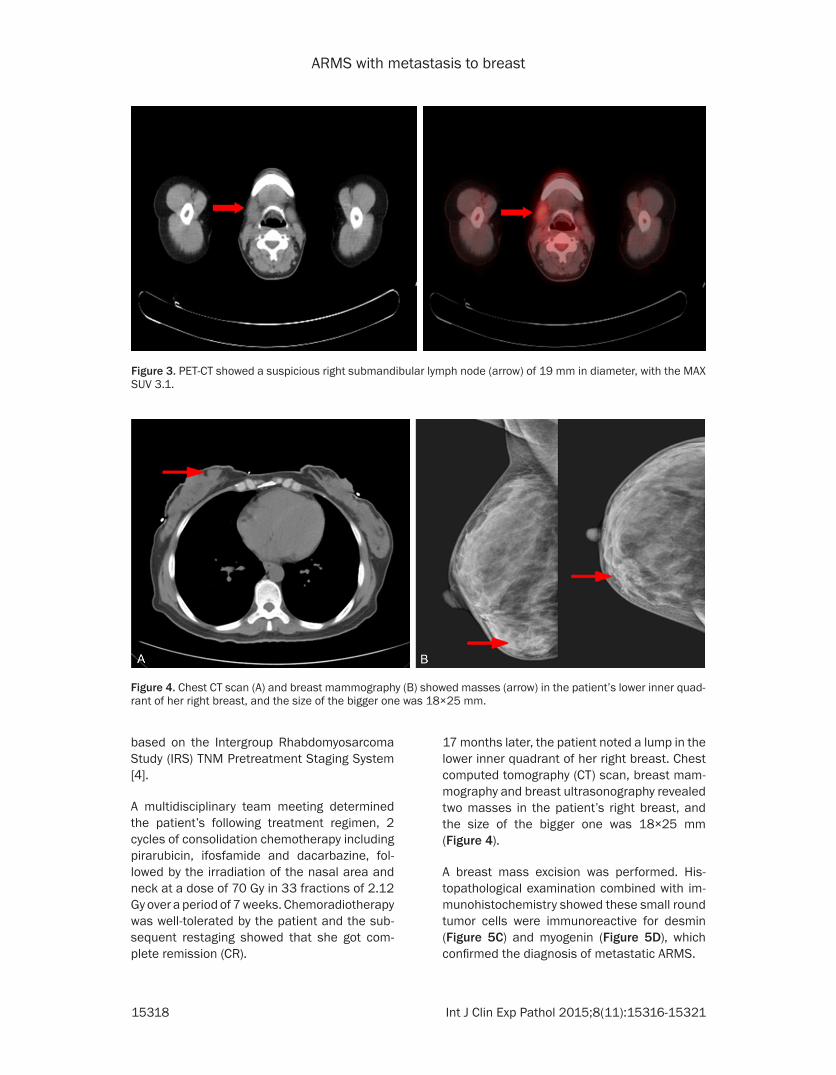
These were all normal except for a suspicious right submandibular lymph node of 19 mm in
diameter, with the MAX SUV 3.1 (Figure 3). It confirmed the diagnosis of a stage III ARMS
Figure 1. The head computed tomography (CT) scan showed a destructive soft-tissue mass (arrow) in the right eth-moid sinus and frontal sinus.
Figure 2. A. Histologic examination showed sheets of small round cells arranged in an alveolar pattern. (H&E, ×200). B. Immunostaining revealed positive result of desmin in the tumor cells. (×200) C. Immunostaining revealed posi-tive result of myogenin in the tumor cells. (×200) D. Immunostaining revealed positive result of CD-56 in the tumor cells. (×200).
ARMS with metastasis to breast
15318 Int J Clin Exp Pathol 2015;8(11):15316-15321
based on the Intergroup Rhabdomyosarcoma Study (IRS) TNM Pretreatment Staging System [4].
A multidisciplinary team meeting determined the patient’s following treatment regimen, 2 cycles of consolidation chemotherapy including pirarubicin, ifosfamide and dacarbazine, fol-lowed by the irradiation of the nasal area and neck at a dose of 70 Gy in 33 fractions of 2.12 Gy over a period of 7 weeks. Chemoradiotherapy was well-tolerated by the patient and the sub-sequent restaging showed that she got com-plete remission (CR).
17 months later, the patient noted a lump in the lower inner quadrant of her right breast. Chest computed tomography (CT) scan, breast mam-mography and breast ultrasonography revealed two masses in the patient’s right breast, and the size of the bigger one was 18×25 mm (Figure 4).
A breast mass excision was performed. His- topathological examination combined with im- munohistochemistry showed these small round tumor cells were immunoreactive for desmin (Figure 5C) and myogenin (Figure 5D), which confirmed the diagnosis of metastatic ARMS.
Figure 3. PET-CT showed a suspicious right submandibular lymph node (arrow) of 19 mm in diameter, with the MAX SUV 3.1.
Figure 4. Chest CT scan (A) and breast mammography (B) showed masses (arrow) in the patient’s lower inner quad-rant of her right breast, and the size of the bigger one was 18×25 mm.

ARMS with metastasis to breast
15319 Int J Clin Exp Pathol 2015;8(11):15316-15321
After the incomplete breast mass excision, she received 4 cycles of second-line chemotherapy, consisting of pirarubicin, cisplatin and dacarba-zine. And the right breast beam concurrent radiotherapy was performed, at a dose of 60 Gy in 30 fractions of 2 Gy, using sIMRT. After 8 months, Chest computed tomography (CT) scan and the breast ultrasonography showed no mass in her breasts. At the time of this paper written, the patient is still doing well and gets CR again since she was diagnosed with breast metastasis.
Discussion
RMS is the most common soft tissue sarcoma in childhood and adolescence, comprising 10-12% of all malignant solid tumors in chil-dren [5]. There are two major histological sub-types of RMS: alveolar rhabdomyosarcoma (ARMS) and embryonal rhabdomyosarcoma
(ERMS) [6]. Alveolar rhabdomyosarcoma (ARMS) is an aggressive myogenic childhood cancer, of which prognosis is poor [7]. For ARMS in adults, the survival probability is even worse, not to mention those with breast metas-tasis [8]. Because most patients with breast metastasis often have already got widely dis-seminated diseases, who relapsed only in the unilateral breast as our patient are extremely rare.
Lung is the most common metastatic site of RMS, and then followed by bone, bone marrow and liver [9]. Breast involvement from distant foci is seldom. The incidence of breast metas-tasis in malignancies has been reported accounting for only 0.5-2% [1, 2]. Of the 1,399 female RMS patients, according to Intergroup Rhabdomyosarcoma Study (IRS), there were 19 breast metastatic individuals except for 7 pri-mary breast RMS, comprising 1.4%. And the
Figure 5. A. Histologic examination showed sheets of small round cells arranged in an alveolar pattern, a few re-sidual breast ducts were presented on the up-left side. (H&E, ×200). B. Histologic examination showed nuclei with various morphological variations in these small round cells. (H&E, ×400). C. Immunostaining revealed positive result of desmin in the tumor cells. (×200). D. Immunostaining revealed positive result of myogenin in the tumor cells. (×200).
ARMS with metastasis to breast
15320 Int J Clin Exp Pathol 2015;8(11):15316-15321
most common primary RMS metastatic to breast is from extremities, followed by naso-pharynx & paranasal sinuses and trunk [10].
ARMS with metastasis to breast often occurred in the pubertal girls. Daniel M et al reported all of the 26 IRS cases were female with ages ranging from 11.5 to 20.2 years, and 24 patients of them were ARMS [10]. W. H. Kwan et al also said that female with an alveolar sub-type of RMS in the pubertal period may place a patient at higher risk of developing breast metastasis [11]. It is plausible that the increas-ing vascularity and rapidly growing mammary tissue during the pubertal development phase may be related with the preferential metastasis to breast, as most of these patients were younger women [12]. Yaren A. et al reported that a pregnant woman got metastatic rhabdo-myosarcoma to the breast, which further sup-ported that vascularity may be associated with the metastasis priority selection [13]. But the hypothesis above cannot be enough to explain why breast metastasis of ARMS happened in a 42-year-old woman in our case.
Histopathology manifested that the tumor cells were small and round cells which arranged in an alveolar pattern (Figures 2A and 5A). Its probable misdiagnosed as other malignancies, such as malignant lymphoma, leukemia, and small cell carcinoma, for the lack of specific characteristics [9, 13, 14]. In our case, the non-Hodgkin lymphoma was once considered when the naive lymphocyte was found at beginning. Fortunately, with the use of immunohistochem-istry (IHC) and FISH, final diagnosis of ARMS was made. RMS should be considered for this kind of histopathological manifestation, espe-cially in the children and teen.
The accurate diagnose is the fundament of subsequent treatments. As classification based on histopathology does not always allow accurate diagnoses for an unambiguous sub-classification, we combined IHC and FISH to distinguish ARMS from other soft tissue tumors and make a definite diagnosis. As it’s not easy to make an accurate diagnosis for breast metastasis, we distinguish it from other tumors clinically and histologically.
Desmin and myogenin are particularly useful in identifying tumors with myogenic differentia-tion [6]. Dias P. et al reported ARMS expressed
at least three folds more myogenin than ERMS averagely, which can be used for distinguishing between alveolar and embryonal RMS [15]. About 80% of ARMS are identified with chromo-somal translocations (t(1;13) (p36;q14) or (t(2;13) (q35;q14)), which generates PAX7-FOXO1 or PAX3-FOXO1 fusion genes [16-18]. The FOXO1 gene is also called FKHR previously. Our patient in the case found the FOXO1 (FKHR) mutation, which provided a strong and precise evidence to guarantee the accurate diagnosis of ARMS from ERMS and other tumors. The genetic testing may play a much more impor-tant role in the future and guide the clinical application of prospective targeted therapy.
ARMS with breast metastasis case is very uncommon, but we should also pay attention to the breast lumps with a previous history of malignant tumor. The routine breast examina-tions, including breast ultrasonography, Chest CT scan, mammography or MRI, should be sug-gested in the follow-up assessment. Perlet et al reported that contrast-enhanced MRI can be successful in the detection of malignant hyper-vascular tumors, with sensitivity up to 95% [19]. Gene detection in conjunction with histo-pathology and IHC can be successful to ascer-tain the diagnosis and assess the prognosis. With these methods above, an appropriate, rational and effective therapeutic schedule would be provided without delay.
Conclusion
Here we report the case of 42-year-old woman presenting with metastatic ARMS of nasophar-ynx and paranasal sinuses in breast, which is extremely rare in adults. Histology, IHC and FISH have been used to confirm the diagnosis. A comprehensive therapy of surgery, chemo-therapy and radiotherapy are performed to the patient. Although metastatic ARMS to the breast often presents a dismal prognosis, which has been described in most reports, the patient in our case has achieved complete remission (CR) and obtained a satisfactory outcome.
Acknowledgements
The authors would like to thank Dr. Hongying Zhang et al, Department of Pathology, West China Hospital, Medical School, Sichuan University, for assistance and advice on the his-topathological diagnosis.
ARMS with metastasis to breast
15321 Int J Clin Exp Pathol 2015;8(11):15316-15321
Disclosure of conflict of interest
None.
Address correspondence to: Dr. Meijuan Huang, Department of Thoracic Oncology, Cancer Center, West China Hospital, Medical School, Sichuan University, Chengdu 610041, China. Tel: +86 1 8980602026; Fax: +86 2885423571; E-mail: [email protected]
References
[1] Toombs BD and Kalisher L. Metastatic disease to the breast: clinical, pathologic, and radio-graphic features. AJR Am J Roentgenol 1977; 129: 673-676.
[2] Hajdu SI and Urban JA. Cancers metastatic to the breast. Cancer 1972; 29: 1691-1696.
[3] Li FP and Fraumeni JF Jr. Rhabdomyosarcoma in Children: Epidemiologic Study and Identification of a Familial Caneer Syndrome. J Natl Cancer Inst 1969; 43: 1365-1373.
[4] Pedrick TJ, Donaldson S and Cox R. Rhabdomyosarcoma: the Stanford experience using a TNM staging system. J Clin Oncol 1986; 4: 370-378.
[5] Binokay F, Soyupak SK, Inal M, Celiktas M, Akgul E and Aksungur E. Primary and meta-static rhabdomyosarcoma in the breast: report of two pediatric cases. Eur J Radiol 2003; 48: 282-284.
[6] Parham DM and Ellison DA. Rhabdomyo- sarcomas in adults and children: an update. Arch Pathol Lab Med 2006; 130: 1454-1465.
[7] Dagher R and Helman L. Rhabdomyosarcoma: an overview. Oncologist 1999; 4: 34-44.
[8] Sultan I, Qaddoumi I, Yaser S, Rodriguez-Galindo C and Ferrari A. Comparing adult and pediatric rhabdomyosarcoma in the surveil-lance, epidemiology and end results program, 1973 to 2005: an analysis of 2,600 patients. J Clin Oncol 2009; 27: 3391-3397.
[9] Tan GC, Shiran MS, Hayati AR, Sharifah NA, Nuru AS and Rohaizak M. Alveolar rhabdomyo-sarcoma of the left hand with bilateral breast metastases in an adolescent female. J Chin Med Assoc 2008; 71: 639-642.
[10] Hays DM, Donaldson SS, Shimada H, Crist WM, Newton WA Jr, Andrassy RJ, Wiener E, Green J, Triche T and Maurer HM. Primary and metastatic rhabdomyosarcoma in the breast: neoplasms of adolescent females, a report from the Intergroup Rhabdomyosarcoma Study. Med Pediatr Oncol 1997; 29: 181-189.
[11] Kwan WH, Choi PH, Li CK, Shing MK, Chik KW, Yuen P and Chow LT. Breast metastasis in ado-lescents with alveolar rhabdomyosarcoma of the extremities: report of two cases. Pediatr Hematol Oncol 1996; 13: 277-285.
[12] Persic M and Roberts JT. Alveolar rhabdomyo-sarcoma metastatic to the breast: long-term survivor. Clin Oncol 1999; 11: 417-418.
[13] Yaren A, Guclu A, Sen N, Erdem E and Demirkan F. Breast metastasis in a pregnant woman with alveolar rhabdomyosarcoma of the upper ex-tremity. Eur J Obstet Gynecol Reprod Biol 2008; 140: 131-133.
[14] Nogi H, Kobayashi T, Kawase K, Tabei I, Toriumi Y, Suzuki M, Kawakami M, Morikawa T and Uchida K. Primary rhabdomyosarcoma of the breast in a 13-year-old girl: report of a case. Surg Today 2007; 37: 38-42.
[15] Dias P, Chen B, Dilday B, Palmer H, Hosoi H, Singh S, Wu C, Li X, Thompson J, Parham D, Qualman S and Houghton P. Strong immunos-taining for myogenin in rhabdomyosarcoma is significantly associated with tumors of the al-veolar subclass. Am J Pathol 2000; 156: 399-408.
[16] Davicioni E, Finckenstein FG, Shahbazian V, Buckley JD, Triche TJ and Anderson MJ. Identification of a PAX-FKHR gene expression signature that defines molecular classes and determines the prognosis of alveolar rhabdo-myosarcomas. Cancer Res 2006; 66: 6936-6946.
[17] Sorensen PH, Lynch JC, Qualman SJ, Tirabosco R, Lim JF, Maurer HM, Bridge JA, Crist WM, Triche TJ and Barr FG. PAX3-FKHR and PAX7-FKHR gene fusions are prognostic indicators in alveolar rhabdomyosarcoma: a report from the children’s oncology group. J Clin Oncol 2002; 20: 2672-2679.
[18] Williamson D, Missiaglia E, de Reyniès A, Pierron G, Thuille B, Palenzuela G, Thway K, Orbach D, Laé M, Fréneaux P, Pritchard-Jones K, Oberlin O, Shipley J and Delattre O. Fusion gene-negative alveolar rhabdomyosarcoma is clinically and molecularly indistinguishable from embryonal rhabdomyosarcoma. J Clin Oncol 2010; 28: 2151-2158.
[19] Perlet C, Sittek H, Forstpointner R, Kessler M and Reiser M. Metastases to the breast from rhabdomyosarcoma: appearances on MRI. Eur Radiol 1999; 9: 1113-1116.