Borangic c. falx dacica. i. propunere pentru o tipologie a armelor curbe dacice, nemvs, i, 1-2, 2006
REFERENCES
1. Hartley J. Acute cervical pain associated with retropharyngeal calcium deposit: a case report. J Bone Joint Surg [Am] 1964;46 :1453-1454
2. Sutro CJ. Calcification of the anterior atlanto-axial ligament as cause for painful swelling and for painful neck. Bull Hosp J Dis Orthop Inst 1967;28: 1-3
3. Newmark H, Forrester DM, Brown JC, Robinson A, Olken SM, Bledsoe R. Calcific tendinitis of the neck. Radiology 1978;128 :355-358
4. Hall FM, Docken WP, Curtis HW. Calcific tendinitis of the longus colli : diagnosed by CT. AJR 1986;147 :742-743
Central Pontine Myelinolysis: Report of a Case with Distinctive Appearance on MR Imaging
We report a case of central pontine myelinolysis (CPM) in which MR imaging showed a distinctive trident-shaped configuration of the pontine lesion not noted on previous reports [1, 2]. This pattern reflects the known pattern of pathologic involvement.
Case Report
A 53-year-old-alcoholic man was brought to the emergency department because of general debility. On admission his vital signs were within normal limits. A general physical examination revealed a thin , disheveled man, but was otherwise unremarkable. Neurologic examination revealed 4/5 strength , normal sensation, and normal deep tendon reflexes.
The serum Na+ was 104 mEq/1 and K+ was 2.9 mEq/1. The serum sodium was corrected to 130 during the next 24 hr by IV administration of normal saline. By the next day the patient felt stronger and generally improved, but during the next several days he became gradually disoriented. A CT scan done at that time showed only atrophy. The results of a lumbar puncture were normal.
The patient deteriorated further and became dysarthric and quadriplegic. A second CT scan on the 25th day of hospitalization revealed a low-density area in the basis pontis with sparing of the periphery (Fig. 1). No abnormality in the basal ganglia was evident. The appearance was thought to be most compatible with CPM.
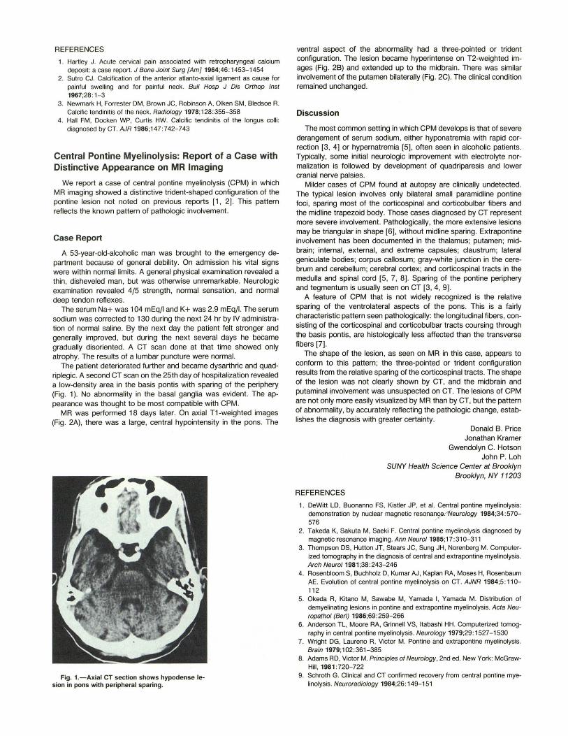
MR was performed 18 days later. On axial T1-weighted images (Fig . 2A), there was a large, central hYPointensity in the pons. The
Fig. 1.-Axial CT section shows hypodense lesion in pons with peripheral sparing.
ventral aspect of the abnormality had a three-pointed or trident configuration . The lesion became hyperintense on T2-weighted images (Fig. 2B) and extended up to the midbrain. There was similar involvement of the putamen bilaterally (Fig. 2C). The clinical condition remained unchanged.
Discussion
The most common setting in which CPM develops is that of severe derangement of serum sodium, either hyponatremia with rapid correction [3, 4] or hypernatremia [5], often seen in alcoholic patients. Typically, some initial neurologic improvement with electroly1e normalization is followed by development of quadriparesis and lower cranial nerve palsies.
Milder cases of CPM found at autopsy are clinically undetected. The typical lesion involves only bilateral small paramidline pontine foci, sparing most of the corticospinal and corticobulbar fibers and the midline trapezoid body. Those cases diagnosed by CT represent more severe involvement. Pathologically, the more extensive lesions may be triangular in shape [6], without midline sparing. Extrapontine involvement has been documented in the thalamus; putamen; midbrain; internal, external, and extreme capsules; claustrum; lateral geniculate bodies; corpus callosum; gray-white junction in the cerebrum and cerebellum; cerebral cortex; and corticospinal tracts in the medulla and spinal cord [5, 7, 8]. Sparing of the pontine periphery and tegmentum is usually seen on CT [3, 4, 9].
A feature of CPM that is not widely recognized is the relative sparing of the ventrolateral aspects of the pons. This is a fairly characteristic pattern seen pathologically: the longitudinal fibers , consisting of the corticospinal and corticobulbar tracts coursing through the basis pontis , are histologically less affected than the transverse fibers [7].
The shape of the lesion, as seen on MR in this case, appears to conform to this pattern; the three-pointed or trident configuration results from the relative sparing of the corticospinal tracts. The shape of the lesion was not clearly shown by CT, and the midbrain and putaminal involvement was unsuspected on CT. The lesions of CPM are not only more easily visualized by MR than by CT, but the pattern of abnormality, by accurately reflecting the pathologic change, establishes the diagnosis with greater certainty.
REFERENCES
Donald B. Price Jonathan Kramer
Gwendolyn C. Hotson John P. Loh
SUNY Health Science Center at Brooklyn Brooklyn, NY 11203
1. DeWitt LD, Buonanno FS, Kistler JP, et al. Central pontine myelinolysis: demonstration by nuclear magnetic resonancemeurology 1984;34:570-576 , c
2. Takeda K, Sakuta M, Saeki F. Central pontine myelinolysis diagnosed by magnetic resonance imaging. Ann Neurol 1985;17: 31 0-311
3. Thompson DS, Hutton JT, Stears JC, Sung JH, Norenberg M. Computerized tomography in the diagnosis of central and extrapontine myelinolysis. Arch Neuro/1981 ;38 :243-246
4. Rosenbloom S, Buchholz D, Kumar AJ, Kaplan RA, Moses H, Rosenbaum AE. Evolution of central pontine myelinolysis on CT. AJNR 1984;5: 11 0-112
5. Okeda R, Kitano M, Sawabe M, Yamada I, Yamada M. Distribution of demyelinating lesions in pontine and extrapontine myelinolysis. Acta Neuropathol (Berl) 1986;69 :259-266
6. Anderson TL, Moore RA, Grinnell VS, Itabashi HH. Computerized tomography in central pontine myelinolysis. Neurology 1979;29 :1527-1530
7. Wright DG, Laureno R, Victor M. Pontine and extrapontine myelinolysis. Brain 1979;102:361 - 385
8. Adams RD, Victor M. Principles of Neurology, 2nd ed. New York: McGrawHill , 1981 :720-722
9. Schroth G. Clinical and CT confirmed recovery from central pontine myelinolysis. Neuroradiology 1984;26: 149- 151
AJNR:8, May/June CORRESPONDENCE 577
A B c Fig. 2.-A, T1-weighted MR image at same level as Fig. 1. Trident-shaped hypointense area in pons. Ventrolateral sparing is in location of corticospinal
and corticobulbar tracts. B, T2-weighted image at same level as part A. C, T2-weighted image shows abnormal signal in right putamen. Similar involvement on left not seen on this slice because of head till.
MR Demonstration of Falx Ossification: Recognition and Differential Diagnosis
An area of high signal has been observed as an incidental finding on routine T1-weighted sequences of the brain . Correlation with CT scans and/or plain skull films revealed the source to be within areas of ossification of the falx cerebri. The marrow within this ossification may led to high signal , such as is seen within the diploic space of the calvaria and other marrow spaces of the body. Appreciation of this finding should prevent confusion with intracerebral hemorrhage and other cerebral diseases (i.e., lipomatous lesions) that may also present with bright signal on Tl-weighted sagittal images.
Case Report
A 64-year-old woman with a history of colonic carcinoma and radiation therapy for a pontine mass has been followed by MR since 1984. The sagittal Tl-weighted image has persistently shown a high frontal area of increased signal (Fig. lA, on next page), which becomes isointense on the T2-weighted sequence (Fig. 1 B, on next page). Correlation with CT has shown a densely mineralized falx in this area, which has not changed in appearance on sequential studies.
Discussion and Differential Diagnosis
It has previously been demonstrated, histologically , that mineralization of the falx cerebri in humans is caused by ossification and not just calcification. These areas of membranous bone formation are complete with marrow elements that may also be subject to metastatic or leukemic infiltrates [1) . It is expected, therefore, that marrow and fat elements within areas of dural ossification have high signal on T1-weighted sequences similar to that demonstrated within the diploic space. A similar high Tl-signal phenomenon has been observed within a densely mineralized osteoma that abutted the inner table. The lesion, which was hyperdense on CT, appeared very bright
on Tl-weighted sequences and essentially isointense to brain on T2-weighted images. The high signal on Tl-weighted spin-echo sequences is again likely to be related to marrow and/or fat elements. Osteomas, while commonly appearing very dense on CT, may consist of highly vascular, fatty , or hematopoetic elements pathologically. If the falx cerebri were calcified or ossified with compact bone, without marrow elements, one could predict a relative absence of signal on all MR sequences because of the paucity of mobile protons.
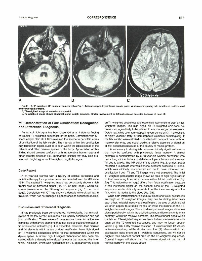
It is necessary to distinguish between clinically significant lesions that may be confused with physiologic falcial marrow. A clinical example is demonstrated by a 68-year-Old woman outpatient who had a long clinical history of definite multiple sclerosis and a recent fall due to ataxia. The MR study in this patient (Fig . 2, on next page) revealed a subacute interhemispheric subdural collection of blood, which was clinically unsuspected and could have mimicked falx ossification if both Tl and T2 images were not evaluated. The initial Tl -weighted parasagittal image shows an area of high signal similar to that emanating from fatty marrow within falcial ossification (Fig. 2A). This lesion (hemorrhage) differs from falcial ossification because it has increased signal on the second echo of the T2-weighted sequences and is distinctly separate from the linear low signal of the falx , which is medial to the blood (Fig. 2B).
While both interhemispheric subdural blood and falcial ossification are bright on Tl-weighted images, they can be distinguished from each other. In falcial marrow and ossification, the area of bright signal will often appear to straddle the falx or cross the midline on the Tl weighted coronal images. The spin-density coronal images show low signal peripherally, within the ossified cortex , and isointensity to brain centrally, within the marrow elements. The area of bright signal within the falx on Tl-weighted sequences tends to become isointense with brain on the T2-weighted sequences, and may no longer appear visible (Fig. 1 B). Fatty marrow has short Tl values and its T2 values, while relatively long, will be shorter than blood [2) . Marrow within falx ossification looks bright on Tl-weighted sequences, but will not be brighter than adjacent normal brain on the T2-weighted sequences. Coronal images will show that the marrow signal mirrors that of normal marrow in the diploic space.