
Case Report A Case of Thanatophoric Dysplasia Type I with...
5
Case Report A Case of Thanatophoric Dysplasia Type I with Fetal Hydrops in the First Trimester Giannina Calongos, 1 Masateru Hori, 1 Mai Ogino, 1 and Hideaki Sawai 2 1 Department of Obstetrics and Gynecology, Meiwa General Hospital, Nishinomiya 663-8186, Japan 2 Department of Obstetrics and Gynecology, Hyogo College of Medicine, Nishinomiya 663-8501, Japan Correspondence should be addressed to Giannina Calongos; [email protected] Received 6 January 2016; Revised 26 January 2016; Accepted 26 January 2016 Academic Editor: Giovanni Monni Copyright © 2016 Giannina Calongos et al. is is an open access article distributed under the Creative Commons Attribution License, which permits unrestricted use, distribution, and reproduction in any medium, provided the original work is properly cited. During a routine prenatal exam, a 36-year-old female in her third pregnancy was diagnosed with fetal hydrops at 11 weeks of gestation. e pregnancy was monitored with periodic ultrasounds; however, spontaneous resolution was not observed. Amniotic fluid examination at 16 weeks of gestation showed a normal karyotype; however, macrocephaly, a narrow thorax, and shortening of the long bones were observed on ultrasonography. With the strong suspicion of a fetal skeletal disease, specifically thanatophoric dysplasia (TD), and aſter extensive genetic counseling, termination of the pregnancy was performed per the parents’ wishes with mechanical cervical dilation and gemeprost (PGE1) administration. Following delivery, the fetus was found to have macrocephaly, a narrow bell-shaped thorax, and a protuberant abdomen, as well as curved long bones, H-shaped platyspondyly, and curved clavicles on skeletal radiography. As a result, the fetus was diagnosed with TD type I. is case illustrates that although TD is a rare disease, an accurate prenatal diagnosis can be made with the use of ultrasonography. 1. Introduction anatophoric dysplasia (TD) is a rare and lethal skeletal dysplasia with an estimated incidence of 1 in 20000 to 40000 births [1] and was first described by Maroteaux et al. in 1967 [2]. TD can be classified into two types: type I is characterized by micromelia with bowed femurs and, uncommonly, the presence of cloverleaf skull deformity; type II is characterized by micromelia with straight femurs and moderate to severe cloverleaf skull deformity [3]. A common feature to both types is the presence of a narrow thorax which causes respiratory failure shortly aſter birth [4, 5]. Fetal hydrops is defined as an abnormal fluid collection in two or more areas of the fetal body. It can be further classified as immune or nonimmune. Immune fetal hydrops develops due to fetal hemolysis secondary to incompatibility of maternal and fetal blood type. Nonimmune hydrops, on the other hand, can result from a large number of causes [6, 7]. However, 20–30% of hydrops is of unknown etiology. Here, we report a case of TD type I with fetal hydrops diagnosed in the first trimester and thereaſter shortening of the long bones and macrocephaly. Confirmation of the diagnosis was made by clinical examination and radiologic studies aſter delivery. 2. Case Presentation A 36-year-old female in her third pregnancy came to our hospital at 8 weeks of gestation for a routine prenatal check. e couple and their previous children’s past and family histories were unremarkable. Ultrasonographic examination at 8 weeks of gestation was unremarkable, with a crown-rump length (CRL) of 15.9 mm. At 11 weeks of gestation, although the CRL was 49.6 mm corresponding to gestational age, fetal hydrops was evident on ultrasonography (Figure 1(a)). Since screening for immune hydrops and congenital infections were negative, a transvaginal ultrasound was performed the following week for further evaluation. At 12 weeks of gestation, appropriate fetal growth was observed with a CRL of 60 mm; however, fetal hydrops was still present. e couple was counseled on the different etiologies of fetal hydrops; however, at this point the cause was not clear. Hindawi Publishing Corporation Case Reports in Obstetrics and Gynecology Volume 2016, Article ID 1821230, 4 pages http://dx.doi.org/10.1155/2016/1821230
Transcript of Case Report A Case of Thanatophoric Dysplasia Type I with...

Case ReportA Case of Thanatophoric Dysplasia Type I withFetal Hydrops in the First Trimester
Giannina Calongos,1 Masateru Hori,1 Mai Ogino,1 and Hideaki Sawai2
1Department of Obstetrics and Gynecology, Meiwa General Hospital, Nishinomiya 663-8186, Japan2Department of Obstetrics and Gynecology, Hyogo College of Medicine, Nishinomiya 663-8501, Japan
Correspondence should be addressed to Giannina Calongos; [email protected]
Received 6 January 2016; Revised 26 January 2016; Accepted 26 January 2016
Academic Editor: Giovanni Monni
Copyright © 2016 Giannina Calongos et al. This is an open access article distributed under the Creative Commons AttributionLicense, which permits unrestricted use, distribution, and reproduction in any medium, provided the original work is properlycited.
During a routine prenatal exam, a 36-year-old female in her third pregnancy was diagnosed with fetal hydrops at 11 weeks ofgestation. The pregnancy was monitored with periodic ultrasounds; however, spontaneous resolution was not observed. Amnioticfluid examination at 16 weeks of gestation showed a normal karyotype; however, macrocephaly, a narrow thorax, and shortening ofthe long bones were observed on ultrasonography. With the strong suspicion of a fetal skeletal disease, specifically thanatophoricdysplasia (TD), and after extensive genetic counseling, termination of the pregnancy was performed per the parents’ wishes withmechanical cervical dilation and gemeprost (PGE1) administration. Following delivery, the fetus was found to havemacrocephaly, anarrow bell-shaped thorax, and a protuberant abdomen, as well as curved long bones, H-shaped platyspondyly, and curved clavicleson skeletal radiography. As a result, the fetus was diagnosed with TD type I. This case illustrates that although TD is a rare disease,an accurate prenatal diagnosis can be made with the use of ultrasonography.
1. Introduction
Thanatophoric dysplasia (TD) is a rare and lethal skeletaldysplasia with an estimated incidence of 1 in 20000 to40000 births [1] and was first described by Maroteaux etal. in 1967 [2]. TD can be classified into two types: typeI is characterized by micromelia with bowed femurs and,uncommonly, the presence of cloverleaf skull deformity; typeII is characterized by micromelia with straight femurs andmoderate to severe cloverleaf skull deformity [3]. A commonfeature to both types is the presence of a narrow thorax whichcauses respiratory failure shortly after birth [4, 5].
Fetal hydrops is defined as an abnormal fluid collectionin two or more areas of the fetal body. It can be furtherclassified as immune or nonimmune. Immune fetal hydropsdevelops due to fetal hemolysis secondary to incompatibilityof maternal and fetal blood type. Nonimmune hydrops, onthe other hand, can result froma large number of causes [6, 7].However, 20–30% of hydrops is of unknown etiology.
Here, we report a case of TD type I with fetal hydropsdiagnosed in the first trimester and thereafter shortening
of the long bones and macrocephaly. Confirmation of thediagnosis was made by clinical examination and radiologicstudies after delivery.
2. Case Presentation
A 36-year-old female in her third pregnancy came to ourhospital at 8 weeks of gestation for a routine prenatal check.The couple and their previous children’s past and familyhistories were unremarkable. Ultrasonographic examinationat 8weeks of gestationwas unremarkable, with a crown-rumplength (CRL) of 15.9mm. At 11 weeks of gestation, althoughthe CRL was 49.6mm corresponding to gestational age, fetalhydrops was evident on ultrasonography (Figure 1(a)). Sincescreening for immune hydrops and congenital infectionswere negative, a transvaginal ultrasound was performedthe following week for further evaluation. At 12 weeks ofgestation, appropriate fetal growth was observed with a CRLof 60mm; however, fetal hydrops was still present.The couplewas counseled on the different etiologies of fetal hydrops;however, at this point the cause was not clear.
Hindawi Publishing CorporationCase Reports in Obstetrics and GynecologyVolume 2016, Article ID 1821230, 4 pageshttp://dx.doi.org/10.1155/2016/1821230

2 Case Reports in Obstetrics and Gynecology
(a) (b)
(c) (d)
Figure 1: Fetal ultrasoundwith skin edema in the posterior part of the head and vertebral column (→) (a). A longitudinal view of the head andthe thorax demonstrating skin edema in the posterior part of the neck (→), macrocephaly, and narrow thorax (b). Fetal ultrasound showingshort curved femur (→) (c). A three-dimensional image with a view of the upper limbs showing bowed humerus (→) (d).
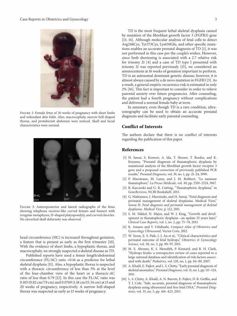
At 13 weeks of gestation the biparietal diameter (BPD)was measured to be 27.4mm, corresponding to the 95thpercentile. From this week on macrocephaly was observedand a narrow thorax was suspected on subsequent ultra-sonographies (Figure 1(b)). At 16 weeks of gestation, amnioticfluid examination showed a normal karyotype (46XX); how-ever, a routine and a four-dimensional ultrasound revealedshortening and bowing of the long bones (femur length(FL) 11.6mm) (Figures 1(c) and 1(d)) with no subsequentimprovement. As a result, a fetal skeletal disease, specificallyTD, was strongly suspected. At 20 weeks of gestation, the FLwas 1.18 cm, compatiblewith less than the 5th percentile. Also,the BPD was 5.3 cm, corresponding to more than the 95thpercentile. During genetic counseling, a molecular analysiswas suggested to the parents in order to make an accurateprenatal diagnosis of TD; however, they opted not to pursuethis exam and decided on termination of the pregnancy,instead. The patient was then hospitalized and underwentmechanical cervical dilation. The following day gemeprost(PGE1) was administered intravaginally every three hours.A 400 g female fetus was delivered dead at 20 weeks and2 days of gestation (Figure 2). All limbs were noted to beextremely short with redundant skin folds. Macrocephalywas evident. A narrow bell-shaped thorax with short ribs
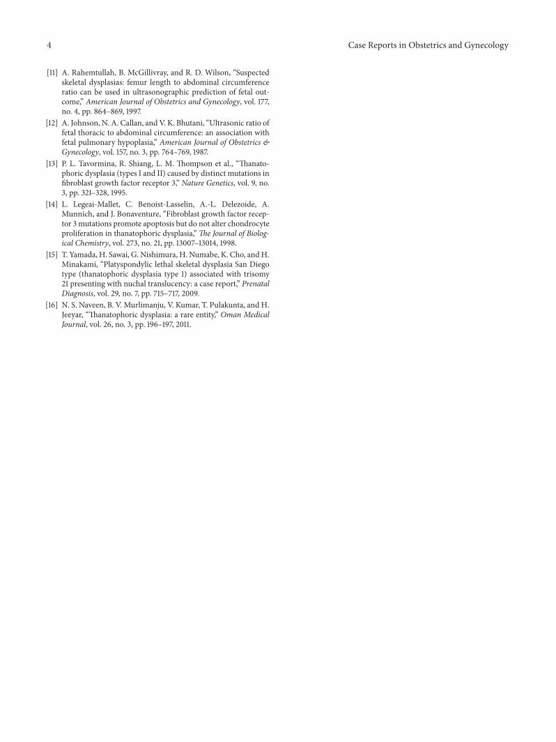
and a protuberant abdomen were noticed; however, the skulland facial characteristics were within normal limits. Skeletalradiography showed telephone receiver-like curved femursand humeri accompanied by irregular metaphyses, an H-shaped platyspondyly, and curved clavicles (Figure 3). Nocloverleaf skull deformity was observed.These characteristicsconfirmed the diagnosis of TD type I.
3. Discussion
The incidence of immune hydrops has decreased due toroutine screening and prophylaxis; however, the mortalityrate of nonimmune hydrops, during either the fetal or theneonatal period, is up to 75.5% [8]. Although fetal hydropsis considered to be a nonspecific finding on obstetric ultra-sounds, previous reports showed that an increased nuchaltranslucency (NT) and hydrops are common features ofserious skeletal dysplasia [9]. Moreover, previous cases of TDreported a NT of 3.4–6.5mm by 14 weeks of gestation whichcorrelates with the 5.7mm observed in this case [9].
As previous studies reported, 40–80% of TD cases canbe correctly diagnosed by ultrasonography in the prenatalperiod [10]. Limb shortening in TD is also sonographicallyapparent from as early as 13 weeks of gestation. Similarly,
Case Reports in Obstetrics and Gynecology 3
Figure 2: Female fetus of 20 weeks of pregnancy with short limbsand redundant skin folds. Also, macrocephaly, narrow bell-shapedthorax, and protuberant abdomen were noticed. Skull and facialcharacteristics were normal.
Figure 3: Anteroposterior and lateral radiographs of the fetusshowing telephone receiver-like curved femurs and humeri withirregularmetaphyses, H-shaped platyspondyly, and curved clavicles.No cloverleaf skull deformity was observed.
head circumference (HC) is increased throughout gestation,a feature that is present as early as the first trimester [10].With the evidence of short limbs, a hypoplastic thorax, andmacrocephaly, we strongly suspected a skeletal disease as TD.
Published reports have used a femur length/abdominalcircumference (FL/AC) ratio <0.16 as a predictor for lethalskeletal dysplasia [11]. Also, a hypoplastic thorax is suspectedwith a thoracic circumference of less than 5% at the levelof the four-chamber view of the heart or a thoracic-ACratio of less than 0.79 [12]. In this case the FL/AC ratio was0.103 (0.82 cm/7.9 cm) and 0.0769 (1.18 cm/15.34 cm) at 13 and20 weeks of pregnancy, respectively. A narrow bell-shapedthorax was suspected as early as 13 weeks of pregnancy.
TD is the most frequent lethal skeletal dysplasia causedby mutation of the fibroblast growth factor 3 (FGFR3) gene[13, 14]. Although molecular analysis of fetal cells to detectArg248Cys, Tyr373Cys, Lys650Glu, and other specific muta-tions enables an accurate prenatal diagnosis of TD [1], it wasnot performed in this case per the couple’s wishes. However,since limb shortening is associated with a 2.7 relative riskfor trisomy 21 [4] and a case of TD type I presented withtrisomy 21 was reported previously [15], we considered anamniocentesis at 16 weeks of gestation important to perform.TD is an autosomal dominant genetic disease; however, it isalmost always caused by a de novomutation in FGFR3 [3]. Asa result, a general empiric recurrence risk is estimated in only2% [16]. This fact is important to consider in order to relieveparental anxiety over future pregnancies. After counseling,the patient had a fourth pregnancy without complicationsand delivered a normal female baby at term.
In summary, even though TD is a rare condition, ultra-sonography can be used to obtain an accurate prenataldiagnosis and facilitate early parental counseling.
Conflict of Interests
The authors declare that there is no conflict of interestsregarding the publication of this paper.
References
[1] H. Sawai, S. Komori, A. Ida, T. Henmi, T. Bessho, and K.Koyama, “Prenatal diagnosis of thanatophoric dysplasia bymutational analysis of the fibroblast growth factor receptor 3gene and a proposed correction of previously published PCRresults,” Prenatal Diagnosis, vol. 19, no. 1, pp. 21–24, 1999.
[2] P. Maroteaux, M. Lamy, and J. M. Robbert, “Le nanismethanatophore,” La Presse Medicale, vol. 49, pp. 2519–2524, 1967.
[3] B. Karczeski and G. R. Cutting, “Thanatophoric dysplasia,” inGeneReviews, NCBI Bookshelf, 2013.
[4] G. Nishimura, J. Murotsuki, and H. Sawai, “Fetal diagnosis andperinatal management of skeletal dysplasias. Medical View,”Sawai H. Fetal diagnosis and perinatal management of skeletaldysplasias. Medical View, p. 122, 2011.
[5] S. M. Nikkel, N. Major, and W. J. King, “Growth and devel-opment in thanatophoric dysplasia—an update 25 years later,”Clinical Case Reports, vol. 1, no. 2, pp. 75–78, 2013.
[6] K. Amano and Y. Ushibashi, Compact Atlas of Obstetrics andGynecology Ultrasound, Vector Core, 2012.
[7] W. Yeom, E. S. Paik, J.-J. An et al., “Clinical characteristics andperinatal outcome of fetal hydrops,” Obstetrics & GynecologyScience, vol. 58, no. 2, pp. 90–97, 2015.
[8] M. E. Abrams, K. S. Meredith, P. Kinnard, and R. H. Clark,“Hydrops fetalis: a retrospective review of cases reported to alarge national database and identification of risk factors associ-ated with death,” Pediatrics, vol. 120, no. 1, pp. 84–89, 2007.
[9] A. Khalil, E. Pajkrt, and L. S. Chitty, “Early prenatal diagnosis ofskeletal anomalies,”Prenatal Diagnosis, vol. 31, no. 1, pp. 115–124,2011.
[10] L. S. Chitty, A. Khalil, A. N. Barrett, E. Pajkrt, D. R. Griffin, andT. J. Cole, “Safe, accurate, prenatal diagnosis of thanatophoricdysplasia using ultrasound and free fetal DNA,” Prenatal Diag-nosis, vol. 33, no. 5, pp. 416–423, 2013.
4 Case Reports in Obstetrics and Gynecology
[11] A. Rahemtullah, B. McGillivray, and R. D. Wilson, “Suspectedskeletal dysplasias: femur length to abdominal circumferenceratio can be used in ultrasonographic prediction of fetal out-come,” American Journal of Obstetrics and Gynecology, vol. 177,no. 4, pp. 864–869, 1997.
[12] A. Johnson, N. A. Callan, and V. K. Bhutani, “Ultrasonic ratio offetal thoracic to abdominal circumference: an association withfetal pulmonary hypoplasia,” American Journal of Obstetrics &Gynecology, vol. 157, no. 3, pp. 764–769, 1987.
[13] P. L. Tavormina, R. Shiang, L. M. Thompson et al., “Thanato-phoric dysplasia (types I and II) caused by distinct mutations infibroblast growth factor receptor 3,” Nature Genetics, vol. 9, no.3, pp. 321–328, 1995.
[14] L. Legeai-Mallet, C. Benoist-Lasselin, A.-L. Delezoide, A.Munnich, and J. Bonaventure, “Fibroblast growth factor recep-tor 3mutations promote apoptosis but do not alter chondrocyteproliferation in thanatophoric dysplasia,”The Journal of Biolog-ical Chemistry, vol. 273, no. 21, pp. 13007–13014, 1998.
[15] T. Yamada, H. Sawai, G. Nishimura, H. Numabe, K. Cho, andH.Minakami, “Platyspondylic lethal skeletal dysplasia San Diegotype (thanatophoric dysplasia type 1) associated with trisomy21 presenting with nuchal translucency: a case report,” PrenatalDiagnosis, vol. 29, no. 7, pp. 715–717, 2009.
[16] N. S. Naveen, B. V. Murlimanju, V. Kumar, T. Pulakunta, and H.Jeeyar, “Thanatophoric dysplasia: a rare entity,” Oman MedicalJournal, vol. 26, no. 3, pp. 196–197, 2011.

Submit your manuscripts athttp://www.hindawi.com
Stem CellsInternational
Hindawi Publishing Corporationhttp://www.hindawi.com Volume 2014
Hindawi Publishing Corporationhttp://www.hindawi.com Volume 2014
MEDIATORSINFLAMMATION
of
Hindawi Publishing Corporationhttp://www.hindawi.com Volume 2014
Behavioural Neurology
EndocrinologyInternational Journal of
Hindawi Publishing Corporationhttp://www.hindawi.com Volume 2014
Hindawi Publishing Corporationhttp://www.hindawi.com Volume 2014
Disease Markers
Hindawi Publishing Corporationhttp://www.hindawi.com Volume 2014
BioMed Research International
OncologyJournal of
Hindawi Publishing Corporationhttp://www.hindawi.com Volume 2014
Hindawi Publishing Corporationhttp://www.hindawi.com Volume 2014
Oxidative Medicine and Cellular Longevity
Hindawi Publishing Corporationhttp://www.hindawi.com Volume 2014
PPAR Research
The Scientific World JournalHindawi Publishing Corporation http://www.hindawi.com Volume 2014
Immunology ResearchHindawi Publishing Corporationhttp://www.hindawi.com Volume 2014
Journal of
ObesityJournal of
Hindawi Publishing Corporationhttp://www.hindawi.com Volume 2014
Hindawi Publishing Corporationhttp://www.hindawi.com Volume 2014
Computational and Mathematical Methods in Medicine
OphthalmologyJournal of
Hindawi Publishing Corporationhttp://www.hindawi.com Volume 2014
Diabetes ResearchJournal of
Hindawi Publishing Corporationhttp://www.hindawi.com Volume 2014
Hindawi Publishing Corporationhttp://www.hindawi.com Volume 2014
Research and TreatmentAIDS
Hindawi Publishing Corporationhttp://www.hindawi.com Volume 2014
Gastroenterology Research and Practice
Hindawi Publishing Corporationhttp://www.hindawi.com Volume 2014
Parkinson’s Disease
Evidence-Based Complementary and Alternative Medicine
Volume 2014Hindawi Publishing Corporationhttp://www.hindawi.com


![Thanatophoric dwarfism - dds.nl · dwarfism but also as an isolated phenomenon [3]. So far as we know, radio- ulnar synostosis has been observed only once in a thanatophoric dwarf](https://static.fdocuments.in/doc/165x107/5fe5a68cd0871340043c1206/thanatophoric-dwarfism-ddsnl-dwarfism-but-also-as-an-isolated-phenomenon-3.jpg)