Case presentation Torsade de Pointes. C15 Case Mr X presented to ED from Bosbokrandt (Nelspruit...
35
Case presentation Torsade de Pointes
-
Upload
phebe-murphy -
Category
Documents
-
view
216 -
download
0
Transcript of Case presentation Torsade de Pointes. C15 Case Mr X presented to ED from Bosbokrandt (Nelspruit...

Case presentation
Torsade de Pointes
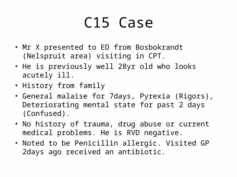
C15 Case
• Mr X presented to ED from Bosbokrandt (Nelspruit area) visiting in CPT.
• He is previously well 28yr old who looks acutely ill.• History from family• General malaise for 7days, Pyrexia (Rigors),
Deteriorating mental state for past 2 days (Confused).• No history of trauma, drug abuse or current medical
problems. He is RVD negative. • Noted to be Penicillin allergic. Visited GP 2days ago
received an antibiotic.
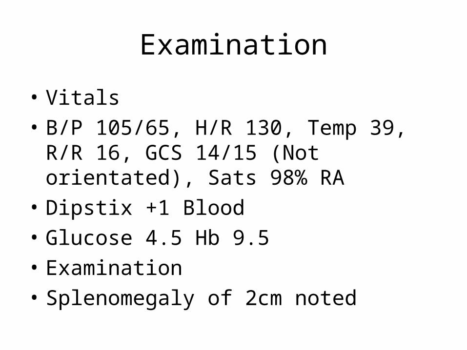
Examination
• Vitals
• B/P 105/65, H/R 130, Temp 39, R/R 16, GCS 14/15 (Not orientated), Sats 98% RA
• Dipstix +1 Blood
• Glucose 4.5 Hb 9.5
• Examination
• Splenomegaly of 2cm noted

Special investigations
• FBC
• Plasmodium PCR
• Giemsa stained thick / thin smear
• CXR
• LP was deferred until platelet count returned
• Elected not to CT at this stage
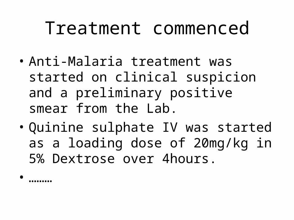
Treatment commenced
• Anti-Malaria treatment was started on clinical suspicion and a preliminary positive smear from the Lab.
• Quinine sulphate IV was started as a loading dose of 20mg/kg in 5% Dextrose over 4hours.
• ………

Patient deteriorated 2hours later
• Pulse rate 180/min with B/P 75/46.
• GCS 10/15
• And an ECG that looks like this….

ECG

Treatment
• Patient was given Midazolam 5mg IV and was cardioverted at 200J Biphasic with return of sinus tachycardia.
• Quinine infusion was stopped and urgent gas was done and CEU + Magnesium was sent to lab.
• Later restarted in ICU.

Polymorphic VT
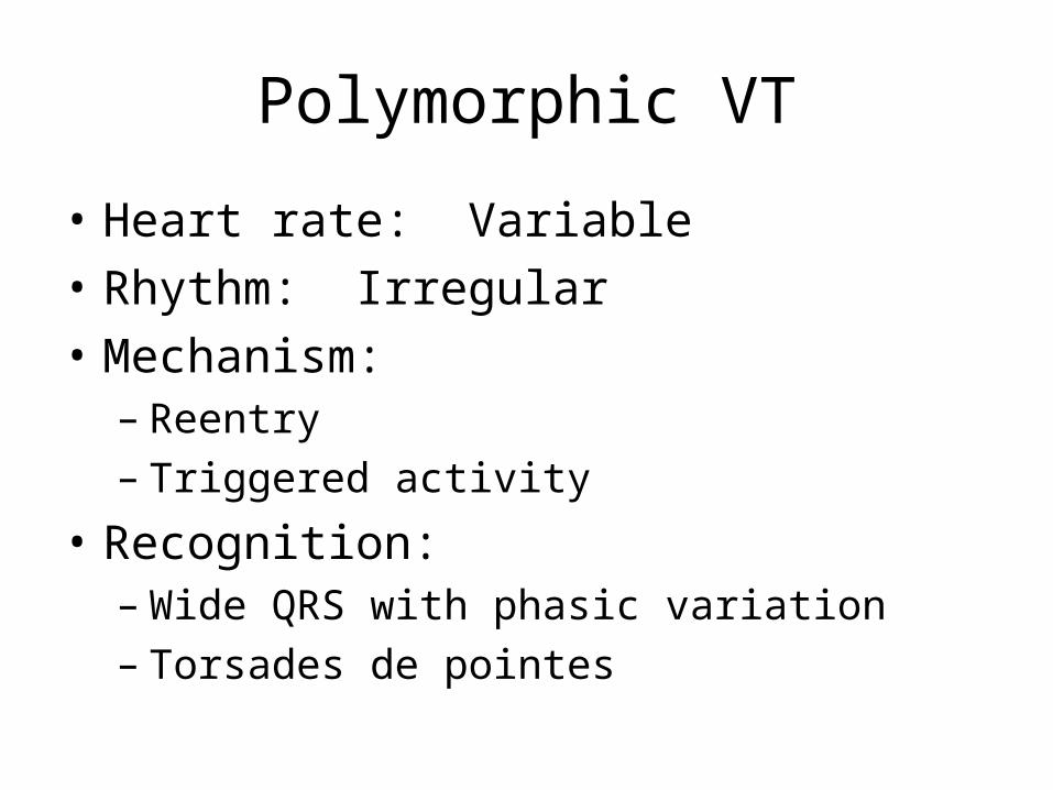
Polymorphic VT
• Heart rate: Variable
• Rhythm: Irregular
• Mechanism:– Reentry– Triggered activity
• Recognition:– Wide QRS with phasic variation– Torsades de pointes

Reentrant
• Reentrant ventricular arrhythmias– Premature ventricular complexes– Idiopathic left ventricular tachycardia– Bundle branch reentry– Ventricular tachycardia and fibrillation when
associated with chronic heart disease:• Previous myocardial infarction• Cardiomyopathy

Triggered
• Triggered activity ventricular arrhythmias– Pause-dependent triggered activity
• Early afterdepolarization (phase 3)• Polymorphic ventricular tachycardia
– Catechol-dependent triggered activity• Late afterdepolarizations (phase 4)• Idiopathic right ventricular tachycardia
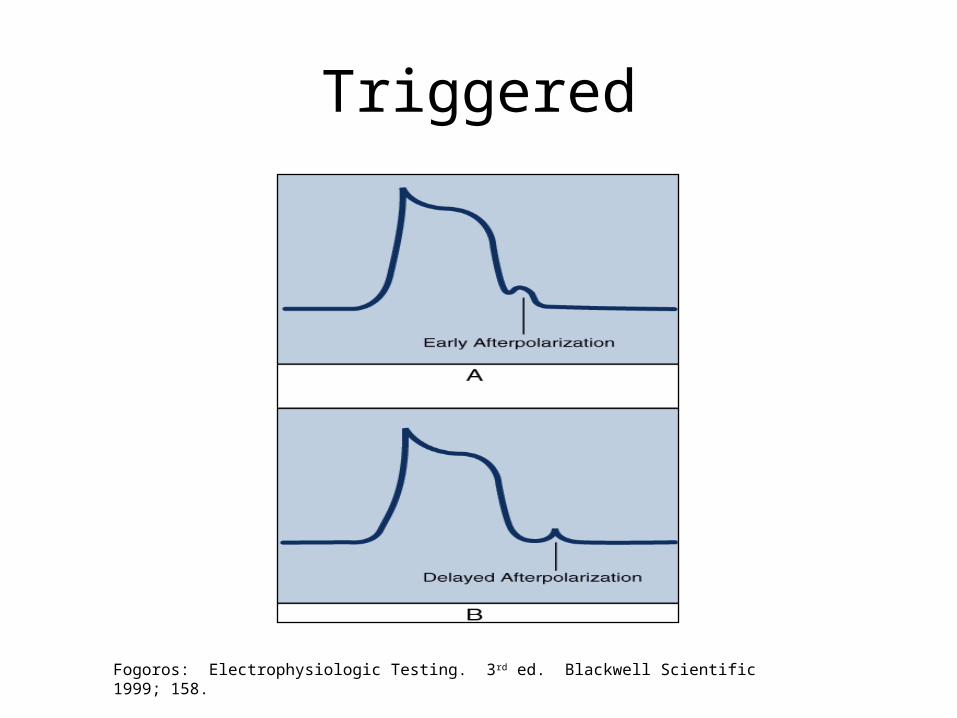
Triggered
Fogoros: Electrophysiologic Testing. 3rd ed. Blackwell Scientific 1999; 158.
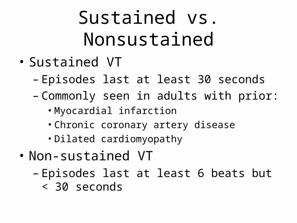
Sustained vs. Nonsustained
• Sustained VT– Episodes last at least 30 seconds– Commonly seen in adults with prior:
• Myocardial infarction• Chronic coronary artery disease• Dilated cardiomyopathy
• Non-sustained VT– Episodes last at least 6 beats but < 30
seconds
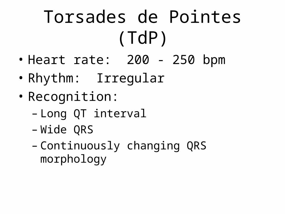
Torsades de Pointes (TdP)
• Heart rate: 200 - 250 bpm
• Rhythm: Irregular
• Recognition:– Long QT interval– Wide QRS– Continuously changing QRS morphology

Causes
• Congenital long QT syndromes (adrenergic-dependent) • Jervell and Lange-Nielsen syndrome • Acquired long QT syndromes • Antiarrhythmic drugs
– Class 1A - Quinidine, disopyramide, procainamide – Class III - Sotalol, amiodarone (rare), ibutilide, dofetilide,
almokalant• Histamine1-receptor antagonists - Terfenadine, astemizole • Cholinergic antagonists - Cisapride, organophosphates (pesticides) • Antibiotics - Erythromycin, clarithromycin, trimethoprim-
sulfamethoxazole, clindamycin, pentamidine, amantadine, chloroquine, halofantrine
• Electrolyte abnormalities - Hypokalemia, hypomagnesemia, hypocalcemia

Pathophysiology
• The association between torsade and a prolonged QT interval has long been known, but the mechanisms involved at the cellular and ionic levels have been made clearer in approximately the last decade.

Pathophysiology
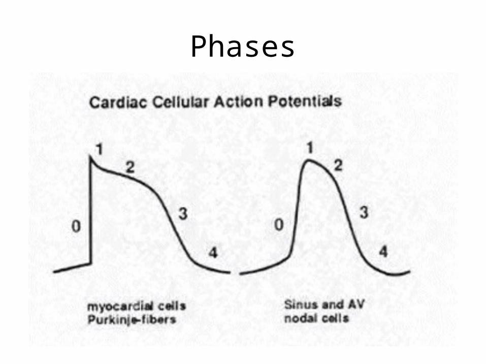
Phases
Phase 1
• Phase 1: During initial upstroke of action potential in a normal cardiac cell, a rapid net influx of positive ions (Na+ and Ca++) occurs, which results in the depolarization of the cell membrane. This is followed by a rapid transient outward potassium current (Ito), while the influx rate of positive ions (Na+, Ca++) declines. This represents the initial part of the repolarization, or phase 1.

Phase 2
• Phase 2 is characterized by the plateau, the distinctive feature of which is the cardiac repolarization. The positive currents flowing inward and outward become almost equal during this stage.
Phase 3
• Phase 3 of the repolarization is mediated by activation of the delayed rectifier potassium current (IK) moving outward while the inward positive current decays. If a slow inactivation of the Ca++ and Na+ currents occurs, this inward "window" current can cause single or repetitive depolarization during phases 2 and 3 (ie, EADs). These EADs appear as pathologic U waves on a surface ECG, and, when they reach a threshold, they may trigger ventricular tachyarrhythmias.
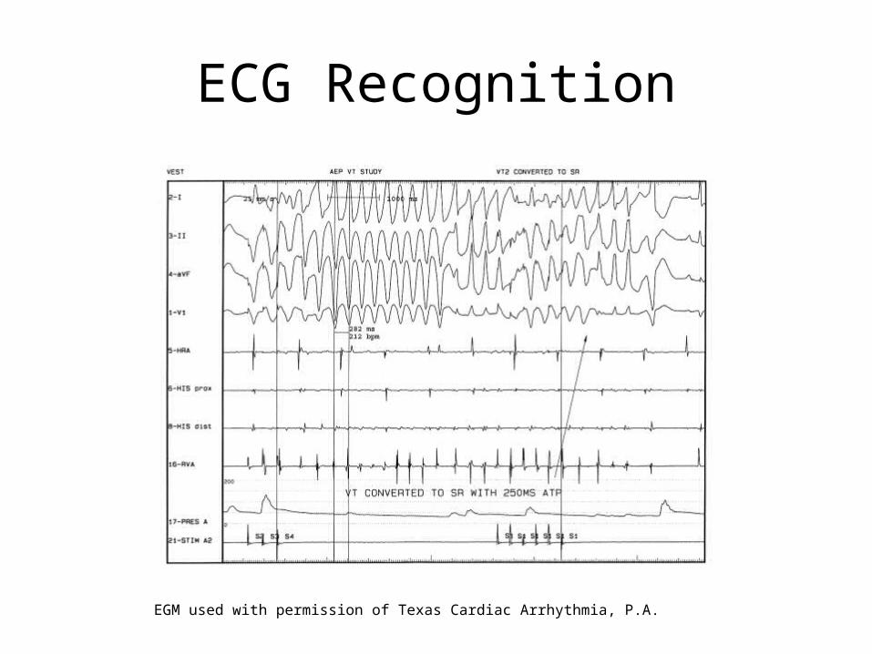
ECG Recognition
EGM used with permission of Texas Cardiac Arrhythmia, P.A.
ECG changes
• Patients have paroxysms of 5-20 beats, with a heart rate faster than 200 bpm; sustained episodes occasionally can be seen.
• Progressive change in polarity of QRS about the isoelectric line occurs.
• Complete 180° twist of QRS complexes in 10-12 beats is present.
• Usually, a prolonged QT interval and pathological U waves are present, reflecting abnormal ventricular repolarization. The most consistent indicator of QT prolongation is a QT of 0.60 s or longer or a QTc (corrected for heart rate) of 0.45 s or longer.
• A short-long-short sequence between the R-R interval occurs before the trigger response.

ECG – Long QT• Marked QT prolongation in an asymptomatic patient on
erythromycin. Patient also was found to be profoundly hypomagnesemic and hypokalemic.
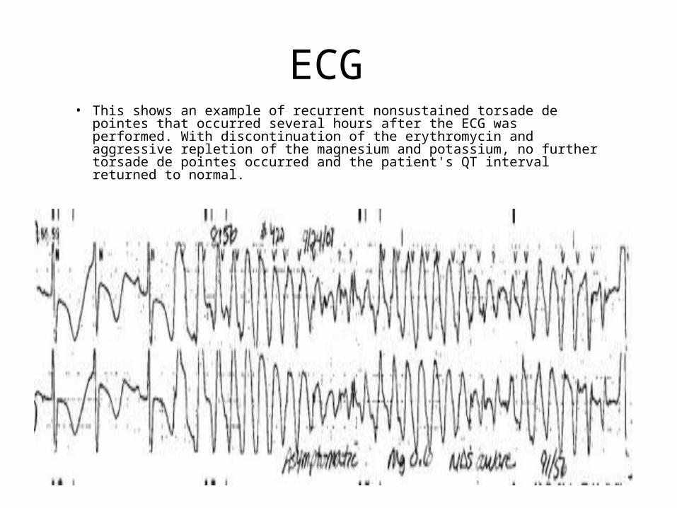
ECG • This shows an example of recurrent nonsustained torsade de pointes
that occurred several hours after the ECG was performed. With discontinuation of the erythromycin and aggressive repletion of the magnesium and potassium, no further torsade de pointes occurred and the patient's QT interval returned to normal.
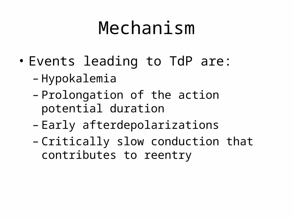
Mechanism
• Events leading to TdP are:– Hypokalemia– Prolongation of the action potential duration– Early afterdepolarizations– Critically slow conduction that contributes to
reentry
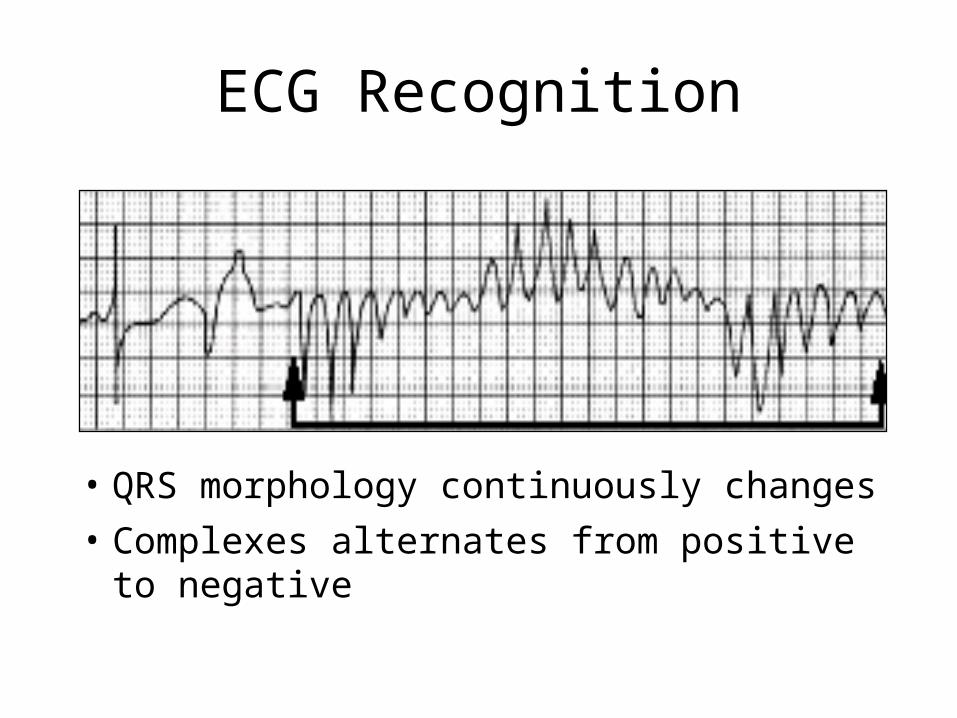
ECG Recognition
• QRS morphology continuously changes
• Complexes alternates from positive to negative

Treatment
• Pharmacologic therapy:– Potassium– Magnesium– Isoproterenol– Possibly class Ib drugs (lidocaine) to
decrease refractoriness/shorten length of action potential
• Overdrive ventricular pacing
• Cardioversion

Overdrive pacing
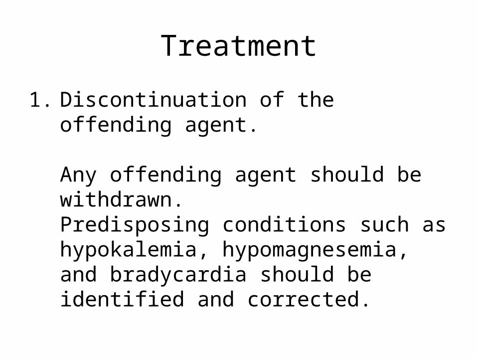
Treatment
1. Discontinuation of the offending agent. Any offending agent should be withdrawn. Predisposing conditions such as hypokalemia, hypomagnesemia, and bradycardia should be identified and corrected.

Treatment
2. Suppression of early after depolarizations.
Magnesium is the drug of choice for suppressing EADs and terminating the arrhythmia.
This is achieved by decreasing the influx of calcium, thus lowering the amplitude of EADs. Magnesium can be given at 1-2 g IV initially in 30-60 seconds, which then can be repeated in 5-15 minutes. Alternatively, a continuous infusion can be started at a rate of 3-10 mg/min. Magnesium is effective even in patients with normal magnesium levels.
Treatment
3. Isoproterenol This drug can be used in bradycardia-dependent torsade that usually is associated with acquired long QT syndrome (pause-dependent). It should be administered as a continuous IV infusion to keep the heart rate faster than 90 bpm. Isoproterenol accelerates AV conduction and decreases the QT interval by increasing the heart rate and reducing temporal dispersion of repolarization. Beta-adrenergic agonists are contraindicated in the congenital form of long QT syndrome (adrenergic-dependent).

New ACLS Algorithm

END