Cascade L-shell soft-x-ray emission as incident x-ray ...johnf/g777/Sokaras_2011.pdf · D....
12
PHYSICAL REVIEW A 83, 052511 (2011) Cascade L-shell soft-x-ray emission as incident x-ray photons are tuned across the 1s ionization threshold D. Sokaras, 1 A. G. Kochur, 2 M. M¨ uller, 3 M. Kolbe, 3 B. Beckhoff, 3 M. Mantler, 4 Ch. Zarkadas, 1,5 M. Andrianis, 1 A. Lagoyannis, 1 and A. G. Karydas 1,6 1 Institute of Nuclear Physics, N.C.S.R. “Demokritos,” Aghia Paraskevi, GR-15310, Athens, Greece 2 Rostov State University of Transport Communication, 344038, Rostov-na-Donu, Russia 3 Physikalisch-Technische Bundesanstalt, Abbestrasse 2-12, D-10587, Berlin, Germany 4 Technische Universit¨ at Wien, A-1040, Vienna, Austria 5 PANalytical B.V., 7600 AA Almelo, The Netherlands 6 Nuclear Spectrometry and Applications Laboratory, International Atomic Energy Agency (IAEA), A-2444, Seibersdorf, Austria (Received 20 April 2010; published 19 May 2011) The cascade L-shell x-ray emission as an incident polarized and unpolarized monochromatic radiation overpass the 1s ionization threshold is investigated for the metallic Fe by means of moderate resolution, quantitative x-ray spectrometry. A full ab initio theoretical investigation of the L-shell x-ray emission processes is performed based on a detailed straightforward construction of the cascade decay trees within the Pauli-Fock approximation. The agreement obtained between experiments and the presented theory is indicated and discussed with respect to the accuracy of advanced atomic models as well as its significance for the characterization capabilities of x-ray fluorescence (XRF) analysis. DOI: 10.1103/PhysRevA.83.052511 PACS number(s): 32.30.Rj, 31.15.A−, 81.07.−b I. INTRODUCTION A single photoionization event produces a short-lived atomic inner-shell-vacancy state. This excitation decays very fast (∼fs) through cascades of electronic transitions, until an ionic ground state is finally reached [1]. Each transition within the cascade is accompanied by an emission of either a photon (radiative transitions) or an electron (Auger and Coster-Kronig transitions). For core-level initial vacancies, the cascades can be very complex having up to millions of branches; thus their complete reliable theoretical description turns to be a rather sophisticated procedure. The cascade decay of an inner-shell vacancy is a funda- mental atomic phenomenon which should be considered in all the processes related to inner-shell vacancies. In x-ray spectroscopy, cascade decay manifests itself in the emission from less-deep shells when an initial vacancy is previously created in a deeper shell. In this case, the so-called cascade x-ray emission occurs [i.e., the x-ray emission from a shell where a vacancy (or vacancies) is created by the cascade decay processes and not directly as by photoionization or a charged-particle impact]. Although the cascade decays of inner-shell vacancy states, or vacancy cascades, have been being studied since the 1960s, the studies devoted to the cascade emission spectra are scarce [2–8]. In these terms, the development and the assessment of advanced theoretical models that have the potential to describe in a quantitatively reliable manner the high complexity fundamental atomic processes (i.e., the cascade relaxation) can be of great interest for the atomic physics field of research toward achieving an extensive understanding. Further on, the accurate study of the cascade x-ray emission through the theoretical description of atomic relaxation mechanisms, * [email protected] besides having a profound interest in atomic physics, can also be of great importance in the applied physics research. X-ray fluorescence (XRF) spectroscopy can be considered as an advanced analytical method providing quantitative information on homogeneous, stratified, or even depth gra- dient materials in a wide field of applications [9]. The quantitative XRF analysis of unknown samples is usually based on the so-called fundamental parameters (FPs) method [10–12]. The FP method determines the unknown elemental concentrations or mass depositions through the iterative reconstruction of the detected x-ray fluorescence intensities, accounting for all the atomic processes contributing to the x-ray fluorescence as well as the given experimental setup characteristics (through the calibration constants). Knowing well the instrumental parameters by means of appropriate calibration procedures, the quantification analysis accuracy is mainly hampered by the reliability and the uncertainties of the tabulated FPs. Nevertheless, inconsistencies within FPs can be partly compensated within the calibration procedure utilizing certified reference materials and standards. One step further, the so-called reference-free XRF analysis [13,14], based on absolutely calibrated experimental setups, has the unique advantage not to rely on any relative measurement involving calibration specimens and, thus tends to serve the emerging needs for the characterization and quality control of advanced technological materials for which appropriate standards do not exist. However, in this case, inconsistencies and uncertainties in the FPs data base directly affect the accuracy of the quantitative analysis supporting the need for an assessment of the theoretical predictions with respect to experimentally determined FP values. Within the XRF analysis of bulk materials, besides the well-known x-ray fluorescence induced by the single pho- toionization process, a number of second-order processes may also contribute to the observed x-ray emission. Various processes that involve the surrounding atoms (apart from 052511-1 1050-2947/2011/83(5)/052511(12) ©2011 American Physical Society
Transcript of Cascade L-shell soft-x-ray emission as incident x-ray ...johnf/g777/Sokaras_2011.pdf · D....
PHYSICAL REVIEW A 83, 052511 (2011)
Cascade L-shell soft-x-ray emission as incident x-ray photonsare tuned across the 1s ionization threshold
D. Sokaras,1 A. G. Kochur,2 M. Muller,3 M. Kolbe,3 B. Beckhoff,3 M. Mantler,4 Ch. Zarkadas,1,5 M. Andrianis,1
A. Lagoyannis,1 and A. G. Karydas1,6
1Institute of Nuclear Physics, N.C.S.R. “Demokritos,” Aghia Paraskevi, GR-15310, Athens, Greece2Rostov State University of Transport Communication, 344038, Rostov-na-Donu, Russia3Physikalisch-Technische Bundesanstalt, Abbestrasse 2-12, D-10587, Berlin, Germany
4Technische Universitat Wien, A-1040, Vienna, Austria5PANalytical B.V., 7600 AA Almelo, The Netherlands
6Nuclear Spectrometry and Applications Laboratory, International Atomic Energy Agency (IAEA), A-2444, Seibersdorf, Austria(Received 20 April 2010; published 19 May 2011)
The cascade L-shell x-ray emission as an incident polarized and unpolarized monochromatic radiation overpassthe 1s ionization threshold is investigated for the metallic Fe by means of moderate resolution, quantitative x-rayspectrometry. A full ab initio theoretical investigation of the L-shell x-ray emission processes is performed basedon a detailed straightforward construction of the cascade decay trees within the Pauli-Fock approximation. Theagreement obtained between experiments and the presented theory is indicated and discussed with respect tothe accuracy of advanced atomic models as well as its significance for the characterization capabilities of x-rayfluorescence (XRF) analysis.
DOI: 10.1103/PhysRevA.83.052511 PACS number(s): 32.30.Rj, 31.15.A−, 81.07.−b
I. INTRODUCTION
A single photoionization event produces a short-livedatomic inner-shell-vacancy state. This excitation decays veryfast (∼fs) through cascades of electronic transitions, until anionic ground state is finally reached [1]. Each transition withinthe cascade is accompanied by an emission of either a photon(radiative transitions) or an electron (Auger and Coster-Kronigtransitions). For core-level initial vacancies, the cascades canbe very complex having up to millions of branches; thus theircomplete reliable theoretical description turns to be a rathersophisticated procedure.
The cascade decay of an inner-shell vacancy is a funda-mental atomic phenomenon which should be considered inall the processes related to inner-shell vacancies. In x-rayspectroscopy, cascade decay manifests itself in the emissionfrom less-deep shells when an initial vacancy is previouslycreated in a deeper shell. In this case, the so-called cascadex-ray emission occurs [i.e., the x-ray emission from a shellwhere a vacancy (or vacancies) is created by the cascadedecay processes and not directly as by photoionization ora charged-particle impact]. Although the cascade decays ofinner-shell vacancy states, or vacancy cascades, have beenbeing studied since the 1960s, the studies devoted to thecascade emission spectra are scarce [2–8].
In these terms, the development and the assessment ofadvanced theoretical models that have the potential to describein a quantitatively reliable manner the high complexityfundamental atomic processes (i.e., the cascade relaxation)can be of great interest for the atomic physics field of researchtoward achieving an extensive understanding. Further on,the accurate study of the cascade x-ray emission throughthe theoretical description of atomic relaxation mechanisms,
besides having a profound interest in atomic physics, canalso be of great importance in the applied physics research.X-ray fluorescence (XRF) spectroscopy can be consideredas an advanced analytical method providing quantitativeinformation on homogeneous, stratified, or even depth gra-dient materials in a wide field of applications [9]. Thequantitative XRF analysis of unknown samples is usuallybased on the so-called fundamental parameters (FPs) method[10–12]. The FP method determines the unknown elementalconcentrations or mass depositions through the iterativereconstruction of the detected x-ray fluorescence intensities,accounting for all the atomic processes contributing to thex-ray fluorescence as well as the given experimental setupcharacteristics (through the calibration constants). Knowingwell the instrumental parameters by means of appropriatecalibration procedures, the quantification analysis accuracyis mainly hampered by the reliability and the uncertaintiesof the tabulated FPs. Nevertheless, inconsistencies within FPscan be partly compensated within the calibration procedureutilizing certified reference materials and standards. One stepfurther, the so-called reference-free XRF analysis [13,14],based on absolutely calibrated experimental setups, has theunique advantage not to rely on any relative measurementinvolving calibration specimens and, thus tends to serve theemerging needs for the characterization and quality controlof advanced technological materials for which appropriatestandards do not exist. However, in this case, inconsistenciesand uncertainties in the FPs data base directly affect theaccuracy of the quantitative analysis supporting the need foran assessment of the theoretical predictions with respect toexperimentally determined FP values.
Within the XRF analysis of bulk materials, besides thewell-known x-ray fluorescence induced by the single pho-toionization process, a number of second-order processesmay also contribute to the observed x-ray emission. Variousprocesses that involve the surrounding atoms (apart from
052511-11050-2947/2011/83(5)/052511(12) ©2011 American Physical Society
D. SOKARAS et al. PHYSICAL REVIEW A 83, 052511 (2011)
the one initially ionized), like the secondary fluorescenceenhancement or the photoelectron or Auger-electron- inducedsecondary ionization, have also been identified to contributeto the primary x-ray fluorescence [15–20]. On the otherhand, intraatomic effects like the resonant Raman scattering[21–26] or the cascade x-ray emission [18] are also expectedto contribute significantly to the detected x-ray intensities,especially when monochromatic excited XRF analysis isemployed at synchrotron radiation facilities.
In the present work, we thoroughly investigate the L-shellsoft-x-ray emission upon photoionization by monochromaticpolarized and unpolarized x-ray radiation tuning the photonenergy across the 1s ionization threshold. Advanced abinitio calculations are explicitly performed both for thedirect and the cascade-produced L emission spectra, throughthe straightforward construction of the cascade decaytrees developed within the Pauli-Fock approximation. Theprocesses of single- and multielectron photoionizations aredistinguished and individually considered. The secondaryeffects, mainly related to the ejected electrons inducingionizations of neighboring atoms, are carefully evaluated bymeans of detailed Monte Carlo calculations. As a case study,the metallic Fe is selected exemplarily.
II. THEORY
A. Cascade-produced photon spectra
The cascade-emitted x-ray spectra are calculated based on astraightforward construction of the cascade de-excitation treesvia the method described in detail elsewhere [27,28], therefore,only a brief description is given below. An initial inner-shellvacancy produced by photoionization is short lived and it candecay through radiative and/or radiationless transitions into anumber of ionic states forming in this way the set of first-generation cascade ionic states. Some of these, in their turn,can decay further forming the second-generation states and soon until all the vacancies are in the outermost shells and candecay no further. Initial and intermediate ionic states are thebranching points in a cascade de-excitation tree, while eachcascade transition from a given branching point is a branch inthis tree. Every branch in the de-excitation tree is characterizedby the branching ratio, defined as
χ(C
(n)k → C(n+1)
m
) = �(C
(n)k → C(n+1)
m
)∑
m �(C
(n)k → C
(n+1)m
) . (1)
Here C(n)k is an ionic configuration which appeared after
the nth decay step, C(n+1)m is the set of the next-decay-step
configurations reached from C(n)k , and �(C(n)
k → C(n+1)m ) is
the partial width (transition rate) of respective transition. Thesummation is performed over all the possible final statesC(n+1)
m reached from the branching point C(n)k considering all
radiative and radiationless (Auger, Coster-Kronig) transitionsbeing energetically allowed. The partial transition widths arecalculated in the configuration-average approximation usingthe Pauli-Fock (PF) wave functions [29] while the transitionenergies are calculated as differences of the mean total energiesof initial and final ionic configurations.
The multivacancy ionic configurations of the cascade oftenhave very complex multiplet structures due to electrostatic andspin-orbital interactions. The multiplets of initial and final con-figurations of some low-energy transitions may overlap, so thatsome term-to-term transitions are forbidden energetically. Onthe other hand, some term-to-term transitions are sometimesallowed even if the center of gravity of final configuration ishigher in energy than that of the initial one. To account forthat, the configuration multiplets are simulated with Gaussianprobability density distributions with variances calculated viathe methods described in [30]. This enables the modification ofthe partial widths when accounting for the multiplet overlaps.
To calculate the cascade-produced photon spectra, theenergy interval of interest was split into the channels ofequal width (0.2 eV), the energy of each radiative transitionin a cascade was analyzed, and the transition probabilitywas accumulated in respective energy channel. The transitionprobabilities (P ) are calculated according to
P (C1 → C2) = P (C1) χ (C1 → C2) , (2)
where C1 and C2 are initial and final configurations of thetransition, P (C1) is the probability for the emitting configu-ration C1 to appear during the cascade development, and χ
is the branching ratio as defined in Eq. (1). The probabilityP (C1) is a product of all consecutive branching ratios of theconsecutive branches leading from the initial inner-vacancystate to the emitting configuration C1.
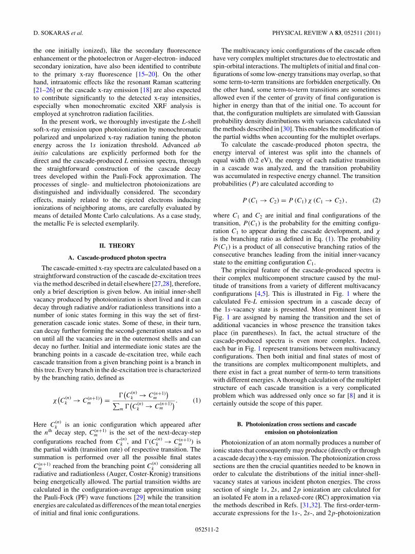
The principal feature of the cascade-produced spectra istheir complex multicomponent structure caused by the mul-titude of transitions from a variety of different multivacancyconfigurations [4,5]. This is illustrated in Fig. 1 where thecalculated Fe-L emission spectrum in a cascade decay ofthe 1s-vacancy state is presented. Most prominent lines inFig. 1 are assigned by naming the transition and the set ofadditional vacancies in whose presence the transition takesplace (in parentheses). In fact, the actual structure of thecascade-produced spectra is even more complex. Indeed,each bar in Fig. 1 represent transitions between multivacancyconfigurations. Then both initial and final states of most ofthe transitions are complex multicomponent multiplets, andthere exist in fact a great number of term-to term transitionswith different energies. A thorough calculation of the multipletstructure of each cascade transition is a very complicatedproblem which was addressed only once so far [8] and it iscertainly outside the scope of this paper.
B. Photoionization cross sections and cascadeemission on photoionization
Photoionization of an atom normally produces a number ofionic states that consequently may produce (directly or througha cascade decay) the x-ray emission. The photoionization crosssections are then the crucial quantities needed to be known inorder to calculate the distributions of the initial inner-shell-vacancy states at various incident photon energies. The crosssection of single 1s, 2s, and 2p ionization are calculated foran isolated Fe atom in a relaxed-core (RC) approximation viathe methods described in Refs. [31,32]. The first-order-term-accurate expressions for the 1s-, 2s-, and 2p-photoionization
052511-2
CASCADE L-SHELL SOFT-X-RAY EMISSION AS . . . PHYSICAL REVIEW A 83, 052511 (2011)
cross sections in the length form, are
σ1s→εp = 4
3π2αa2
0ES21s
[〈1s|r|εp+〉 − 〈1s|r|2p+〉〈2p|εp+〉
〈2p|2p+〉 − 〈1s|r|3p+〉〈3p|εp+〉〈3p|3p+〉
]2
, (3)
σ2s→εp = 4
3π2αa2
0ES22s
[〈2s|r|εp+〉 − 〈2s|r|2p+〉〈2p|εp+〉
〈2p|2p+〉 − 〈2s|r|3p+〉〈3p|εp+〉〈3p|3p+〉
]2
, (4)
σ2p→εs = 4
3π2αa2
0ES22p
[〈2p|r|εs+〉 − 〈2p|r|1s+〉〈1s|εs+〉
〈1s|1s+〉 − 〈2p|r|2s+〉〈2s|εs+〉〈2s|2s+〉 − 〈2p|r|3s+〉〈3s|εs+〉
〈3s|3s+〉]2
, (5)
σ2p→εd = 8
3π2αa2
0ES22p
[0.4〈2p|r|εd+〉2 + 0.6
(〈2p|r|εd+〉 − 〈2p|r|3d+〉〈3d|εd+〉
〈3d|3d+〉)2]
. (6)
Here α is the fine structure constant, a0 is the Bohr radius, E isthe incident photon energy, and ε is the energy of the ejectedphotoelectron. All the 〈nl| atomic orbitals are optimized in theground-state configurations while the |{n,ε}+〉 orbitals are inrespective single-vacancy configurations; this is indicated bythe “+” subscript. The terms Snl are the products of the overlap
FIG. 1. (Color online) Fe-L emission spectrum produced bya cascade decay of the 1s−1 state (up). Most of the componentsrepresent the transitions in the presence of additional vacancies(shown in parentheses). Calculated x-ray emission spectra uponmultielectron ionizations (1s−14s−1, 1s−13d−1, 1s−13p−1) are alsopresented as discussed in Sec. II B.
integrals between the same atomic orbitals not involved in aphotoelectron transitions, that is,
Snl =∏
i
〈nili |nili+〉Ni−δ(ni li ,nl), (7)
where Ni are the occupation numbers in the ground-stateconfiguration nil
Ni
i . As shown in [31,32] the expressions(3)–(6) are valid if the photoelectron wave functions |εl+〉 areoptimized being orthogonal to the same-symmetry lower-lying|nil+〉 atomic orbitals of the core-ionized atom.
Upon 1s and 2s photoionization, according to dipoleselection rules, photoelectrons can have only p symmetry. Inthe case of 2p ionization both s and d channels are allowed,then the 2p cross section is the sum of Eqs. (5) and (6).However, the d channel is dominating, contributing about97% of the total 2p-subshell cross section. The 2p-subshellconfiguration-average photoionization cross section does notaccount for the spin-orbital split of the 2p−1 level. This canbe introduced by splitting the total 2p cross sections σ2p
into σL2 = 13σ2p and σL3 = 2
3σ2p according to the statisticalweights.
The ionization threshold energies are calculated in anisolated-atom approximation as differences of total PF en-ergies of nl-ionized and ground-state configurations. Thecorresponding energies of the L2 and L3 thresholds arecalculated using the spin-orbit constant (ζ2p) obtained for the2p−1 configuration: E(L2) = EPF (2p−1) − EPF (0) + ζ2p,E(L3) = EPF (2p−1) − EPF (0) − 0.5ζ2p. All the calculatedthreshold energies are presented in Table I and compared withthe corresponding values given in the Elam et al. database[34] and previous theoretical studies [35,36]. One can seethat the calculated values are systematically higher than theexperimental ones. This is due to extra-atomic relaxation uponcreation of an inner-shell vacancy in a solid, a phenomenonevidently absent in a free atom [33]. Within further study,the calculated cross sections are shifted to the experimentalthreshold positions.
The calculated photoionization cross sections for the Kand L shells of the iron atom are presented in Fig. 2 andcompared with the widely used cross sections by Scofield [39].As seen from Fig. 2, the obtained cross sections (continuouslines) are systematically lower than those by Scofield. The
052511-3

D. SOKARAS et al. PHYSICAL REVIEW A 83, 052511 (2011)
TABLE I. Calculated and experimental threshold energies (in eV)for Fe.
Theory
Shell Present work Previous work [33] Experiment [34]
K 7134.3 7135ab 7112.0c
L1 861.9 863ab 844.6d
L2 735.5 736ab 719.9d
L3 722.6 722ab 706.8d
aReference [35].bReference [36].cReference [37].dReference [38].
reason for this discrepancy is in different approximationsused in the calculations. In contrast to the relaxed-core (RC)approximation used here, a frozen-core (FC) approximationwas employed by Scofield [39]. In FC approximation, uponphotoionization all the atomic orbitals remain the same as inthe ground state. This means that the overlap integrals presentin Eqs. (3)–(6) are either unity (for the same orbitals) or zero(for different orbitals). Then, all the additional rearrangementterms in parentheses in Eqs. (3)–(6) disappear, and, mostimportant, S2
nl terms [Eq. (7)] are now unity. However, in RCapproximation these terms are S2
1s = 0.7311, S22s = 0.7653,
and S22p = 0.7601, being the reason for lower cross sections
in the RC approximation.It should be noted that the S2
nl terms have a definitephysical sense. The creation of an nl vacancy in an atomleads to the so-called shake processes (i.e., to additionalexcitations or ionizations of other, mostly outermost atomicsubshells). In “sudden” approach [40], the squared overlapintegral 〈nili |nili+〉2 is the probability for the nili electron toremain in its orbital upon the change of the potential causedby the appearance of the deep nl vacancy. Then, S2
nl definedin Eq. (7) is the probability that all the electrons of the atomexcept the ionized one will stay where they had been before
FIG. 2. (Color online) Photoionization cross section for free Featom. Single-shell and cumulative ionization cross sections based onrelaxed core approximation (this work) are compared with frozencore calculations by Scofield [39] (dash lines).
nl ionization. In other words, it is the probability of single nl
ionization. Since the probability of any process to happen isunity, 1 − S2
nl is then a combined probability of all multipleexcitation or ionization processes accompanying nl ionization.The absence of the S2
nl terms in the cross section formulasof the FC approximations means that the FC cross sectionscontain implicitly all multiple ionization processes. In this way,far from the ionization thresholds FC cross sections normallycompare well with the experiment [41].
In this work the shake processes are considered explicitly.Within the sudden approach [40] the relative probability ofadditional excitation or ionization of an n1l1 subshell inducedby a sudden vacancy creation in the nl subshell is
w(nl,n1l1) = σ (nl−1n1l−11 )
σ (nl−1)= N1(〈n1l1|n1l1+〉−2 − 1).
(8)
Here N1 is the population of the n1l1 subshell, while 〈n1l1|and |n1l1+〉 wave functions in the overlap integral are thoseoptimized in the ground state and in the nl−1 configura-tion, respectively. Direct calculations of σ (nl−1n1l
−11 ) cross
sections near nl thresholds (see, e.g., [42]) showed that thesudden limit, expressed by Eq. (8), being independent ofthe exciting photon energy, is reached at incident photonenergies above the nl threshold by about three times the n1l1ionization energy. This condition is fulfilled for the excitingenergies considered within the present study (see Sec. III). Thecalculated relative shake probabilities of additional excitationor ionization of 3p, 3d, and 4s electrons upon 1s, 2s, and 2p
ionization are presented in Table II and compared with thecalculations of Mukoyama and Taniguchi [43] that are basedon Hartree-Fock-Slater (HFS) wave functions. The relativeprobabilities of the shake processes compare well with thoseof Ref. [43] while the discrepancies seen are due mainly todifferent approximations used for the atomic wave functionsin this work (PF) and in Ref. [43] (HFS) as discussed in detailelsewhere [44].
It should be noted that the probabilities of the shakeprocesses given by Eq. (8) are combined probabilities ofshake-up (SU) and shake-off (SO) processes. In SU processan outer-shell n1l1 electron is excited to a higher-lying excitedbound state, for example, 3d → 4d while in SO processesthe n1l1 electron is ejected from the atom. The relativecontributions from the SU and the SO processes to the totalshake probability [Eq. (8)] can vary noticeably depending on
TABLE II. Relative probabilities w(nl,n1l1) of shake processesinvolving the subshells n1l1 upon creation of initial vacancy in theinner subshell nl. The results of Ref. [43] are shown in parenthesesfor comparison.
Initial Additional vacancy n1l1
Vacancy nl 3p 3d 4s
1s 0.0410 (0.0480) 0.1132 (0.1321) 0.1446 (0.1451)2s 0.0180 (0.0210) 0.1231 (0.1361) 0.1274 (0.1256)2p 0.0192 (0.0240) 0.1218 (0.1386) 0.1327 (0.1276)
aThe absolute probabilities of [43] were divided by respectiveprobabilities of single ionizations S2
nl .
052511-4
CASCADE L-SHELL SOFT-X-RAY EMISSION AS . . . PHYSICAL REVIEW A 83, 052511 (2011)
the atom, the initial vacancy nl, and the affected subshelln1l1 [45]. A detailed analysis of the cascade decay of the1s−14p1 excited state in Argon has shown that the excitedelectron in a bound Rydberg state affects the cascade littlebeing mostly a spectator of rapid transitions involving the coreelectrons [46]. It is expected that this is a general situation,and the cascades originating from the SU states will not bevery different from those starting from respective SO states.Therefore the total shake probabilities [Eq. (8)] are attributedto the formation of nl−1n1l
−11 states.
The consideration of multielectron ionization processesupon a photoabsorption ending up in a direct or a cascadex-ray emission, can be of great importance toward the exactcalculation of the x-ray emission spectra. The presence ofspectator vacancies can considerably affect the x-ray spectraas these satellite emission lines can vary from few up to severalelectron volts with respect to the diagram emission lines.
Let Spct(C) denote the x-ray spectrum emitted froma given ionic configuration C [for instance, Spct(1s−1),Spct(1s−14s−1), Spct(1s−13d−1), and Spct(1s−13p−1) arepresented in Fig. 1]. Each component in Spct(C) is a dimen-sionless probability of a photon emission upon the decay ofthe configuration C. In this way the cumulative x-ray spectrumemitted after photoabsorption at a given exciting energy E0 is
Spct(E0) =∑C
σC(E0)Spct(C), (9)
where the summation is performed over the core ionicconfigurations C produced by photoionization [with crosssection σC(E0)]. For this study, the x-ray emission spectrafor all the core ionic configurations C = nl−1, nl−13p−1,nl−13d−1, nl−14s−1, with nl = 1s, 2s, 2p1/2, 2p3/2 have beenexplicitly calculated. The components in Spct(E0) are nowthe cross sections of lines emission. The emission spectra(Eq. (9)) can be very complex having multitudes of satellitecomponents, especially if the energy is enough for the 1s-shellionization, and this can play an important role for the accuratecalculation of self-attenuation of the emitted x rays within thesample.
It is introduced here a quantity ωnl(C) which is a generalizedfluorescence yield (GFY) (i.e., the fluorescence yield ofthe subshell nl for any given configuration C). Standardfluorescence yield is the probability of photon emission uponcreation of a single vacancy in the subshell nl [i.e., inthe present notation, ωnl = ωnl(nl−1)]. As discussed above,the generalized fluorescence yield ωnl(C) can be noticeablydifferent from ωnl if the initial configuration C contains avacancy (or vacancies) deeper than nl.
Evidently, the GFY can be split into the partial contributionscoming from specific transitions,
ωnl(C) =∑
i
ωnl→ni li (C). (10)
Here the quantities ωnl→ni li (C) are partial generalized fluo-rescence yields (PGFY) for a given ionic configuration C,nl → nili being the radiative transitions from the nl subshellwhich may happen during the decay of the configuration C.
It is worthwhile to introduce the PGFY associated withphotoionization of a specific inner subshell n0l0, ω
n0l0nl→ni li
.As discussed above, n0l0 photoionization produces not only
a singly ionized n0l−10 state, but also a number of doubly
ionized n0l−10 n1l
−11 states with additional vacancies in outer
n1l1 subshells. Each of the ionic states produced by n0l0photoionization gives its contribution to the PGFY. Thesecontributions are proportional to the probabilities of thosestates to appear in the n0l0-photoionization process. The PGFYassociated with an n0l0 photoionization is then determined as
ωn0l0nl→ni li
=[
1 +∑n1l1
w(n0l0,n1l1)
]−1[ωnl→ni li
(n0l
−10
)
+∑n1l1
w(n0l0,n1l1)ωnl→ni li
(n0l
−10 n1l
−11
)]. (11)
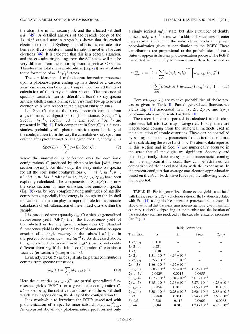
Here w(n0l0,n1l1) are relative probabilities of shake pro-cesses given in Table II. Partial generalized fluorescenceyields Eq. (11) associated with 1s, 2s, 2p1/2, and 2p3/2
photoionization are presented in Table III.The uncertainties incorporated in calculated atomic char-
acteristics fall into two major categories. Firstly, there areinaccuracies coming from the numerical methods used inthe calculation of atomic quantities. These can be controlledby setting the accuracy parameters for the iteration routineswhen calculating the wave functions. The atomic data reportedin this section and in Sec. V are numerically accurate inthe sense that all the digits are significant. Secondly, andmost importantly, there are systematic inaccuracies comingfrom the approximations used; they can be estimated viacomparison of the calculated data with the experiment. Inthe present configuration-average one-electron approximationbased on the Pauli-Fock wave functions the following effectsare neglected:
TABLE III. Partial generalized fluorescence yields associatedwith 1s, 2s, 2p1/2, and 2p3/2 photoionization of the Fe atom calculatedwith Eq. (11) taking double ionization processes into account. Itshould be noted that the x-ray emission energy for a given transitioncan vary noticeably depending on the number and the location ofthe spectator vacancies produced by the cascade relaxation processes(see Fig. 1).
Initial ionization
Transition 1s 2s 2p1/2 2p3/2
1s-2p1/2 0.110 – – –1s-2p3/2 0.221 – – –1s-3p 0.038 – – –2s-2p1/2 1.31×10⁻⁶ 4.34×10⁻⁶ – –2s-2p3/2 3.55×10⁻⁶ 1.16×10⁻⁵ – –2s − 3p 1.86×10⁻⁴ 4.37×10⁻⁴ – –2p1/2-3s 2.88×10⁻⁴ 1.55×10⁻⁴ 4.52×10⁻⁴ –2p1/2-3d 0.0029 0.0015 0.0055 –2p1/2-4s 1.87×10⁻⁵ 1.04×10⁻⁵ 3.01×10⁻⁵ –2p3/2-3s 5.45×10⁻⁴ 3.36×10⁻⁴ 7.27×10⁻⁷ 4.26×10⁻⁴
2p3/2-3d 0.0056 0.0033 9.05×10⁻⁶ 0.00522p3/2-4s 3.58×10⁻⁵ 2.25×10⁻⁵ 2.60×10⁻⁸ 2.86×10⁻⁵
3s-3p 0.0068 0.0013 9.74×10⁻⁶ 9.66×10⁻⁶
3p-3d 0.338 0.113 0.0065 0.00653p-4s 0.084 0.013 4.23×10⁻⁵ 4.23×10⁻⁵
052511-5

D. SOKARAS et al. PHYSICAL REVIEW A 83, 052511 (2011)
(i) Solid-state effects which may affect the probabilities oftransitions with the participation of the outer atomic subshells,and absolute energies of ionic states.
(ii) Many-electron effects which in certain cases may affectemission spectra [47–50].
(iii) Multiplet splitting which affects the profiles of theemission spectra (note that the effect of multiplet splittingis effectively included when calculating the branching ratios).
Neglect of the solid-state effects is the most severe of theapproximations adopted. As discussed in more detail in Sec. V,some decay channels, which are forbidden in an isolated-ionapproximation, may open in solid state thus modifying thedecay tree. As for the transition energies, they are expected tobe affected since the solid-state extra-atomic relaxation effectis larger for the initial vacancy than for the final vacancy stateof a transition. In the present work the calculated emissionspectra were shifted by −5.9 eV in order for the calculatedLα1 emission line to be in compliance with the correspondingexperimental one [51] for the metallic iron. It should be notedthat the shifts in emission spectra are less as compared withthe shifts of the single levels (see ionization thresholds inTable I). This is because during transitions the extra-atomicrelaxation—however different—is present both in initial andfinal states, and cancels out partially.
As for the many-electron and multiplet splitting effects,they are not expected to affect the cumulative cascade Lspectra significantly. Indeed, these phenomena do not affectthe integral intensities of the spectra, although sometimesaffecting their profiles substantially: The intensity is split into alarge number of components on wide energy intervals. This isof no importance in the present moderate energy resolutionexperiment registering the integral intensity of a bunch oftransitions.
It should be noticed that similar approximations have beenapplied earlier in Ref. [7] in the description of the cumulativeresonantly excited cascade 5d-4d emission from metalliclanthanum, as the exciting energy was scanned across the3d-4f discrete excitation thresholds. A good agreement withthe experiment obtained in Ref. [7] supports that the adoptedtheoretical approximations should be also applicable to thecase of the L cascade emission in metallic iron.
C. Calculating Fe-L Yield
The total Fe-L detected intensity, IFeL, when a pure ironsample is irradiated with photons of energy E0 and flux I0, inthe so-called parallel beam approximation, is given as
IFeL = I0�
4π
∑i
Pi(E0) M(Eo,Ei)ε(Ei)1
cos θb
. (12)
The sum runs over all the allowed transitions i [i.e., all theindividual components of Spct(E0); Eq. (9)] with energy Ei
and probability of emission Pi (cm2 g−1). � (sr) is the solidangle of detection confined by the aperture placed in frontof the detector, ε is the detector efficiency at energy Ei , θb
the angle of incidence with respect to the sample normal,and M(Eo,Es) (g cm−2) stands for the absorption correctionfactor accounting for the incident and the fluorescence x-rays
self-attenuation within the iron sample defined according toits thickness as follows:
M(E0,Ei) ={ 1−e−ξµT
µT, intermediate thickness
1µT
, infinite thickness.(13)
ξ (g cm−2) is the thickness of the sample and µT is given as
µT = µ(E0)
cosθb
+ µ(Ei)
cosθd
, (14)
whereas µ (cm2 g−1) stands for the iron sample mass atten-uation coefficient at the corresponding energies and θd theemitted photons take-off angle with respect to the samplenormal.
III. EXPERIMENT
The experimental study of the Fe-L cascade emission hasbeen carried out by two individual series of measurementsemploying proton-induced x-ray beams and synchrotron ra-diation, respectively. The former series of experiments wererealized at the novel proton-induced XRF chamber installedand operated at the 5.5-MV Tandem Van de Graaf acceleratorlaboratory of the Institute of Nuclear Physics at NationalCentre for Scientific Research, “Demokritos,” Athens. Theprinciple of the proton-induced XRF technique has beenpreviously demonstrated and applied toward fundamental aswell as analytical x-ray spectrometry studies over low-energyparticle accelerator facilities [22–24,26]; the irradiation of athick pure primary target by a few MeV high current protonbeam forms a high intensity unpolarized x-ray source ofselectable energy and high monochromaticity composed of theprimary target characteristic radiation to be used for furtherx-ray-related studies. The two-level proton-induced XRFchamber hosts in the lower part up to six primary targets usedfor ion-beam irradiation selecting in this way the energy of theincident x-ray beam. In the upper level, a six-position rotatablesample holder hosts the samples considered for analysis (allincident and take-off angles are set to 45◦). The proton-inducedx-ray beam is guided from the primary target to the sampleposition through a proper collimator whereas a filter can beinserted within its path in order to eliminate the backscatteredprotons and selectively absorb various spectral components ofthe incident x-ray beam. The chamber associates an energydispersive ultrathin window (AP 1.7) Si(Li) spectrometer[full-width at half maximum (FWHM) 136 eV at 5.89 keV;Gresham Sirius] capable of detecting photons even below theC-Kα fluorescence line. A double collimator system, placedin front of the detector window, confines the solid angle ofdetection to about 23 msr while the incorporated rare-earthmagnets configuration prevents the photoelectrons or Augerelectrons (up to ∼20 keV) to reach and deposit energy into thedetector crystal. For monitoring the incident x-ray beam flux,the electrically isolated holder of the primary targets enablescharge measurement of the impinging ion-beam current (thatit is proportional related to the x-ray beam flux) whereas inaddition, a p-i-n diode x-ray detector, placed at the lowerlevel of the chamber, monitors during each measurement theprimary proton-induced x-ray emission.
052511-6
CASCADE L-SHELL SOFT-X-RAY EMISSION AS . . . PHYSICAL REVIEW A 83, 052511 (2011)
TABLE IV. Proton-induced x-ray beams: Primary targets, filters,incident proton, and x-ray beam energies.
Primary Proton beam X-ray beamtarget (MeV) Filtera (keV)
Al 1.00 15 µm LDPEb 1.49Si 1.00 15 µm LDPEb 1.74NaCl 1.25 50 µm Kapton 2.65Ti 1.75 10 µm Al & 4.51
43 µm KaptonV 1.50 12.5 µm Ti & 4.95
25 µm LDPEb &10 µm HDPEc
Cr 1.75 10 µmAl & 5.4143 µm Kapton
Mn 1.50 20 µmAl & 5.8925 µm Kapton
Fe 1.50 20 µm Mn 6.40Co 1.50 23 µm Fe 6.93Ni 1.50 17.5 µm Co 7.48Cu 1.50 20 µm Ni 8.04Zn 1.75 10 µm Cu & 8.63
13 µm Kapton
aAdditionally a permanent filter, 9 µm of Kapton (DuPont), separatesthe two levels of the proton-induced XRF chamber.bLow-density polyethylene.cHigh-density polyethylene.
For the Fe-L cascade x-ray emission study, a thick, high-purity, freshly polished, metallic iron target was irradiatedwith incident x-ray beams of selectable photon energiesacross the Fe 1s shell ionization threshold. The various x-raybeams in the energy range 1.49–8.63 keV were formed byemploying a large set of high-purity primary targets presentedin Table IV. The properties that the primary targets had tofulfill are the high endurance over the high-current (∼µA)proton beam irradiation conditions and a certain degree ofelectrical conductivity (the surface charging effects inducedby the accumulation of the secondary electrons have to beeliminated, since they may lead to the emission of intense x-raybremsstrahlung). As a proton-induced x-ray beam is based onthe inner-shell ionizations, it consequently consists of a lot ofcharacteristic x-ray lines emitted during the de-excitation ofthe target element. In this way, in order to produce highlymonochromatic incident x-ray beams, proper filters wereinstalled between the primary target and the iron sample thatfavor the strong attenuation of Kβ with respect to Kα radiation.These filters acted also as appropriate absorbers of the L orouter shells emission lines, and for the continuum radiationinduced by the proton beam interactions with the primarytarget atoms. However, for certain primary targets (Ti, Cr, Mn),no such filters that favor the Kβ attenuation were introduced, asthe energy of their Kβ line was lying energetically very closeto the Kα of the very next in the periodic table target element;in this way, its contribution within the total Fe-L emissioncould be subtracted extracting thus the net contribution of theKα line. For the lower atomic number primary targets utilizedin the experiment (Al and Si), there is no filter available toproduce a selective attenuation of the Kβ line since the Kα
and Kβ characteristic lines are relatively close with respect
to their energy; however, the incident x-ray beam can beconsidered rather monochromatic as the relative Kα intensityis 97%–98%. For all the cases, proper polymer filters werealso introduced in order to eliminate completely the flux of thebackscattered protons in the direction of the incident x-raybeam. The proton beam energy was selected by assessingand combining together experimental and simulation data foreach primary target in such a way as to optimize the Fe-Lintensity versus background. The Fe-L intensity is obviouslydirectly proportional to the available x-ray flux incident at theiron sample, accounting properly for the thick primary targetx-ray emission yield and the absorption in the filters. Thebackground below the Fe-L peak is mainly due to the Comptonscattering of high-energy γ rays into the Si(Li) crystal inducedby nuclear reactions between the proton beam and the primarytarget nuclei. Summarizing, the total x-ray flux on the sampleposition is estimated to be generally 106–107 photons s−1 forall the primary targets when a 1.0 µA proton beam is utilized.
As for the synchrotron radiation-based experiments,the monochromatic radiation provided by the four-crystalmonochromator (FCM) beamline in the PTB laboratory [52] atthe electron storage ring BESSY II was employed, optimizedfor metrology purposes. The beamline’s leg ends up at acylindrical ultrahigh vacuum chamber where an incident beamspot of 300 × 300 µm2 with flux ∼1010 photons s−1 isdelivered. The samples considered for irradiation are properlyaligned in the center of the chamber forming a 45◦/45◦geometry, with respect to the sample normal, as the incidentand the take-off angles are concerned. Furthermore, since theplane of detection coincides with the polarization plane ofthe incident radiation, the detectable contribution of Comptonand Rayleigh scattered photons is minimized. The chamberassociates an absolutely calibrated ultrathin window Si(Li)spectrometer (FWHM = 139 eV at 5.89 keV; Rontec) [53]capable of detecting efficiently photons down to the B-Kα
fluorescence line. The detector is placed behind a calibratedaperture defining precisely the solid angle of detection. Morespecifically for the purposes of the current study the aperture’sdiameter was set at 1.501 ± 0.004 mm and placed 59.4 ± 0.2mm away from the sample center. Furthermore, a calibratedphotodiode [54] placed behind the chamber and across theincident x-ray beam path, enables the radiant power to bedetermined absolutely.
For this study a high-purity metallic iron foil was employedand irradiated across an extended energy range (2.5–9.7 keV).In order to obtain the exact thickness of the foil (450 ± 14 nm),transmittance measurements were performed within the rangeof 4–10 keV incorporating the mass attenuation coefficientsgiven in the Elam et al. database [34].
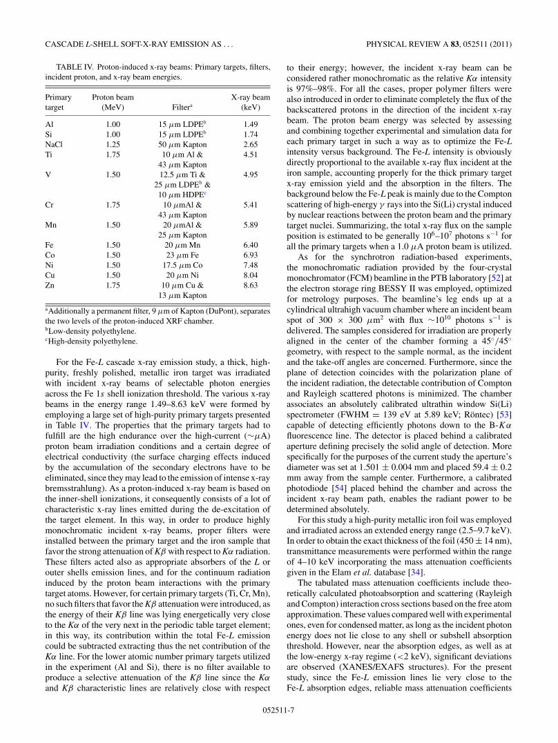
The tabulated mass attenuation coefficients include theo-retically calculated photoabsorption and scattering (Rayleighand Compton) interaction cross sections based on the free atomapproximation. These values compared well with experimentalones, even for condensed matter, as long as the incident photonenergy does not lie close to any shell or subshell absorptionthreshold. However, near the absorption edges, as well as atthe low-energy x-ray regime (<2 keV), significant deviationsare observed (XANES/EXAFS structures). For the presentstudy, since the Fe-L emission lines lie very close to theFe-L absorption edges, reliable mass attenuation coefficients
052511-7
D. SOKARAS et al. PHYSICAL REVIEW A 83, 052511 (2011)
FIG. 3. (Color online) Experimental (this work) and theoretical[34] mass attenuation coefficients for metallic Fe in the energyregion 500–1550 eV. The solid line represents the smoothed meanexperimental values obtained by multiple transmittance energy scans.
are of great importance in order to account properly forthe self-attenuation in the Fe sample. For this purpose, theexperimental mass attenuation coefficients were determinedfor the metallic Fe through transmittance measurementsof monochromatic undulator radiation within the range of500–1550 eV (with an energy step of 0.5–1 eV, the fineststeps were selected across the Fe-L absorption thresholds)at the plane grating monochromator (PGM) beamline [55](PTB laboratory at BESSY II), optimized also for metrologypurposes. Employing an absolutely calibrated photodiode afterthe sample (the metallic Fe foil) and across the incidentx-ray beam path, the transmitted radiant power was deter-mined. The transmittance measurements were converted toexperimental mass attenuation coefficients after accountingfor the thickness of the Fe foil, measured, as discussedabove, through another series of transmittance measurementsat higher photon energies where the tabulated-theoreticalmass attenuation coefficients are more reliable. The overalluncertainty for the determined mass attenuation coefficientswithin the energy range of 500–1550 eV is estimated to bewithin 5%−10%. These experimental values, obtained bymultiple transmittance energy scans, as well as their meansmoothed value (continuous line) are plotted in Fig. 3 andcompared to the tabulated ones by Elam et al. [34]. Apparently,very important deviations near the Fe-L absorption thresholdsare observed, while the deviations are gradually eliminated atthe higher photon energies.
IV. DATA ANALYSIS
The moderate resolution x-ray spectrometry studies providethe total detected Fe-L intensity for each of the employedincident x-ray beams. Common strategy in both sets of mea-surements is the absolute determination of the Fe-L intensitiesat every exciting x-ray beam energy per incident photonand steradian. At the same time, since metallic targets ofinfinite or intermediate thickness were employed during bothexperimental studies, all the secondary effects contributing to
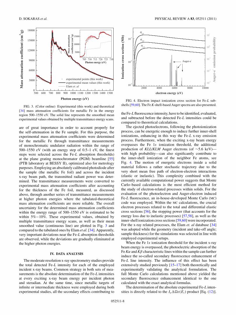
FIG. 4. Electron impact ionization cross section for Fe-L sub-shells [59,60]. The Fe K-shell-based Auger spectra are also presented.
the Fe-L fluorescence intensity, have to be identified, evaluated,and subtracted before the detected Fe-L intensities could becompared to theoretical calculations.
The ejected photoelectrons, following the photoionizationprocess, can be energetic enough to induce further inner-shellionizations, enhancing in this way the Fe-L x-ray emissionprocess. Furthermore, when the exciting x-ray beam energyoverpasses the Fe 1s ionization threshold, the additionalproduction of KLL/KLM Auger electrons (of ∼5.6 keV)—with high probability—can also significantly contribute tothe inner-shell ionization of the neighbor Fe atoms, seeFig. 4. The motion of energetic electrons inside a solidmaterial follows a rather stochastic trajectory due to thevery short mean free path of electron-electron interactions(elastic or inelastic). This complexity combined with thecurrently available computational power suggests that MonteCarlo–based calculations is the most efficient method forthe study of electron-related processes within solids. For theevaluation of the photoelectron and Auger-electron inducedFe-L fluorescence, an in-house-developed Monte Carlo (MC)code was employed. Within the MC calculations, the crucialelectron processes related to the total and differential elasticcross sections [56], the stopping power (that accounts for theenergy loss due to inelastic processes) [57,58], as well as theinner-shell ionization cross sections [59,60] were incorporated.For the x-ray related processes, the Elam et. al database [34]was adopted while the geometry (incident and take-off angle;sample thickness) for the simulations was selected in line withemployed experimental setups.
When the Fe 1s ionization threshold for the incident x-raybeam energy is overpassed, the photoelectric absorption of theFe Kα and Kβ characteristic lines within the sample can furtherinduce the so-called secondary fluorescence enhancement ofFe-L line intensity. The influence of this effect has beenextensively studied previously [15–17] both theoretically andexperimentally validating the analytical formulation. Thefull Monte Carlo calculations mentioned above yielded thesecondary fluorescence enhancement identical to the onecalculated with the exact analytical formulas.
The determination of the absolute experimental Fe-L inten-sities is directly related to the Io�ε(Ei) product [Eq. (12)].
052511-8
CASCADE L-SHELL SOFT-X-RAY EMISSION AS . . . PHYSICAL REVIEW A 83, 052511 (2011)
For the synchrotron radiation setup, being optimized formetrology purposes, each of these factors are determinedabsolutely. For the proton-induced XRF setup, this product hadto be found experimentally; the Io� term was determinedwith a 4% precision through fluorescence measurements ofpure monoelemental or well-characterized compound samples(down to fluorine Kα = 677 eV employing NaCl, Al, Si, CaF2,Ti) for each of the proton-beam irradiated primary targets.The detector efficiency for the energies of the Fe-L lines(mostly 700 eV) and upward was calculated based on theexperimentally measured transmission for the AP 1.7 ultrathinwindow (including the supporting silicon grid transmittance)provided by the manufacturer [61] and the nominal values forthe nickel contact (120 A) and the silicon dead layer (150 nm).The good precision of 4% in the determination of the Io�
term in the energy range covered by the fluorescence linesof the aforementioned calibration targets supports the modeladopted for the detector efficiency calculation.
V. RESULTS AND DISCUSSION
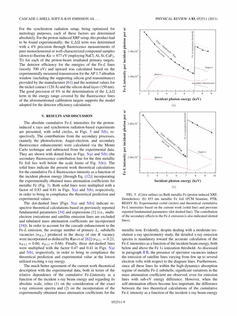
The absolute cumulative Fe-L intensities for the proton-induced x rays and synchrotron radiation-based experimentsare presented, with solid circles, in Figs. 5 and 5(b), re-spectively. The contributions from the secondary processes,(namely the photoelectron, Auger-electron, and secondaryfluorescence enhancement) were calculated via the MonteCarlo technique and subtracted from the experimental data.They are shown with dotted lines in Figs. 5(a) and 5(b) (thesecondary fluorescence contribution line for the thin metallicFe foil lies well below the scale limits of Fig. 5(b)). Thesolid lines indicate the present work theoretical calculationsfor the cumulative Fe-L fluorescence intensity as a function ofthe incident photon energy [through Eq. (12)] incorporatingthe experimentally obtained mass attenuation coefficients formetallic Fe (Fig. 3). Both solid lines were multiplied with afactor of 0.83 and 0.81 in Figs. 5(a) and 5(b), respectively,in order to bring in compliance the theoretical prediction andexperimental values.
The dot-dashed lines [Figs. 5(a) and 5(b)] indicate re-spective theoretical calculations based on previously reportedfundamental parameters [34] and expressions [1] (i.e., multi-electron ionizations and satellite emission lines are excludedand tabulated mass attenuation coefficients are incorporated[34]). In order to account for the cascade enhancement to theFe-L emission, the average number of primary Li subshellsvacancies (nKLi) produced in the decay of one K vacancywere incorporated as deduced by Rao et al. [62] (nKL1 = 0.21,nKL2 = 0.60, nKL3 = 0.66). Finally, these dot-dashed lineswere multiplied with the factor 0.47 and 0.41 in Figs. 5(a)and 5(b), respectively, in order to bring in compliance thetheoretical prediction and experimental value at the lowestutilized exciting x-ray energy.
The much better agreement of the current work theoreticaldescription with the experimental data, both in terms of therelative dependence of the cumulative Fe-Lintensity as afunction of the incident x-ray beam energy and regarding itsabsolute scale, relies (1) on the consideration of the exactx-ray emission spectra and (2) on the incorporation of theexperimentally obtained mass attenuation coefficients for the
(a)
(b)
FIG. 5. (Color online) (a) Bulk metallic Fe (proton induced XRF,Demokritos). (b) 453 nm metallic Fe foil (FCM beamine, PTB,BESSY II). Experimental (solid circles) and theoretical cumulativeFe-L intensities based on the current work (solid line) and previousreported fundamental parameters (dot-dashed line). The contributionof the secondary effects to the Fe-L emission is also indicated (dottedlines).
metallic iron. Evidently, despite dealing with a moderate res-olution x-ray spectrometry study, the detailed x-ray emissionspectra is mandatory toward the accurate calculation of theFe-L intensities as a function of the incident beam energy, bothbelow and above the Fe 1s ionization threshold. As discussedin paragraph II B, the presence of spectator vacancies inducethe emission of satellite lines varying from few up to severalelectron volts with respect to the diagram lines. Furthermore,since all these lines lie within the high-dynamics absorptionregime of metallic Fe-L subshells, significant variations in themass attenuation coefficient are observed, even for emissionlines with sub-eV energy difference. However, when theself-attenuation effects become less important, the differencebetween the two theoretical calculations of the cumulativeFe-L intensity as a function of the incident x-ray beam energy
052511-9
D. SOKARAS et al. PHYSICAL REVIEW A 83, 052511 (2011)
becomes less prominent. This becomes evident from the resultsfor the thin metallic Fe foil utilized at the synchrotron radiationexperiments [Fig. 5(b)], even though the difference in theabsolute scale still remains. It is interesting to note that thediscrepancy between the two theories increases as the incidentphoton energy varies from 1.5 to 7 keV [this is pronouncedmore in the bulk Fe case, see Fig. 5(a)]. It is evident thatin the pre-K-edge region the slope of the Fe-L intensityagainst exciting photon energy is determined by L1-, L2- andL3-photoionization cross sections. It is seen from Fig. 2 thatthe relative contribution of the L1 ionization increases with thegrowth of the exciting photon energy. The characteristic x-raylines emitted upon L1 photoionization are mainly emitted inthe presence of an additional vacancy (created by an L1L2 orL1L3 Coster Kronig transition); they lie energetically abovethe L3 ionization threshold and are thus strongly absorbed.Only accurate calculation of the cascade-produced emissionline energies allows one to account for this effect and to get anL-intensity dependence close to the experiment.
The cumulative uncertainties for the experimental absoluteFe-L intensities are estimated to be less than 10% in bothsets of experimental measurements. These come from theindividual uncertainties in the mass attenuation coefficients,in the thickness of the intermediate thickness Fe sample, in thecalibration of the proton-induced XRF setup, and the databasesfor the electron-related processes considered in the MonteCarlo simulations. Therefore, the absolute difference amongtheory and experiment, being 17%–19%, clearly exceeds the
TABLE V. Fundamental parameters for Fe as calculated in thiswork and compared with selected previously reported values.
This work Previous works
Fluorescence yieldsωK 0.369 0.351a
ωL1 0.000 45 0.000 40a
ωL2 0.005 94 0.0054a (0.0036)b
ωL3 0.005 64 0.0059a
Coster-Kronig transitionsf13 0.65 0.68a
f12 0.28 0.27a
f23 0.00 0.00a (0.42)b
Relative intensities1s-2p1/2 (Kα2) 0.298 0.297c
1s-2p3/2 (Kα1) 0.599 0.581c
1s-3p (Kβ) 0.104 0.122c
2s-2p1/2 (f ′12) 0.010 –
2s-2p3/2 (f ′13) 0.026 0.021d
2s-3p (Lβ2,3) 0.965 0.979d
2p1/2-3s (Ln) 0.076 0.082d
2p1/2-3d (Lβ1) 0.919 0.910d
2p1/2-4s 0.005 0.007d
2p3/2-3s (Ll) 0.075 0.086d
2p3/2-3d (Lα1,2) 0.920 0.907d
2p3/2-4s (Lβ6) 0.005 0.008d
aReferences [63] and [64].bSemiempirical corrections for condensed matter [65].cReference [66].dReference [67].
estimated experimental uncertainties which might be partiallyassigned to solid-state-related phenomena. Through the cal-culated partial generalized fluorescence yields (Table III), thefundamental atomic parameters for Fe, as obtained throughthe configuration-average Pauli-Fock approximation, havebeen extracted and compared with selected reported valuesin Table V. In general, a good agreement is observed withthe respective theoretically deduced fundamental parameterswhich are mainly based on Dirac-Hartree-Slater [63,64] andDirac-Fock [66,67] approximation. However, when comparedto certain semiempirically extracted values [65] (shown inTable V in parentheses) some significant discrepancies areseen. It has been previously reported [65,68–70] that althoughthe L2L3 Coster-Kronig process is energetically forbidden forthe Fe free atoms (as well as for some neighbor transitionmetals with unfilled 3d shell), this transition is allowed incondensed matter due to solid-state effects. This discrepancyinevitably affects also the other competitive relaxation pro-cesses and thus the fluorescence yield of the L2 shell (ωL2),too, which consequently also reduces the intensity of the Fe-Lfluorescence lines.
Another important observation is related to the Ll/Lα
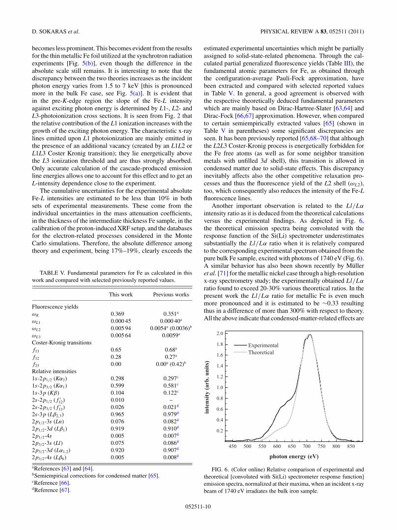
intensity ratio as it is deduced from the theoretical calculationsversus the experimental findings. As depicted in Fig. 6,the theoretical emission spectra being convoluted with theresponse function of the Si(Li) spectrometer underestimatessubstantially the Ll/Lα ratio when it is relatively comparedto the corresponding experimental spectrum obtained from thepure bulk Fe sample, excited with photons of 1740 eV (Fig. 6).A similar behavior has also been shown recently by Mulleret al. [71] for the metallic nickel case through a high-resolutionx-ray spectrometry study; the experimentally obtained Ll/Lα
ratio found to exceed 20-30% various theoretical ratios. In thepresent work the Ll/Lα ratio for metallic Fe is even muchmore pronounced and it is estimated to be ∼0.33 resultingthus in a difference of more than 300% with respect to theory.All the above indicate that condensed-matter-related effects are
FIG. 6. (Color online) Relative comparison of experimental andtheoretical [convoluted with Si(Li) spectrometer response function]emission spectra, normalized at their maxima, when an incident x-raybeam of 1740 eV irradiates the bulk iron sample.
052511-10
CASCADE L-SHELL SOFT-X-RAY EMISSION AS . . . PHYSICAL REVIEW A 83, 052511 (2011)
most probably responsible for these observed deviations, sincethey can affect considerably outermost-shell-related atomicprocesses which are not being described by theoretical modelsbased on the free atom approximation.
VI. CONCLUSIONS
In this work, through moderate resolution x-rayspectrometry, the cumulative L-shell soft-x-ray emission of Fewas found to be independent from the polarization state of theincident radiation–within the experimental uncertainities–inaccordance with the dipole approximation. The detailed abinitio theoretical approach, based on the direct constructionof the cascade de-excitation trees within the Pauli-Fockapproximation, showed that the exact calculation of the directand the cascade x-ray emission spectra is mandatory forthe accurate description of the absolute Fe-L x-ray emissionintensity when the exciting radiation energy is scanned acrossthe Fe 1s ionization threshold. Multielectron ionizations andcascade autoionization processes leading toward multivacancystates, induce the emission of satellite lines which incertain cases experience substantially different (higher)
self-attenuation as compared to the diagram emission lines. Inorder to correctly calculate the self-attenuation for the L-shellsoft x rays, experimental mass attenuation coefficients arefound to be essential. Further on, secondary ionizations due toenergetic electron motion can give considerable contribution tothe emission and have to be appropriately accounted for.An isolated-atom approximation sets certain limitationsregarding the description of specific inner-shell processeswhen compared to experimental observations in the metallicphase. The latter indicates the need for a complete overallab initio theory incorporating core-electron processesand condensed matter or chemical environment effects,especially when open-shell-related processes are investigated.The overall insights presented here are also expected toimprove considerably the reference-free x-ray fluorescencequantitative analysis applied for the characterization ofadvanced technological materials.
ACKNOWLEDGMENTS
One of the authors (A.G.K.) thanks his colleagues from“Demokritos” for their hospitality during his stay in Athens.
[1] W. Bambynek, B. Crasemann, R. W. Fink, H.-U. Freund,H. Mark, C. D. Swift, R. E. Price, and P. V. Rao, Rev. Mod.Phys. 44, 716 (1972).
[2] E. T. Verkhovtseva, E. V. Gnatchenko, P. S. Pogrebnyak, andA. A. Tkachenko, J. Phys. B 19, 2089 (1986).
[3] E. T. Verkhovtseva and P. S. Pogrebnjak, J. Phys. B 13, 3535(1980).
[4] A. G. Kochur, Ye. B. Mitkina, and V. L. Sukhorukov, J. Phys. B31, 5293 (1998).
[5] A. G. Kochur, V. L. Sukhorukov, and Ye. B. Mitkina, J. Phys. B33, 2949 (2000).
[6] G. Omar and Y. Hahn, Z. Phys. D 25, 41 (1992).[7] A. Moewes, R. G. Wilks, A. G. Kochur, and E. Z. Kurmaev,
Phys. Rev. B 72, 075129 (2005).[8] A. G. Kochur, S. Bruhl, I. D. Petrov, and Ye. B. Mitkina, Eur.
Phys. J. Special Topics 169, 51 (2009).[9] M. West, A. T. Ellis, P. Kregsamer, P. J. Potts, C. Streli,
C. Vanhoff, and P. Wobrauschek, J. Anal. At. Spectrom. 23,1409 (2008).
[10] M. Mantler, J. P. Willis, G. R. Lachance, B. A. R. Vrebos, K.-E.Mauser, N. Kawahara, R. M. Rousseau, and P. N. Brouwer,in Handbook of Practical X-Ray Fluorescence Analysis, editedby B. Beckhoff, B. Kanngiesser, N. Langhoff, R. Wedell, andH. Wolff (Springer, New York, 2006), pp. 309–410.
[11] R. M. Rousseau, X-Ray Spectrom. 13, 115 (1984).[12] R. M. Rousseau, X-Ray Spectrom. 13, 121 (1984).[13] B. Beckhoff, J. Anal. At. Spectrom. 23, 845 (2008).[14] B. Beckhoff, R. Fliegauf, M. Kolbe, M. Muller, J. Weser, and
G. Ulm, Anal. Chem. 79, 7873 (2007).[15] D. K. G. de Boer, X-ray Spectrom. 19, 145 (1990).[16] M. Mantler, Anal. Chim. Acta 188, 25 (1986).[17] A. G. Karydas, X-ray Spectrom. 34, 426 (2005).[18] N. Kawahara, T. Shoji, T. Yamada, Y. Kataoka, B. Beckhoff,
G. Ulm, and M. Mantler, Adv. X-ray Anal. 45, 511 (2002).
[19] B. Beckhoff, M. Kolbe, O. Hahn, A. G. Karydas, Ch. Zarkadas,D. Sokaras, and M. Mantler, X-ray Spectrom. 37, 462 (2008).
[20] A. Owens, B. Beckhoff, M. Kolbe, M. Krumrey, A. Mantero,M. Mantler, A. Peacock, M.-G. Pia, D. Pullan, U. G. Schneider,and G. Ulm, Anal. Chem. 80, 8398 (2008).
[21] C. J. Sparks Jr., Phys. Rev. Lett. 33, 262 (1974).[22] A. G. Karydas, S. Galanopoulos, Ch. Zarkadas, T. Paradellis,
and N. Kallithrakas-Kontos, J. Phys. Condens. Matter 14, 12367(2002).
[23] A. G. Karydas and Th. Paradellis, J. Phys. B 30, 1893 (1997).[24] Ch. Zarkadas, A. G. Karydas, M. Muller, and B. Beckhoff,
Spectrochim. Acta, Part B 61, 189 (2006).[25] M. Mueller, B. Beckhoff, G. Ulm, and B. Kanngiesser, Phys.
Rev. A 74, 012702 (2006).[26] D. Sokaras, M. Muller, M. Kolbe, B. Beckhoff, Ch. Zarkadas,
and A. G. Karydas, Phys. Rev. A 81, 012703 (2010).[27] A. G. Kochur, A. I. Dudenko, V. L. Sukhorukov, and I. D. Petrov,
J. Phys. B 27, 1709 (1994).[28] A. G. Kochur, V. L. Sukhorukov, A. I. Dudenko, and Ph. V.
Demekhin, J. Phys. B 28, 387 (1995).[29] R. Kau, I. D. Petrov, V. L. Sukhorukov, and H. Hotop, Z. Phys.
D 39, 267 (1997).[30] R. Karaziya, Sums of Atomic Quantities and Mean Characteris-
tics of Spectra (Mokslas, Vilnius, 1991).[31] V. L. Sukhorukov, V. F. Demekhin, V. V. Timoshevskaya, and
S. V. Lavrentev, Opt. Spectrosc. (USSR) 47, 407 (1979).[32] V. L. Sukhorukov, V. F. Demekhin, V. A. Yavna, A. I. Dudenko,
and V. V. Timoshevskaya, Opt. Spectrosc. (USSR) 55, 229(1983).
[33] D. A. Shirley, R. L. Martin, S. P. Kowalczyk, F. R. McFeely,and L. Ley, Phys. Rev. B 15, 544 (1977).
[34] W. T. Elam, B. D. Ravel, and J. R. Sieber, Radiat. Phys. Chem.63, 121 (2002).
[35] K. Siegbahn et al., Nova Acta R. Soc. Sci. Ups 20, (1967).
052511-11
D. SOKARAS et al. PHYSICAL REVIEW A 83, 052511 (2011)
[36] U. Gelius, Phys. Scr. 9, 133 (1974).[37] J. A. Bearden and A. F. Burr, Rev. Mod. Phys. 39, 125 (1967).[38] J. C. Fuggle and N. Martensson, J. Electron Spectrosc. Relat.
Phenom. 21, 275 (1980).[39] J. H. Scofield, Lawrence Livermore Laboratory, Report No.
UCRL-51326, 1973.[40] V. P. Sachenko and V. F. Demekhin, Sov. Phys. JETP 22, 532
(1966).[41] H. Ebel, R. Svagera, M. F. Ebel, A. Shaltout, and J. H. Hubbell,
X-ray Spectrom. 32, 442 (2003).[42] A. G. Kochur, A. M. Nadolinsky, and V. F. Demekhin, J. de
Phys. Colloque C8 47, C8 (1986).[43] T. Mukoyama and K. Taniguchi, Phys. Rev. A 36, 693 (1987).[44] A. G. Kochur and V. A. Popov, J. Phys. B 39, 3335 (2006).[45] A. G. Kochur and V. A. Popov, Radiat. Phys. Chem. 75, 1525
(2006).[46] A. G. Kochur, A. I. Dudenko, I. D. Petrov, and V. F. Demekhin,
J. Electron Spectrosc. Relat. Phenom. 156, 78 (2007).[47] V. F. Demekhin, V. L. Sukhorukov, V. A. Yavna, S. A. Kulagina,
S. A. Prosandeyev, and Yu. I. Bayrachny, Bull. Acad. Sci. USSRPhys. Ser. 40, 28 (1976).
[48] V. L. Sukhorukov, S. A. Yavna, V. F. Demekhin, and B. M.Lagutin, Koordinatsionnaya Khimiya (USSR) 22, 510 (1985).
[49] V. L. Sukhorukov, I. D. Petrov, V. F. Demekhin, and S. V.Lavrentiev, Izvesyiya Akademii Nauk USSR. Seriya Fizich-eskaya 49, 1463 (1985).
[50] V. L. Sukhorukov, I. D. Petrov, B. M. Lagutin, S. A. Yavna, andV. F. Demekhin, Koordinatsionnaya Khimiya (USSR) 12, 205(1986).
[51] R. D. Deslattes et al., Rev. Mod. Phys. 75, 35 (2003).[52] B. Beckhoff, A. Gottwald, R. Klein, M. Krumrey, R. Muller,
M. Richter, F. Scholze, R. Thornagel, and G. Ulm, Phys. StatusSolidi B 246, 1415 (2009).
[53] F. Scholze and M. Procop, X-Ray Spectrom. 30, 69 (2001).[54] A. Gottwald, U. Kroth, M. Krumrey, M. Richter, F. Scholze, and
G. Ulm, Metrologia 43, 125 (2006).[55] F. Senf, U. Flechsig, F. Eggenstein, W. Gudat, R. Klein,
H. Rabus, and G. Ulm, J. Synchrotron Radiat. 5, 780(1997).
[56] Z. Czyzewski, D. O. MacCallum, A. Romig, and D. C. Joy,J. Appl. Phys. 68, 3066 (1990).
[57] J. M. Fernandez-Varea, R. Mayol, D. Liljequist, and F. Salvat,J. Phys. Condens. Matter 5, 3593 (1993).
[58] A. Jablonski, S. Tanuma, and C. J. Powell, Surf. Interface Anal.38, 76 (2006).
[59] D. Bote, F. Salvat, A. Jablonski, and C. J. Powell, At. Data Nucl.Data Tables 95, 871 (2009).
[60] D. Bote and F. Salvat, Phys. Rev. A 77, 042701 (2008).[61] P. Smith, e2v Scientific Instruments (private communication).[62] P. Venugopala Rao, M. H. Chen, and B. Crasemann, Phys. Rev.
A 5, 997 (1972).[63] M. H. Chen, B. Crasemann, and H. Mark, Phys. Rev. A 21, 436
(1980).[64] M. H. Chen, B. Crasemann, and H. Mark, Phys. Rev. A 24, 177
(1981).[65] M. O. Krause, J. Phys. Chem. Ref. Data 8, 307 (1979).[66] J. H. Scofield, Phys. Rev. A 9, 1041 (1974).[67] J. H. Scofield, Phys. Rev. A 10, 1507 (1974).[68] S. L. Sorensen, S. J. Schaphorst, S. B. Whitfield, B. Crasemann,
and R. Carr, Phys. Rev. A 44, 350 (1991).[69] S. Iacobucci, F. Sirotti, M. Sacchi, and G. Stefani, J. Electron
Spectrosc. Relat. Phenom. 123, 397 (2002).[70] M. O. Krause, C. W. Nestor, C. J. Sparks, and E. Ricci, Oak
Ridge National Laboratory, Report No. ORNL-5399, 1978.[71] M. Muller, B. Beckhoff, R. Fliegauf, and B. Kanngiesser, Phys.
Rev. A 79, 032503 (2009).
052511-12