Carmel X-series - Calmar Lasercalmarlaser.com/docs/Carmel White Paper Rev 1.2 Feb 2016.pdf · The...
12
©2016 Calmar Laser, Inc www.calmarlaser.com Carmel X-series Microscopy and Biophotonics Applications Using Fiber-Based Femtosecond Lasers White Paper Revision 1.2 January 2016
Transcript of Carmel X-series - Calmar Lasercalmarlaser.com/docs/Carmel White Paper Rev 1.2 Feb 2016.pdf · The...

©2016 Calmar Laser, Inc www.calmarlaser.com
Carmel X-series Microscopy and Biophotonics Applications Using
Fiber-Based Femtosecond Lasers
White Paper
Revision 1.2 January 2016
200-1000-01 Using Fiber-Based Femtosecond Lasers in Microscopy and Biophotonics 2
Overview
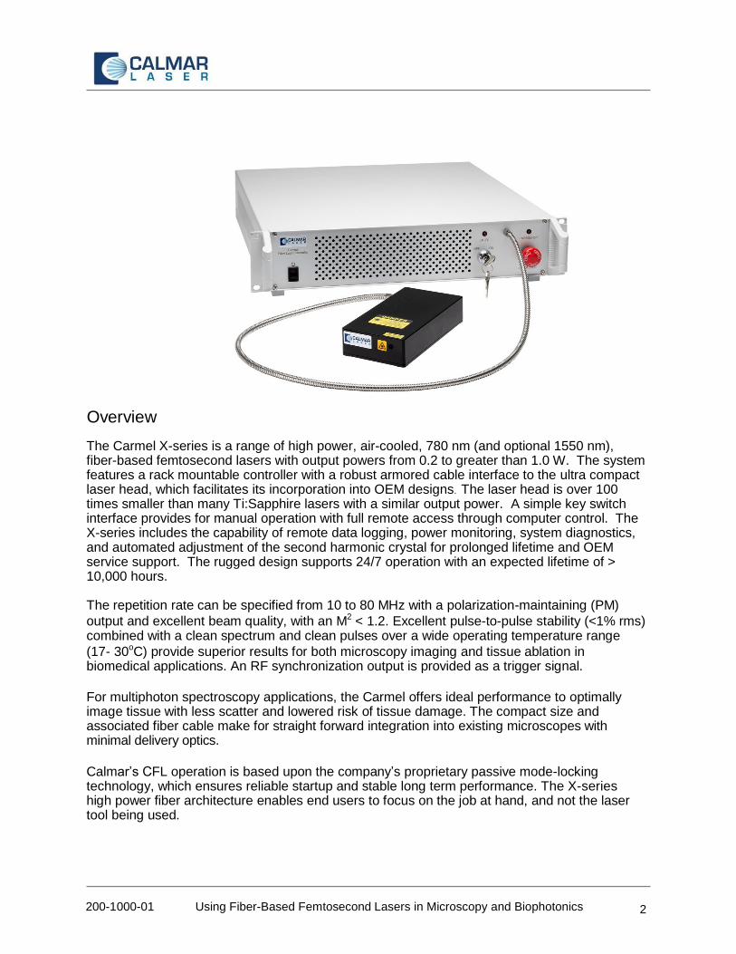
The Carmel X-series is a range of high power, air-cooled, 780 nm (and optional 1550 nm), fiber-based femtosecond lasers with output powers from 0.2 to greater than 1.0 W. The system features a rack mountable controller with a robust armored cable interface to the ultra compact laser head, which facilitates its incorporation into OEM designs. The laser head is over 100 times smaller than many Ti:Sapphire lasers with a similar output power. A simple key switch interface provides for manual operation with full remote access through computer control. The X-series includes the capability of remote data logging, power monitoring, system diagnostics, and automated adjustment of the second harmonic crystal for prolonged lifetime and OEM service support. The rugged design supports 24/7 operation with an expected lifetime of > 10,000 hours. The repetition rate can be specified from 10 to 80 MHz with a polarization-maintaining (PM)
output and excellent beam quality, with an M2 < 1.2. Excellent pulse-to-pulse stability (<1% rms) combined with a clean spectrum and clean pulses over a wide operating temperature range
(17- 30oC) provide superior results for both microscopy imaging and tissue ablation in biomedical applications. An RF synchronization output is provided as a trigger signal.
For multiphoton spectroscopy applications, the Carmel offers ideal performance to optimally image tissue with less scatter and lowered risk of tissue damage. The compact size and associated fiber cable make for straight forward integration into existing microscopes with minimal delivery optics.
Calmar’s CFL operation is based upon the company’s proprietary passive mode-locking technology, which ensures reliable startup and stable long term performance. The X-series high power fiber architecture enables end users to focus on the job at hand, and not the laser tool being used.

200-1000-01 Using Fiber-Based Femtosecond Lasers in Microscopy and Biophotonics 3
Features
Field proven product reliability
Ultra compact laser head
Easy to use
Wavelength 780 nm
Pulse energy > 12 nJ
Pulse widths < 90 fs with negligible pulse pedestal
RF synchronization output
High repetition rate up to 80 MHz
Air cooled – no chiller required
Low power consumption
Excellent beam pointing and pulse energy stability
Full computer control and data logging capability
Remote system diagnostics
Expected lifetime > 10,000 hours
Background Since 1996, Calmar has been a technology leader in the design and manufacture of all-fiber ultrafast lasers for the ophthalmology, microelectronics, solar, telecom, and scientific research markets. Fiber lasers offer the advantages of long life semiconductor diode combined with the efficiency, reliability, performance and low cost of optical fiber amplification. These key advantages have created a significant momentum to adopt fiber laser technology (versus legacy laser technologies) for a wide range of research and industrial applications.
Calmar’s femtosecond laser sources are passively mode-locked fiber lasers. Passive mode- locking makes these lasers easier to operate than actively mode-locked lasers, as no external RF clock signal is required, and little or no warm-up time is needed. Temperature control is also less of an issue with passive mode-locked lasers.
Calmar’s passively-mode-locked lasers produce pulses as narrow as 80 fs wide. Repetition rates are fixed in the range 10 - 100 MHz. The peak output power of a femtosecond laser is, of course, high due to the short pulse durations, and peak powers up to 10 kW can be achieved using an integrated fiber amplifier (based on Erbium (EDFA) or Ytterbium fibers (YDFA). Figure 1 shows a simplified schematic of a passively mode-locked fiber laser.
Figure 1 – Schematic of Passive Mode-locked Fiber Laser

200-1000-01 Using Fiber-Based Femtosecond Lasers in Microscopy and Biophotonics 4
Since Calmar’s fiber lasers are manufactured from discrete components, dispersive and non-linear effects can be carefully controlled. Pulse shape is transform-limited, and the pedestal is typically 20dB lower than the signal.
Calmar’s lasers are recognized for their stability, as demonstrated by their low timing jitter and low amplitude noise, thereby ensuring that the quality of the laser output meets even the most stringent test requirements

200-1000-01 Using Fiber-Based Femtosecond Lasers in Microscopy and Biophotonics 5
Carmel X-series Specifications
The following table provides specifications for the Carmel X-series Femtosecond Fiber laser.

200-1000-01 Using Fiber-Based Femtosecond Lasers in Microscopy and Biophotonics 6
Carmel X-series Dimensions
The Laser Head and Controller are connected by a flexible shielded fiber with length of 80 cm. Laser Head
Scale in inches
Controller
Scale in inches

200-1000-01 Using Fiber-Based Femtosecond Lasers in Microscopy and Biophotonics 7
Carmel X-series Performance The following test results provide an indication of the optical performance of the X-series.

200-1000-01 Using Fiber-Based Femtosecond Lasers in Microscopy and Biophotonics 8
Beam Quality
Power Stability
200-1000-01 Using Fiber-Based Femtosecond Lasers in Microscopy and Biophotonics 9
Carmel Applications
The Carmel femtosecond laser has many applications:
Multiphoton microscopy
Biophotonics
Ti:sapphire oscillator
Materials characterization
Optical coherent tomography
Non-linear optical studies
Optical metrology
Terahertz radiation
Intra-Tissue Refractive Index Shaping Multi-Photon Microscopy
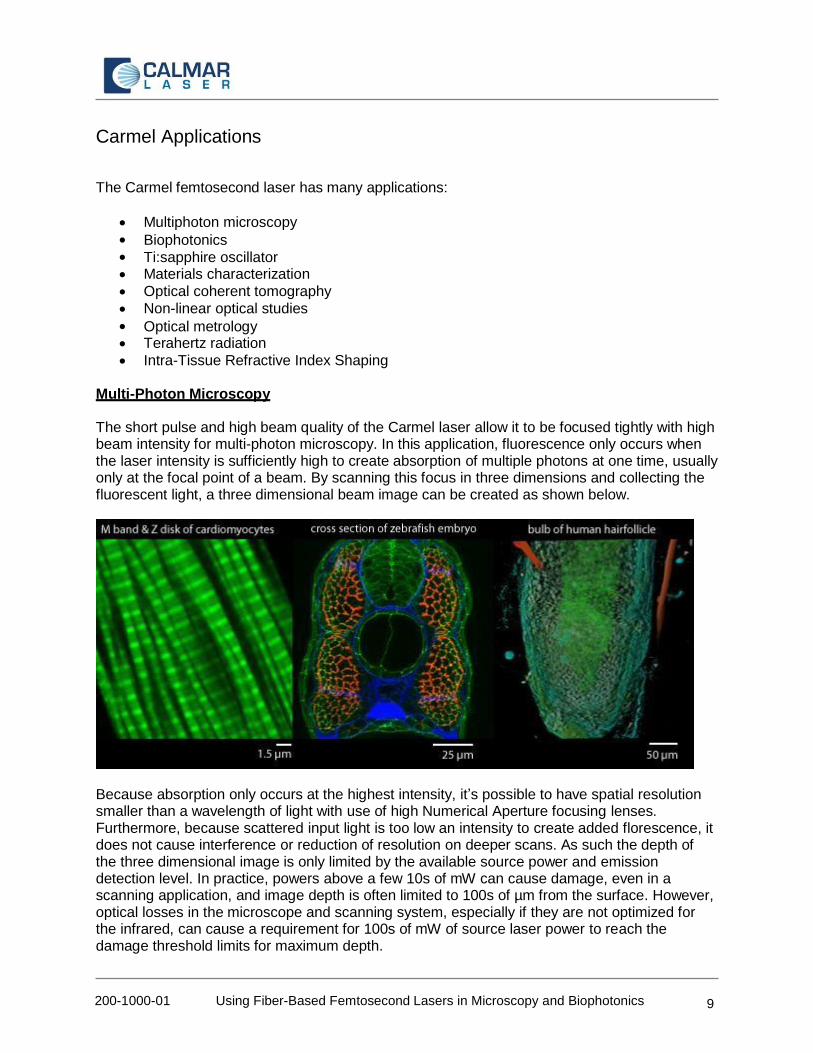
The short pulse and high beam quality of the Carmel laser allow it to be focused tightly with high beam intensity for multi-photon microscopy. In this application, fluorescence only occurs when the laser intensity is sufficiently high to create absorption of multiple photons at one time, usually only at the focal point of a beam. By scanning this focus in three dimensions and collecting the fluorescent light, a three dimensional beam image can be created as shown below.
Because absorption only occurs at the highest intensity, it’s possible to have spatial resolution smaller than a wavelength of light with use of high Numerical Aperture focusing lenses. Furthermore, because scattered input light is too low an intensity to create added florescence, it does not cause interference or reduction of resolution on deeper scans. As such the depth of the three dimensional image is only limited by the available source power and emission detection level. In practice, powers above a few 10s of mW can cause damage, even in a scanning application, and image depth is often limited to 100s of µm from the surface. However, optical losses in the microscope and scanning system, especially if they are not optimized for the infrared, can cause a requirement for 100s of mW of source laser power to reach the damage threshold limits for maximum depth.

200-1000-01 Using Fiber-Based Femtosecond Lasers in Microscopy and Biophotonics 10
Compared to Confocal Microscope Imaging, Multi-photon Imaging relies on a nonlinear absorption process which offers the following advantages:
• Inherent z-axis (depth) resolution • Higher resolution- close or even beyond the diffraction limit (multiphoton excitation) • Fast – capable of following physiological processes • Higher sensitivity - no confocal aperture that blocks up to 99% of emission • Increased penetration depth due to reduced interference from scattered light. • Reduced photodamage/ photobleaching • In-vivo imaging with reduced photodamage to sample. • Possibility of fluorescent lifetime imaging • A Confocal Microscope system is complex and for UV excitation needs fused silica optics
Of note, new dyes and florophores have become readily available with improved absorption in the 780 nm range.
References for Multi-Photon Imaging
Warren R. Zipfel, Rebecca M. Williams, Richard Christie, Alexander Yu Nikitin, Bradley T. Hyman,and Watt W. Webb, “Live tissue intrinsic emission microscopy using multiphoton-excited native fluorescence and second harmonic generation”, PNAS
Fritjof Helmchen & Winfried Denk, “Deep Tissue two-photon microscopy”, Nature Methods, Vol. 2, No. 12, Dec. 2005, p. 932
Related Web Links:
Live tissue intrinsic emission microscopy using multiphoton-excited native fluorescence and second harmonic generation, Warren R. Zipfel, Rebecca M. Williams, Richard Christie, Alexander Yu Nikitin, Bradley T. Hyman,and Watt W. Webb*
Small animal in vivo imaging market to reach $1.55B in 2017, says report
Microscopy technique heightens understanding of brain cell death during stroke
Two-photon excited fluorescence imaging is useful for identifying ovarian tumors in mice

200-1000-01 Using Fiber-Based Femtosecond Lasers in Microscopy and Biophotonics 11
Intra-Tissue Refractive Index Shaping (IRIS)
Another femtosecond laser application for vision correction is Gradient Index Microlenses generated by scanning femtosecond laser pulses inside the cornea for in situ creation of customized Intraocular Lenses (IOLs). This technique allows correction for errors in cataract surgeries that are can occasionally have more than 1 Diopter of residual refractive error.
Using femtosecond laser pulses to target a predetermined 3-D space can typically cause a 1% change in refractive index. Using a 500-mW laser, an inscribing speed of 120 mm/sec and higher could be achieved
with 0.8-µm resolution of the inscribing pattern. [1]
Ohthalmic hydrogel polymers are micro- machined with near-infrared femtosecond laser pulses. Refractive index changes up to +0.05 have been obtained, and lateral gradient index refractive structures are written into the flat polymers. By measuring the transmitted wavefront of the micromachined polymer, astigmatism as high as 0.8 diopters can be induced in the micromachined region. [2]
Interferogram of polymer sample with cylindrical lens written in dotted rectangular area. The solid curve represent the trend of fringe of the bulk sample. The additional curve in rectangular area shows additional parabolic phase added to the bulk sample fringes. [2]
References for IRIS:
1. “Laser advances custom intraocular lenses in situ”, Ophthalmology Times, Feb 15, 2011.
2. “Lateral gradient index microlenses written in ophthalmic hydrogel polymers by femtosecond laser micromachining”, Optical Materials Express, Vol. 1, Issue 8, pp. 1416- 1424 (2011)

200-1000-01 Using Fiber-Based Femtosecond Lasers in Microscopy and Biophotonics 12
For more information on our Compact Fiber Laser, Picosecond Fiber Laser series, Femtosecond Fiber Laser series, or any other Calmar products, please contact us.
E-mail:
Telephone:
+1 650.272.6980, extension 110
Fax:
+1 650.272.6988
Address:
Calmar Laser 951 Commercial Street Palo Alto, CA 94303 USA