Carlos Adriano Albuquerque Andrade de Matos
209
Transcript of Carlos Adriano Albuquerque Andrade de Matos


Carlos Adriano Albuquerque Andrade de Matos
Regulation of ataxin-3 by phosphorylation:
Relevance for Machado-Joseph disease
Tese de Doutoramento em Biologia, na especialidade de Biologia Celular, orientada pela Professora Doutora Ana Luísa Monteiro de Carvalho e
pela Professora Doutora Sandra de Macedo Ribeiro e apresentada ao Departamento de Ciências da Vida da Faculdade de Ciências e Tecnologia
da Universidade de Coimbra
Julho de 2014


O trabalho aqui apresentado foi realizado no Centro de Neurociências e
Biologia Celular – CNC, Universidade de Coimbra, Portugal e no Instituto de Biologia
Molecular e Celular – IBMC, Porto, Portugal e financiado pela Fundação para a Ciência
e Tecnologia.
The work hereby presented was performed at the Center for Neuroscience and
Cell Biology – CNC, University of Coimbra, Coimbra, Portugal and at the Institute for
Molecular and Cell Biology – IBMC, Porto, Portugal and funded by the Portuguese
Foundation for Science and Technology.
SFRH/BD/47160/2008; PTDC/SAU-NMC/110602/2009; PEst-C/SAU/LA0001/2013-2014

Nota de capa
Pormenor de Sorrowing Old Man (At Eternity’s Gate), de Vincent van Gogh. A
imagem encontra-se no domínio público.
Cover nove
Detail of Sorrowing Old Man (At Eternity’s Gate), by Vincent van Gogh. The
image is in the public domain.

A toda a minha família
e em especial aos Andrades


“He no longer saw the face of his friend Siddhartha, instead he saw other faces, many, a long sequence, a flowing river of faces, of hundreds, of thousands, which all came and disappeared, and yet all seemed to be there simultaneously, which all constantly changed and renewed themselves, and which were still all Siddhartha. He saw (…) all of these figures and faces in a thousand relationships with one another, each one helping the other, loving it, hating it, destroying it, giving re-birth to it, each one was a will to die, a passionately painful confession of transitoriness, and yet none of them died, each one only transformed, was always re-born, received evermore a new face, without any time having passed between the one and the other face—and all of these figures and faces rested, flowed, generated themselves, floated along and merged with each other (…).”
in Shiddhartha by Hermann Hess
“All we have to decide is what to do with the time that is given to us.”
- Gandalf, in The Lord of the Rings By J.R.R. Tolkien
Agradecimentos
Quando se procura agradecer a alguém por uma contribuição para um trabalho de
anos (cinco, neste caso), poderá torna-se difícil escolher a pessoa a quem se dirigem as
primeiras palavras. No meu caso, porém, considero-me com sorte, pois a escolha foi sempre
clara para mim desde o início. Começo, pois, por agradecer à minha orientadora, a Professora
Ana Luísa Carvalho. Em primeiro – em primeiríssimo – lugar, agradeço-lhe pelas aulas de
Biologia Celular e Molecular a que tive o prazer de assistir no anfiteatro do antigo
Departamento de Zoologia, há quase dez anos. Quando decidi entrar na Universidade de
Coimbra para estudar Biologia, o meu gosto por esta ciência estava muito direccionado para as
áreas da Biodiversidade e da Ecologia, embora me tenham sempre fascinado e interessado
também as áreas mais moleculares da Biologia. Foram talvez (ou talvez certamente) as suas
aulas que me começaram a direccionar para aquele que viria a ser um percurso académico
voltado para a Biologia Celular e Molecular, na esperança de um dia poder vir eu próprio a
fazer investigação e a contribuir para esta área do conhecimento científico. Em segundo lugar
agradeço-lhe assim a oportunidade que me deu de trabalhar consigo durante o meu projecto
de Mestrado e de me ter permitido expandir esse trabalho para o Doutoramento sobre cujo
trabalho versa esta tese. Agradeço-lhe todos os conselhos, todas as observações pertinentes,
todas as sugestões certeiras, todo o conhecimento que me transmitiu e todo o espírito
científico que procura incutir aos seus alunos e que tão bem exemplifica. Agradeço a
motivação que sempre me transmitiu, o ânimo, a confiança nas minhas capacidades que
sempre reconheci e que espero honrar com o presente trabalho. Finalmente, ficarei para
sempre grato pelo facto de ter sempre tolerado os meus erros, as minhas falhas e as minhas
incertezas e por ter sempre reconhecido que, não obstante o dever do trabalho que
desenvolvemos, é importante viver também outras coisas. Graças a isso tive a oportunidade
de perseguir outros sonhos, e de cumprir um ou dois.
Agradeço de igual modo à Doutora Sandra Ribeiro, cuja distância durante este período
de Doutoramento foi unicamente física, estando na verdade sempre presente durante todo o
processo. Agradeço-lhe toda a orientação que me deu durante estes anos, bem como o facto
de me ter permitido obter alguns dos materiais cruciais para muitas das experiências aqui
mencionadas. Agradeço-lhe a confiança que depositou em mim e o seu modo inspirador de
olhar para os problemas científicos. Não posso deixar também de agradecer a oportunidade
que me deu de trabalhar noutros projectos para além daquele que aqui apresento.
Devo o maior dos agradecimentos aos meus pais. Para além de serem os responsáveis
por eu ter tido as condições e a oportunidade para estudar e para ingressar no Douramento,
são sem dúvida os principais responsáveis pelo meu gosto pela ciência e pela liberdade que
tive em escolher as vias que escolhi. Agradeço-lhes o modo como cuidam de mim e se
preocupam e me fazem sentir amado e capaz de fazer qualquer coisa neste mundo. Vocês são
os maiores e esta tese é para vocês.

À minha maninha Marcela agradeço o apoio, a preocupação, a alegria, a sensatez e a
ligação que sempre manteve comigo e que é única. Agradeço-lhe as risadas, as conversas
sérias e a introspecção que delas deriva. Um grande abraço fica aqui também para o Eduardo,
a quem agradeço toda a amizade e ajuda para tantas coisas. Ao meu maninho João estarei
sempre grato pelo companheirismo, pela amizade, pela boa energia que transmite a quem o
rodeia e em especial a mim. Sempre foi o meu melhor amigo, e assim vai continuar a ser.
Manos, esta tese é para vocês também.
À minha avó Ilda agradeço um amor que nunca lhe conseguirei agradecer por palavras,
à minha avó Fernanda o cuidado de tantos anos, às minhas tias Ni e Zi a vivacidade e o
interesse com que fazem tudo e com que me motivaram para a ciência (ainda que talvez não o
saibam). Agradeço a todo o resto da minha família o omnipresente apoio e interesse e um
amor puro e único, nomeadamente ao meu tio Cá, à minha tia Anabela, à minha tia Lúcia, ao
Acácio e a todos os meus primos que menciono aqui por ordem meramente etária e de quem
lamento não ter estado mais próximo nestes anos que passaram: Inês, João, Beatriz, Ana
Carolina, Maria Inês, Maria Carolina.
A ti, Clévio, agradeço por muito mais do que seria possível conter nesta página.
Obrigado por tudo o que fizeste por mim nestes últimos anos, por todo o companheirismo,
pela amizade, pela aventura, pela alegria e pelo gosto de viver. Obrigado por me teres
suportado nestes últimos tempos, por seres compreensivo e teres contribuído para que eu
tenho acabado a escrita desta tese sem ter perdido o juízo. Obrigado pelas partidas de squash,
pelos jantares e pelos momentos de diversão. Agradeço-te também por acreditares em mim e
por seres um grande exemplo de competência e de espírito de trabalho. E agradeço, claro está,
pela ajuda – pela enorme ajuda – com as experiências com o modelo lentiviral de rato. Sem o
teu auxílio esta tese seria bem mais pobre.
Agradeço ao Doutor Carlos Duarte tudo que me ensinou acerca de Neurobiologia, todo
o contributo técnico para o trabalho aqui apresentado e a boa disposição com que sempre me
motivou a investir nos meus sonhos. Estou muito grato também ao Doutor Ramiro Almeida e
ao Doutor João Peça pela discussão interessante e perspicaz do meu trabalho. Ao Doutor Luís
almeida agradeço a oportunidade que tive no seu laboratório com o modelo lentiviral de MJD
em ratos, pelo seu interesse, pela sua apreciação e pela sua boa disposição.
Devo o maior agradecimento a todos os meus colegas de laboratório, sem excepção. A
todos vós estou grato pelo ânimo, pela energia criadora e compassiva que todos
demonstraram para comigo nestes quase 6 anos. Agradeço à Tatiana as palhaçadas e as
conversas construtivas, ao Luís o seu companheirismo e o exemplo da sua dedicação ao
trabalho, à Sandra a alegria e toda a ajuda e interesse, ao Graciano (o pórpio) a boa disposição,
ao Rui, ao Michelle e ao Ivan a amizade, à Miranda, à Gladys, à Dominique e à Mariline a
simpatia, à Marta as risadas e ao Pedro Afonso a sua boa disposição contagiante e a sua
sinceridade. Deixo um agradecimento também à Joana Ferreira pelo seu interesse, pela sua
ajuda, pela grande paciência e pelo facto de ter estado quase sempre a meu lado, literalmente.
Devo um agradecimento especial também à Joana Fernandes, pela amizade que tivemos e da
qual espero continuar a ser merecedor. Agradeço à Susana o enorme interesse que sempre

demonstrou no meu trabalho, a paciência, e o modo crucial como me ajudou com as
experiências que utilizaram as culturas de córtex. Sem ti esta tese estaria incompleta.
Agradeço à Rita o facto de me ter ensinado a fazer os ensaios com a Ub-AMC e a Ub-VS e à
Doutora Luísa Cortes a ajuda com a microscopia. Um obrigado não fica esquecido para a
Mariliza e a sua contribuição para os estudos de agregação. Fica aqui ainda um grande beijinho
para a Dona Céu e para a Elisabete, por toda a simpatia e pelo facto de tornarem o trabalho no
laboratório mais fácil.
Agradeço às princesas Mariana, Marisa e Sara a boa disposição e a preocupação e à
Célia a alegria e a ajuda na formatação desta tese. Deixo também um agradecimento ao Mário
Laço pela ajuda com os ensaios de actividade com cadeias de polyUb e à Joana Santos e ao
Bruno Almeida pelo auxílio com a produção e atx3 recombinante. Agradeço à Doutora Patrícia
Maciel a oportunidade para aprender e trabalhar no seu laboratório, à Andreia Castro e à Ana
Jalles toda a ajuda com as C. elegans e à Dulce Almeida, à Fátima Ramalhosa, à Francisca e às
outras meninas do Minho o modo como me acarinharam.
Agradeço aos meus grandes amigos Carlos, por acreditar em mim e nas minhas
capacidades, pelos momentos de alegria e diversão e pelas conversas sobre trabalho e sobre
os mistérios do mundo. Ao Pedro serei sempre grato pelo companheirismo e pela sabedoria e
pela grande e eterna amizade. Deixo um obrigado à Renata, por ser uma amiga tão antiga mas
sempre tão presente e à Joana Vindeirinho pela alegria e por ser sempre compincha. Para o
maior viola de fado, Alexis Simões, para o maior guitarrista, Hugo Paiva de Carvalho, e para o
rouxinol José Branco fica um grande e Minervino abraço de obrigado pela vossa amizade e
pelo vosso interesse no meu trabalho.
Finalmente, agradeço a todas as pessoas que fazem de mim uma pessoa feliz e que
eventualmente não estejam mencionados nos parágrafos acima incluídos. Deste modo, e
muito resumidamente, um obrigado à malta de Biologia por me ligarem a um passado saudoso,
à malta do japonês, aos meus amigos do Japão e, claro está, aos meus amigos de sempre e
para sempre: André, Bekas, Filipe, Joana, Joana.
ओं मणिपदे्म ह ं


1
Table of contents
Table of contents 1
Abbreviations 5
Resumo 9
Abstract 11
Chapter 1 – Introduction 15
1.1 Neurodegenerative diseases 15
1.2 Machado-Joseph disease 16
Description and epidemiology 16
Clinical features and neuropathology 16
Genetic causes 18
1.3 Polyglutamine diseases 19
Machado-Joseph disease in the context of trinucleotide repeat disorders 19
Repeat length and pathology 19
Repeat instability 21
Aggregation and inclusion bodies 22
Toxicity mechanisms 23
The role of protein context 24
1.4 Ataxin-3 25
Distribution and ubiquity 25
Intracellular localization 25
Structure, domains and enzymatic activity 26
Clues to ataxin-3 function and biologic role 28
A. Ubiquitination 29
B. Deubiquitinating enzymes 30
C. Ataxin-3 deubiquitinase activity and ubiquitin binding – possible involvement with the
ubiquitin-proteasome pathway 31
D. Ataxin-3 interactors and protein homeostasis systems 34
E. Ataxin-3 and E3 ubiquitin ligases 36
F. Ataxin-3 and transcription regulation 38
G. Roles of ataxin-3 in the organization of the cytoskeleton and myogenesis 39
H. Summary of ataxin-3 proposed roles 40
Intracellular transport and shuttling 42
Atx3 degradation and turnover 42
1.5 Toxic consequences of ataxin-3 polyglutamine expansion 43
Polyglutamine sequences as causes of toxicity 43
Intracellular inclusion bodies 44
Polyglutamine aggregation and toxicity 45
Mechanisms of ataxin-3 aggregation 45

2
Proteolytic cleavage and toxic fragment hypothesis 47
Polyglutamine expansion-induced changes in ataxin-3 features 50
A. Effects on deubiquitinase activity and ubiquitin interaction 50
B. Effects on ataxin-3 interactions 51
C. Effects on transcription regulation 52
D. Aberrant interactions as the source of toxicity 53
Ataxin-3 as a protector against polyglutamine toxicity 53
The role of the nuclear environment 54
1.6 Ataxin-3 toxicity mechanisms, cellular context and neuronal damage 55
Mechanisms of cellular toxicity 55
Neuronal toxicity and dysfunction 56
Deubiquitinases and ubiquitination in the nervous system 58
A. Deubiquitinases and neuronal structure 59
B. Deubiquitinases and synaptic transmission 60
C. Possible links between ataxin-3 biologic role and neuronal function 61
1.7 Post-translational modifications as regulators of protein activity and toxicity 62
Cell context and post-translational modifications 62
Regulation of deubiquitinating enzymes by post-translational modifications 64
Modulation of polyglutamine toxicity by post-translational modifications 66
A. Phosphorylation of huntingtin 67
B. Phosphorylation of other polyglutamine disease-causing proteins 70
Post-translational modifications of ataxin-3 72
1.8 Objectives 75
Chapter 2 – Materials and Methods 79
Expression plasmids 79
Site-directed mutagenesis 79
Antibodies 80
Mass spectrometry 80
Generation of the phospho-specific antibody 83
Mammalian cell line culture and transfection 84
Primary cortical neuron culture 84
Transfection of cortical neurons 85
Chemical stimuli 85
Bacterial expression and purification of recombinant proteins 86
Production of lentiviral vectors 87
Infection of cortical neurons – ataxin-3 silencing 88
Preparation of cell extracts 88
Lambda phosphatase reaction 88
SDS-PAGE and Western blot 89

3
Protein structure analysis, sequence alignment and molecular weight estimation 90
Activity assays with Ub-AMC 90
Activity assays with polyUb chains 91
Active deubiquitinase-labelling assays with Ub-VS 91
Immunocytochemistry and fluorescence microscopy 91
Generation of the lentiviral rat model 92
Histological processing 93
Immunohistochemistry 93
Quantitative analysis of immunohistochemistry images 94
Statistical analysis 94
Chapter 3 – Phosphorylation of serine 12 of ataxin-3: validation and functional
consequences 97
3.1 Introduction – Detection of serine 12 as a phosphorylation site of ataxin-3 by mass
spectrometry 97
3.2 Detection of phosphorylated ataxin-3 in cell lines and neurons 98
Generation of the phospho-specific antibody anti-Patx3 98
Detection of serine 12 phosphorylation with the anti-Patx3 antibody 100
Confirming phosphorylation – Lambda phosphatase experiments 103
Confirming ataxin-3 detection – Ataxin-3 silencing strategies 103
3.3 Ataxin-3 structure analysis and functional predictions 105
3.4 Generation of ataxin-3 phosphomutants 109
3.5 Ataxin-3 phosphorylation at serine 12 affects its proteolytic activity 110
In vitro activity assays with Ub-AMC 110
In vitro activity assays with polyUb chains 111
Ub-VS binding assays 114
Chapter 4 – Phosphorylation of serine 12 of ataxin-3: consequences for
polyglutamine-induced toxicity 121
4.1 Ataxin-3 phosphorylation at serine 12 prevents the decrease in neuronal dendritic
length caused by polyglutamine expansion 121
4.2 Ataxin-3 phosphorylation at serine 12 prevents excitatory synapse loss triggered by
polyglutamine expansion 127
4.3 Ataxin-3 phosphorylation at serine 12 affects its aggregation in cortical neurons 133
4.4 Phosphomutation of serine 12 reduces aggregation and neurodegeneration in a
lentiviral rat model of MJD 137
Chapter 5 – Final considerations 119
5.1 General discussion 119
Phosphorylation of ataxin-3 fragments 120

4
Phosphorylation of serine 12 and atx3 deubiquitinase activity 123
Ataxin-3 in the cerebral cortex 126
PolyQ-expansion and phosphorylation of serine 12 as determinants of neuronal morphology 128
Phosphorylation of serine 12 as a protector against expanded ataxin-3-induced toxicity 130
5.2 Future perspectives 134
5.3 Conclusion 137
References 139

5
Abbreviations
6His hexahistidine
ALP alkaline phosphatase
AMPA α-amino-3-hydroxy-5-methyl-4-isoxazolepropionic acid
AMPAR AMPA receptor
ANOVA analysis of variance
AR androgen receptor
ATM protein kinase ataxia-telangiectasia mutated
atx3 ataxin-3
atx3L ataxin-3-like protein
BACE457Δ β-sile amyloid precursor protein cleaving enzyme
BCA bicinchoninic acid
BLAST Basic Local Alignment Seach Tool
BSA bovine serum albumin
CAA cytosine-adenine-adenine
CACNA1A calcium channel, voltage dependent, P/Q type, α1A subunit
CAG cytosine-adenine-guanine
CaMK calcium/calmodulin-dependent protein kinase
CaMK2 calcium/calmodulin-dependent protein kinase 2
CBP CREB-binding protein
Cdc2 cell division cycle 2 kinase
Cdk1 cycling-dependent kinase 1
Cdk5 cyclin-dependent kinase 5
CHIP C-terminus of Hsp70-interacting protein
CK1 casein kinase 1
CK2 casein kinase 2
CLAP chymostatin, pepstatin, antipain and leupeptin
CNS central nervous system
CREB cAMP response element-binding protein
CRM1 chromosome region maintenance 1
CRMP-2 collapsing response mediator protein-2
DARPP-32 dopamine- and cyclic AMP-regulated phosphoprotein of 32 kDa
DIV days in vitro
DMEM Dulbecco’s Modified Eagle’s Medium
DNA PK DNA-dependent protein kinase
DRPLA dentatorubral-pallidoluysian atrophy
DTT dithiothreitol
DUB deubiquitinase; deubiquitinating enzyme
DUB-1 Ub C-terminal hydrolase
DUBA deubiquitinating enzyme A
ECF Enhanced Chemifluorescence substrate
EMBL-EBI European Molecular Biology Laboratory – European Bioinformatics
Institute

6
ER endoplasmic reticulum (in figures, only)
ERAD endoplasmic reticulum-associated degradation
FBS fetal bovine serum
FOXO forkhead box class O
FOXO4 FOXO transcription factor 4
FRAP fluorescence recovery after photobleaching
FSK forskolin
GABA γ-aminobutyric acid
GABAR GABA receptor
GFP green fluorescent protein
GluR6 glutamate receptor channel subunit β2
GSK3 glycogen synthase kinase 3
GSK3β glycogen synthase kinase 3β
HBSS Hank’s balanced salt solution
HD Huntington’s disease
HDAC3 histone deacetylase 3
HDAC6 histone deacetylase 6
HEK human embryonic kidney
HEPES 4-(2-hydroxyethyl)-1-piperazineethanesulfonic acid
Hsp70 70 kDa heat shock protein
htt huntingtin
IGF1 insulin-like growth factor
IgG immunoglobulin G
IH immunohistochemistry (in figures, only)
IKK IκB kinase
InsP3R1 type 1 inositol 1,4,5-trisphosphate receptor
IPB immunoprecipitation buffer
iPSCs induced pluripotent stem cells
JAMM JAB1/MPN/MOV34 metalloenzymes
JD Josephin domain
JNK c-Jun N-terminal kinase
KLH keyhole limpet hemocyanin
KO knockout
LB Lysogeny Broth
LC liquid chromatography
LC-ES-MS LC followed by ion spray MS
LTF long-term synaptic facilitation
MAP mitogen-activated protein
MAP2 microtubule-associated protein 2
MAPK MAP-kinase
MEM Minimum Essential Medium
mGluR1 metabotropic glutamate receptor subtype 1
MITOL mitochondrial Ub ligase
MJD Machado-Joseph disease
MMP2 matrix-metalloproteinase 2
mono. monoclonal
monoUb monoubiquitin
mRNA messenger RNA
MS mass spectrometry
MTOC microtubule-organizing center

7
n.d. none described
NCBI National Center for Biotechnology Information
NCor nuclear receptor co-repressor
NEAA non-essential amino acids
NEDD8 neural precursor cell expressed developmentally downregulated
gene 8
NES nuclear export signals
NF-κB nuclear factor kappa-light-chain-enhancer of activated B cells
NI nuclear inclusion
NLS nuclear localization signal
NMDA N-methyl-D-aspartate receptors
NMDAR NMDA receptor
NMR nuclear magnetic resonance
OA okadaic acid
ON overnight
OTU ovarian tumor protease
p38MAPK p38 mitogen-activated protein kinase
Patx3 atx3 phosphorylated at S12 (when referring to the phospho-specific
antibody)
PBS phosphate buffer saline
PCAF p300/CBP-associated factor
PDB Protein Data Bank
PICK1 protein interacting with C kinase
PKA protein kinase A
PKB Protein kinase B
PKC protein kinase C
PKG protein kinase G
PMA phorbol 12-myristate 13-acetate
PMSF phenylmethanesulphonyl fluoride
poly. polyclonal
polyQ polyglutamine
polyUb polyubiquitin
PP phosphatases (in figures, only)
PP1 protein phosphatase 1
PP2A protein phosphatase 2A
PSD postsynaptic density
PSD-95 postsynaptic density protein 95
PTM post-translation modification
PVDF polyvinylidene difluoride
RCSB Research Collaboratory for Structural Bioinformatics
RFU relative fluorescence units
RIPA radioimmunoprecipitation buffer
RSK ribosomal s6 kinase
RT room temperature
SBMA spinal and bulbar muscular atrophy
SCA spinocerebellar ataxia
SCA1,2,3,6,7,17 spinocerebellar ataxia type 1, 2, 3, 6, 7 or 17
SDS sodium dodecyl sulfate
SDS-PAGE SDS-polyacrylamide gel electrophoresis
SEM standard error of the mean

8
SGK serum/glucocorticoid regulated kinase
shRNA short hairpin RNA
SOD2 manganese superoxide dismutase
SOV sodium orthovanadate
STS staurosporine
SYK spleen tyrosine kinase
TARF2 tumor necrosis factor receptor-associated factor 2
TBP TATA box binding protein
TBS-T Tris-buffered saline-Tween 20
TCRα T-cell receptor α
TEV tobacco etch virus
UAF USP1-associated factor
Ub ubiquitin
Ub-AMC Ub-C-terminal 7-amino-4-methylcoumarin
UBP2 Ub C-terminal hydrolase 2
Ub-VS Ub-vinylsulfone
UCH Ub C-terminal hydrolase
UCHL1 Ub C-terminal hydrolase isozyme L1
UIMs Ub-interacting motifs
UPP Ub-proteasome pathway
USP Ub-specific processing protease
USP1,2,4,7,8,10,14,21,25,33,36 Ub C-terminal hydrolase 1, 2, 4, 7, 8, 10, 14, 21, 25, 33 or 36
USP9X Ub-specific peptidase 9
VCP valosin-containing protein
VGLUT1 vesicular glutamate transporter subtype 1
VGLUT2 vesicular glutamate transporter subtype 2
WB Western blot (in figures, only)
WT wild-type (in respect to residue at position 12 of atx3)
YAC yeast artificial chromosome
λPP lambda phosphatase (in figures, only)

9
Resumo
A doença de Machado-Joseph (DMJ), também designada por ataxia espinocerebelosa
tipo 3, é a forma mais comum de ataxia hereditária dominante no mundo. Apesar de ser uma
doença neurodegenerativa rara, apresenta considerável prevalência em Portugal,
especialmente em algumas regiões do vale do Tejo e no arquipélago dos Açores. A DMJ
manifesta-se clinicamente por uma perda progressiva da capacidade de coordenação de
movimentos voluntários e por outros sintomas motores e posturais que se agravam
progressivamente; a doença é invariavelmente fatal e nenhum tratamento eficaz foi até hoje
desenvolvido.
O factor genético responsável pela DMJ é conhecido desde há décadas: uma expansão
anormal de uma sequência repetitiva de CAG na região codificante do gene MJD1. O gene
codifica a ataxina-3 (atx3), uma enzima com actividade de desubiquitinase (DUB) que contém
uma sequência de poliglutaminas (poliQ) resultante dos trinucleótidos de CAG, que quando
expandida para lá de um valor crítico torna a atx3 tóxica. Embora os mecanismos que
estabelecem a ligação entre esta expansão anormal da atx3 e as características
neurodegenerativas da DMJ sejam ainda grandemente desconhecidos, acredita-se que o
estabelecimento de interacções aberrantes entre proteínas e a agregação de atx3 serão
importantes intervenientes na patogénese da DMJ, levando possivelmente ao compromisso de
sistemas celulares importantes. Com efeito, apesar de a atx3 ser ubiquamente expressa em
diferentes tecidos e tipos celulares, a toxicidade da atx3 expandida afecta unicamente células
neuronais, o que indica que os mecanismos patogénicos ocorrem especificamente em
neurónios. Sabe-se que a mutação que causa a DMJ – a expansão de poliQ – é também
responsável por outras doenças neurodegenerativas, mas as proteínas nelas envolvidas não
partilham qualquer homologia para lá da sequência de poliQ. Tendo em conta que cada uma
das nove doenças de poliQ é caracterizada por uma degeneração neuronal selectiva que
ocorre segundo um padrão distinto, considera-se que a função biológica de cada uma das
proteínas que as causam seja um factor importante na definição das consequências toxicas da
expansão. Deste modo, os domínios proteicos para lá da sequência de polyQ deverão ser
moduladores importantes na toxidade e poderão estar envolvidos nas vias responsáveis pela
selectividade celular. O papel biológico da atx3 continua por apurar, mas as informações
disponíveis sugerem que esta proteína participa em mecanismos celulares que utilizam sinais
de ubiquitinação, como sejam sistemas de controlo de qualidade, ou na regulação da
transcrição genética. As modificações pós-traducionais (MPTs) são mecanismos que regulam
diversas funções das proteínas e por este motivo acredita-se que possam influenciar a
toxicidade das proteínas com sequências de poliQ, através da modificação de domínios que
estejam fora da sequência expandida. Para além disso, uma diferente regulação destas
proteínas por MPTs poderá ajudar a explicar a especificidade celular dos padrões de
neurodegenração. Uma melhor compreensão da função da atx3 e do modo como a proteína é

10
regulada será, por estes motivos, importante para melhor definir os mecanismos patogénicos
da DMJ.
A fosforilação é uma forma conhecida de MPT que se sabe modificar diversas
proteínas envolvidas em doenças de poliQ e interferir com a sua agregação e toxicidade.
Tendo isto em conta, procurámos caracterizar quais são os efeitos da fosforilação da serina 12
da atx3, uma MPT que identificámos recentemente por espectrometria de massa usando atx3
purificada a partir de linhas celulares humanas. Nesse sentido, gerámos um anticorpo fosfo-
específico direccionado para a detecção de serina 12 fosforilada e determinámos que é
possível identificar espécies fosforiladas derivadas de atx3 em linhas celulares de mamíferos e
em neurónios corticais de rato. Uma análise estrutural da atx3 demonstrou que a serina 12 se
encontra no domínio catalítico da proteina, próximo do local catalítico, fazendo deste resíduo
um provável agente regulador da actividade da atx3. Concordantemente, ensaios in vitro
demonstraram que mimetizando a fosforilação da serine 12 através da mutação deste resíduo
para aspartato reduz a actividade da atx3 contra substratos modelo de ubiquitina, o que indica
que esta MPT poderá ser importante na modulação da função da atx3.
Demonstrámos também pela primeira vez que a expressão de atx3 com uma expansão
de poliQ em neurónios corticais perturba a morfologia dendrítica e diminui o número de
sinapses glutamatérgicas funcionais; este tipo de alterações poderá estar envolvido na DMJ. A
mimetização da fosforilação reverteu, em certa medida, aquele fenótipo neuromorfológico, o
que sugere que a fosforilação da serina 12 poderá ter um papel protector, eventualmente
relacionado com algum tipo de função da atx3 na manutenção da estrutura neuronal que
nunca fora descrito. No mesmo modelo celular, a mutação da serina 12 para aspartato ou
alanina reduz também a formação de agregados de atx3. Os efeitos que a fosforilação da
serina 12 tem na toxicidade induzida pela atxs3 expandida foram ainda investigados num
modelo lentiviral de DMJ em rato, no qual se demonstrou que a mutação da serina 12 leva a
uma diminuição da formação de agregados e a uma redução da depleção de neurónios.
Os resultados deste estudo sugerem que a serina 12 da atx3 define a toxicidade da
proteína e que a modificação deste aminoácido por fosforilação interfere com as
consequências tóxicas da expansão de poliQ. Este trabalho estabeleceu um modelo de cultura
de neurónios com aplicações promissoras para o estudo das modificações neuronais
resultantes da expansão da atx3 e representa ainda a primeira vez em que os efeitos da
fosforilação da atx3 são estudados in vivo.
11
Abstract
Machado-Joseph disease (MJD), otherwise known as spinocerebellar ataxia type 3, is
the most common form of dominantly-inherited ataxia in the world. Despite being a rare
neurodegenerative disease, it is remarkably prevalent in Portugal, particularly in some regions
of the Tagus river valley and in the Azorean archipelago. Clinically, MJD is characterized by a
progressive impairment of the coordination of voluntary movements and others motor and
postural symptoms that aggravate progressively; the disease is invariably fatal and to this date
no effective treatment has been developed.
The genetic cause of MJD has been known for two decades: an abnormal expansion of
a CAG repeat sequence in the coding region of the MJD1 gene. The gene codifies ataxin-3
(atx3), a deubiquitinating enzyme (DUB) that contains a polyglutamine (polyQ) tract encoded
by the CAG trinucleotides which upon expansion beyond a critical threshold renders the
protein toxic. Though the precise mechanisms linking the abnormal polyQ expansion to the
neurodegenerative features of MJD remain to be elucidated, aberrant protein interactions and
aggregation are envisioned as important players in pathogenesis, possibly leading to the
compromise of important cellular systems. Importantly, though atx3 displays ubiquitous
expression throughout diverse tissues and cell types, polyQ-expanded atx3 toxicity affects only
neuronal cells, indicating that disease mechanisms are neuronal-specific. The same type of
mutation – polyQ expansion – is known to cause other neurodegenerative diseases, but the
causative proteins in each of these cases share no homology outside the polyQ tract.
Considering that each of the nine polyQ diseases is characterized by distinct patterns of
selective neuronal demise, is it believed that the concrete biologic role of each disease-causing
protein plays a role in defining toxicity outcomes. Accordingly, protein domains outside the
polyQ tract are admitted to be important modulators of toxicity, participating in the
mechanisms responsible for cell selectivity. The precise biologic role of atx3 remains to be
elucidated, but available evidence suggest a participation in cellular mechanisms utilizing
ubiquitination signals, such as protein quality control systems, or transcription regulation.
Post-translational modifications (PTMs) are a group of mechanisms that regulate diverse
protein functions and are consequently admitted to influence polyQ toxicity by modifying the
domains outside the expanded tract. Furthermore, differential regulation of disease-causing
proteins between cells by PTMs may help explain the cell-specificity patterns of polyQ-induced
neurodegeneration. A better understanding of atx3 function and of how the protein is
regulated is crucial for a better characterization of MJD pathogenesis mechanisms.
Phosphorylation is a well characterized PTM that has been shown to modify diverse
polyQ disease-causing proteins and to interfere with aggregation and toxicity. We thus set out
to characterize the effects of phosphorylation of atx3 at serine 12, a PTM we recently
identified by a mass spectrometry analysis of atx3 purified from human cell lines. We
generated a phospho-specific antibody directed against that modified residue and observed
that atx3-derived species are phosphorylated in both mammalian cells lines and in rat cortical

12
neurons. Structural analysis of atx3 revealed that serine 12 is localized in the catalytic domain
of the protein, close to the catalytic site, making it a good candidate for an activity regulator.
Accordingly, in vitro assays demonstrated that mimicking phosphorylation by mutating serine
12 to aspartate decreases its activity against ubiquitin model substrates, indicating that this
PTM may be important in defining atx3 function.
We demonstrated for the first time that expression of expanded atx3 is cortical
neurons impairs dendritic morphology and diminishes the number of functional glutamatergic
synapses, two neuron-specific alterations that may be involved in MJD. Mimicking atx3
phosphorylation limited this neuromorphologic phenotype, suggesting a protective role of
serine 12 phosphorylation possibly linked to a yet undescribed function of atx3 in the
maintenance of neuronal structure. In the same cell model, mutating serine 12 to aspartate or
alanine also reduced atx3 aggregate formation. The effects of serine 12 phosphorylation on
expanded atx3-induced toxicity were further explored in a lentiviral rat model of MJD, where
mutation of the residue was shown to decrease both aggregate formation and neuronal
depletion.
The results of this study suggest that serine 12 of atx3 plays a role in defining atx3
toxicity and that modification of this amino acid residue by phosphorylation interferes with the
toxic outcomes of polyQ expansion. The current work established a neuronal culture model
with promising applications for the further study of neuron-specific changes deriving from atx3
expansion and represents the first time the outcomes of atx3 phosphorylation are studied in
vivo.
Keywords: Machado-Joseph disease, ataxia, neurodegenerative diseases, ataxin-3,
deubiquitinases, polyglutamine diseases, phosphorylation

Chapter 1
Introduction


Introduction
15
Chapter 1 – Introduction
1.1 Neurodegenerative diseases
Neurological diseases have a recognizable and often dramatic impact on the life of
human beings. Since the nervous system is responsible for the control and coordination of a
vast number of organic activities, pathology can lead to the compromise of many human
functions, including body movement, cognition and sensory acquisition, expressed in patients
by a wide range of symptoms and disabilities. In the last decades, the increase in longevity,
observed specially in industrialized countries, along with the scientific and technical
achievements witnessed in the fields of biologic sciences and medicine, have shed light into
the diversity of progressive diseases of the nervous system that develop later in life. These
disorders are grouped under the term of neurodegenerative diseases (Jellinger, 2009;
Jennekens, 2014), and have been one of the most important focus of recent biomedical
research. Diseases of the nervous system are usually considered degenerative when they
involve a chronic progression of symptoms, caused by a matching progressive loss of neuronal
structure and functionality (Bredesen et al., 2006; Jennekens, 2014; Palop et al., 2006). Some
of the diseases obeying to this criterion are in fact not limited to old age, starting at earlier
phases in life.
Disorders that are commonly classified as neurodegenerative include dementias such
as Alzheimer’s and prion diseases, movement disorders like Parkinson’s, Huntington’s disease
(HD) and several ataxias, and motor neuron pathologies like amyotrophic lateral sclerosis.
Neurodegenerative diseases usually have devastating effects and so far only symptomatic
treatments have been developed. They thus remain, to this date, incurable. The lack of
effective treatments may be largely due to the incomplete knowledge of the pathologic
mechanisms involved, despite the effort put into investigating their causes. However, though
some of these diseases are in fact described as idiopathic or as resulting from a complex
interplay between genetic and environmental factors, many are known to arise from discrete
genetic factors. Understanding how these pathogenic genes and their eventual protein
products give rise to the observed neurodegeneration features – and how to interfere with
those mechanisms – is bound to help future development of efficient treatments.

Chapter 1
16
1.2 Machado-Joseph disease
Description and epidemiology
Machado-Joseph disease (MJD) is a hereditary disorder of the nervous system caused
by mutation of the ATXN3 gene (also known as MJD or MJD1), localized in the long arm of
chromosome 14 (14q32.1) (Kawaguchi et al., 1994; Takiyama et al., 1993). The disease, also
known as spinocerebellar ataxia type 3 (SCA3), was first described during the 1970s, in two
families of Portuguese immigrants in the United States, both originally from two islands of the
Azorean archipelago: Flores and São Miguel (Nakano et al., 1972; Rosenberg et al., 1976;
Woods and Schaumburg, 1972). Currently, with the molecular diagnose tools now available,
MJD has been identified worldwide, and in spite of being a rare disease, it is nonetheless the
most common form of autosomal dominant spinocerebellar ataxia (SCA), accounting for about
21 % of cases (Figure 1A) (Schöls et al., 2004). Particularly, MJD is the most predominant form
of SCA in Portugal (3,1/100.000 habitants, in the mainland; Figure 1B), with the highest
prevalence in the world being found in the Azores – 1/239 habitants, in the island of Flores
(Bettencourt and Lima, 2011; Coutinho et al., 2013). However, MJD accounts for an important
fraction of SCA cases in other countries as well, not only in North America, in the United States
and Canada, but also in Mexico and Brazil, in European countries such as France, the
Netherlands and Germany, in Australia and in Asian countries like Japan, China, Taiwan,
Singapore and India (Bettencourt and Lima, 2011; Durr, 2010; Martins et al., 2007; Martins et
al., 2012; Ruano et al., 2014).
Though the dispersal of the disease mutation was initially believed to originate from
the Portuguese maritime expansion during the 15th and 16th centuries and also to latter
Portuguese emigration phenomena in the last few centuries, MJD has been found in families
with diverse ethnic backgrounds (Gaspar et al., 2001). The characterization of those
backgrounds has suggested the existence of two main genetic lineages of MJD: the most
ancestral one (~6.000 years) apparently originated in Asia and later spread to Europe and to
the rest of the world, while the other, more recent lineage (~1.500 years), was possibly
initiated in mainland Portugal and then dispersed through Portuguese emigration (Martins et
al., 2007).
Clinical features and neuropathology
Patients with MJD have diverse clinical presentations, but one of the most recognized
signs, and frequently one the earliest, is the hallmark ataxia, i. e., an impairment of the
coordination of voluntary movements. Other symptoms include postural instability, dystonia,
proprioceptive loss, tremor, paucity of movements (bradikinesia or akinesia), disorders of
muscle tone and contraction (fasciculations, spasticity and rigidity), amyotrophy, neuropathy,
visual (nystagmus, eyelid retraction, ophthalmoparesis, dipoplia) and speech problems

Introduction
17
Figure 1: MJD is the most prevalent form of autosomal dominantly-inherited ataxias. A The worldwide relative prevalence of dominantly-inherited ataxias is based on the percentages presented by Schöll and collaborators (2004). B The percentages presented for Portugal were calculated based on the study by Bettencourt and coworkers (2013). SCA17 and ataxias unrelated to polyQ expansions are grouped under other mutations. Unknown genetic causes reflects diseases causes by unidentified genetic factors.
(dysarthria), swallowing difficulties (dysphagia) and weight loss without loss of appetite,
incontinence, restless legs syndrome, sleep disorders and depression (Bettencourt and Lima,
2011; Colomer Gould, 2012; Riess et al., 2008; Vale et al., 2010). Cognitive difficulties and
dementia have been rarely described (Burk et al., 2003; Ishikawa et al., 2002). The observance
of each of the above clinical signs differs greatly between patients, including in individuals
within the same family. The age of onset is also highly variable, and although MJD is often
described as a late-onset disease, symptoms may in fact appear between ages of 4 and 70
years, with a mean age of onset of about 40 years (Bettencourt and Lima, 2011). The great
pleomorphism of MJD has led clinicians to classify the diverse disease phenotypes into 3 to 5
clinical types, grouped according to the collection of symptoms observed, the age of onset and
the degree of progressiveness (Bettencourt and Lima, 2011; Colomer Gould, 2012).
MJD is progressive in all cases and ultimately fatal; the mean survival time is 21 years
(Bettencourt and Lima, 2011; Kieling et al., 2007). At advanced stages patients suffer from
cachexia and pulmonary complications and the major terminal illness reported is aspiration
pneumonia, admittedly arising from dysphagia (Colomer Gould, 2012; Rüb et al., 2006a; Schöls
et al., 2000).
The clinical features of MJD patients result from the structural and functional
compromise of several discrete regions of the central nervous system (CNS). The observed
symptoms and numerous studies investigating brain regional atrophy, changes in metabolism
and functionality, neuronal loss and other pathological markers demonstrated that
neurodegeneration involves predominantly a) the cerebellum, including the dentate nucleus,
spinocerebellar tracts and the medial cerebellar peduncle; b) the brainstem, namely the pons
and medulla oblongata (including the vestibular nucleus, the locus coeruleus, and the medial

Chapter 1
18
and lateral lemniscus); c) the basal ganglia, namely the substantia nigra, the globus pallidus
and the striatum (including the caudate nucleus and the putamen); d) the thalamus; e) cranial
nerves, especially the oculomotor (III) and the hypoglossoal (XII) nerves; and f) the spinal cord,
namely the anterior horn, the Clarke’s column and the dorsal columns (Bettencourt and Lima,
2011; Matos et al., 2011; Riess et al., 2008; Rosenberg, 1992; Rüb et al., 2013; Sudarsky and
Coutinho, 1995; Wullner et al., 2005). Other brain regions less frequently implicated in MJD
but increasingly considered important include the cerebral and cerebellar cortices, the
Purkinje cells and the inferior olive. Overall, observations have thus suggested the pathological
compromise of diverse neuronal pathways, including: a) somatomotor loops (the
cerebellothalamocortical and the basal ganglia-thalamocortical motor loops), dysfunctionally
leading to ataxia and other motor and postural symptoms; b) sensory systems (somatosensory,
visual and auditory systems); c) the vestibular system, causing impaired body balance; d) the
oculomotor system, causing eye-movement related symptoms; e) the ingestion-related
brainstem system; f) the precerebellar brainstem system, g) the midbrain dopaminergic system,
causing the parkinsonism-like symptoms (paucity of movements and rigidity) ; h) the midbrain
cholinergic system; and the i) pontine noradrenergic system (Colomer Gould, 2012; Rüb et al.,
2008; Rüb et al., 2013).
To this date, MJD remains incurable. Apart from therapeutic strategies aiming at
alleviating some of the symptoms or helping patients cope with disabilities, no effective
causative treatment – capable of preventing or curing the disease, delaying its onset, or
stalling progression – has been developed (Bettencourt and Lima, 2011; Riess et al., 2008; Rüb
et al., 2013).
Genetic causes
The biologic mechanisms connecting mutation of the ATXN3 gene to the development
of the neurodegenerative features and clinical signs of MJD are still not completely understood.
Nonetheless, it has been known for 2 decades that the mutation causing MJD consists of an
abnormal repetition of cytosine-adenine-guanine (CAG) trinucleotides in the coding region of
the gene (Kawaguchi et al., 1994). In the human population, the number of CAGs
trinucleotides in that particular sequence of the ATXN3 gene varies significantly, and only the
expansion beyond a critical threshold is associated with MJD. Healthy individuals have been
known to carry up to 44 of those repeats, while longer sequences, with 61-87 CAG repeats,
have been connected with the development of the disease (Maciel et al., 2001). Alleles with
CAG sequences of intermediates sizes have been rarely described and display a lower
penetrance, since their pathologic outcome is not as certain. In fact, although the shortest
repeat number associated with MJD was 45, healthy individuals carrying 51 CAGs have also
been described (Bettencourt and Lima, 2011; Maciel et al., 2001; Riess et al., 2008; Rüb et al.,
2013).

Introduction
19
The protein product of the ATXN3 gene is an ubiquitous deubiquitinating enzyme
(DUB) named ataxin-3 (atx3) (Kawaguchi et al., 1994). Since the CAG trinucleotide codifies the
amino acid glutamine, the abnormally repetitive form of the gene is translated as an aberrantly
elongated protein, containing an expanded sequence of repeated glutamine residues. The way
this expanded glutamine tract of atx3 leads to the development of MJD is the subject of much
of the scientific research aiming at comprehending this devastating pathology.
1.3 Polyglutamine diseases
Machado-Joseph disease in the context of trinucleotide repeat disorders
MJD is not the only disorder caused by an abnormal repeat expansion. Nucleotide
repeat expansions are a common genetic cause of many hereditary diseases and, in fact, the
majority of autosomal dominant SCAs to which a mutation has been ascribed are known to
result from this type of modification, and most of these cases correspond to trinucleotide
repeat disorders (Rüb et al., 2013). Out of these, apart from MJD/SCA3, five SCAs are known to
result from CAG repeat expansions in coding regions of the respective causative genes; this is
true for spinocerebellar ataxias type 1, 2, 6, 7 and 17 (SCA1, 2, 6, 7, 17) (Cummings and Zoghbi,
2000; Rüb et al., 2013). Three other neurodegenerative diseases – HD, dentatorubral-
pallidoluysian atrophy (DRPLA) and spinal and bulbar muscular atrophy (SBMA) – also arise
from exaggerated CAG repetitions in genes that are translated into proteins containing
sequences of expanded glutamines. For this reason, the 9 disorders are collectively known as
polyglutamine (polyQ) diseases (Figure 2; Table I).
All polyQ diseases involve progressive neuronal demise restricted to selective
populations of neurons, though the specific critical threshold of repeat number, the
degeneration pattern and the associated clinical signs are characteristic of each disorder. In
spite of the specific traits of each disease, the fact that the same type of alterations in
otherwise unrelated genes and proteins lead to neurodegeneration has prompted researchers
to look into polyQ diseases as a group, in search of common pathological mechanism (Bauer
and Nukina, 2009; Cummings and Zoghbi, 2000; Gatchel and Zoghbi, 2005; Shao and Diamond,
2007; Zoghbi and Orr, 2000).
Repeat length and pathology
An important common feature of the group is the positive correlation existing
between the CAG (or glutamine) repeat number and both the severity and precocity of
symptoms: lengthier repeat sequences have been recurrently reported to be associated with
aggravated clinical presentations and to earlier ages of disease onset (Zoghbi and Orr, 2000).
Both aspects constitute evidence to the prominence of the expanded sequences in the disease

Chapter 1
20
Figure 2: CAG trinucleotide repeat expansions and polyQ diseases. The genes causatively involved in polyQ diseases contain a CAG repeat sequence in their protein-coding region. The CAG trinucleotide encodes the amino acid glutamine, thus being translated into a protein product that includes a tract containing repeated glutamine residues – the polyQ sequence. When the CAG trinucleotides are repeated beyond a critical threshold the polyQ-expanded protein leads to neurodegeneration, whose regional selectivity varies depending on the particular protein involved.
mechanisms of polyQ diseases. In the case of MJD, the CAG repeat size explains 50-75% of the
variation observed in the age at which symptoms first manifest and a link between the severity
of clinical presentation and the number of CAG repetitions has also been suggested for the
disease, with longer expansions giving rise to harsher symptoms (Bettencourt and Lima, 2011;
Maciel et al., 1995; Maruyama et al., 1995; Riess et al., 2008).
In fact, there appears to be a correlation between at least some of the described MJD
clinical types and the size of CAG sequences carried by patients: considering the first three
types characterized, lengthier sequences appear to associate with the most severe and earlier
form – type 1 –, while smaller and intermediate sequences are associated with milder
presentations – types 3 and 2, respectively (Colomer Gould, 2012; Maciel et al., 1995;
Maruyama et al., 1995; Riess et al., 2008). Cases of homozigoty of expanded ATXN3 alleles

Introduction
21
Table I: Biologic features of polyQ diseases
Disease name
Mutated gene
Protein product
Putative function
CAG repeat number Regions most
affected Normal Pathogenic
HD HD Huntingtin Signalling, transport, transcription
6-34 36-121 Striatum, cerebral cortex
DRLPA DRPLA Atrophin 1 Transcription 7-34 49-88 Cerebellum, cerebral cortex, basal ganglia, Luys body
SBMA AR Androgen receptor
Steroid-hormone receptor
9-36 38-62 Anterior horn and bulbar neurons, dorsal root ganglia
SCA1 SCA1 Ataxin-1 Transcription 6-39 40-82 Cerebellar Purkinje cells, dentate nucleus, brainstem
SCA2 SCA2 Ataxin-2 RNA metabolism 15-24 32-200 Cerebellar Purkinje cells, brainstem, frontotemporal lobes
MJD/ SCA3
ATXN3/MJD/MJD1
Ataxin-3
Deubiquitinating activity and transcription regulation
10-51 45-87
Cerebellar dentate neurons, basal ganglia, brainstem, spinal cord
SCA6 CACNA1A CACNA1A P/Q-type α1A calcium channel subunit
4-20 20-29 Cerebellar Purkinje cells, dentate nucleus, inferior olive
SCA7 SCA7 Ataxin-7 Transcription 4-35 37-306 Cerebellum, brainstem, macula, visual cortex
SCA17 SCA17 TBP Transcription 25-42 47-63 Cerebellar Purkinje cells, inferior olive
(adapted from Gatchel and Zoghbi, 2005; Matos et al., 2011; and Shao and Diamond, 2007)
Abbreviations: HD, Huntington’s disease; DRPLA, dentatorubral-pallidoluysian atrophy; SBMA, spinal
and bulbar muscular atrophy; SCA, spinocerebellar ataxia; MJD, Machado-Joseph disease; CACNA1A,
calcium channel, voltage dependent, P/Q type, α1A subunit; TBP, TATA box binding protein.
have been described and shown to result in very early disease onsets (an onset at 4 years has
been reported) and very severe clinical features, thus suggesting a gene dosage effect
(Carvalho et al., 2008; Lang et al., 1994; Lerer et al., 1996; Sobue et al., 1996).
Repeat instability
Another phenomenon recurrently observed in polyQ diseases is anticipation, i. e., the
propensity of successive generations to present worsened disease manifestations, involving
earlier ages of onset and/or more severe symptoms (Cummings and Zoghbi, 2000; Durr, 2010;
Schöls et al., 2004). This is generally attributed to the further increase in CAG repeat number

Chapter 1
22
that tendentially occurs with each new generation, resulting from the significant instability of
the pathogenic expansion (Rüb et al., 2013; Zoghbi and Orr, 2000). Interestingly, this
intergenerational instability is more pronounced in the case of paternal transmissions of the
disease gene, more often leading to further trinucleotide expansions. Accordingly, in MJD,
non-expanded alleles are usually transmitted without modifications (Bettencourt et al., 2008),
but anticipation, and the increased instability and risk of further expansion by patrilineal
transmission, have been observed (Bettencourt and Lima, 2011; Maruyama et al., 1995; Riess
et al., 2008).
Furthermore, the instability of the polyQ disease alleles is not limited to the germline,
extending to somatic cells as well (Riess et al., 2008; Zoghbi and Orr, 2000). Somatic mosaicism
has been documented in MJD, though an increased number of CAG repeats does not correlate
with increased vulnerability of particular CNS structures to degeneration (Cancel et al., 1998;
Ito et al., 1998; Lopes-Cendes et al., 1996).
Aggregation and inclusion bodies
A conspicuous characteristic of every polyQ disease is the formation of microscopic
intraneuronal inclusion bodies in discrete regions of the CNS of patients (Gatchel and Zoghbi,
2005; Williams and Paulson, 2008; Zoghbi and Orr, 2000). In the majority of polyQ diseases,
inclusions are found in the nucleus (nuclear inclusions – NIs), but in some cases also in the
cytoplasm (HD, SBMA, SCA2 and MJD) or exclusively in the cytoplasm (SCA6) (Gatchel and
Zoghbi, 2005; Schöls et al., 2004; Todd and Lim, 2013). These macromolecular aggregates are
known to contain the pathogenic proteins related with each disorder, as well as ubiquitin (Ub)
and frequently other proteins involved in quality control systems of the cell, namely other
components of the Ub-proteasome pathway (UPP) and molecular chaperones, which are
usually recruited in response to misfolding and protein aggregation. These observations led to
the hypothesis that polyQ diseases could arise from pathologic mechanisms involving protein
aggregation, caused by misfolding events triggered by the aberrant polyQ expansions (Williams
and Paulson, 2008; Zoghbi and Orr, 2000). In effect, polyQ-containing proteins are prone to
aggregate in vitro, and aggregation has been continuously defined as an important toxicity
mechanism in the context of polyQ diseases. In spite of their considerable diversity,
neurodegenerative disorders are often defined as protein misfolding diseases or
proteinopathies, since they are recurrently admitted to involve protein conformation changes,
aggregation and formation of protein deposits (Jellinger, 2009).
Nonetheless, though inclusion bodies undoubtedly constitute a polyQ disease hallmark,
their direct causative involvement in pathogenic pathways has been a matter of long debate
(Todd and Lim, 2013). Surmounting evidence from diverse studies of cell culture-based
systems, animal models or polyQ disease patients have demonstrated that inclusion formation
does not necessarily correlate with cell loss or atrophy – in some cases, inclusion formation is
even associated with cell survival (Arrasate et al., 2004; Bauer and Nukina, 2009; Gatchel and

Introduction
23
Zoghbi, 2005; Saudou et al., 1998; Schöls et al., 2004; Slow et al., 2005). Consequently,
inclusions are now admitted to be either a byproduct of upstream toxicity events or in fact
even the result of protective mechanisms moved by the cell in order to deal with the actual
toxic species (Ross and Poirier, 2004; Todd and Lim, 2013; Williams and Paulson, 2008). In the
case of MJD, though the presence of NIs has been known for some time (Paulson et al., 1997b),
this disease markers have been detected not only in regions suffering neurodegeneration, but
also in structures normally considered as spared in the disease, such as the autonomic ganglia,
the interstitial nucleus of Cajal and several thalamic nuclei that do not degenerate (Riess et al.,
2008; Rüb et al., 2006b; Yamada et al., 2001). The presence of inclusions in MJD patients’
neurons does not predict the actual fate of the respective cells, playing no clear protective or
deleterious role (Riess et al., 2008).
Toxicity mechanisms
The identity of the actual toxic species causing neurodegeneration in polyQ diseases
remains, to some extent, a mystery. It has been speculated that even the expanded messenger
RNA (mRNA) molecules resulting from the transcription of pathogenic CAG repeat-containing
genes might be toxic (Evers et al., 2014; Gatchel and Zoghbi, 2005). In fact, expression of non-
translated mRNAs containing CAG expansions in Drosophila leads to neuronal degeneration
and, conversely, altering every other CAG trinucleotide to cytosine-adenine-adenine (CAA,
which also codifies glutamine), in an expanded CAG-containing truncated atx3 gene, decreases
its regular toxicity (Li et al., 2008). However, these observations are yet to be thoroughly
explored, and the involvement of mRNA toxicity in polyQ diseases is considered controversial
(La Spada and Taylor, 2010). On the other hand, the theoretical and empirical body of research
demonstrating the toxicity of the translated polyQ-expanded protein products of CAG-
containing genes is highly and convincingly developed. Prominent observations implicating
proteins in the toxicity mechanisms of polyQ diseases derive from studies exploring the role of
the subcellular localization of the pathogenic proteins, the importance of their molecular
interactions, the mechanisms and outcomes of aggregation and the effects elicited by
interference with protein quality control systems.
The presence of an expanded polyQ stretch in the context of each disease-associated
protein is believed to introduce a toxic gain of function, thereby turning the otherwise normal
proteins into toxic entities (Bauer and Nukina, 2009; Gatchel and Zoghbi, 2005; La Spada and
Taylor, 2010). The molecular and cellular consequences of the polyQ-induced alterations are
believed to constitute the basis of the pathogenic mechanisms they trigger. The several
changes possibly caused by polyQ expansion include: a) modifications of host protein function;
b) aberrant molecular interactions; c) increase in the propensity to form toxic aggregates
(distinct from the macromolecular inclusions bodies described above); d) proteolytic
generation of toxic fragments; e) transcriptional alterations; f) impaired axonal transport; g)
abnormalities in neurotransmission; h) proteotoxic stress resulting from disruption of quality
Chapter 1
24
control systems; i) mitochondrial dysfunction, leading to oxidative stress and bioenergetic
defects; j) dysregulation of intracellular calcium homeostasis; and k) impairment of DNA
quality control systems (Hands et al., 2008; Shao and Diamond, 2007; Takahashi et al., 2010;
Williams and Paulson, 2008). More than one of these mechanisms is admitted to contribute to
each polyQ disease, and it is expectable that some of them (primarily changes in protein
function and molecular interactions) underlie many of the others.
PolyQ-expanded proteins tend to suffer conformational changes, and, as mentioned
before, they tend to aggregate, with longer polyQ sequences increasing this propensity
(Williams and Paulson, 2008). Accordingly, as hinted by the putative mechanisms of polyQ
toxicity stated above, the actual agents of toxicity have been variously defined as a) the full-
length, monomeric, pathogenic protein; b) the oligomeric intermediates of its aggregation
pathway; c) the microaggregates and protofibrils formed with the process; d) or truncated
fragments of the protein, which in turn may also aggregate (Bauer and Nukina, 2009;
Takahashi et al., 2010; Todd and Lim, 2013; Williams and Paulson, 2008). Yet again, it is
possible that all or at least some of these agents mediate toxicity pathways, though ultimately
stemming from the same alterations introduced by the polyQ expansions. The general
adult/late onset of polyQ diseases may be connected to these misfolding-related events.
Possibly, the decline of quality control systems and the accompanying increase in protein
damage that occur with ageing lead to further misfolding and aggregation of expanded polyQ
proteins, causing neurodegeneration and the development of symptoms (Hands et al., 2008;
Kirkwood, 2008).
The role of protein context
Though it is tempting to try to envision an unifying set of mechanisms that is
responsible for expansion-derived toxicity in every polyQ disease, the differences existing
between them suggest that other players are also important in defining the toxicity of the
expanded proteins. PolyQ-containing proteins display a widespread distribution in the CNS and
other tissues, but the neurodegeneration profile and the clinical features (including symptoms
and age of onset) observed in each disorder don’t overlap, even in the case of the six SCAs
(Rüb et al., 2013; Takahashi et al., 2010; Zoghbi and Orr, 2000). Additionally, the pathogenic
threshold of glutamine repeat number varies between diseases, clearly showing that the
expanded sequence is not the only factor defining the disease outcome (Williams and Paulson,
2008).
In fact, each polyQ disorder is caused by proteins that are otherwise unrelated, sharing
no significant homology outside the polyQ stretch. For this reason, authors consensually point
out another factor to be considered when addressing polyQ disease toxicity – the role of
protein context (Gatchel and Zoghbi, 2005; Takahashi et al., 2010). Each polyQ-containing
protein is assumed to have varying biochemical properties and to play different cellular
functions; presumably, the alterations introduced by expansion will thus have different toxic

Introduction
25
outcomes. Toxicity has even been proposed to be actually driven by the normal function of the
proteins, with the polyQ tracts playing a modulatory role (Gatchel and Zoghbi, 2005).
Accordingly, in order to deepen the understanding of the pathological pathways involved in
polyQ diseases and their cell type selectivity, it is important to look also at the regions outside
the polyQ tract, comprehend the properties and function of each protein and explore how the
polyQ expansion affects them. These aspects have frequently been the target of much of the
scientific research developed in the field of polyQ diseases. Aggregation of polyQ-containing
proteins, for example, has repeatedly been shown to be modulated by the sequences flanking
the polyQ tract (Almeida et al., 2013; Bauer and Nukina, 2009; Saunders and Bottomley, 2009).
In order to expose the hypotheses explaining the causes of MJD, the following sections
will focus on what is currently known about the molecular and biologic characteristics of atx3
and address the consequences deriving from polyQ expansion of this protein. These segments
are based on previously published work (Matos et al., 2011), hereby appropriately expanded
and updated.
1.4 Ataxin-3
Distribution and ubiquity
Atx3 is a protein of wide distribution in the Eukarya domain of Life, with homologs
identified in diverse groups of eukaryotic organisms, including plants, fungi and animals – from
flatworms and nematodes to mollusks, arthropods and chordates (according to the National
Center for Biotechnology Information – NCBI, USA). This pervasive conservation in
considerably diverse clades suggests that atx3 is a protein of important biologic role (Tzvetkov
and Breuer, 2007).
In mice, rats and humans, atx3 presents an ubiquitous expression, being found in
different tissues and cell types, including in the brain, despite the selective neuronal demise its
mutation causes in MJD patients (Costa et al., 2004; Ichikawa et al., 2001; Nishiyama et al.,
1996; Paulson et al., 1997a; Schmidt et al., 1998; Trottier et al., 1998). Atx3 expression varies
between different brain regions and cellular types, but increased levels at the transcript or
protein level do not correlate with the admitted specific vulnerability to degeneration (Bauer
and Nukina, 2009; Ichikawa et al., 2001; Trottier et al., 1998; Zoghbi and Orr, 2000).
Furthermore, mRNA levels in the brain do not differ between MJD patients and controls
(Nishiyama et al., 1996).
Intracellular localization
In addition to the ubiquitous distribution of atx3 among different tissues, the protein
seems to be widely, though heterogeneously, distributed within the cells themselves, being
found in the cytoplasm (mitochondria included) and the nucleus, with varying degrees of

Chapter 1
26
predominance depending on the cell type (Macedo-Ribeiro et al., 2009; Paulson et al., 1997b;
Pozzi et al., 2008; Reina et al., 2010; Tait et al., 1998; Trottier et al., 1998; Wang et al., 1997).
In human brain cells atx3 is predominantly cytoplasmic, generally displaying a somatodendritic
and axonal distribution, though it is also occasionally found in the nucleus (Paulson et al.,
1997b; Schmidt et al., 1998; Trottier et al., 1998). The heterogeneity observed suggests that
regulation of atx3 expression levels and localization may be functionally important (Trottier et
al., 1998).
Structure, domains and enzymatic activity
Atx3 is a protein of about 40-43 kDa, fundamentally constituted by a structured
globular N-terminal domain, named Josephin domain (JD), followed by a more flexible C-
terminal tail (Masino et al., 2003). The JD (residues 1-182, in the human protein) is a catalytic
subunit displaying Ub protease activity, i. e., the ability to cleave isopeptide bonds between Ub
monomors. The C-terminal tail includes two or three Ub-interacting motifs (UIMs) and the
polyQ stretch of variable repeat number and length, whose expansion beyond the critical
threshold of about 44 glutamines has been associated with the development of MJD (Albrecht
et al., 2004; Burnett et al., 2003; Nicastro et al., 2005) (Figure 3).
Studies have reported the existence of different isoforms of atx3 (Goto et al., 1997;
Kawaguchi et al., 1994; Schmidt et al., 1998). The two most extensively characterized are
undoubtedly isoforms MJD1a (also termed ataxin-3a) and MJD1-1 (otherwise known as ataxin-
3c), which result from alternative splicing and differ in the architecture of their respective C-
terminal tails: isoform MJD1a has only two UIMs, followed by the polyQ sequence and a C-
terminal hydrophobic stretch, while the MJD1-1 isoform displays an additional UIM after the
polyQ stretch instead (Colomer Gould, 2012; Goto et al., 1997; Harris et al., 2010; Kawaguchi
et al., 1994). Both forms are expressed in the human brain, though it was reported that the
3UIM-containg isoform is the predominant one (Harris et al., 2010; Schmidt et al., 1998). It is
possible, however, that the observed prevalence is relative only to the soluble levels of the
atx3. In actual fact, more than 50 transcripts of the ATXN3 gene have been described in
humans, at least a part of them admittedly deriving from alternative splicing (Bettencourt et
al., 2009; Ichikawa et al., 2001). The actual biological relevance of all these variants remains
undisclosed, but some of the uncharacterized ones have been predicted to be translated into
protein products (Bettencourt and Lima, 2011).
According to its nuclear magnetic resonance (NMR) structure, the JD is mainly
composed of two subdomains: a globular catalytic subdomain and a flexible helical hairpin
(Figure 4A) (Mao et al., 2005; Nicastro et al., 2005). The surface of the JD includes two
hydrophobic Ub-binding sites: site 1 is localized close to the catalytic cleft existing between the
two subdomains, and site 2, contiguous to the other one, is placed on the opposite surface of
the JD (Figure 4C and D) (Nicastro et al., 2009). The Ub protease activity was first predicted
trough an integrative bioinformatics analysis of atx3 primary sequence (Scheel et al., 2003) and

Introduction
27
Figure 3: Domain architecture of atx3. Atx3 is mainly composed by a structured N-terminal catalytic domain with DUB activity, the JD, followed by a flexible C-terminal tail containing 2 UIMs capable of interacting with Ub and the polyQ region of variable length (Qn). Alleles occurring in the healthy population contain up to 44 glutamine residues, while sequences with 61-87 repeats have been associated with the development of MJD. Alleles with intermediate sizes have been found in healthy individuals and in MJD patients. Depending on the isoform, a third UIM may follow the polyQ sequence. Domain mapping is based on the UniProt reference sequence of human atx3 (code P54252) with 3 UIMs and a polyQ sequence with 14 repeats and uses the annotations described by Almeida and collaborators (2013).
later attested biochemically using several Ub model substrates and Ub protease-specific
inhibitors (Burnett et al., 2003; Mao et al., 2005; Nicastro et al., 2005). The JD is a motif with a
high degree of conservation between species (Albrecht et al., 2003; Masino et al., 2003) and,
in fact, proteins other than atx3 have also been found to possess this type of catalytic domain
(Nijman et al., 2005). These observations have established atx3 and those other JD-containing
proteins as DUBs (Burnett et al., 2003; Scheel et al., 2003; Tzvetkov and Breuer, 2007).
According to structural comparisons with other proteases, atx3 is a papain-like
cysteine protease. The amino acids of the catalytic triad – cysteine 14, histidine 119 and
asparagine 134 – are strictly conserved in relation to other groups of DUBs (see following
subsection) (Mao et al., 2005; Nicastro et al., 2005) (Figure 4B). The glutamine residue at
position 9 is also recognized as being important for the catalytic activity of atx3.
Apart from the JD, atx3 tertiary structure remains largely unknown. The C-terminal tail
presents a decreased degree of conservation among species and is considered to be less
structured and complex (Albrecht et al., 2004; Albrecht et al., 2003; Masino et al., 2003). Still,
NMR analysis of the two UIMs located upstream of the polyQ stretch (the ones conserved in
the two major atx3 isoforms) has shown that they fold into two α-helixes, separated by a short
flexible linker region (Almeida et al., 2013; Song et al., 2010) (Figure 5). The polyQ region is
admitted to be polymorphic: while isolated polyQ sequences possibly lack regular secondary
structures, it has been suggest that, when they are part of a protein, polyQ tracts may have
multiple conformations, varying between α-helical structures, random coils and extended
loops, depending on their size (glutamine number), the actual regions flanking them (the

Chapter 1
28
Figure 4: Tertiary strucutre of the JD. A The catalytic domain of atx3 is mainly constituted by a globular subdomain and a flexible hairpin. Between them lies the catalytic cleft (PDB code: 1YZB). B The arrangement of the amino acids of the catalytic triad (in red) and glutamine 9 (in purple) is presented in detail. The JD is able to interact with two Ub molecules through two Ub-binding sites: C site 1, close to the catalytic cleft, and D site 2, on the opposite surface (PDB code: 2JRI).
protein context), possible molecular interactions and the cellular environment (Almeida et al.,
2013; Kim et al., 2009).
Clues to ataxin-3 function and biologic role
Even twenty years after atx3 was described for the first time, the precise functions of
this disease-causing protein remain, to some extent, shrouded in mystery. Several hints to the
cellular mechanisms with which atx3 may be involved have derived from studies exploring its
enzymatic activity and its interactions, each one contributing to the drawing of the larger
picture of atx3 biologic role. Currently, atx3 is admitted to participate in cellular pathways
utilizing Ub signals and has been predominantly associated with protein quality control
systems and transcription regulation.

Introduction
29
Figure 5: Tertiary structure of UIM1 and UIM2. The first two UIMs of atx3 form two α-helixes,
separated by a flexible linker region (PDB code: 2KLZ).
A. Ubiquitination
Ubiquitination (or ubiquitylation) is a reversible mechanism of post-translational
processing that consists on the covalent binding of Ub, a small and ubiquitous protein, to a
lysine residue of another protein (Pickart and Eddins, 2004; Pickart and Fushman, 2004; Reyes-
Turcu et al., 2009; Wilkinson et al., 2005). Conjugation of Ub is known to be implicated in the
regulation of many protein properties, including enzymatic activity, stability and subcellular
localization, and to have outcomes in countless cellular processes, ranging from protein
degradation and endocytosis to DNA repair, chromatin remodeling and gene expression, cell
cycle progression, pathway signaling, endocytosis and secretory pathway component sorting
(Weissman, 2001; Welchman et al., 2005).
Ub is one the most versatile molecular signals in eukaryotic cells, and the actual
outcome of ubiquitination depends on the type of arrangements produced by Ub conjugation:
Ub can be conjugated in a monomeric form (monoubiquitination), to one or several lysine
residues of a protein, or form chains constituted by several Ub moieties (polyubiquitination)
(Ikeda and Dikic, 2008). PolyUb chains are formed by isopeptide bonds established between a
lysine of the last Ub monomer of a growing chain and the C-terminal glycine of a newly added
monomer (Weissman, 2001). PolyUb chains are very diverse since they can have varying
lengths and because conjugation between Ub monomers can be mediated by seven possible
lysines. In living organisms, polyUb chains can be assembled with diverse topologies resulting
from different linkage modes existing between Ub monomers. In vivo, the best characterized
chains are the ones described as homotypic, resulting from the repetition of the linkage to a
particular residue, but other types of arrangements have been also described, including chains
of mixed-linkage types, branched chains and the recently uncovered linear chains, resulting
from conjugation between the N-terminal methionine and the C-terminal glycine of Ub
monomers (Ikeda and Dikic, 2008; Rieser et al., 2013; Ye and Rape, 2009). The most familiar,
classic, polyUb chains are the ones linked by lysine 48, whose well documented function is the
targeting of proteins for proteasomal degradation; the UPP is one of the main mechanisms of

Chapter 1
30
short-lived or damaged protein turnover. The in vivo functions of the other, atypical, chains
are being progressively unveiled, but many have been shown to mediate non-degradative
pathways, such as the ones described above. In particular, K63-linked polyUb chains, probably
the best studied chains after K48-linked ones, have been implicated in DNA repair mechanisms,
endocytosis and protein sorting (Ikeda and Dikic, 2008; Komander et al., 2009; Weissman,
2001; Ye and Rape, 2009).
Conjugation of Ub to a target protein is a multistep process mainly involving three
types of enzymes: Ub-activating (E1) enzymes, Ub-conjugating (E2) enzymes and Ub protein
(E3) ligases. In brief, initially the E1 enzymes bind (or activate) Ub in a process requiring ATP,
the E2 enzymes then conjugate Ub onto themselves, acting as carrier proteins, and the E3
ligases finally transfer Ub to the side chain of the targeted lysine residue of the substrate
protein (Fang and Weissman, 2004; Pickart and Eddins, 2004; Weissman, 2001). E3 ligases (and
E2 enzymes, to a lesser extent) are admitted to be responsible for the selectivity in substrate
selection and for controlling the assembly of polyUb chains, with specific linkage types (Ikeda
and Dikic, 2008). The formation of polyUb chains is widely accepted to occur by sequential
addition of Ub monomers to substrate-bound Ub, but this idea is currently challenged by
evidence demonstrating that polyUb chains can be assembled by E2 and E3 enzymes and only
then conjugated onto substrate proteins (Li et al., 2007).
B. Deubiquitinating enzymes
Ubiquitination is a regulated process that is not only reversible but also dynamic, being
terminated and modulated by enzymes that counter the action of the Ub-conjugating
machinery – the deubiquitinating enzymes (also termed deubiquitinases and hereby generally
abbreviated as DUBs). The human genome encodes about 79 putative functional DUBs
(Komander et al., 2009; Nijman et al., 2005). These enzymes are proteases directed at the
isopeptide bonds formed by Ub, and according to the mechanism of catalysis and homology
they have been subdivided into five families: i) the Ub C-terminal hydrolases (UCHs); ii) the Ub-
specific processing proteases (USPs); iii) the ovarian tumor proteases (OTUs); iv) the Josephins;
and v) the JAB1/MPN/Mov34 metalloenzymes (JAMMs). The Josephin family of DUBs includes
atx3 and three other human proteins, all of them containing a JD: the atx3-like protein (atx3L),
Josephin-1 and Josephin-2 (Tzvetkov and Breuer, 2007).
Most DUBs are cysteine proteases, the exception being the JAMM family, which is
constituted by zinc metalloproteases (Komander et al., 2009; Nijman et al., 2005). Although
cysteine protease DUBs of different families share only a limited sequence and structural
similarity, the structure of the catalytic core and the active site amino acids closely resemble
those of the classical cysteine protease papain (Eletr and Wilkinson, 2014; Nijman et al., 2005).
The conserved catalytic triad is constituted by a cysteine, a histidine and an aspartate. Catalysis
is mediated by the thiol group of cysteine, whose deprotonation is assisted by the histidine
residue, in turn polarized and oriented by the aspartate. The cysteine performs a nucleophilic
Introduction
31
attack on the carbonyl of the peptide bond and the intermediate is stabilized in an oxyanion
hole, generally provided by hydrogen bond donors (glutamine, in the case of atx3), and the
catalytic cysteine. The target protein is released and a covalent intermediate is formed
between the enzyme and, in the case of DUBs, Ub, which is then release upon reaction of the
intermediate with water (Eletr and Wilkinson, 2014; Nijman et al., 2005).
DUBs may play different roles: a) they may remove Ub from substrates, terminating a
Ub signal (possibly rescuing them from degradation); b) they are responsible for shortening
polyUb chains, editing an ubiquitination arrangement and exchanging Ub signals; c) they may
modulate the rate of ubiquitination, d) they may recognize and regulate entry of proteins into
the proteasome (proofreading); e) they disassemble bound and unbound polyUb-chains and
recycle Ub; f) and they are even implicated in processing immature Ub, which is synthetized as
different fusion proproteins (Komander et al., 2009; Weissman, 2001). In the cell, DUBs are
subject to several mechanisms of regulation that define the timing and localization of cleavage
and that modulate their enzymatic activity (usually low in vitro) in order to avoid cleavage of
unwanted bonds, being generally considered to take part in multiprotein complexes that
define and coordinate and their actions (Marfany and Denuc, 2008; Reyes-Turcu et al., 2009).
The tight regulation of deubiquitination is also illustrated by the specificity or preferences that
some DUBs display, in terms of both ubiquitinated substrates and particular types of Ub signals
(Komander et al., 2009). Given the fact that DUBs participate in the control of Ub signals, it is
not surprising that this group of enzymes plays a part in several cellular pathways, though
current evidence demonstrates that most individual DUBs are involved in a limited number of
systems (Nijman et al., 2005). Over the years, DUBs have been implicated in regulating the
stability of other proteins (by interfering with the UPP and lysosomal degradation), cell cycle
regulation, gene expression, DNA repair, enzyme activation, endocytosis, modulation of
kinases and signal transduction pathways (Clague and Urbe, 2006; Eletr and Wilkinson, 2014;
Nijman et al., 2005; Reyes-Turcu et al., 2009).
C. Ataxin-3 deubiquitinase activity and ubiquitin binding – possible involvement with
the ubiquitin-proteasome pathway
The fact that atx3 displays DUB activity and possesses regions able to interact with Ub
points out to an involvement in pathways that rely on Ub signals, and starting from the first
studies trying to explore atx3 function, significant evidence has connected atx3 to the UPP
(Doss-Pepe et al., 2003). Atx3 interacts with ubiquitinated species (Doss-Pepe et al., 2003) and
is able to bind polyUb chains trough the UIMs located in the C-terminal tail, being capable of
interacting with both K48 and K63-linked chains (Burnett et al., 2003; Chai et al., 2004;
Donaldson et al., 2003; Winborn et al., 2008). When binding mono- or polyUb, the affinity of
the first two tandem UIMs is greater than that of each individual UIM, suggesting a
cooperative action. The binding is stronger with lengthier polyUb chains, likely resulting from
structural modifications of the linker region existing between the UIMs and that may also

Chapter 1
32
modulate the interaction with chains of different linkage type (Song et al., 2010). Additionally,
there is a preference for chains of at least four Ub monomers and, perhaps not coincidently,
K48-linked polyUb chains of four and more monomers are the ones involved in targeting
proteins for proteasomal degradation (Burnett et al., 2003; Mao et al., 2005; Winborn et al.,
2008). Importantly, atx3 has been shown to be able to bind polyubiquitinated proteins in both
non-neural and neural cell lines (Berke et al., 2005).
Regarding its activity as a DUB, results suggest that atx3 functions as a protease
directed at editing polyUb chains, rather than favoring their complete disassembly (Burnett et
al., 2003; Burnett and Pittman, 2005; Kuhlbrodt et al., 2011; Mao et al., 2005; Nicastro et al.,
2010; Winborn et al., 2008). In vitro assays with isolated polyUb chains or ubiquitinated
proteins reveal that atx3 decreases the levels of lengthier species, shortening Ub chains rather
than promoting the recycling of free monoUb (Burnett et al., 2003; Kuhlbrodt et al., 2011;
Nicastro et al., 2010; Winborn et al., 2008). Additionally, atx3 shows a preference for cleaving
K63-linked chains or chains of mixed K48 and K63 linkage, compared to pure K48-linked chains,
in vitro (Winborn et al., 2008).
The first in vivo clues to atx3 function derive from the characterization of an atx3
knockout (KO) mouse (Schmitt et al., 2007). These animals showed no significant
morphological or behavioral differences, compared to wild-type animals, but had a notable
increase in the levels of ubiquitinated proteins, detectable in the testis and in the brain. This
establishes atx3 as an active DUB, in vivo. The absence of deleterious physiological
consequences was suggested to result from possible redundancy existing among DUBs
(Schmitt et al., 2007).
In cells, when atx3 activity is inhibited through mutation of its catalytic cysteine to an
aspartate residue, there is an accumulation of polyubiquitinated proteins (Berke et al., 2005;
Mao et al., 2005). This effect requires intact UIMs, indicating that the accumulation results
from substrate trapping by the catalytic inactive atx3, and thus suggesting a link between the
catalytic activity of the JD and the UIMs. These motifs are admitted to help recruit the
polyubiquitinated substrates and present them to the catalytic site in a way that allows for
sequential editing (Mao et al., 2005; Winborn et al., 2008). Accordingly, it was also observed
that the UIMs are required for atx3 to exhibit the cleavage polyUb linkage type preferences
reported (Winborn et al., 2008). The particular contribution of the possible third UIM is not
very clear, considering that, in vitro, the two major isoforms (MJD1a and MJD1-1) have similar
Ub protease activity (Harris et al., 2010).
The two Ub-interacting surfaces present in the JD display a low level of affinity, but are
nonetheless important in binding polyUb chains and positioning them for cleavage (Nicastro et
al., 2009; Nicastro et al., 2010). Experiments using mutants of these sites revealed that site 1,
next to the catalytic cleft, is indispensable for cleavage of polyUb chains, being possibly
required for the correct placing of Ub relatively to the catalytic site. Its negative charge may
facilitate the docking of the positively-charged C-terminus of Ub (Almeida et al., 2013; Nicastro
et al., 2010; Todi et al., 2010). Site 2 is not as critical for enzymatic activity and acts differently
depending on the Ub linkage type: mutation does not alter the cleavage of K63-linked chains,

Introduction
33
but impairs activity against K48-linked ones. This suggests that site 2 is probably more involved
in determining cleaving preferences. Additionally, the cooperative interplay between these
two Ub-binding sites of the JD and the UIMs is admitted to be crucial for the cleavage of Ub
chains by atx3, in cells. The UIMs may recruit and bind polyUb chains, while the two sites at
the JD surface may adjust the position of the isopeptide bonds relatively to the catalytic site,
allowing proteolysis (Nicastro et al., 2009; Nicastro et al., 2010). Curiously, the structure of the
JD apparently only accommodates K48-linked polyUb and, in fact, the isolated JD associates
more strongly with K48-linked polyUb chains and cleaves them more efficiently than K63-
linked ones, in contrast to the full-length protein (Nicastro et al., 2010; Winborn et al., 2008).
This particularity of the free JD may be related to the observation that the activity of full-
length atx3 was greatest against polyUb chains of mixed K48 and K63 linkage (Nicastro et al.,
2010; Winborn et al., 2008).
In the experiments described before, when atx3 DUB activity is inhibited by mutation,
there is an increase in polyubiquitinated species of a considerable range of molecular sizes,
localized primarily in the nucleus (Berke et al., 2005). This indicates that atx3 presumably binds
and eventually acts upon a broad range of ubiquitinated substrates. Furthermore, the changes
provoked by the mutation are reportedly similar to what is elicited when the proteasome is
pharmacologically inhibited (Berke et al., 2005), suggesting that atx3 is possibly involved with
ubiquitinated proteins that are targeted for degradation by the UPP. Intriguingly though,
current studies present contradictory results regarding the actual effect of atx3 on protein
turnover. Atx3 is able to edit K48-linked polyUb chains conjugated to a model protein
(lysozyme) in vitro, at the same time blocking its proteasome-dependent degradation (Burnett
and Pittman, 2005). The authors of this report proposed that atx3 may partially deubiquitinate
proteins and prevent their degradation by binding through the UIMs, while maintaining their
degradation signals. The same group had previously reported that, in cells, expression of atx3
increased the stability of a short-lived green fluorescent protein (GFP)-tagged with a sequence
that targets it for proteasomal degradation (Burnett et al., 2003). However, it has also been
reported that lysates from Caenorhabditis elegans lacking the worm atx3 ortholog were better
at stabilizing GFP-tagged Ub-fusion degradation substrate (UbiV-GFP) (Kuhlbrodt et al., 2011).
The preference for atypical polyUb chains suggests that atx3 may function as a
regulator of topologically complex polyUb chains (Winborn et al., 2008), but it is challenging to
draw any definite conclusion regarding the particular systems in which atx3 DUB activity is
involved, seeing as the actual physiologic targets of atx3 enzymatic activity are yet to be
determined. DUBs are known to display specificity at several levels, being able to distinguish
not only between different Ub linkage types and chain structures, but also between different
ubiquitinated substrates (Komander et al., 2009; Reyes-Turcu et al., 2009). Importantly, the in
vitro assays evaluating atx3-mediated proteolysis usually only employ model substrates such
as Ub derivatives, Ub fusion proteins or polyUb chains, and the observed activity is generally
low (Burnett et al., 2003; Chow et al., 2004b; Durcan et al., 2011; Nicastro et al., 2010; Weeks
et al., 2011; Winborn et al., 2008). This suggests that external factors or the association with
the endogenous substrate(s) of atx3 may be required for optimal proteolysis. Most DUBs are

Chapter 1
34
known to be cryptic, requiring proper substrate(s) in order to acquire a catalytic-competent
conformation (Komander et al., 2009; Reyes-Turcu et al., 2009; Reyes-Turcu and Wilkinson,
2009). As detailed below, growing evidence from recent studies exploring atx3 molecular
interactions is starting to suggest that some E3 Ub ligases may interact with atx3 in a
substrate-like manner (see section below).
D. Ataxin-3 interactors and protein homeostasis systems
Atx3 has been shown to interact with several other proteins (Figure 6). The studies
describing the outcomes of these interactions have provided additional hints into the possible
participation of atx3 in protein homeostasis systems.
Atx3 was initially shown to associate with the proteasome through its N-terminal
region (Doss-Pepe et al., 2003), though a latter study reported that this interaction may not be
very strong or even direct (Todi et al., 2007). Another report described an in vitro direct
interaction with the p45 subunit of the proteasome, but related it with the turnover of atx3
itself (Wang et al., 2007). Regardless, two of the best characterized atx3 interactions favor the
idea of a functional involvement with the UPP: the ones atx3 establishes directly with valosin-
containing protein (VCP)/p97 and with the human homologs of the yeast DNA repair protein
Rad23 A and B (HHR23A/B) (Burnett et al., 2003; Doss-Pepe et al., 2003; Laço et al., 2012a;
Mao et al., 2005). Atx3 interacts with VCP/p97 through its C-terminal region (Boeddrich et al.,
2006; Hirabayashi et al., 2001; Mao et al., 2005; Matsumoto et al., 2004; Zhong and Pittman,
2006) and with Ub-like domains of HHR23A/B through the Ub-binding site 2 of the JD (Figure 6)
(Nicastro et al., 2005; Wang et al., 2000). Both proteins have been implicated in diverse
biological processes, including the UPP, as they have both been linked to the shuttling of
polyubiquitinated substrates to the proteasome for degradation (Albrecht et al., 2004; Burnett
et al., 2003; Doss-Pepe et al., 2003). It has been proposed that, functioning with these
interactors, atx3 may act in a number of different ways: a) trimming polyUb chains of a
substrate, thus facilitating the subsequent disassembly of the chain by proteasome-associated
DUBs; b) editing polyUb chains in order to guarantee that the substrate is correctly targeted
for degradation; or c) functioning as a transiently-associated subunit of the proteasome and
recognizing some of its substrates (Boeddrich et al., 2006; Doss-Pepe et al., 2003; Wang et al.,
2008). The interaction with VCP/p97 (but not with hHR23A) was recently shown to enhance
atx3 DUB activity in vitro, against both K48- and K63-lined polyUb chains (Laço et al., 2012a;
Nicastro et al., 2010). It was proposed that, in a cellular context, VCP-p97 could activate atx3,
which would preferentially process K63 linkages present in polyubiquitinated substrates,
yielding polyUb chains enriched in K48 linkages that would target those substrates for
transference to the proteasome (Laço et al., 2012a). In C. elegans, the majority of UbiV-GFP-
derived polyubiquitinated species that accumulated when both atx3 and VCP/p97 orthologs
were knocked down do not present K48-linkages, supporting the idea that atx3 prefers chains
of atypical topology (Kuhlbrodt et al., 2011).

Introduction
35
Figure 6: Mapping of atx3 direct interactions and subcellular targeting sequences. Atx3 has been shown to interact directly with several proteins. The figure uses continuous lines to represent interactions known to be mediated by limited regions within atx3 domains: HHR23A/B interacts with Ub-binding site 2 of the JD, while VCP/p97 interacts with a part of the linker region between UIM2 and the polyQ sequence (Qn). Interrupted lines represent interactions which have been mapped with lesser detail: NEDD8 and tubulin have been demonstrated to interact with atx3 JD, histones bind atx3 sequence N-terminally of the polyQ region, CHIP interacts with atx3 C-terminally of amino acid 133, PCAF, p300 and CBP bind a polyQ-containing C-terminal region and parkin interacts with both the JD and the UIMs. One functional NLS was described in the vicinity of the region interacting with VCP/p97 and two NES were reported in the JD. The figure uses the same domain annotations as figure 3.
Particularly, VCP/p97 and HHR23A/B constitute important players in the endoplasmic
reticulum-associated degradation (ERAD), the system mediating the ubiquitination of
misfolded proteins and unassembled complex constituents present in the secretory pathway
and consequent exportation to the cytosol for degradation by the proteasome (Amm et al.,
2014; Hirsch et al., 2009; Meusser et al., 2005). While atx3 has been described to be involved
with the ERAD, once again there are contradictory results regarding whether it promotes or
decreases degradation by this pathway. In transfected cells, while one study showed that atx3
decreases the degradation of CD3δ, a transmembrane ERAD substrate (Zhong and Pittman,
2006), other authors reported instead that inactivation of atx3 by C14A mutation produced a
better stabilization of other ERAD substrates – transmembrane T-cell receptor α (TCRα) and
the luminal β-sile amyloid precursor protein cleaving enzyme (BACE457Δ) (Wang et al., 2006).
Nonetheless, results point out a role for atx3 in regulating the flow of substrates through the
ERAD pathway. It was recently proposed that DUB activity in general, regardless of its agent,
may be an important player imparting discrimination between putative substrates of the ERAD
(Zhang et al., 2013). Targeting for degradation involves several factors, including the encounter

Chapter 1
36
of the substrate with the enzymatic machinery mediating ubiquitination, the length of time of
the interaction between them, the action of countering DUBs and several downstream steps.
The interplay between ubiquitination and deubiquitination may dynamically define the length
of dwell time of the Ub chains, thus dynamically defining the degradation signals. It was
further suggested that, considering that DUBs may be important in downstream events after
commitment to degradation, modulation of deubiquitination in slightly different ways
(possibly through different procedures such as in the experiments mentioned above) can have
different outcomes in terms of substrate degradation (Zhang et al., 2013).
Neural precursor cell expressed developmentally downregulated gene 8 (NEDD8), a
Ub-like protein, interacts with atx3 in a substrate-like manner (Ferro et al., 2007; Mori et al.,
2005). NEDD8 is conjugated to other proteins in a way reminiscent of ubiquitination and this
attachment, termed neddylation, has been reported to regulate target protein’s functions (van
der Veen and Ploegh, 2012). The interaction between atx3 and NEDD8, along with the
observed deneddylase activity of atx3, may interfere with the neddylation state of other
proteins, influencing the processes with which this modification is involved (Ferro et al., 2007).
Atx3 has also been linked to aggresome formation. Aggresomes are misfolded protein
aggregates that form near the microtubule-organizing center (MTOC) when the proteasome is
not able to deal with misfolded proteins. These structures seem to be of physiologic
importance, since they concentrate defective proteins that are then degraded by lysosomes,
contributing to the maintenance of cellular homeostasis (Kopito, 2000; Markossian and
Kurganov, 2004). Endogenous atx3 seems to play a role in the regulation of aggresome
formation, having been shown to colocalize with aggresome and preaggresome particles and
to be important in the formation of aggresomes themselves (Burnett and Pittman, 2005). Both
atx3 catalytic activity and UIMs mediate this last function. Atx3 was additionally shown to
associate with: dynein and histone deacetylase 6 (HDAC6), two known constituents of the
complex responsible for the transport of misfolded protein to the MTOC (Burnett and Pittman,
2005), ubiquilin-1, a recently described player in aggresome formation (Heir et al., 2006), and
directly with tubulin, one of the main constituents of the cytoskeleton (Mazzucchelli et al.,
2009; Zhong and Pittman, 2006). Though it has been proposed that atx3 may protect
misfolded protein before they reach the MTOC or stabilizes proteins involved in that transport
(Burnett and Pittman, 2005), atx3 was recently shown to play a role in generating unanchored
Ub chains, whose C-terminal region binds to HDAC6, signalizing misfolding proteins for
transport to forming aggresomes (Ouyang et al., 2012).
E. Ataxin-3 and E3 ubiquitin ligases
DUBs are frequently found to interact with E3 Ub ligases (Eletr and Wilkinson, 2014;
Sowa et al., 2009; Ventii and Wilkinson, 2008), the enzymes responsible for the formation of
the isopeptide bonds between an Ub moiety and a lysine residue of the target protein –
promoting ubiquitination – or of another Ub molecule – forming polyUb chains (Pickart and
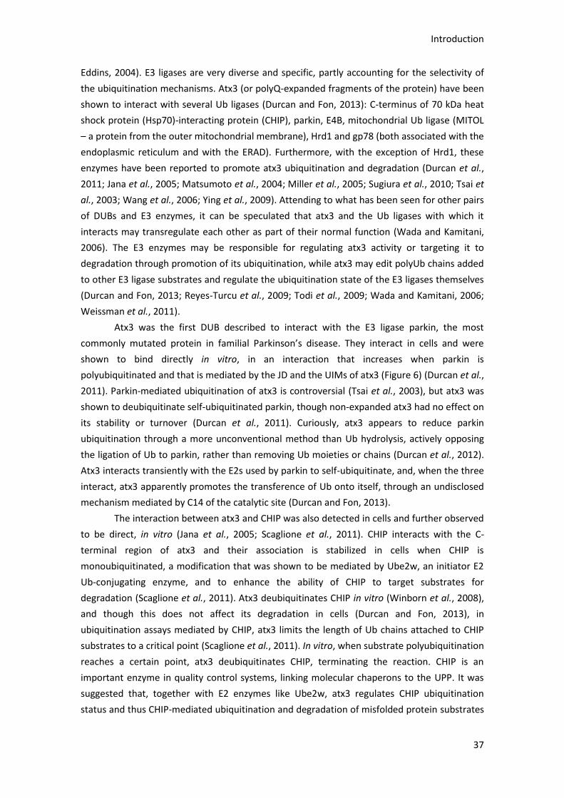
Introduction
37
Eddins, 2004). E3 ligases are very diverse and specific, partly accounting for the selectivity of
the ubiquitination mechanisms. Atx3 (or polyQ-expanded fragments of the protein) have been
shown to interact with several Ub ligases (Durcan and Fon, 2013): C-terminus of 70 kDa heat
shock protein (Hsp70)-interacting protein (CHIP), parkin, E4B, mitochondrial Ub ligase (MITOL
– a protein from the outer mitochondrial membrane), Hrd1 and gp78 (both associated with the
endoplasmic reticulum and with the ERAD). Furthermore, with the exception of Hrd1, these
enzymes have been reported to promote atx3 ubiquitination and degradation (Durcan et al.,
2011; Jana et al., 2005; Matsumoto et al., 2004; Miller et al., 2005; Sugiura et al., 2010; Tsai et
al., 2003; Wang et al., 2006; Ying et al., 2009). Attending to what has been seen for other pairs
of DUBs and E3 enzymes, it can be speculated that atx3 and the Ub ligases with which it
interacts may transregulate each other as part of their normal function (Wada and Kamitani,
2006). The E3 enzymes may be responsible for regulating atx3 activity or targeting it to
degradation through promotion of its ubiquitination, while atx3 may edit polyUb chains added
to other E3 ligase substrates and regulate the ubiquitination state of the E3 ligases themselves
(Durcan and Fon, 2013; Reyes-Turcu et al., 2009; Todi et al., 2009; Wada and Kamitani, 2006;
Weissman et al., 2011).
Atx3 was the first DUB described to interact with the E3 ligase parkin, the most
commonly mutated protein in familial Parkinson’s disease. They interact in cells and were
shown to bind directly in vitro, in an interaction that increases when parkin is
polyubiquitinated and that is mediated by the JD and the UIMs of atx3 (Figure 6) (Durcan et al.,
2011). Parkin-mediated ubiquitination of atx3 is controversial (Tsai et al., 2003), but atx3 was
shown to deubiquitinate self-ubiquitinated parkin, though non-expanded atx3 had no effect on
its stability or turnover (Durcan et al., 2011). Curiously, atx3 appears to reduce parkin
ubiquitination through a more unconventional method than Ub hydrolysis, actively opposing
the ligation of Ub to parkin, rather than removing Ub moieties or chains (Durcan et al., 2012).
Atx3 interacts transiently with the E2s used by parkin to self-ubiquitinate, and, when the three
interact, atx3 apparently promotes the transference of Ub onto itself, through an undisclosed
mechanism mediated by C14 of the catalytic site (Durcan and Fon, 2013).
The interaction between atx3 and CHIP was also detected in cells and further observed
to be direct, in vitro (Jana et al., 2005; Scaglione et al., 2011). CHIP interacts with the C-
terminal region of atx3 and their association is stabilized in cells when CHIP is
monoubiquitinated, a modification that was shown to be mediated by Ube2w, an initiator E2
Ub-conjugating enzyme, and to enhance the ability of CHIP to target substrates for
degradation (Scaglione et al., 2011). Atx3 deubiquitinates CHIP in vitro (Winborn et al., 2008),
and though this does not affect its degradation in cells (Durcan and Fon, 2013), in
ubiquitination assays mediated by CHIP, atx3 limits the length of Ub chains attached to CHIP
substrates to a critical point (Scaglione et al., 2011). In vitro, when substrate polyubiquitination
reaches a certain point, atx3 deubiquitinates CHIP, terminating the reaction. CHIP is an
important enzyme in quality control systems, linking molecular chaperons to the UPP. It was
suggested that, together with E2 enzymes like Ube2w, atx3 regulates CHIP ubiquitination
status and thus CHIP-mediated ubiquitination and degradation of misfolded protein substrates

Chapter 1
38
(Scaglione et al., 2011). Atx3 may be important in limiting the length of the polyUb chain to the
minimum needed for degradation (four monomers) or modulate the rate of proteasomal
degradation through the interplay between cycles of Ub conjugation and removal.
Interestingly, though atx3 has been shown to cleave long Ub chains preferentially, in this case
it is able to remove Ub from monoubiquitinated CHIP, though only in the presence of a CHIP
polyubiquitinated substrate. This suggests that atx3 may be recruited when a substrate is
polyubiquitinated, in order to deubiquitinate CHIP and terminate the process (Scaglione et al.,
2011).
F. Ataxin-3 and transcription regulation
A different aspect of atx3 function concerns its involvement in transcription regulation.
Characterization of glutamine repeat-containing proteins has demonstrated that many of them
are involved in regulating signalling at the transcriptional level, the polyQ sequence mediating
important molecular interactions (Hands et al., 2008). Atx3 has been shown to influence the
regulation of the expression of many genes (Evert et al., 2003; Rodrigues et al., 2007). A
microarray analysis using two different C. elegans KO strains lacking the worm ortholog of atx3
identified 290 genes (1.4% of the worm total genome) that are differentially expressed
comparing to wild-type animals. 143 of these had unknown function, but the other 147 genes
were grouped according to their proposed biological roles: cell structure and mobility
components (50%), signal transduction (20%), the UPP (8%), and other cell processes (22%),
which include genes encoding enzymes, transporters, receptors and channels involved in
diverse pathways. Of the 290 differentially expressed genes, 253 were upregulated and 37
were downregulated in KO animals (Rodrigues et al., 2007). Even though there was a clear
transcriptional dysregulation in the KO strains, in resemblance with the KO mice mentioned
above, the animals were viable and had no overt phenotype, possibly due to functional
redundancy and adaptative modifications.
Atx3 function as a transcription regulator is supported by in vitro and in situ molecular
interactions studies. Atx3 is known to interact with: a) three transcription activators – cAMP
response element-binding protein (CREB)-binding protein (CBP), p300 and p300/CBP-
associated factor (PCAF) – through its C-terminal region (Li et al., 2002); b) two transcription
repressors – histone deacetylase 3 (HDAC3) and nuclear receptor co-repressor (NCor) –
through an interaction involving its UIMs (Evert et al., 2006a); and histones, mainly through
the N-terminal region (Li et al., 2002). Interaction with CBP, p300, PCF and histones was
demonstrated to be direct (Figure 6) (Li et al., 2002). The same studies report that atx3
represses the transcription mechanisms mediated by these proteins through different
processes. CBP, p300 and PCAF are transcriptional co-activators that form complexes with
other co-activators and proteins with acetyltransferase activity and acetylate histones; atx3
seems to repress transcription mediated by these co-activators by binding them through its C-
terminal region. Furthermore, binding of histones through the N-terminal of atx3 seems to
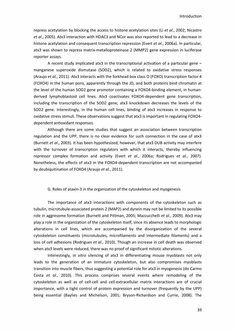
Introduction
39
repress acetylation by blocking the access to histone acetylation sites (Li et al., 2002; Nicastro
et al., 2005). Atx3 interaction with HDAC3 and NCor was also reported to lead to a decrease in
histone acetylation and consequent transcription repression (Evert et al., 2006a). In particular,
atx3 was shown to repress matrix-metalloproteinase 2 (MMP2) gene expression in luciferase
reporter assays.
A recent study implicated atx3 in the transcriptional activation of a particular gene –
manganese superoxide dismutase (SOD2), which is related to oxidative stress responses
(Araujo et al., 2011). Atx3 interacts with the forkhead box class O (FOXO) transcription factor 4
(FOXO4) in the human pons, apparently through the JD, and both proteins bind chromatin at
the level of the human SOD2 gene promotor containing a FOXO4-binding element, in human-
derived lymphoblastoid cell lines. Atx3 coactivates FOXO4-dependent gene transcription,
including the transcription of the SOD2 gene; atx3 knockdown decreases the levels of the
SOD2 gene. Interestingly, in the human cell lines, binding of atx3 increases in response to
oxidative stress stimuli. These observations suggest that atx3 is important in regulating FOXO4-
dependent antioxidant responses.
Although there are some studies that suggest an association between transcription
regulation and the UPP, there is no clear evidence for such connection in the case of atx3
(Burnett et al., 2003). It has been hypothesized, however, that atx3 DUB activity may interfere
with the turnover of transcription regulators with which it interacts, thereby influencing
repressor complex formation and activity (Evert et al., 2006a; Rodrigues et al., 2007).
Nonetheless, the effects of atx3 in the FOXO4-dependent transcription are not accompanied
by deubiquitination of FOXO4 (Araujo et al., 2011).
G. Roles of ataxin-3 in the organization of the cytoskeleton and myogenesis
The importance of atx3 interactions with components of the cytoskeleton such as
tubulin, microtubule-associated protein 2 (MAP2) and dynein may not be limited to its possible
role in aggresome formation (Burnett and Pittman, 2005; Mazzucchelli et al., 2009). Atx3 may
play a role in the organization of the cytoskeleton itself, since its absence leads to morphologic
alterations in cell lines, which are accompanied by the disorganization of the several
cytoskeleton constituents (microtubules, microfilaments and intermediate filaments) and a
loss of cell adhesions (Rodrigues et al., 2010). Though an increase in cell death was observed
when atx3 levels were reduced, there was no proof of significant mitotic alterations.
Interestingly, in vitro silencing of atx3 in differentiating mouse myoblasts not only
leads to the generation of an immature cytoskeleton, but also compromises myoblasts
transition into muscle fibers, thus suggesting a potential role for atx3 in myogenesis (do Carmo
Costa et al., 2010). This process comprises several events where remodeling of the
cytoskeleton as well as of cell-cell and cell-extracellular matrix interactions are of crucial
importance, with a tight control of protein expression and turnover (frequently by the UPP)
being essential (Baylies and Michelson, 2001; Bryson-Richardson and Currie, 2008). The

Chapter 1
40
observed effect of atx3 silencing was proposed to be mediated by integrins and, in agreement,
silencing atx3 decreases the levels of a5 and a7 integrin subunits, which are known
participants in myogenesis (do Carmo Costa et al., 2010; Mayer, 2003). Furthermore, atx3
interacts with integrin subunit a5 and represses its degradation, and several proteins related
to integrin-mediated responses were reported to have altered levels in myoblasts when atx3
was silenced (do Carmo Costa et al., 2010). It is noteworthy, however, that no overt phenotype
concerning cytoskeleton organization or muscle differentiation was reported in atx3 KO
animals. This may be due to compensation or to subtle changes in the cytoskeleton that were
not identified in the analyses performed (do Carmo Costa et al., 2010; Rodrigues et al., 2007;
Rodrigues et al., 2010; Schmitt et al., 2007).
H. Summary of ataxin-3 proposed roles
The biochemical characteristics of atx3, the interactions described so far and the
cellular outcomes of both features suggest that the biological role of atx3 may be connected to
three major processes: a) protein homeostasis pathways; b) transcription regulation; c)
cytoskeleton organization and myogenesis (Figure 7). Considering in particular the
participation of the protein in quality control systems, atx3 may be important in: (i) regulating
the formation, maintenance and termination of Ub signals, namely in polyUb chains of atypical
morphology and possibly involving interactions with E3 enzymes like parkin and CHIP; (ii)
shuttling substrates to the proteasome for degradation or, conversely, (iii) protecting them
from proteasomal degradation; (iv) modulating the targeting and flux of ERAD substrates; and
(v) aggresome formation.
As discussed above, atx3 participation in these diverse pathways may result from a
more limited cell role, in a restricted number of mechanisms and interactions. For example,
atx3 participation in the UPP may make it responsible for the regulation of the half-life of
several different targets, which in turn take part in diverse cell functions (do Carmo Costa et al.,
2010; Evert et al., 2006a; Rodrigues et al., 2007; Rodrigues et al., 2010). The fine-tuning of Ub
signals, with outcomes possibly unrelated to degradation, may also be a promising mechanism
through which atx3 mediates several cellular functions. Ubiquitination is increasingly regarded
as a very complex and dynamic process, resulting from the intricate balances between Ub
conjugation and deubiquitination (Komander et al., 2009; Marfany and Denuc, 2008; Nguyen
et al., 2014; Zhang et al., 2013). The observed preference for processing of atypical chains, the
opposing effects in actual protein degradation described, and the mechanistic interaction
between atx3 and E3 in ligases polyUb chains support this hypothesis. Considering the
complexity of Ub signals and the myriad of pathways they intervene, regulation mediated by
atx3 possibly may be also relevant in mechanisms in which atx3 has thus far not been
implicated, such as DNA repair mechanisms, endocytosis and transport (Eletr and Wilkinson,
2014; Nijman et al., 2005; Reyes-Turcu et al., 2009). Interestingly, atx3 regulates protein stress
responses, protein degradation and ageing in C. elegans, and the observed effects were

Introduction
41
Figure 7: Atx3 activity, interactions and proposed biologic roles. A Atx3 displays DUB activity and has been shown to be able to edit polyUb chains, possibly regulating Ub signals in tandem with E3 ligases such as parkin and CHIP. Atx3 is believed to participate in quality control systems, interfering with B proteasomal degradation particularly in C the ERAD, by means of its interaction with HHR23A/B and VCP/p97. D Atx3 has been further linked to aggresome formation, interacting with tubulin and dynein, and with E the organization of the cytoskeleton itself. F Interactions with regulators of histone acetylation support many reports describing atx3 as a participant in transcription regulation.
described to require the synergistic action of VCP and involve the editing the ubiquitination
state of substrates of the insulin/insulin-like growth factor (IGF1) (Kuhlbrodt et al., 2011). This

Chapter 1
42
suggests that regulation of Ub signals by atx3 may be very important in specific biological
mechanisms (Scaglione et al., 2011).
Intracellular transport and shuttling
As mentioned before, atx3 is found in both the cytoplasm and the nucleus. Several
studies demonstrated that atx3 is actively transported across the nuclear envelope, being
actively shuttled from the cytoplasm to the nucleus and vice versa (Antony et al., 2009; Chai et
al., 2002; Macedo-Ribeiro et al., 2009).
Regarding nuclear importation, a functional nuclear localization signal (NLS) upstream
of the polyQ sequence (R282-R285) has been predicted (Boeddrich et al., 2006; Tait et al.,
1998) and later shown to be active, promoting atx3 transport to the cell nucleus (Figure 6)
(Macedo-Ribeiro et al., 2009). Nuclear exportation appears to integrate both chromosome
region maintenance 1 (CRM1)-dependent and independent mechanisms (Macedo-Ribeiro et
al., 2009) and, as of yet, two apparently functional nuclear export signals (NES) have been
reported (I77-Y99 and E141-E158), though without consensus (Figure 6) (Antony et al., 2009;
Macedo-Ribeiro et al., 2009). There is compelling evidence supporting the idea that at least
the CMR1-independent mechanism of nuclear export is mediated by a N-terminal
conformational motif comprised by regions in both the JD and the UIMs (Macedo-Ribeiro et al.,
2009).
Atx3 shifts to the nucleus in response to proteotoxic stimuli, such as oxidative stress
(Araujo et al., 2011) and heat shock, but independently from the identified NLS (Reina et al.,
2010). Two other regions seem important for this transport instead: the JD, namely S111, and
the C-terminal region comprising the polyQ sequence and the rest of the C-terminal (but not
the UIMs) (Reina et al., 2010). This observation adds to other recent evidence suggesting that
atx3 biological function may be important in cellular systems of stress response (Araujo et al.,
2011; Kuhlbrodt et al., 2011; Mueller et al., 2009; Todi et al., 2009). Atx3 activity as a DUB
participating in protein homeostasis pathways is in agreement with this possibility, as atx3 is
likely important in dealing with misfolded proteins that arise as a consequence of stress, or
that may also exacerbate cellular stress. The responses may also be triggered at a
transcriptional level, considering the observed coactivation of FOXO4-mediated SOD2
expression by atx3 (Araujo et al., 2011).
Atx3 degradation and turnover
Both the UPP and autophagy, two of the main protein degradation pathways existing
in animal cells, have been shown to modulate atx3 turnover (Harris et al., 2010). Atx3 has been
shown to be polyubiquitinated and to be degraded by the proteasome (Berke et al., 2005; Jana
et al., 2005; Matsumoto et al., 2004; Todi et al., 2007; Tsai et al., 2003; Wang et al., 2007); as

Introduction
43
mentioned above, many E3 Ub ligases were reported to interact with atx3 and to promote its
ubiquitination and degradation. However, the proportional importance of the UPP apparently
depends on the particular atx3 isoform in question. It was suggested that proteasomal
degradation is more important only in the case of the atx3 isoform lacking the third UIM
(MJD1a), which is a less stable isoform. The three UIM-containing atx3 is more stable and is
mainly degraded by autophagy (Harris et al., 2010).
Interestingly, atx3 catalytic activity appears to regulate its turnover, since catalytically
inactive atx3 has higher steady state levels in cells, partially resulting from a reduction on
proteasomal degradation (Todi et al., 2007). Strikingly, however, in this case and in several
other studies (Berke et al., 2005; Matsumoto et al., 2004; Miller et al., 2004), increased
ubiquitination did not correlate with increased degradation, thereby indicating that
proteasomal degradation independent of ubiquitination may also be relevant (Todi et al.,
2007).
1.5 Toxic consequences of ataxin-3 polyglutamine expansion
Polyglutamine sequences as causes of toxicity
The physiologic role of homopeptidic repeats such as polyQ sequences is not clearly
understood, but it has been pointed out that this type of motives is usually associated with
multimolecular complexes and that they are most likely important in regulating or mediating
molecular interactions (Almeida et al., 2013; Fiumara et al., 2010; Hands et al., 2008; Schaefer
et al., 2012). Homopeptidic regions are considered to be unstructured, becoming structured
when establishing molecular interactions (Almeida et al., 2013). It has recently been shown
that polyQ tracts and adjacent regions have a propensity to engage in intermolecular
interactions via formation of coiled-coils, in every disease-associated polyQ protein (Fiumara et
al., 2010). The flexibility of the polyQ sequence may facilitate the establishment of protein-
protein interactions with the polyQ tract itself or with other protein regions, and stabilize them
(Schaefer et al., 2012).
To date, the mechanisms by which polyQ-expand atx3 leads to MJD pathogenesis have
not been clarified, though, as for every other polyQ disease, the unstable polyQ expansion is
considered to be of causal importance. There are several observations supporting this idea: a)
the polyQ disease protein sequences are dissimilar outside the polyQ tract; b) disease severity
increases with glutamine repeat number; c) age of onset decreases with the increase of repeat
number; d) several transgenic models expressing polyQ sequences outside the natural gene
context present neurodegenerative phenotypes (Ordway et al., 1999; Takahashi et al., 2010;
Williams and Paulson, 2008; Zoghbi and Orr, 2000). As stated above, however, though the
polyQ sequence expansion appears to be the triggering factor leading to the development of
MJD, atx3 regions outside the polyQ region and the protein properties they determine are sure

Chapter 1
44
to define the development of MJD and to determine its particular aspects (Gatchel and Zoghbi,
2005; Robertson and Bottomley, 2010).
Intracellular inclusion bodies
The formation of large inclusion bodies containing the pathogenic protein is one
hallmark of every polyQ expansion disease. Even though wild-type atx3 displays a wide
distribution within cells and is predominantly cytoplasmic in neurons, in MJD patients’ brains,
atx3 is commonly known to amass in the form of NIs present only in neurons (Paulson et al.,
1997b). Cytoplasmic inclusions have occasionally been reported (Hayashi et al., 2003; Paulson
et al., 1997b; Yamada et al., 2001) and recently axonal inclusions have also been observed in
patients brains, in fibers known to degenerate in MJD (Seidel et al., 2010). Apart from the
polyQ-expanded atx3, NIs and the axonal inclusions contain several other proteins, including
members of the cell quality control systems, like proteasome constituents, Ub and molecular
chaperones, which may be mobilized against atx3 misfolding and aggregation (Chai et al.,
1999a; Chai et al., 1999b; Chow et al., 2004a; Paulson et al., 1997b; Schmidt et al., 2002; Seidel
et al., 2010).
Intracellular inclusions have been often linked to the pathogenesis of polyQ diseases as
a major means of polyQ toxicity because of their ability to sequester different molecules,
provoking cytotoxicity through several possible mechanisms: a) hindrance of transcription,
through sequestration of molecules involved in transcription regulation (Chai et al., 2002;
McCampbell et al., 2000); b) a general disturbance of the quality control systems of the cells,
due to sequestration of proteasome constituents and molecular chaperones (Chai et al.,
1999a; Chai et al., 1999b; Ferrigno and Silver, 2000; Muchowski et al., 2000; Paulson et al.,
1997b; Schmidt et al., 2002; Warrick et al., 1999); c) hindrance of axonal transport, resulting
from motor protein titration and physical blocking (Gunawardena et al., 2003); d) other
disturbances caused by recruitment of Ub-binding proteins (since inclusions are heavily
ubiquitinated) or other polyQ-containing proteins (Donaldson et al., 2003; Perez et al., 1998).
As explained before, the importance of these large microscope-visible macromolecular
aggregates to disease pathogenesis is now disputed and many authors suggest that they are in
fact just the result of protective mechanisms moved by the cell in order to cope with the
toxicity of the expanded proteins (Ross and Poirier, 2004; Todd and Lim, 2013; Williams and
Paulson, 2008). However, studies concerning MJD demonstrate that there is no perfect overlap
between the brain regions that degenerate and the regions presenting NIs (Riess et al., 2008;
Rüb et al., 2006b; Yamada et al., 2001). Additionally, it was also shown that, in the pons,
patients with more severe neuronal loss presented a smaller amount of neurons with NIs
(Evert et al., 2006b).

Introduction
45
Polyglutamine aggregation and toxicity
Studies suggest that expanded atx3 and every other polyQ-expanded protein tend to
form aggregates, as a result of polyQ expansion-induced misfolding and consequent transition
to aggregation-prone conformations (Bevivino and Loll, 2001; Chow et al., 2004c; Jana and
Nukina, 2004; Muchowski and Wacker, 2005; Nagai et al., 2007; Tanaka et al., 2001; Williams
and Paulson, 2008). Aggregates formed by atx3 or other polyQ-containing proteins were
shown to contain β-rich fibrillar structures of amyloid nature, reminiscent of the amyloid
aggregates observed in Alzheimer’s and prion diseases (Almeida et al., 2013; Bevivino and Loll,
2001; Chen et al., 2002). As for most amyloid-forming proteins, several pathways may drive
the conversion of the soluble protein to amyloid aggregates, through the formation of
different conformationally altered monomeric or self-assembled multimeric species (Uversky,
2010), being the small aggregates or oligomers the ones most commonly envisioned as the
actual toxic species causing cytotoxicity (Takahashi et al., 2010). It is suggested that the soluble
amyloid oligomers have an inherent toxicity caused by mechanisms common to all of them
(Kayed et al., 2003), for example being able to destabilize the lipid bilayer, increasing its
conductivity, or to sequester quality control system components and transcription factors,
causing proteotoxic stress and transcriptional dysregulation (Figure 8) (Demuro et al., 2005;
Kayed et al., 2004; Schaffar et al., 2004; Shao and Diamond, 2007). It is also possible that the
cell-mediated formation of NIs, though directed as an adaptive response, may trigger a
snowball effect, in which important proteins are sequestered and more cellular systems
become dysfunctional.
Mechanisms of ataxin-3 aggregation
Investigation has focused on a search to comprehend the mechanisms underlying atx3
aggregation and on trying to identify the toxic misfolded monomeric or oligomeric species
(Takahashi et al., 2010). In vitro, polyQ-expanded atx3 aggregates into amyloid-like fibrils
(Bevivino and Loll, 2001; Ellisdon et al., 2007; Ellisdon et al., 2006) but, curiously, even non-
expanded atx3 and the JD alone display a tendency to aggregate (albeit at slower rates than
the expanded forms), underlying the relevance of regions outside the polyQ tract for protein
self-assembly and aggregate formation (Chow et al., 2004a; Chow et al., 2004b; Gales et al.,
2005; Masino et al., 2011; Masino et al., 2004). Aggregation of atx3 is admitted to consist of
two steps: the first step is independent from the polyQ sequence and involves a monomeric
thermodynamic nucleus formed by a single misfolded protein to which monomers are added,
consequently leading to elongation and fibril formation; in the case of the expanded protein
there is a second aggregation step that leads to the formation of more stable sodium dodecyl
sulfate (SDS)-insoluble aggregates (Ellisdon et al., 2007; Saunders and Bottomley, 2009).
The JD plays a central role in the early conformational changes (Gales et al., 2005),
modulating the aggregation of both expanded and non-expanded atx3 (Ellisdon et al., 2006;

Chapter 1
46
Figure 8: Putative mechanisms mediating expanded atx3 toxicity. PolyQ expansion of atx3 beyond 45 glutamines is known to lead to the development of MJD, though the identity of the actual toxic species is still a matter of debate. The effect the pathogenic mutation may have on the physiological function of atx3, be it at the level of protein quality control or transcription regulation, could affect important cellular activities and lead to cellular demise. Expanded atx3 is known to aggregate and produce large macromolecular aggregates, and it is believed that soluble amyloid-like oligomers that serve as intermediaries of the self-aggregation pathway may be deleterious to the cells, possibly destabilizing the cell membrane and/or recruiting important proteins with consequent disruption of important cellular systems. Proteotoxic stress may arise from atx3 misfolding, aggregation, aberrant interactions or the compromise of its native function as a player in protein homeostasis maintenance. Though without consensus, proteolytic cleavage of atx3 and generation of toxic protein fragments may be an important step contributing to toxic events such as oligomerization. The figure uses continuous black lines to represent changes produced on atx3 upon polyQ expansion and interrupted black lines to represent events related with proteolytic cleavage. The gray arrows link putative toxic species to some of mechanisms that might make them deleterious.

Introduction
47
Masino et al., 2004; Saunders and Bottomley, 2009). Interestingly, the surfaces involved in self-
association of this domain overlap with the functionally relevant Ub binding sites 1 and 2
(Masino et al., 2011; Nicastro et al., 2009). These observations provide a direct link between
protein function and aggregation and expose a role for intracellular interactors in protecting
against atx3 self-assembly, especially given the fact that Ub reduces in vitro aggregation of the
JD (Masino et al., 2011). The C-terminal region of atx3 may also represent a bridge between
physiological interactions and aggregation. In atx3, the regions predicted to form coiled-coils
partially includes the α-helical UIMs and regions shown to interact with molecular partners
such as VCP domains (Boeddrich et al., 2006; Fiumara et al., 2010; Song et al., 2010). When
expanded, the polyQ region may provoke aberrant protein interactions leading to atx3
aggregation. Overall, the regions outside the polyQ tract seem to predispose atx3 to
aggregation, and the expansion of the polyQ sequence precipitates the formation of highly
stable amyloid aggregates (Almeida et al., 2013). The connection between normal molecular
interactions and aggregation may help explain the failure of non-expanded atx3 to self-
aggregate in the crowded cell environment and provide clues to understand the delayed onset
of symptoms in patients (Gales et al., 2005).
Taking these observations into account, one can speculate about the possible causes
for the persistent atx3 aggregation occurring in MJD patients’ brain and for the adult/late
onset of the disease. In healthy people, the slow aggregation kinetics of the non-expanded
atx3, the interaction with its macromolecular partners, and the high instability of the
aggregates formed ensure an effective break down by the cell’s intrinsic quality control
mechanisms. Conversely, in MJD patients, the enhanced kinetics of the expanded atx3
aggregation, eventual polyQ expansion-induced alterations in protein-protein interactions, as
well as the great stability of the aggregates formed lead to their aberrant accumulation inside
the cells. This accumulation may increase with aging, as a result of the progressive failure of
quality control mechanisms (Ellisdon et al., 2007; Ellisdon et al., 2006; Gales et al., 2005;
Masino et al., 2011; Masino et al., 2004).
Proteolytic cleavage and toxic fragment hypothesis
Several studies suggest that the proteolytic cleavage of polyQ disease proteins
contributes to pathogenesis, the resulting fragments being of crucial importance for their
aggregation and cytotoxicity. This idea is referred to as the toxic fragment hypothesis
(Takahashi et al., 2010; Tarlac and Storey, 2003; Wellington et al., 1998). Proteolytic cleavage
of atx3 has been reported in several cell and animal models and in MJD patients’ brains,
though results are considerably diverse.
Atx3 has been shown to produce polyQ-containing C-terminal fragments of about 34-
37 kDa (Goti et al., 2004; Jung et al., 2009; Simões et al., 2012; Simões et al., 2014). In
transgenic mice, a 36 kDa fragment was enriched in the nuclear fraction of expanded atx3-
expressing animals’ brain homogenates, but was not detected in other tissues (Goti et al.,

Chapter 1
48
2004). A fragment with similar characteristics was also enriched in the nuclear fraction of MJD
patients’ brains and was even identified as more abundant in the dentante nuclei and the
substantia nigra, two brain regions known to be affected in the disease (Goti et al., 2004).
Furthermore, studies using transfected cell lines reported that expression of C-terminal
fragments of atx3 containing the expanded polyQ sequence leads to strong atx3 aggregation
and cell death (Breuer et al., 2010; Goti et al., 2004; Haacke et al., 2006; Ikeda et al., 1996;
Paulson et al., 1997b; Yoshizawa et al., 2000). Severe toxicity, characterized by
neurodegeneration and motor phenotypes, is observed in animal models expressing C-
terminal fragments of atx3 containing expanded polyQ regions (Ikeda et al., 1996; Torashima
et al., 2008; Warrick et al., 1998).
Fragments of other molecular sizes and/or composition have been also described,
albeit more varyingly. In COS-7 cells, endogenous atx3 was shown to generate a 28 kDa
fragment described to contain the C-terminal region of atx3 and including the polyQ tract
(Berke et al., 2004), possibly corresponding to a non-expanded form of the 34-37 kDa fragment
mentioned above. Another study, using transfected COS-7 cells, mentioned a greater variety of
fragments. Non-expanded atx3 was shown to generate four C-terminal fragments (with about
18, 21, 28 and 42 kDa) and two N-terminal fragments (with 37 and 42 kDa), while the
expanded protein generated only two C-terminal fragments (a 37 kDa fragment and another
with about 50 kDa). The 42 kDa fragments were found only in the cytoplasm, but the other
ones were found also in the nucleus (Pozzi et al., 2008). In an in vitro proteolysis of purified
non-expanded human atx3 using a postnuclear supernatant preparation derived from a mice
neural cell line, five main atx3 fragments were identified: one, with about 29 kDa, derived from
the endogenous mouse protein, and four others derived from the human protein, with about
16, 20, 24 and 34 kDa respectively (at least some of them containing N-terminal regions)
(Haacke et al., 2007). Recent reports described an atx3 fragment of about 26 kDa present in
the brain of lentiviral MJD mouse models, that includes the N-terminus of the protein that is
also detected in the brains of animals transduced with non-expanded atx3 (Simões et al., 2012;
Simões et al., 2014). Another report observed that incubation of lysates with calpains gives rise
to an N-terminal fragment of ~30 kDa in both transfected human cell lines and a transgenic
model, derived from either expanded and non-expanded overexpressed human atx3, in the
cell lines, as well as from the endogenous mouse protein and expanded atx3, in the model
(Hübener et al., 2013). Studies performed in neuronal cultures generated from human-derived
induced pluripotent stem cells (iPSCs) revealed a 34 kDa atx3 fragment, which was curiously
present (and had the same size) in both MJD patients and controls, possibly indicating that it is
a N-terminal fragment (Koch et al., 2011). Other fragments, of diverse molecular sizes (25-45
kDa) were also shown to be generated and to aggregate, but only in patient-derived neurons.
The great diversity of fragments mentioned here may be overstated, and some of them may
admittedly result from common proteolytic processes, though yielding apparently different
products in the corresponding studies. These differences may result from the employment of
considerably different experimental models, and possibly from different sampling conditions.
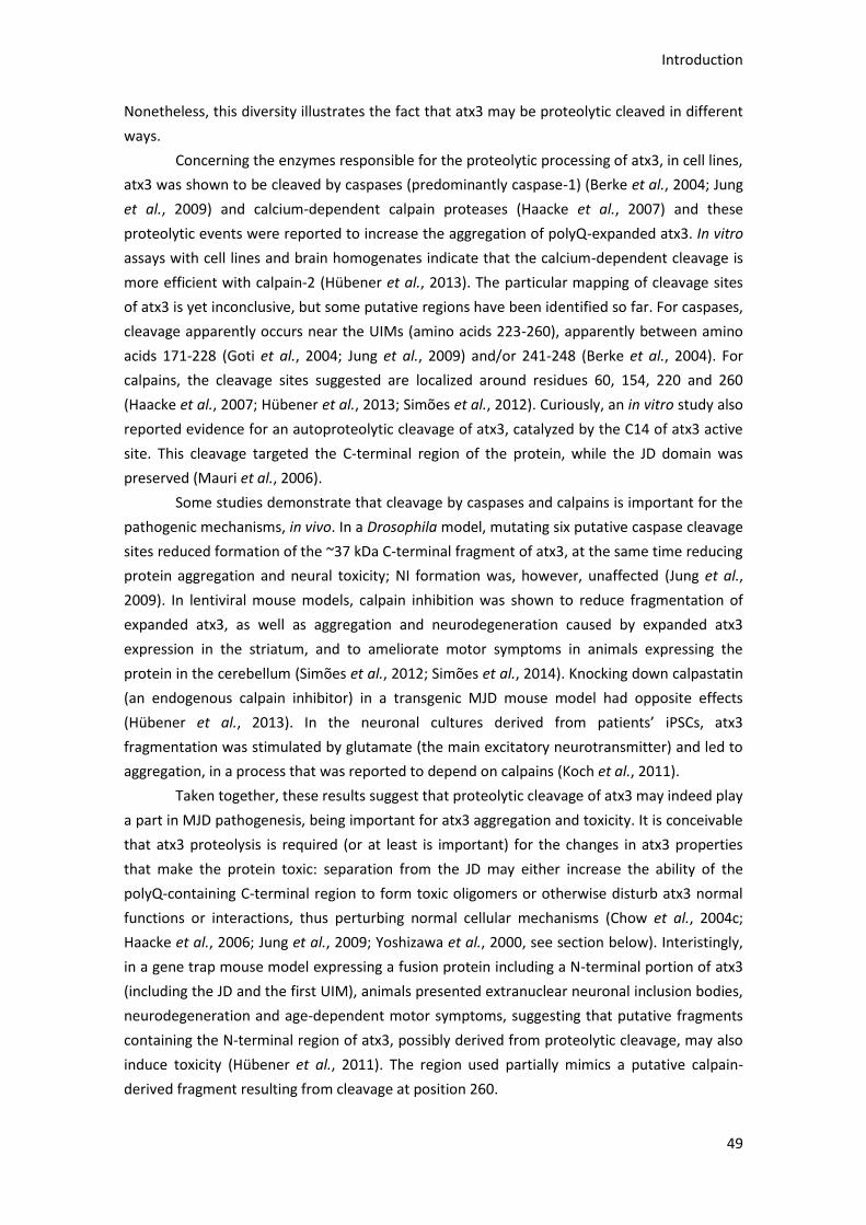
Introduction
49
Nonetheless, this diversity illustrates the fact that atx3 may be proteolytic cleaved in different
ways.
Concerning the enzymes responsible for the proteolytic processing of atx3, in cell lines,
atx3 was shown to be cleaved by caspases (predominantly caspase-1) (Berke et al., 2004; Jung
et al., 2009) and calcium-dependent calpain proteases (Haacke et al., 2007) and these
proteolytic events were reported to increase the aggregation of polyQ-expanded atx3. In vitro
assays with cell lines and brain homogenates indicate that the calcium-dependent cleavage is
more efficient with calpain-2 (Hübener et al., 2013). The particular mapping of cleavage sites
of atx3 is yet inconclusive, but some putative regions have been identified so far. For caspases,
cleavage apparently occurs near the UIMs (amino acids 223-260), apparently between amino
acids 171-228 (Goti et al., 2004; Jung et al., 2009) and/or 241-248 (Berke et al., 2004). For
calpains, the cleavage sites suggested are localized around residues 60, 154, 220 and 260
(Haacke et al., 2007; Hübener et al., 2013; Simões et al., 2012). Curiously, an in vitro study also
reported evidence for an autoproteolytic cleavage of atx3, catalyzed by the C14 of atx3 active
site. This cleavage targeted the C-terminal region of the protein, while the JD domain was
preserved (Mauri et al., 2006).
Some studies demonstrate that cleavage by caspases and calpains is important for the
pathogenic mechanisms, in vivo. In a Drosophila model, mutating six putative caspase cleavage
sites reduced formation of the ~37 kDa C-terminal fragment of atx3, at the same time reducing
protein aggregation and neural toxicity; NI formation was, however, unaffected (Jung et al.,
2009). In lentiviral mouse models, calpain inhibition was shown to reduce fragmentation of
expanded atx3, as well as aggregation and neurodegeneration caused by expanded atx3
expression in the striatum, and to ameliorate motor symptoms in animals expressing the
protein in the cerebellum (Simões et al., 2012; Simões et al., 2014). Knocking down calpastatin
(an endogenous calpain inhibitor) in a transgenic MJD mouse model had opposite effects
(Hübener et al., 2013). In the neuronal cultures derived from patients’ iPSCs, atx3
fragmentation was stimulated by glutamate (the main excitatory neurotransmitter) and led to
aggregation, in a process that was reported to depend on calpains (Koch et al., 2011).
Taken together, these results suggest that proteolytic cleavage of atx3 may indeed play
a part in MJD pathogenesis, being important for atx3 aggregation and toxicity. It is conceivable
that atx3 proteolysis is required (or at least is important) for the changes in atx3 properties
that make the protein toxic: separation from the JD may either increase the ability of the
polyQ-containing C-terminal region to form toxic oligomers or otherwise disturb atx3 normal
functions or interactions, thus perturbing normal cellular mechanisms (Chow et al., 2004c;
Haacke et al., 2006; Jung et al., 2009; Yoshizawa et al., 2000, see section below). Interistingly,
in a gene trap mouse model expressing a fusion protein including a N-terminal portion of atx3
(including the JD and the first UIM), animals presented extranuclear neuronal inclusion bodies,
neurodegeneration and age-dependent motor symptoms, suggesting that putative fragments
containing the N-terminal region of atx3, possibly derived from proteolytic cleavage, may also
induce toxicity (Hübener et al., 2011). The region used partially mimics a putative calpain-
derived fragment resulting from cleavage at position 260.

Chapter 1
50
Contrastingly though, Pozzi and collaborators observed that expanded atx3 is cleaved
to a lesser extent when compared with the normal protein, in COS-7 cells (Pozzi et al., 2008).
The authors suggested that the expanded polyQ stretch can mask some cleavage sites and that
pathogenesis can result from accumulation of uncleaved expanded atx3.
Polyglutamine expansion-induced changes in ataxin-3 features
Apart from inducing the formation of aggregates, the polyQ expansion of atx3 may
interfere with the protein at the singular level, producing changes in its function, interactions
or localization that lead to cellular toxicity and thereby play a part in the development of MJD.
A. Effects on deubiquitinase activity and ubiquitin interaction
It has initially been hypothesized that atx3 expansion leads to a loss of protein function
and that this would be sufficiently significant to cause the MJD. However, the mouse and C.
elegans atx3 KOs present no detectable overt phenotype, indicating that atx3 function is not
crucial or at least that it can been overcome by other proteins, in these systems (Rodrigues et
al., 2007; Schmitt et al., 2007). Another hypothesis, with better experimental support, is that
the expanded polyQ affects the protein’s function in a way that alters its cellular activity,
actively promoting cell toxicity.
Atx3 has been linked to protein homeostasis pathways and, in line with what has been
said above, disturbance of these mechanisms is a possible cause of toxicity as it might interfere
with the normal turnover and degradation of important molecules. Generally, studies have
demonstrated that there are not many significant differences in the deubiquitinating activity
between normal and expanded atx3 and that there is no change in polyUb chain linkage
preferences depending on polyQ length (Berke et al., 2004; Burnett et al., 2003; Winborn et al.,
2008). One study (Winborn et al., 2008), however, described that the ability of expanded atx3
to reduce general cellular protein ubiquitination was decreased, when compared to that of the
non-expanded form. This decrease can have consequences in the cellular pathways that use
Ub signals influenced by atx3 DUB activity. In yeast, expression of expanded atx3 (but not the
non-expanded protein) stabilized an ubiquitinated model substrate degraded by the UPP
(Doss-Pepe et al., 2003). Expanded atx3 retains its ability to bind polyubiquitinated substrates
in a UIM-dependent manner, with no global differences comparing to the normal protein
(Berke et al., 2004) but, nonetheless, the possibility that the two forms may differentially bind
and/or proteolyse particular substrates is not ruled out.
Atx3 participation in protein homeostasis systems adds an interesting aspect to MJD:
atx3 could interact with misfolded and aggregated proteins as part of its normal function, but
possible stable interactions between them and the expanded protein may potentiate protein
aggregation and constitute a cause for disturbance of those systems (Burnett et al., 2003;

Introduction
51
Burnett and Pittman, 2005). Interestingly, the presence of Ub and proteasome constituents in
atx3 inclusions can partially be explained by their physiologically relevant interactions with
atx3 (Doss-Pepe et al., 2003).
B. Effects on ataxin-3 interactions
Another aspect where an expanded polyQ tract may alter atx3 properties, possibly
influencing its physiological role, is in the interactions atx3 establishes. HHR23A and HHR23B
binding is not affected by the polyQ expansion, but HHR23A colocalizes with expanded atx3-
containing aggregates in cell lines (Wang et al., 2000). PolyQ expansion of atx3 increases the
extent of the interaction with VCP/p97, which also colocalizes with the NIs in MJD patients
(Boeddrich et al., 2006). These changes are likely to cause an altered distribution pattern for
these proteins and influence their activity and other molecular interactions (Boeddrich et al.,
2006; Higashiyama et al., 2002; Hirabayashi et al., 2001; Wang et al., 2000). Interestingly, the
enhancement of atx3 DUB activity elicited by VCP/p97 is abrogated when atx3 is expanded
(Laço et al., 2012a). Impairment of the ERAD caused by expansion of atx3 may induce
endoplasmic reticulum proteotoxic stress that may contribute to the degenerative
mechanisms (Wang et al., 2006; Zhong and Pittman, 2006). Curiously, mice expressing an atx3
fusion protein containing just its N-terminal region (including the JD, but lacking the region
that interacts with VCP/p97) display an increased susceptibility to endoplasmic reticulum
stress (Hübener et al., 2011).
In spite of the many studies unable to detect polyQ-induced changes in atx3 enzymatic
activity, expanded atx3 was recently shown to deubiquitinate parkin to a greater extent than
non-expanded atx3 and to induce its degradation by autophagy through an unknown
mechanism (Durcan et al., 2011). Interestingly, the increase in deubiquitination was specially
directed against chains of K27- and K29-linkage, which may be important in the mechanisms
leading to degradation. Parkin levels were also reduced in an expanded atx3-expressing mouse
model. Considering that parkin has been described as having neuroprotective effects and the
fact that its mutation causes a familial form of Parkinson’s disease, the expanded atx3-induced
decrease of parkin levels may be related to symptoms of Parkinsonism observed in some MJD
patients. PolyQ expansion of atx3 does not change atx3 enzymatic activity towards CHIP, but
alters their interaction, increasing the affinity of their binding but turning the potentiating
effect of ubiquitination of CHIP redundant (Scaglione et al., 2011). Furthermore, the levels of
CHIP were also reduced in the brain of the above transgenic model, comparing to wild-type
animals and to atx3 KOs. This could be the result of the altered dynamics in the atx3-CHIP
interaction, caused by the polyQ-induced increase in affinity (Scaglione et al., 2011). These
observations indicate that disturbances caused by polyQ expansion of atx3 may have a
negative impact on the cellular homeostasis mechanisms mediated by parkin and CHIP, which
have been otherwise shown to be protectors against polyQ-induced toxicity (Durcan and Fon,
2013; Miller et al., 2005; Tsai et al., 2003).

Chapter 1
52
C. Effects on transcription regulation
PolyQ expansion might also affect the proposed function of atx3 as a transcription
regulator. Expansion may alter atx3 binding properties and compromise its ability to establish
transcription repressor complexes, leading to the increased expression of certain genes, some
of which may contribute to MJD development.
When compared to non-expanded atx3, expanded atx3 appears to bind chromatin in
different regions and to aberrantly increase transcription in cells (Evert et al., 2006a; Evert et
al., 2003). Histone acetylation is increased in cells expressing expanded atx3 and in human
MJD brain, although both normal and expanded forms associate with HDAC3 and NCoR (Evert
et al., 2006a). Interestingly, however, though this interaction is normally mediated by the UIMs
of atx3, the aberrant transcriptional activation is instead mediated by cysteine 14 of atx3
catalytic site. Expanded atx3 is also reported to bind transcriptional co-activators (CBP, p300
and PCAF) to a greater extent than the non-expanded form, though its inhibitory effect on
transcription was similar (Li et al., 2002).
Importantly, MJD patients’ brains have been reported to show transcriptional
dysregulation of several genes (Evert et al., 2006a; Evert et al., 2001; Wen et al., 2003). A
direct example of the negative effect on transcription regulation comes from studies relating
apoptosis with the cell loss observed in MJD, as expanded atx3 was reported to upregulate the
Bax protein (a pro-apoptotic protein), while downregulating Bcl-xL levels (an anti-apoptotic
protein), in cultured neurons (Chou et al., 2006).
Expression of several genes involved in cell signaling and synaptic transmission was
also shown to be altered in the brain of MJD model mice expressing expanded atx3, suggesting
that synaptic dysfunction resulting from transcription dysregulation may play a part in toxicity
mechanisms of MJD (Chou et al., 2008). Importantly, in accordance with the mechanisms
proposed for atx3-mediated genetic repression – but contrasting to studies of human MJD
brain (Evert et al., 2006a) –, these transcriptional alterations have been related to decreased
histone acetylation, and the pharmacological reversal of this state by administration of the
deacetylase inhibitor sodium butyrate was reported to counter transcriptional dysregulation
and simultaneously ameliorate the disease phenotype of this MJD model mice (Chou et al.,
2011).
In the case of the FOXO4-mediated coactivation of SOD2, though atx3 interacts with
FOXO4 independently from the polyQ tract length, the expanded form shows an impaired
effect on transcription and, concordantly, SOD2 has a decreased expression in MJD patients’
pons (Araujo et al., 2011). SOD2 expression is normally activated by FOXO transcription factors
in response to oxidative stress, and, accordingly, in patient-derived lymphoblastoid cell lines,
there is an increased susceptibility to reactive oxygen species formation and decreased
viability upon oxidative stress stimuli. Binding of atx3 to the SOD2 gene promoter region is
increased in patient-derived cells, and both FOXO4 and atx3 binding becames unresponsive to
the oxidative stress stimuli, indicating that these oxidative stress responses involve binding of

Introduction
53
both FOXO4 and atx3 to SOD2 gene promoter, and suggesting that expanded atx3 prevents the
FOXO4 upregulation of SOD2 upon oxidative stress (Araujo et al., 2011).
D. Aberrant interactions as the source of toxicity
Though the deleterious effects on transcription and quality control systems may
logically derive from modifications of atx3 endogenous functions, through its effect in
modulating Ub signals, it is tempting to hypothesize that all the cellular changes originate
actually from aggregation and aberrant interactions that destabilize not only atx3 function but
other cellular systems as well. In fact, all the toxicity mechanisms caused by polyQ expansion,
including aggregation itself, are increasingly envisioned as ultimately stemming from the
tendency of the pathogenic proteins to interact aberrantly (Schaefer et al., 2012; Williams and
Paulson, 2008). Some interactions with physiologic partners may be enhanced and others
diminished, and the advent of abnormal associations may recruit different proteins and disturb
a wide range of cellular systems (Gatchel and Zoghbi, 2005; Williams and Paulson, 2008).
Aggregation would arise from aberrant interactions between specimens of a particular polyQ-
expanded protein.
Misfolding of the expanded polyQ-containing proteins may alter interactions that are
mediated by different protein regions, including the polyQ tract and, in fact, the propensity of
the polyQ tracts and adjacent regions to engage in intermolecular interactions via formation of
coiled-coils is significantly increased upon polyQ expansion (Fiumara et al., 2010). These
regions may also be implicated in protein aggregation, likely representing a transition state
between normal protein conformation and aberrant β-sheath aggregation. Aberrant
interactions with other coiled-coil forming proteins (e.g., CHIP) might recruit them and
destabilize cellular homeostasis (Fiumara et al., 2010).
Ataxin-3 as a protector against polyglutamine toxicity
It has been determined that, apart from expanded atx3, protein aggregates seen in
MJD also contain normal, non-expanded protein (Fujigasaki et al., 2000). This is possible
because MJD is an autosomal dominant disease, and so both normal and expanded forms can
be present in the same cell, in the case of heterozygous individuals. The co-aggregation of non-
expanded atx3 may reflect the importance of regions outside the polyQ tract in the
aggregation mechanism or result from conformational changes that the misfolded expanded
atx3 induces in the non-expanded protein, leading to its recruitment to the assembling
aggregates (Chow et al., 2004a).
Another possibility, however, is that atx3 is recruited to aggregates because of UIM-
mediated recruitment to the highly ubiquitinated inclusions and/or because of its involvement
in the UPP and other protein homeostasis systems (Burnett et al., 2003; Burnett and Pittman,

Chapter 1
54
2005; Donaldson et al., 2003; Fujigasaki et al., 2001; Warrick et al., 2005) . Consistent with this,
a striking feature of atx3 is that it normally colocalizes with aggregates (or inclusion bodies)
formed in other polyQ expansion diseases (Uchihara et al., 2001) and other neurodegenerative
states (Burnett et al., 2003; Fujigasaki et al., 2000; Pountney et al., 2003; Seilhean et al., 2004;
Takahashi et al., 2001). Warrick and collaborators (2005) made important observations that
strongly suggested this protective role of atx3 against polyQ toxicity. They reported that flies
expressing both non-expanded and expanded atx3 (or non-expanded atx3 and other expanded
polyQ-disease proteins) did not present the phenotypes that result from expression of the
expanded polyQ protein. This suggested that atx3 was able to mitigate toxicity in several
Drosophila polyQ diseases models, including a MJD model. The protective activity is apparently
linked to atx3 involvement in quality control mechanisms such as the UPP, as it requires
normal proteasome functionality and atx3 Ub-associated activities, including Ub protease
activity and UIM functionality. These considerations may further explain why MJD patients
with two pathogenic alleles have a more severe phenotype than heterozygous patients
(Carvalho et al., 2008; Lang et al., 1994; Lerer et al., 1996; Sobue et al., 1996). The worsened
phenotype of homozygous patients may result not only from the presence of two pathogenic
alleles but also from a loss of the protective function of non-pathogenic atx3 (Warrick et al.,
2005)
Nonetheless, the idea of atx3 functioning as a protector against polyQ toxicity was
recently challenged by several research groups. In contrast to the observations in flies, a
double transgenic mouse coexpressing both expanded and non-expanded atx3 had a similar
disease phenotype (including neurological symptoms and brain NIs) to that of mice expressing
only the extended form of the protein (Hübener and Riess, 2010). Similarly, overexpression of
non-expanded atx3 in a lentiviral rat model of MJD expressing expanded atx3 does not prevent
development of the MJD-related phenotype caused by the pathogenic form of the protein
(Alves et al., 2010). However, the possibility that the contrasting results obtained in flies and
rodents may reflect the use of different animal models cannot be excluded. Until further
studies come to light, it is not possible to conclude if, in humans, the milder phenotype of
heterozygous individuals is due to the protective effect of atx3 or simply to gene dosage.
The role of the nuclear environment
The nucleus is an important player in the pathogenesis of MJD and other polyQ
diseases, seeing as nuclear localization of polyQ proteins is repeatedly reported as important
for the development of polyQ disease-related phenomena (Schöls et al., 2004; Shao and
Diamond, 2007). As mentioned before, though, in neurons atx3 distributes mainly in the
cytoplasm, whereas the most conspicuous atx3 aggregates, the macromolecular inclusions, are
mainly observed in the nucleus (Paulson et al., 1997b; Schmidt et al., 1998). Whether they are
toxic themselves or are formed as a response to the toxicity of other expanded atx3-derived

Introduction
55
species, their presence may be indicative of the negative effects of atx3 in this cellular
compartment.
Considering that atx3 is detected in both the cytoplasm and the nucleus, that it is
actively shuttled between the two compartments and that its biological function has been
related to transcription regulation, the normal cellular role of atx3 appears to involve the
nucleus (Antony et al., 2009; Evert et al., 2006b; Evert et al., 2001; Macedo-Ribeiro et al.,
2009; Wen et al., 2003). Consequently, expanded polyQ-derived modifications of this nuclear
role may be deleterious to the cell. In transgenic mice, it was demonstrated that artificially
targeting expanded atx3 to the nucleus indeed aggravates the disease phenotype, while
forcing nuclear export reduces it (Bichelmeier et al., 2007). Furthermore, inhibition of calpains,
which was observed to reduced toxicity in MJD lentiviral mouse models, was also reported to
prevent nuclear translocation of atx3 in MJD transgenic mice (Simões et al., 2012). These
observations strongly imply that nuclear localization of atx3 is critically important for the
manifestation of MJD symptoms, in vivo.
Clearly, the simple nuclear localization of atx3 is not sufficient to cause MJD, as the
endogenous protein itself is detected in the nucleus (Trottier et al., 1998), but several studies
reported that non-expanded atx3 fragments (Perez et al., 1998) and full-length atx3 (Macedo-
Ribeiro et al., 2009) targeted to the nucleus lead to aggregate formation, suggesting that the
nuclear environment may be important in this event. Moreover, non-expanded atx3 was found
to adopt a unique conformation once in the nucleus, and to bind to the nuclear matrix (Perez
et al., 1999). It was also proposed that the formation of large atx3 aggregates in the nucleus
may be due to a less efficient degradation of the protein in this compartment, when compared
to the cytoplasm (Breuer et al., 2010; Goellner and Rechsteiner, 2003).
Stress-induced nuclear transport of expanded atx3 as a part of its normal physiologic
function would be expected to have deleterious effects on the cell, protecting it from
degradation and accelerating toxicity (Bichelmeier et al., 2007; Breuer et al., 2010; Reina et al.,
2010). Aging and a possible dysfunction of atx3 caused by the unstable expansion may
culminate in an inability for atx3 to protect against proteotoxic stress, leading to cell toxicity
(Reina et al., 2010). Remarkably, the presence of misfolded and/or aggregated expanded atx3,
which may constitute a proteotoxic stimulus by itself, will likely increase nuclear localization of
the protein and thus be responsible for triggering and/or enhancing those effects (Mueller et
al., 2009).
1.6 Ataxin-3 toxicity mechanisms, cellular context and neuronal damage
Mechanisms of cellular toxicity
The considerations presented above demonstrate how toxicity derived from polyQ
expansion of atx3 has been related with diverse biochemical and cellular mechanisms. In short,
and in parallel with the plethora of putative toxic changes involved in polyQ disease

Chapter 1
56
pathogenesis that were presented before, atx3 expansion has been variously shown to: alter
atx3 DUB activity against parkin and other unidentified ubiquitinated species; cause aberrant
interactions, increasing the association with VCP/p97 and CHIP; lead to the formation of toxic
aggregates and toxic fragments; and elicit transcriptional alterations. Induction of proteotoxic
stress is admittedly caused by misfolding and aggregation of expanded atx3, but can also result
from alterations of atx3 interaction with CHIP or changes in the UPP, the ERAD or aggresome
formation (Figure 8).
Other mechanisms of cellular toxicity, apparently less related to atx3 proposed roles,
have also been suggested and supported by growing experimental evidence. Components of
the autophagic pathway have been shown to accumulate in MJD patients’ brains, suggesting
also a compromise of autophagy (Nascimento-Ferreira et al., 2011). Mitochondrial activity is
compromised in several cell-based MJD model systems (Laço et al., 2012b) and expression of
expanded atx3 was shown to cause a decrease in the activity of antioxidant enzymes, which
may be a cause of oxidative stress (Yu et al., 2009). Furthermore, there is evidence for DNA
damage (a possible consequence of oxidative stress) in cellular and animal MJD models and in
patients’ brains (Kazachkova et al., 2013; Yu et al., 2009). Together, these observations
propose a role for mitochondrial dysfunction and oxidative stress in MJD, whose effects may
include impaired bioenergetics and oxidative damage (Evers et al., 2014). Considering the
effects of expanded atx3 in the FOXO4-mediated transcription of the antioxidant enzyme SOD2,
oxidative damage may also arise from impaired stress responses at a transcriptional level
(Araujo et al., 2011). Finally, considering that Ub signals are implicated in a great variety of
cellular systems, changes in undisclosed aspects of atx3 role caused by polyQ expansion
possibly influence other processes, with negative consequences for cell function and survival.
Neuronal toxicity and dysfunction
In spite of the diversity of cellular pathways demonstrated or suggested to be altered
due to atx3 polyQ expansion, it is challenging to try to define a particular cascade of
mechanisms and point out a limited group of upstream players. As exposed before, many
authors believe that aberrant interactions resulting from misfolding are the ulterior source of
toxicity, but the intricacies of MJD pathogenesis (and of the other polyQ diseases) remain
largely undisclosed. What is more, the majority of the proposed mechanisms represent general
toxicity pathways that would affect any cell, independently of type, and are thus unable to
explain why neurons are specifically targeted in MJD. It is conceivable that neurons might be
particularly sensitive to misfolded proteins and the consequences of aberrant interactions and
aggregation, being highly specialized postmitotic cells (Williams and Paulson, 2008). In fact,
some studies demonstrate fragmentation and aggregation limited to neuronal systems (Goti et
al., 2004; Koch et al., 2011). It could also be the case that neurons are more sensitive to the
particular proteotoxic stresses, transcriptional aberrations or other cellular dysfunctions
caused by pathogenic atx3.

Introduction
57
Reports exploring cellular mechanisms that would be specifically deleterious for
neurons are relatively scarce. Additionally, being a neurodegenerative disorder, MJD as well as
the other polyQ diseases are often studied in a context where cell loss is envisioned as both
the result of protein toxicity and the cause of the observed clinical symptoms, but recent
studies suggest that MJD symptoms may arise before significant cell death occurs (Bichelmeier
et al., 2007; Boy et al., 2009; Chou et al., 2008; Goti et al., 2004; Shakkottai et al., 2011; Silva-
Fernandes et al., 2010). The pathogenic mechanisms may include not only pathways that lead
to neuronal death, but also to neuronal dysfunction. Supporting this idea, neuronal
dysfunction has been increasingly linked to many neurodegenerative disorders, including
Alzheimer’s disease (Terry et al., 1991) and polyQ diseases such as HD (Fan and Raymond,
2007). Defects in physiology occurring before actual neuronal loss are admittedly contributing
to clinical presentations and might constitute the actual basis of neuronal damage. The
hypothesis that symptoms develop even before severe neuronal death occurs encourages the
search for therapies that may act before irreversible damage, ideally avoiding it (Boy et al.,
2009; Shakkottai et al., 2011).
According to the alterations observed in a MJD mouse model expressing atx3 in the
CNS, cell signaling and synaptic transmission deficiencies may arise from transcriptional
alterations (Chou et al., 2008). Even before prominent cell loss occurs, mice displayed altered
decreased expression of genes involved in calcium signaling and mitogen-activated protein
(MAP) kinases pathways, as well as glutamatergic (including glutamate receptor channel
subunit β2 – GluR6 – and vesicular glutamate transporter subtype 2 – VGLUT2) and γ-
aminobutyric acid (GABA) neurotransmission. Another study employing a yeast artificial
chromosome (YAC) MJD mouse model, generated using the whole human expanded ATXN3
gene and expressing atx3 throughout the body (Cemal et al., 2002), suggested that a direct
disturbance of calcium signaling, an important player in neurotransmission, may play a role in
MJD (Chen et al., 2008). PolyQ-expanded atx3 (but not the non-expanded form) was found to
associate with the type 1 inositol 1,4,5-trisphosphate receptor (InsP3R1) in cortical lysates, and
to potentiate InsP3-mediated calcium release in both transfected cells and patients fibroblasts.
Treatment of the transgenic mice with dantrolene, a stabilizer of calcium signaling, was shown
to ameliorate many symptoms of the YAC MJD mouse model and to decrease neuronal loss
(Chen et al., 2008).
Impaired axonal transport is one other mechanism that would specifically target
neurons. Neuronal inclusion bodies have been seldom described outside the nucleus, but were
recently observed in axons of human MJD patients’ brains, in fiber tracts connecting regions
known to degenerate in MJD (Seidel et al., 2010). As with NIs, these axonal inclusions
colocalize with proteins involved in regulation of protein turnover, such as Ub. The correlation
between the areas displaying axonal inclusions and the ones that degenerate supports the idea
that axonal aggregates play a role in pathogenesis, possibly interfering with axonal transport
and consequently with delivery of proteins to the synapses, with negative outcomes for cell
function and survival (Gunawardena et al., 2003; Seidel et al., 2010).

Chapter 1
58
In neural cell models expressing human atx3, the expanded protein caused
disturbances of electrophysiological properties – reduction of resting membrane potential and
hyperpolarizing shift of the activation curve of delayed rectifier potassium currents – even
before the onset of drastic cell demise (Jeub et al., 2006). A recently published report using
model mice expressing a C-terminal fragment of polyQ-expanded atx3 showed a severe
compromised dendritic morphology and capacitance in Purkinje cells. This was accompanied
by an altered subcellular distribution of metabotropic glutamate receptor subtype 1 (mGluR1),
which formed unusual dendritic clusters, and an impairment of mGluR signaling (Konno et al.,
2014). The model used presents several limitations: expression is directed uniquely at Purkinje
cells, whose involvement in the disease is disputed; only a fragment of atx3 is being expressed;
and comparison was made with wild-type animals. Nonetheless, it illustrates the putative
contribution of neuronal functionality and synaptic transmission in MJD disease mechanisms.
Interestingly though, in the YAC MJD mouse model expressing atx3 throughout the body,
Purkinje cells exhibited abnormal spontaneous firing, even before neurodegeneration was
observed but concomitantly with mild gait symptoms (Shakkottai et al., 2011). The
electrophysiological changes involved depolarization block, reflecting a reduction in
repolarizing potassium currents associated with a decrease in voltage-activated potassium
channels currents. Interestingly, expanded atx3 was found to affect the kinetics of voltage-
activated potassium channels in cell lines, in a polyQ length-dependent manner.
The pivotal study using cultured neurons generated from human-derived iPSCs
demonstrated that the increased fragmentation and aggregation observed in patient-derived
neurons required stimulation with glutamate and was neuron-specific, since it was not
observed in MJD patients-derived glia, fibroblasts or iPSCs (Koch et al., 2011). Furthermore, it
was also dependent on availability of extracellular calcium, and the functionality of sodium
channels and ionotropic glutamate receptors (α-amino-3-hydroxy-5-methyl-4-
isoxazolepropionic acid receptors – AMPAR – and N-methyl-D-aspartate receptors – NMDAR).
This led the authors to propose a mechanism whereby depolarization-induced calcium influx
and activation of calpains (described to be necessary for the process) leads to cleavage and
aggregation of pathogenic atx3, specifically in neurons (Koch et al., 2011).
Deubiquitinases and ubiquitination in the nervous system
Observing that atx3 is widely expressed in different tissues, the specific targeting of
neurons by the pathological effects of the expanded protein can also be regarded as resulting
from the fact that atx3 is particularly important in this type of cells. Expansion of atx3 and
consequent disturbance of its functions and those of the systems with which atx3 interacts in
neurons could then help explain why they are selectivity affected in MJD. Glutamine repeat
regions are the most prevalent amino acid repetition in humans, and although they have not
been shown to be specially represented in neurologically specialized proteins (Hands et al.,
2008; Huntley and Golding, 2004), they have been found to be significantly represented in

Introduction
59
neurodevelopmental genes (Almeida et al., 2013). In atx3 KO mice, the observed increase on
ubiquitinated proteins is detected in both brain and testis lysates, but curiously the change is
more pronounced in the latter samples (Schmitt et al., 2007). Though this can lead to the
hastened conclusion that atx3 DUB activity is more important in the testis than in the brain, it
illustrates how atx3-mediated regulation of ubiquitination can vary between the brain and
other tissues. Except for this observation and the neuron-specific toxic mechanisms presented
above, the importance of atx3 function in neurons has not been fully explored.
Nonetheless, other clues to the particular relevance of atx3 in neuronal tissues may be
suggested by the particular role of other DUBs and Ub signals in the nervous system, namely
through mechanisms of protein sorting and degradation. Ub-mediated pathways have been
diversely implicated in events that are crucial for neuronal structure and function, such as
neuronal growth and development, synaptic function and plasticity, and neuronal survival
(Hamilton and Zito, 2013; Yi and Ehlers, 2007).
A. Deubiquitinases and neuronal structure
Mutation or knockdown of several components of Ub ligase complexes have been
described to produce changes in the rate and orientation of axonal growth, along with some
morphological aberrations, in neuronal cell cultures and model organisms. The UPP is probably
important in regulating the surface expression of important players determining axonal growth
patterns (like transmembrane receptors) and in controlling the localized dynamics of the
cytoskeleton, necessary for the polarized extension of neurites (Yi and Ehlers, 2007). In
vertebrates, components of the UPP such as Ub, E1 enzymes and the proteasomal machinery
have been implicated in the formation of new growth cones after lesions as well as in the
responsivity of growth cones to external guidance cues, both of which are admitted to rely on
rapid protein synthesis and degradation (Campbell and Holt, 2001; Verma et al., 2005). The
UPP is also important in neuronal pruning, i. e., the phenomenon whereby excessively
branched axonal projections generated during development are refined and matured by
elimination of some of the branches and synapses (Yi and Ehlers, 2007). Interestingly,
overexpression of the yeast DUB Ub C-terminal hydrolase 2 (UBP2) causes loss of pruning in
Drosophila, in a process reported to result from impairment of protein degradation by the
proteasome (Watts et al., 2003). In vertebrates, Ub C-terminal hydrolase 33 (USP33) interacts
with Roundabout 1, a receptor implicated in axonal pathfinding, and silencing of that DUB
destabilizes regular axonal growth patterns (Yuasa-Kawada et al., 2009).
The correct balance between ubiquitination and deubiquitination is important for the
formation of synapses with correct structure and functionality, seeing as the compromise of
cellular components involved in ubiquitination may lead to morphologically aberrant and
dysfunctional synapses (Yi and Ehlers, 2007). Additionally, the UPP is also implicated in synapse
maintenance, as proteasomal degradation of important synaptic components is known to lead
no synaptic loss (Pak and Sheng, 2003).

Chapter 1
60
B. Deubiquitinases and synaptic transmission
Ubiquitination is involved in mediating synapse function itself, possibly through the
regulation of the turnover of synaptic proteins and control of vesicle traffic. The UPP has been
variously demonstrated to play a role in the turnover of several neurotransmitter receptors,
such as glutamate and GABA receptors (GABARs), and degradation of receptors during the
ERAD is predicted to influence the posterior trafficking of synaptic receptors (Yi and Ehlers,
2007). Proteins involved in ubiquitination, such as adaptor proteins and Ub ligases have been
reported to be implicated also in the presentation and endocytosis of neurotransmitter
receptors; for example, proteosomal functionality and ubiquitination of scaffolding proteins of
the synapses have been implicated in activity-dependent endocytosis of AMPAR (Colledge et
al., 2003; Patrick et al., 2003).
Proteasomal degradation is also known to regulate the turnover of presynaptic
proteins, with likely effects on synaptic vesicle priming, release and recycling, but faster
mechanism linking ubiquitination signals to the function of presynaptic terminals may be at
play as well (Yi and Ehlers, 2007). Interestingly, depolarization of synaptosomes (biochemically
isolated synaptic terminals) in the presence of calcium leads to a rapid decrease of
ubiquitinated species, apparently resulting from protein deubiquitination rather than
degradation (Chen et al., 2003a). In epithelial cells, pharmacologically-provoked entry of
calcium also reduces the amount of Ub conjugates, and one of the proteins whose
ubiquitinated state decreases is epsin-1, a protein involved in vesicle formation. Silencing the
DUB FAM/Ub-specific peptidase 9 (USP9X) in epithelial cells causes epsin-1 to remain
monoubiquitinated, which was suggested to affect epsin-1 interactions and, consequently, the
dynamics and sorting of presynaptic vesicles (Chen et al., 2003a). These results point out a
possible mechanism of fast synaptic regulation by ubiquitination. Curiously, in Drosophila,
overexpression of FAM/USP9X ortholog Faf leads to defects in synapse morphology at
neuromuscular junctions, though this effect was regarded as resulting from changes in
development (DiAntonio et al., 2001).
The observation of neurological symptoms in animals that are deficient in certain DUBs
finely illustrates the importance of these enzymes in neuronal mechanisms. Mutant mice with
decreased levels of USP14 (Ub C-terminal hydrolase 14), a proteasome-associated DUB
implicated in yielding exclusively monoUB from substrates (Komander et al., 2009), display an
ataxic phenotype and several defects in synaptic transmission, accompanied by a reduction in
free monoUb, but show no neuronal loss (Crimmins et al., 2006; Wilson et al., 2002). The
observations made in this model support the idea that DUBs and Ub availability play relevant
roles in regulating synaptic activity. Loss of USP14 leads to neurodevelopment defects in the
neuronal part of neuromuscular junctions, including nerve terminal sprouting and impaired
arborization, accompanied by altered morphology of postsynaptic acetylcholine receptor
clusters (Chen et al., 2009). The observed decrease of monoUb in synaptosomal fractions

Introduction
61
suggested that the developmental aberrations result from diminished protein turnover at the
synapse (Chen et al., 2009). Interestingly, the Purkinje cells of mice deficient in USP14 have an
altered GABAergic transmission, with increased levels of GABAergic receptors at the cell
surface and increased GABA-mediated currents. USP14 was shown to interact with a GABAR
subunit, leading to the hypothesis that the observed changes result from decrease in receptor
turnover caused by USP14 subexpression (Lappe-Siefke et al., 2009; Todi and Paulson, 2011).
In fact, increased ubiquitination of GABARs resulting from blockade of neuronal activity has
been reported to lead to a decrease in surface expression dependent on the proteasome,
rooted in a decreased stability at the level of the endoplasmic reticulum (Saliba et al., 2007).
Ub C-terminal hydrolase isozyme L1 (UCHL1), a DUB that is known to stabilize monoUb
(Osaka et al., 2003), is one of the most abundant proteins in the brain (Leroy et al., 1998) and
it has been suggested that its role in maintaining the levels of free Ub is important for the
maintenance of structure and function of the nervous system, particularly at the level of the
synapse (Todi and Paulson, 2011). UCHL1 has been associated with familial forms of PD (Chen
et al., 2010; Leroy et al., 1998) and mice lacking UCHL1 functionality show reduced levels of
monoUb in the brain and develop motor ataxia (Osaka et al., 2003; Saigoh et al., 1999). In
hippocampal cultures, NMDAR activation upregulates UCHL1 and increases the levels of free
Ub, while pharmacological inhibition of this DUB decreases the levels of monoUb and
decreases dendritic spine density but increases both spine size and the size of protein clusters
in both the postsynaptic and presynaptic regions. UCHL1 inhibition was further shown to
decrease protein degradation by the UPP, admittedly through the decrease of monoUb levels
(Cartier et al., 2009). KO mice for UCHL1 develop spasticity and paralysis and display
impairment in synaptic transmission at the neuromuscular junctions, accompanied by loss of
synaptic vesicles and other structural defects (Chen et al., 2010). The authors of this report
suggested that synaptic defects may result from the decreased levels of free Ub, that would
hinder the role of this signalling molecule in membrane protein endocytosis (Chen et al., 2010).
Interestingly, in the sea slug Aplysia, the UCHL1 ortholog AP-Uch has been shown to be
upregulated during long-term synaptic facilitation (LTF), a synaptic plasticity mechanism
related to memory formation and cognition, and to be required for this phenomenon (Hegde
et al., 1997). It has been suggested that AP-Uch potentiates proteasomal degradation of
proteins that would otherwise inhibit the establishment of LTF (Hegde et al., 1997), or that this
effect is due to its action in recycling Ub during degradation of proteins involved in LTF (Todi
and Paulson, 2011).
C. Possible links between ataxin-3 biologic role and neuronal function
Many reports have identified several other DUBs at the synapse, some of which were
demonstrated to be associated with the synaptic proteasome (Kowalski and Juo, 2012). Other
studies employing DUB mutants or protein silencing have variously associated DUBs with
neurodevelopmental changes, neurological symptoms and neuronal survival (Todi and Paulson,
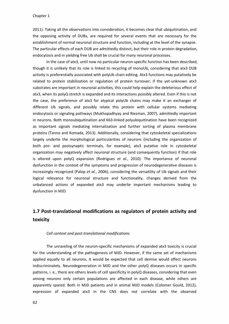
Chapter 1
62
2011). Taking all the observations into consideration, it becomes clear that ubiquitination, and
the opposing activity of DUBs, are required for several events that are necessary for the
establishment of normal neuronal structure and function, including at the level of the synapse.
The particular effects of each DUB are admittedly distinct, but their role in protein degradation,
endocytosis and in yielding free Ub shall be crucial for many neuronal processes.
In the case of atx3, until now no particular neuron-specific function has been described,
though it is unlikely that its role is linked to recycling of monoUb, considering that atx3 DUB
activity is preferentially associated with polyUb chain editing. Atx3 functions may putatively be
related to protein stabilization or regulation of protein turnover; if the yet-unknown atx3
substrates are important in neuronal activities, this could help explain the deleterious effect of
atx3, when its polyQ stretch is expanded and its interactions possibly altered. Even if this is not
the case, the preference of atx3 for atypical polyUb chains may make it an exchanger of
different Ub signals, and possibly relate this protein with cellular systems mediating
endocytosis or signaling pathways (Mukhopadhyay and Riezman, 2007), admittedly important
in neurons. Both monoubiquitination and K63-linked polyubiquitination have been recognized
as important signals mediating internalization and further sorting of plasma membrane
proteins (Tanno and Komada, 2013). Additionally, considering that cytoskeletal specializations
largely underlie the morphological particularities of neurons (including the organization of
both pre- and postsynaptic terminals, for example), atx3 putative role in cytoskeletal
organization may negatively affect neuronal structure (and consequently function) if that role
is altered upon polyQ expansion (Rodrigues et al., 2010). The importance of neuronal
dysfunction in the context of the symptoms and progression of neurodegenerative diseases is
increasingly recognized (Palop et al., 2006); considering the versatility of Ub signals and their
logical relevance for neuronal structure and functionality, changes derived from the
unbalanced actions of expanded atx3 may underlie important mechanisms leading to
dysfunction in MJD.
1.7 Post-translational modifications as regulators of protein activity and
toxicity
Cell context and post-translational modifications
The unraveling of the neuron-specific mechanisms of expanded atx3 toxicity is crucial
for the understanding of the pathogenesis of MJD. However, if the same set of mechanisms
applied equally to all neurons, it would be expected that cell demise would affect neurons
indiscriminately. Neurodegeneration in MJD and the other polyQ diseases occurs in specific
patterns, i. e., there are others levels of cell specificity in polyQ diseases, considering that even
among neurons only certain populations are affected in each disease, while others are
apparently spared. Both in MJD patients and in animal MJD models (Colomer Gould, 2012),
expression of expanded atx3 in the CNS does not correlate with the observed

Introduction
63
neurodegeneration profiles, meaning that the toxicity mechanisms are either specific of
certain cell populations, or that different cell populations show varying degree of resistance to
the effects of expanded atx3. Protein properties and neuronal particularities may conjugate, so
that toxicity spares some neurons but damages others. Additionally, though studies often
describe neurodegeneration as encompassing regional atrophy, cell loss and other
morphological signs of demise, it is increasingly important to consider that cells that have no
visible changes in structure may be dysfunctional and thus contribute to the disease as well.
This is possibly the case, for instance, of Purkinje cells, which have been classically considered
to be spared in MJD, but were reported to be dysfunctional in symptomatic YAC MJD model
(Shakkottai et al., 2011). The same may be true for cell populations presenting aggregates, but
displaying no other overt morphologic defects.
The differential effect of atx3 in different neuronal cell types is undoubtedly mediated
by the protein regions outside the polyQ tract, considering that the other polyQ-containing
proteins associated with neurodegenerative disease lead to different neurodegeneration
profiles, despite the wide distribution and the common pathological expansion. Each of those
proteins shares no homology outside the polyQ tract, and it is logical to consider these regions
as responsible for the specificities of each pathology. Interestingly, a recent study employing a
C. elegans model of MJD expressing expanded atx3 in neurons described that even in this
organism aggregation occurs only in specific neuronal subtypes (Teixeira-Castro et al., 2011),
but the profile was different from what was elicited by expression of isolated polyQ sequences.
Additionally, polyQ sequences of 40 residues produced stronger locomotor deficits than atx3
containing 75 glutamines.
Differential regulation of proteins in different cell types may be mediated by post-
translation modifications (PTMs), a diverse set of mechanisms that regulate protein properties,
frequently in a reversible way (Cohen, 2002; La Spada and Taylor, 2003; Pennuto et al., 2009).
Among the most prevalent (and best studied) PTMs is the covalent conjugation of small
protein modifiers, as is the case of ubiquitination but also of others Ub-like proteins, such as
NEDD8 and small Ub-like modifiers (SUMO), which are added to lysine residues of substrate
proteins through an enzymatic cascade largely resembling that of Ub conjugation (van der
Veen and Ploegh, 2012; Welchman et al., 2005). Many other PTMs exist in eukaryotic cells, and
among the numerous modifications involving conjugation of chemical groups to amino acid
side chains, the most acknowledged is phosphorylation. This modification usually consists in
the addition of a phosphate group to the side chains of amino acids containing a hydroxyl
group. In eukaryotes, phosphorylation normally occurs in serine, threonine and tyrosine
residues, and is catalyzed by enzymes designates as kinases; the reverse effect, termed
dephosphorylation, is catalyzed by phosphatases (Cohen, 2002).
PTMs regulate many different properties of a protein, including enzymatic activity,
molecular interactions, subcellular localization and stability. The changes produced are
considered to result from steric or charge effects or to derive from possible alterations in
protein interactions (Hunter, 2007). PTMs are the basis of many regulatory pathways of the
cell and are consequently implicated in a myriad of biological phenomena. Phosphorylation, in

Chapter 1
64
particular, is considered the most general regulatory device in eukaryotic cells and has been
demonstrated to mediate mechanisms in almost every physiologic event (Cohen, 2002; Hunter,
2012; Pawson and Scott, 2005). Strikingly, it has been predicted that around 30 % of the
human proteins are phosphorylated and that, at any time point in a eukaryotic cell, one-third
of all proteins are phosphorylated (Cohen, 2002). For this reasons, it is logical to hypothesize
that at least some of atx3 features are regulated by PTMs. Furthermore, it is also possible that
PTMs influence the toxicity mechanisms moved upon polyQ expansion of the protein, either by
playing particular roles in atx3 biological function that become deleterious to neurons upon
mutation, or by directly influencing toxic phenomena, such as aggregation, proteolytic
processing or abnormal interactions. Understanding how atx3 PTMs affect the properties of
the protein may thus help decipher the biological role of this protein, but is probably also
bound to contribute to the understanding of MJD pathogenic mechanisms.
Regulation of deubiquitinating enzymes by post-translational modifications
DUBs are themselves responsible for the modulation of one diverse and complex PTM
that is implicated in several cellular mechanisms – ubiquitination. For this reason, these
enzymes are highly regulated, by means of different strategies. The activity of DUBs is known
to be regulated in cells by the proteins with which they interact (including the proper
substrates and scaffolding proteins), by changes in their subcellular localization and,
remarkably, by PTMs that target them (Komander et al., 2009). Understanding the possible
effects that PTMs have on DUBs is perhaps the best way to envisage the possible outcomes of
atx3 modifications.
Proteases are frequently regulated by phosphorylation, a modification that can either
activate or inactivate these enzymes, depending on the particular sites and proteins that are
targeted (Renatus and Farady, 2012). Largely due to high-throughput proteomic studies, many
DUBs are currently known to be phosphorylated, some of them in more than one residue
(Kessler and Edelmann, 2011). However, only a small fraction of these modifications has been
biochemically characterized and their outcomes explored. Phosphorylation of many DUBs has
been demonstrated to modify their enzymatic activity, contributing to the extensive crosstalk
recognized to exist between phospho and Ub signaling (Komander et al., 2009). The majority
of available studies describes phosphorylation events with negative consequences for DUB
activity; this is the case of phosphorylation of Ub C-terminal hydrolase 8 (USP8),
cylindromatosis-associated USP CYLD and otubain-1, OTU enzyme, at amino acids S680, S418
and Y26, respectively (Edelmann et al., 2010; Hutti et al., 2007; Mizuno et al., 2007; Reiley et
al., 2005). The conclusions of these studies were based on observations obtained with
phosphomutated DUBs, where the phosphorylation sites are either mutated to negatively
charged amino acids so as to resemble constitutive phosphorylation states, or to alanine
residues, in order to block phosphorylation. The evaluation of activity was performed through
various methods: USP8 reacted ex vivo against polyUb chains (Mizuno et al., 2007), CYLD
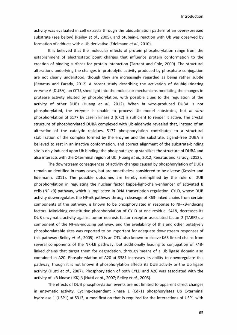
Introduction
65
activity was evaluated in cell extracts through the ubiquitination pattern of an overexpressed
substrate (see below) (Reiley et al., 2005), and otubain-1 reaction with Ub was observed by
formation of adducts with a Ub derivative (Edelmann et al., 2010).
It is believed that the molecular effects of protein phosphorylation range from the
establishment of electrostatic point charges that influence protein conformation to the
creation of binding surfaces for protein interaction (Tarrant and Cole, 2009). The structural
alterations underlying the changes in proteolytic activity produced by phosphate conjugation
are not clearly understood, though they are increasingly regarded as being rather subtle
(Renatus and Farady, 2012) A recent study describing the activation of deubiquitinating
enzyme A (DUBA), an OTU, shed light into the molecular mechanisms mediating the changes in
protease activity elicited by phosphorylation, with possible clues to the regulation of the
activity of other DUBs (Huang et al., 2012). When in vitro-produced DUBA is not
phosphorylated, the enzyme is unable to process Ub model substrates, but in vitro
phosphorylation of S177 by casein kinase 2 (CK2) is sufficient to render it active. The crystal
structure of phosphorylated DUBA complexed with Ub-aldehyde revealed that, instead of an
alteration of the catalytic residues, S177 phosphorylation contributes to a structural
stabilization of the complex formed by the enzyme and the substrate. Ligand-free DUBA is
believed to rest in an inactive conformation, and correct alignment of the substrate-binding
site is only induced upon Ub binding; the phosphate group stabilizes the structure of DUBA and
also interacts with the C-terminal region of Ub (Huang et al., 2012; Renatus and Farady, 2012).
The downstream consequences of activity changes caused by phosphorylation of DUBs
remain unidentified in many cases, but are nonetheless considered to be diverse (Kessler and
Edelmann, 2011). The possible outcomes are hereby exemplified by the role of DUB
phosphorylation in regulating the nuclear factor kappa-light-chain-enhancer of activated B
cells (NF-κB) pathway, which is implicated in DNA transcription regulation. CYLD, whose DUB
activity downregulates the NF-κB pathway through cleavage of K63-linked chains from certain
components of the pathway, is known to be phosphorylated in response to NF-κB-inducing
factors. Mimicking constitutive phosphorylation of CYLD at one residue, S418, decreases its
DUB enzymatic activity against tumor necrosis factor receptor-associated factor 2 (TARF2), a
component of the NF-κB-inducing pathway, and the availability of this and other putatively
phosphorylatable sites was reported to be important for adequate downstream responses of
this pathway (Reiley et al., 2005). A20 is an OTU also known to cleave K63-linked chains from
several components of the NK-kB pathway, but additionally leading to conjugation of K48-
linked chains that target them for degradation, through means of a Ub ligase domain also
contained in A20. Phosphorylation of A20 at S381 increases its ability to downregulate this
pathway, though it is not known if phosphorylation affects its DUB activity or the Ub ligase
activity (Hutti et al., 2007). Phosphorylation of both CYLD and A20 was associated with the
activity of IκB kinase (IKK) β (Hutti et al., 2007; Reiley et al., 2005).
The effects of DUB phosphorylation events are not limited to apparent direct changes
in enzymatic activity. Cycling-dependent kinase 1 (Cdk1) phosphorylates Ub C-terminal
hydrolase 1 (USP1) at S313, a modification that is required for the interactions of USP1 with
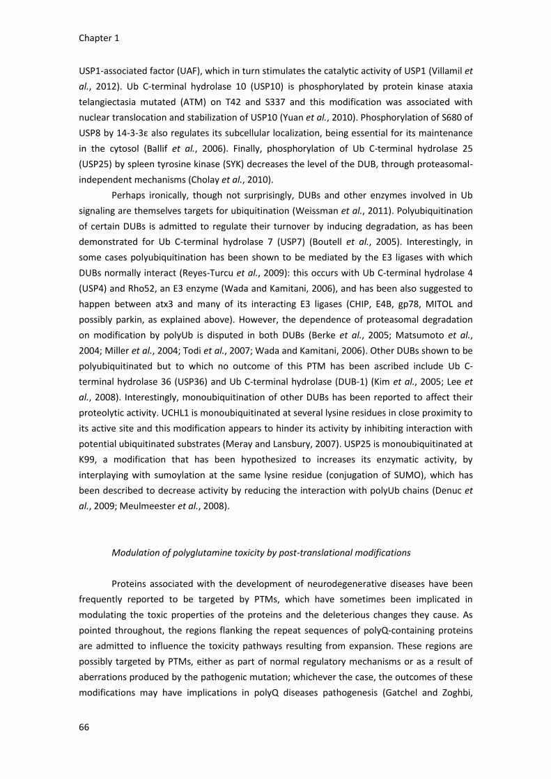
Chapter 1
66
USP1-associated factor (UAF), which in turn stimulates the catalytic activity of USP1 (Villamil et
al., 2012). Ub C-terminal hydrolase 10 (USP10) is phosphorylated by protein kinase ataxia
telangiectasia mutated (ATM) on T42 and S337 and this modification was associated with
nuclear translocation and stabilization of USP10 (Yuan et al., 2010). Phosphorylation of S680 of
USP8 by 14-3-3ε also regulates its subcellular localization, being essential for its maintenance
in the cytosol (Ballif et al., 2006). Finally, phosphorylation of Ub C-terminal hydrolase 25
(USP25) by spleen tyrosine kinase (SYK) decreases the level of the DUB, through proteasomal-
independent mechanisms (Cholay et al., 2010).
Perhaps ironically, though not surprisingly, DUBs and other enzymes involved in Ub
signaling are themselves targets for ubiquitination (Weissman et al., 2011). Polyubiquitination
of certain DUBs is admitted to regulate their turnover by inducing degradation, as has been
demonstrated for Ub C-terminal hydrolase 7 (USP7) (Boutell et al., 2005). Interestingly, in
some cases polyubiquitination has been shown to be mediated by the E3 ligases with which
DUBs normally interact (Reyes-Turcu et al., 2009): this occurs with Ub C-terminal hydrolase 4
(USP4) and Rho52, an E3 enzyme (Wada and Kamitani, 2006), and has been also suggested to
happen between atx3 and many of its interacting E3 ligases (CHIP, E4B, gp78, MITOL and
possibly parkin, as explained above). However, the dependence of proteasomal degradation
on modification by polyUb is disputed in both DUBs (Berke et al., 2005; Matsumoto et al.,
2004; Miller et al., 2004; Todi et al., 2007; Wada and Kamitani, 2006). Other DUBs shown to be
polyubiquitinated but to which no outcome of this PTM has been ascribed include Ub C-
terminal hydrolase 36 (USP36) and Ub C-terminal hydrolase (DUB-1) (Kim et al., 2005; Lee et
al., 2008). Interestingly, monoubiquitination of other DUBs has been reported to affect their
proteolytic activity. UCHL1 is monoubiquitinated at several lysine residues in close proximity to
its active site and this modification appears to hinder its activity by inhibiting interaction with
potential ubiquitinated substrates (Meray and Lansbury, 2007). USP25 is monoubiquitinated at
K99, a modification that has been hypothesized to increases its enzymatic activity, by
interplaying with sumoylation at the same lysine residue (conjugation of SUMO), which has
been described to decrease activity by reducing the interaction with polyUb chains (Denuc et
al., 2009; Meulmeester et al., 2008).
Modulation of polyglutamine toxicity by post-translational modifications
Proteins associated with the development of neurodegenerative diseases have been
frequently reported to be targeted by PTMs, which have sometimes been implicated in
modulating the toxic properties of the proteins and the deleterious changes they cause. As
pointed throughout, the regions flanking the repeat sequences of polyQ-containing proteins
are admitted to influence the toxicity pathways resulting from expansion. These regions are
possibly targeted by PTMs, either as part of normal regulatory mechanisms or as a result of
aberrations produced by the pathogenic mutation; whichever the case, the outcomes of these
modifications may have implications in polyQ diseases pathogenesis (Gatchel and Zoghbi,

Introduction
67
2005; Takahashi et al., 2010). Modification of polyQ-containing proteins may be either
neurotoxic, when they contribute to toxicity-associated mechanisms such as aggregation,
proteolytic cleavage (itself a PTM) and aberrant interactions, or neuroprotective, in cases
where the PTMs lead to degradation of the pathogenic proteins (Pennuto et al., 2009). There
are many concrete examples of PTMs that influence aspects of polyQ-induced toxicity, with
reports focusing frequently on ubiquitination, sumoylation, palmitoylation, acetylation and,
specially, phosphorylation (Bauer and Nukina, 2009; Ehrnhoefer et al., 2011; La Spada and
Taylor, 2010; Pennuto et al., 2009). The following subsections will address the current data
regarding phosphorylation of polyQ-containing proteins and the effects of this PTM on
disease-related mechanisms (Table II).
A. Phosphorylation of huntingtin
Huntingtin (htt), the protein involved in HD, is known to be modified by
phosphorylation in many different residues and it has been extensively demonstrated that this
modifications influences the toxic outcomes caused by polyQ expansion of htt (Ehrnhoefer et
al., 2011). Overall, the modifications that have been so far characterized indicate that
phosphate-conjugation plays a protective role in HD-related events. Htt is a large protein that
has been functionally related to diverse cellular pathways including axonal transport,
transcription and signaling, possibly functioning as a scaffolding protein (Gatchel and Zoghbi,
2005; Williams and Paulson, 2008). Notably, due to its large size, studies frequently utilize htt
truncated forms instead of the full-length protein.
Activation of Akt, shown to phosphorylate S421 in vitro, was initially described to
decrease inclusion formation and cell death in striatal neuron cultures expressing an expanded
htt N-terminal fragment (the striatum is harshly affected in HD) (Humbert et al., 2002). Later,
another kinase – serum/glucocorticoid regulated kinase (SGK) – was also shown to target S421
and to protect striatal neurons against expanded htt-induced cell death (Rangone et al., 2004).
Inhibition of calcineurin, a phosphatase demonstrated to dephosphorylate S421 in vitro and in
cells, was also reported to prevent death of HD cell culture models (Pardo et al., 2006).
Important evidence for the protective role of S421 phosphorylation in vivo was obtained with
lentiviral rat models of HD expressing a fragment of expanded htt in the striatum, in which
mimicking phosphorylation of S421 by mutation of this residue to an aspartate decreased the
neuronal loss elicited by the pathogenic protein fragment (Pardo et al., 2006). Mechanistically,
the protective effects of S421 phosphorylation may be related to htt role in axonal transport:
in cultures of rat cortical neurons, expanded htt hinders the axonal transport of vesicles, but
mimicking phosphorylation of S421 restores transport to the levels observed with non-
expanded htt (Colin et al., 2008; Zala et al., 2008). Phosphorylation of S421 was also shown to
reduce caspase 6-mediated cleavage of expanded htt and its nuclear localization, two events
recognized to be deleterious in HD (Warby et al., 2009). Recent results further suggested that
phosphorylation of S421 may be influenced by excitotoxic events: the levels of S421
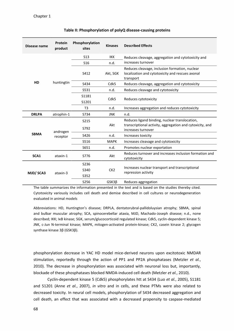
Chapter 1
68
Table II: Phosphorylation of polyQ disease-causing proteins
Disease name Protein
product
Phosphorylation
sites Kinases Described Effects
HD huntingtin
S13 IKK Reduces cleavage, aggregation and cytotoxicity and increases turnover S16 n.d.
S412 Akt, SGK Reduces cleavage, inclusion formation, nuclear localization and cytotoxicity and rescues axonal transport
S434 Cdk5 Reduces cleavage, aggregation and cytotoxicity
S531 n.d. Reduces cleavage and cytotoxicity
S1181 Cdk5 Reduces cytotoxicity
S1201
T3 n.d. Increases aggregation and reduces cytotoxicity
DRLPA atrophin-1 S734 JNK n.d.
SBMA androgen receptor
S215 Akt
Reduces ligand binding, nuclear translocation, transcriptional activity, aggregation and cytoxicity, and increases turnover S792
S426 n.d. Increases toxicity
S516 MAPK Increases cleavage and cytotoxicity
S651 n.d. Promotes nuclear exportation
SCA1 ataxin-1 S776 Akt Reduces turnover and increases inclusion formation and cytotoxicity
MJD/ SCA3 ataxin-3
S236
CK2 Increases nuclear transport and transcriptional repression activity
S340
S352
S256 GSK3β Reduces aggregation
The table summarizes the information presented in the text and is based on the studies thereby cited.
Cytotoxicity variously includes cell death and demise described in cell cultures or neurodegeneration
evaluated in animal models
Abbreviations: HD, Huntington’s disease; DRPLA, dentatorubral-pallidoluysian atrophy; SBMA, spinal
and bulbar muscular atrophy; SCA, spinocerebellar ataxia, MJD, Machado-Joseph disease; n.d., none
described; IKK; IκB kinase; SGK, serum/glucocorticoid regulated kinase; Cdk5, cyclin-dependent kinase 5;
JNK, c-Jun N-terminal kinase; MAPK, mitogen-activated protein-kinase; CK2, casein kinase 2; glycogen
synthase kinase 3β (GSK3β).
phosphorylation decrease in YAC HD model mice-derived neurons upon excitotoxic NMDAR
stimulation, reportedly through the action of PP1 and PP2A phosphatases (Metzler et al.,
2010). The decrease in phosphorylation was associated with neuronal loss but, importantly,
blockade of these phosphatases blocked NMDA-induced cell death (Metzler et al., 2010).
Cyclin-dependent kinase 5 (Cdk5) phosphorylates htt at S434 (Luo et al., 2005), S1181
and S1201 (Anne et al., 2007), in vitro and in cells, and these PTMs were also related to
decreased toxicity. In neural cell models, phosphorylation of S434 decreased aggregation and
cell death, an effect that was associated with a decreased propensity to caspase-mediated

Introduction
69
cleavage (Luo et al., 2005). Phosphorylation of S1181 and S1201 was demonstrated to be
stimulated by DNA damage, and inhibiting their modification by mutating alanine residues
rends the non-expanded htt toxic, leading to cell death of rat striatal neurons similar to what is
elicited by expanded htt (Anne et al., 2007). Contrarily, mutating both S1181 and S1201 of
expanded htt to negatively charged aspartate residues decreases toxicity.
T3 of htt was reported to be phosphorylated in mouse brain and an hht fragment
containing phosphomimetic T3D was reported to lead to stronger aggregation in both cell
cultures and in vivo, in neurons from transgenic Drosophila larvae (Aiken et al., 2009).
Curiously, although the non-phosphorylatable T3A form decreases aggregation, both mutants
were shown to reduced neurodegeneration and lethality in this model, indicating that the T3
residue probably has an intrinsic property leading to toxicity, that decreases upon mutation to
either alanine or aspartate. It was suggested that, given the differences in aggregation
between the two T3 mutants, different rescue mechanisms may be at work. It is possible that,
in accordance with current opinions, the microscopic aggregates observed in this study do not
correspond to the toxic species (Aiken et al., 2009).
Overexpression of IKK in striatal precursor cells leads to the phosphorylation of S13
and S16 of a htt fragment, though in vitro assays indicate that the IKK-catalyzed
phosphorylation is only direct in the case of S13 (Thompson et al., 2009). Importantly,
phosphorylation of S13 was further confirmed to target endogenous htt. In transfected cells,
increased phosphorylation at S13 and S16 was associated with decreased levels of the htt
fragment and, interestingly, phosphomimicking of both residues or overexpression of IKK
decreased polyubiquitination and sumoylation at nearby lysine residues and increased
degradation of the htt fragment by the proteasome and the lysosome. Contrastingly, htt
fragments with expanded glutamines, which were shown to present reduced levels of S13 and
S16 phosphorylation, had a tendency to accumulate in cells, possibly due to decreased clearing
(Thompson et al., 2009). In vitro, the phosphomimetic mutants of similar fragments containing
37 glutamines (the threshold of HD risk) showed decreased tendency to aggregate, comparing
to non-mutated fragments and non-phosphorylatable mutants (Gu et al., 2009; Mishra et al.,
2012). Importantly, in a transgenic mouse model expressing full-length expanded htt, the
disease phenotype is largely preserved when S13 and S16 are mutated to alanines, but is
rescued by mutation to aspartate (phosphomimetic mutant). The disease signs of the alanine-
mutated model include htt aggregation, selective neurodegeneration (cortex and striatum; the
cerebellum is spared) and motor as well as psychiatric-like deficits (Gu et al., 2009). Counter-
intuitively, taking into account these possible protective roles of S13 and S16,
phosphomimicking of both residues increases nuclear localization in transfected cortical
neuronal cultured (Thompson et al., 2009) and, in particular, phosphorylation of S16 has
recently been associated with nuclear accumulation of N-terminal fragments (Havel et al.,
2011).
A mass spectrometry (MS) analysis of full-length htt expressed in cell lines identified
three additional htt phosphorylational sites – S2076, S2653 and S2657 – along with a
monophosphorylated peptide to which an individual phosphorylation site could not be

Chapter 1
70
mapped; phosphorylation can target either S533, S535 or S356 (Schilling et al., 2006). This
amino acids lie on a proteolytic susceptibility domain of htt and S536 in particular has been
identified as a site for calpain cleavage (Gafni et al., 2004). Mutating this residue to aspartate,
but not the other two, reduced the cleavage of C-terminally truncated expanded htt and the
toxicity associated with expression of this protein, in a cell line (Schilling et al., 2006).
Important insights into the relevance of phosphorylation defects in HD derive also
from the analysis of kinases in the brain of HD patients and animal models. Cdk5 purified from
the brains of a HD mouse model displays a reduced activity (Luo et al., 2005) and, in HD
patients brain, Akt is more prone to suffer caspase-mediated proteolytic cleavage, as detected
in the cerebellum, cortex and, specially, in the striatum (Humbert et al., 2002). Contrastingly,
however, the levels of SGK are increased in HD patients’ brain, which nonetheless supports a
role for this kinase in HD-related mechanisms (Rangone et al., 2004).
Finally, there is some important evidence for a correlation between the patterns of
these protective phosphorylation events and the cell selectivity of HD: generally speaking,
reports demonstrate that phosphorylation is often reduced in cell types known to be specially
sensitive to expanded htt toxicity. The levels of phosphorylated T3 of a htt fragment are
reduced in transfected striatal precursor cells when compared to non-neuronal cell lines, and
phosphorylation decreases even more upon polyQ expansion of that htt fragment.
Interestingly, this last difference is only observed in rat striatal precursor cells but not in non-
neuronal cell lines (Aiken et al., 2009). In HD mouse models, the levels of T3 phosphorylation
are specially decreased in the striatum, when compared to the cortex (Aiken et al., 2009). In a
HD cell culture model, phosphorylation of expanded htt at S421 is decreased when compared
to wild-type cell lines (Pardo et al., 2006) and the same was observed for transfected cell lines
expressing expanded or non-expanded truncated htt and for YAC transgenic mice (Warby et al.,
2005). S421 is phosphorylated under physiological conditions in both human and wild-type
mouse brain, but, in the last case, it was further observed that the phosphorylation levels in
the three particular brain regions analyzed varied: it was higher in the cerebellum, less in the
cortex and least in the striatum (Ehrnhoefer et al., 2011; Warby et al., 2005). This correlates
with the neurodegeneration pattern observed in HD (Warby et al., 2005). Interestingly,
stimulation of NMDARs in the striatum of YAC HD model mice decreases the levels of S421
phosphorylation relatively to wild-type animals (Metzler et al., 2010), indicating that excitoxic
stimuli may increase neuronal susceptibility by decreasing the protective effect of S421
phosphorylation.
B. Phosphorylation of other polyglutamine disease-causing proteins
The androgen receptor (AR) is a testosterone-associated steroid hormone receptor
that is widely studied because of its involvement in prostate cancer, though it is also related to
another disease, SBMA, which is caused when the polyQ region of this protein is expanded
(Williams and Paulson, 2008). A large number of phosphorylation sites has been mapped in AR:
Introduction
71
S16, S83, S96, S215, S258, S310, S426, S516, S651, S792, Y223, Y267, Y307, Y346, Y357,
Y362/363, Y393, Y534, Y551 and Y915 (Gioeli et al., 2006; Guo et al., 2006; Palazzolo et al.,
2007; Pennuto et al., 2009). The phosphorylation of some of these residues has been
associated with the functions of AR; for instance, S651 phosphorylation is induced by stress
kinase signaling and was shown to modulate AR transcriptional activity, by increasing its
nuclear exportation (Gioeli et al., 2006). Interestingly, serine phosphorylation of AR,
irrespective of particular serine residues, increases with larger glutamine repeats (LaFevre-
Bernt and Ellerby, 2003), but particular phosphorylation events appear to be either protective
or deleterious, depending on the phosphorylation site. In cell lines, blockade of S516
phosphorylation, which is catalyzed by a mitogen-activated protein-kinase (MAPK), abrogates
polyQ-expanded AR-derived cell death and formation of caspase-3-derived cleavage products,
which have been implicated in the toxicity mechanisms of SBMA (LaFevre-Bernt and Ellerby,
2003). Preventing S426 and S516 phosphorylation also reduces toxicity of the polyQ-expanded
AR, but turns the non-expanded AR toxic (Funderburk et al., 2009). Phosphorylation of S215
and S792 was suggested to be mediated by Akt and to be associated with a reduction in
expanded AR ligand binding, ligand-dependent nuclear translocation, transcriptional activation
and citotoxicity, in cell lines (Palazzolo et al., 2007). This phosphorylation event was further
demonstrated to reduce expanded AR aggregation and increase its degradation by the UPP in
cultured cells, and Akt activation was shown to ameliorate the disease phenotype of a SBMA
mouse model (Palazzolo et al., 2009).
Atrophin-1 is a protein involved in transcription regulation and is causatively related to
the development of DRPLA (Bauer and Nukina, 2009). S734 of atrophin-1 is phosphorylated by
the c-Jun N-terminal kinase (JNK), but the affinity of this enzyme decreases when the polyQ
region of atrophin-1 is expanded. This alteration was suggested to delay neuronal pro-survival
pathways (Okamura-Oho et al., 2003).
Ataxin-1 (atx1) causes SCA1 and its function has been also associated with
transcription regulation. Phosphorylation of S776 has been reported to play a critical role in
the pathogenesis of this SCA1, considering that mutating this residue to alanine renders
expanded atx1 unable to form aggregates in a cell line and, remarkably, transgenic mice
expressing this mutated form in Purkinje cells displayed a considerably milder disease
phenotype, when compared to animals expressing non-mutated expanded atx1 (Emamian et
al., 2003). The ameliorated characteristics included less inclusion number, less degeneration of
Purkinje cells, and increased motor performance. Akt-mediated phosphorylation, which was
shown to target S776, leads to the binding of expanded atx1 and the 14-3-3 protein in vitro
(Chen et al., 2003b). This interaction is considered to be very relevant for pathogenesis: in cell
lines, cotransfection of 14-3-3 was shown to stabilize expanded atx1 and increase inclusion
formation; in transgenic Drosophila, 14-3-3 co-expression increased neurodegeneration
caused by expanded atx1 (Chen et al., 2003b).

Chapter 1
72
Post-translational modifications of ataxin-3
As explained before, DUBs are frequently observed to display a low enzymatic activity
in vitro, and atx3 is no exception (Chow et al., 2004a; Komander et al., 2009). Thus, it is
predicted that atx3 activity, and consequently the biologic function of this protein, may be
regulated by PTMs, either directly or by interfering with important interactions with other
proteins (Almeida et al., 2013; Reyes-Turcu et al., 2009). Furthermore, it is also possible that
other properties of atx3, such as subcellular localization and turnover, both possibly relevant
for its physiological role and for the pathogenesis of MJD, may be as well regulated by these
kinds of mechanisms. The differential regulation of atx3 subcellular localization by PTMs and
other mechanisms in different cells may explain the diversity in intracellular distribution
observed both in cell lines and human brain cells (Trottier et al., 1998). Finally, attending to the
effects of phosphorylation on the toxicity mechanisms caused by other polyQ-containing
proteins, it is plausible to admit that PTMs of atx3 conceivably interfere with the pathways
linking polyQ-expansion of atx3 to the disease outcomes in MJD, either leading to protection
or aggravating the deleterious effect of the pathogenic protein. The fact that the
understanding of regulation of atx3 by PTMs may simultaneously elucidate about its biological
function and the pathogenic mechanisms of MJD justifies the interest of scientific research in
this area.
Atx3 is currently known to be targeted by different PTM mechanisms. As exposed
above when dealing with the turnover and degradation of atx3, polyubiquitination has been
described to target this protein, though there is controversy whether it leads to degradation by
the proteasome; this may not be true, at least in some cases (Berke et al., 2005; Jana and
Nukina, 2004; Matsumoto et al., 2004; Todi et al., 2007; Tsai et al., 2003). One study
demonstrated that the expanded protein is more stable than its non-expanded counterpart
and that it is degraded by the proteasome less efficiently; this would be expected to contribute
to the accumulation of the pathogenic protein and to the persistence of its toxic effects
(Matsumoto et al., 2004). However, increased ubiquitination did not correlate with increased
degradation, i.e., the two forms were ubiquitinated to similar extents. Thus, the differences
observed may be due to an inefficient proteasomal recognition and degradation due to the
expanded polyQ tract (Matsumoto et al., 2004). Nevertheless, it appears that there is not
much consensus on the increased stability of the expanded form and, in effect, another study
reported that both normal and expanded forms of atx3 are degraded with the same efficiency
by the proteasome and that there is no inhibition of the proteasome by soluble expanded atx3
(Berke et al., 2005).
Recent reports demonstrated that atx3 is also monoubiquitinated in cells (including
neural cells), and that this modification increases its DUB activity independent of potential
cofactors or interactors, in vitro (Todi et al., 2009). K117 of the JD, located in the vicinity of the
catalytic site, is the preferentially ubiquitinated lysine residue (Figure 9) both in vitro and in
cells (modification of other lysines was less frequently detected), and ubiquitination of this
residue alone is sufficient for the observed activity enhancement (Todi et al., 2010).

Introduction
73
Interestingly, though the UIMs are important in determining cleaving preferences, the increase
in activity by monoubiquitination occurs independently from the functionality of these motifs
(Todi et al., 2010; Todi et al., 2009). However, when UIM1 and UIM2 are not functional,
monoubiquitinated atx3 inverts its preferences, cleaving K48-linked chains more efficiently
than K63-linked ones (Todi et al., 2010; Todi et al., 2009). Furthermore, availability of K117 for
ubiquitination increases atx3 ability to promote aggresome formation, which is dependent on
its DUB activity (Todi et al., 2010). Taking into consideration that several proteotoxic stressors
enhanced atx3 ubiquitination (mono- and polyubiquitination), authors have suggested that the
upregulation of atx3 activity by ubiquitination may serve as a positive feedback mechanism in
response to proteotoxic stress (Todi et al., 2009). In transgenic Drosophila, coexpression of
atx3 and isolated polyQ sequences reduces degeneration caused by the toxic repeated
sequences, through an undisclosed process that was nonetheless unrelated to a decrease in
toxic polyQ sequence stability (Tsou et al., 2013). It was further demonstrated that contrary to
atx3 where all lysine residues had been mutated, atx3 with only one lysine residue at position
117 – which was shown to be ubiquitinated in Drosophila as well – was sufficient to produce
this effect (Tsou et al., 2013). These results strongly suggest an important role for K177
ubiquitination on the in vivo function of atx3. Importantly, ubiquitination of atx3 is not
targeted by its own DUB activity in trans, arguing against the possibility that atx3 regulates its
own activity by regulating its ubiquitination status (Todi et al., 2007).
Through biochemical analysis of the phosphoamino ac ids present on 32P-
orthophosphate-radiolabled samples of atx3, Mueller and collaborators (2009) showed that
atx3 is phosphorylated on serine residues. In fact, several serine phosphorylation sites have
been mapped, all of them located in the UIMs: S236, in the first UIM, S256 and 260/261 (it is
not clear which of the adjacent serine residues is phosphorylated), located on the second UIM,
and S340 and 352 of the third UIM (Figure 9) (Fei et al., 2007; Mueller et al., 2009). Importantly,
there are already some clues to the possible biological relevance of these modifications. The
study by Mueller and collaborators (2009) showed that stimulating phosphorylation of serine
residues 236, or 340 and 352 simultaneously, by mutating these amino acids to aspartate,
leads to an increased nuclear localization in cells lines, admitted to be important for
aggregation and toxicity of the expanded form, and enhances repression of atx3-regulated
transcription in gene reporter assays of MMP2 expression (Mueller et al., 2009). Curiously the
increased nuclear localization of atx3 occurs independently from the described NLS pathway,
at least for S340 and S352 (Macedo-Ribeiro et al., 2009; Mueller et al., 2009). Additionally, a
phosphomimetic mutant of residues 340 and 352 also increased the stability and half-life of
atx3 (Mueller et al., 2009). CK2 was shown to interact with atx3 and to phosphorylate its UIM-
containing C-terminal region (Mueller et al., 2009; Tao et al., 2008) and pharmacological
inhibition of this kinase not only reduced the amount of nuclear atx3 and activated atx3-
regulated gene transcription, but also decreased NI formation, indicating that CK2-dependent
phosphorylation regulates these properties, admittedly by interference with the
phosphorylated state of atx3 (Mueller et al., 2009). Considering what was discussed about the
conditioning role of the nuclear localization and decreased turnover of atx3 in the deleterious

Chapter 1
74
Figure 9: Mapping and effects of atx3 PTMs. Apart from being polyubiquitinated, atx3 has been shown to be monoubiquitinated at K117, a PTM that was demonstrated to increase its DUB enzymatic activity. Five serine residues present in the UIMs have also been described to be phosphorylated: S236, S256, S260/S261, S340 and S352. Hindering S256 phosphorylation was shown to increase aggregation of polyQ-expanded atx3, suggesting that this modification may be protective against aggregation. Mimicking phosphorylation of residues S340 and S352 was shown to increase atx3 nuclear shuttling and atx3-mediated repression of transcription and to increase the stability of the protein. Mimicking S236 elicited analogous effects on nuclear transport and transcriptional repression (not represented). The figure uses the same domain annotations as figure 3, except for the region C-terminally of UIM2; those annotations are based on what is mentioned by the authors describing the phosphorylation sites on UIM3 (Mueller et al., 2009), using an atx3 sequence with 19 glutamine repeats (the protein sequence corresponds to Uniprot human atx3 isoform 2 - code P54252-2).
mechanisms of MJD, a phosphorylation-mediated increase in stability and nuclear localization
conceivably interferes with protein aggregation and toxicity (Bichelmeier et al., 2007; Breuer et
al., 2010). Phosphorylation of serines 236, 340 and 352 may participate in the nuclear
translocation of expanded atx3 and, in the nucleus, the phosphorylated protein may lead to
aberrant atx3-regulated gene expression (Evert et al., 2006a; Mueller et al., 2009). The
increased stability may contribute to increased aggregation and NI formation. In fact, C-
terminal-containing fragments of atx3 showed an increase of NI formation when serines 340
and 352 were mutated to mimic phosphorylation (Mueller et al., 2009).
Fei and coworkers (2007) reported that S256 is phosphorylated in vitro by glycogen
synthase kinase 3β (GSK3β), but observed a decreased phosphorylation of the expanded form
of the protein. Contrary to the observations indicating a possible nefarious effect for atx3
phosphorylation, these authors observed that preventing phosphorylation of S256 by mutating
this residue to alanine enhanced the aggregation of an expanded form of atx3. These results
indicate that there may be some relation between the phosphorylated state of S256 and the
formation of the toxic protein aggregates. It is not surprising that the effects of
phosphorylation may differ according to the particular residues that are modified.
A recent study suggested that a serine residue of the JD may be important in atx3
responses to proteotoxic stress (Reina et al., 2010). Under normal conditions, nuclear
localization of atx3 is determined by its NLS and by CK2-dependent phosphorylation, but it was

Introduction
75
reported that proteotoxic stimuli such as heat shock and oxidative stress overrides those
signals and increase atx3 nuclear localization independently (Antony et al., 2009; Macedo-
Ribeiro et al., 2009; Mueller et al., 2009; Reina et al., 2010). Two different regions seem
important for this transport: the JD, namely S111, and the C-terminal region comprising the
polyQ sequence and the rest of the C-terminal (but not the UIMs). Considering that S111 was a
putative target for phosphorylation, the authors of this study mutated S111 to an alanine
residue and observed that heat shock no longer induced an increase in atx3 nuclear
localization; mutation to an aspartate, however, was not sufficient for localization in the
nucleus, but responded normally to heat shock (Reina et al., 2010). Although these results
suggest that modification of S111 may be important in regulating the behavior of atx3 after
stress stimuli, phosphorylation at S111, or at any other residue of the JD, remains undescribed.
1.8 Objectives
The toxicity derived from the pathogenic expansion of polyQ-containing proteins is
usually attributed to changes in biologic function, subcellular localization, molecular
interactions and aggregation of the proteins carrying the pathogenic mutation, but it is
extensively recognized that the regions outside the polyQ tract are crucial in determining
toxicity. Phosphorylation is a reversible post-translational mechanism known to regulate
diverse protein properties, and it has been frequently described to target disease-related
proteins outside their repeat sequence and to influence the deleterious alterations the polyQ
expansion causes. The mechanisms involved in MJD and the cellular role of atx3 are still not
clearly understood, but comprehending how this protein is regulated by phosphorylation, and
how this modification affects atx3 when the protein is expanded, is due to yield relevant
contributions. Alterations caused by phosphorylation may increase our understanding of how
atx3 behaves in a cell, but may also help uncover important points of regulation that interfere
with toxicity mechanisms.
In a previous approach to the study of atx3 phosphorylation, we identified a novel
phosphorylation site of the protein through MS, that was localized in the catalytic JD – serine
12 (Matos, 2009). The current work aims at characterizing this modification, both in terms of
the outcomes it might have regarding atx3 activity as a DUB, and in respect to the way S12
phosphorylation might alter aggregation of expanded atx3 and the neurodegeneration it
causes. With these objectives, we moved to confirm that atx3 is phosphorylated in neuronal
cells and determine the effects of this modification on atx3 DUB enzymatic activity in vitro,
using phosphomimetic mutants of atx3. The effects of S12 phosphorylation in the context of
expanded atx3-induced toxicity were analyzed by observing how different forms of atx3
generated by mutation of S12 affected relevant morphological characteristics of cultured
neurons. Finally, the outcomes of those mutations were further evaluated in vivo, using a well
described lentiviral rat model of MJD with striatal pathology.

Chapter 1
76
The current study is the first to characterize the phosphorylation of an amino acid
residue of atx3 present in the catalytic domain of the protein. Additionally, it is also the first to
investigate the effects of atx3 phosphorylation specifically in neuronal cells and in in vivo
animal models of MJD.

Chapter 2
Materials and Methods


Material and Methods
79
Chapter 2 – Materials and Methods
Expression plasmids
Eukaryotic expression pEFGP-C1 plasmids encoding human atx3 variant MJD1a (NCBI
accession number: AAB33571.1) with 28 glutamines (GFP-atx3 28Q) or 84 glutamines (GFP-
atx3 28Q) fused with the GFP in the N-terminal was a kind gift from Dr. Henry Paulson,
University of Iowa (Iowa City, IA USA; plasmids described in Chai et al., 1999b). Bacterial
expression pDEST17 plasmid encoding hexahistidine (6His)-tagged human atx3 variant MJD1-1
(NCBI accession number: NP_004984.2) with 13 glutamines (6His-atx3) has been previously
described by our group (Gales et al., 2005). Site-directed mutagenesis was used as described
elsewhere (Scarff et al., 2013) to introduce a cleavage site for the recombinant tobacco etch
virus (TEV) protease between the open reading frame and the N-terminal sequence coding for
the 6His tag (6His-TEVg-atx3). Eukaryotic expression plasmid SIN-W-PGK encoding human atx3
variant MJD1a with 72 glutamines and a N-terminal Myc tag (Myc-atx3 72Q) and plasmid SIN-
CW-PGK-nls-LacZ-LTR-TRE encoding an universal short hairpin (shRNA) targeting human and
rat atx3 (shRNA-atx3) were a kind gift from Dr. Luís Pereira de Almeida, University of Coimbra
(Coimbra, Portugal; plasmids described in Alves et al., 2008b and Alves et al., 2010,
respectively; shRNA-atx3 is thereby designated as shAtaxUNIV), as were the constructs used in
the viral production (pCMVDR-8.92, pMD.G and pRSV-Rev). Site-directed mutagenesis was
used to mutate serine 12 of atx3 encoded in the expression plasmids to aspartate or alanine,
as described below.
Site-directed mutagenesis
Mutagenesis was performed using the QuickChange Site-Directed Mutagenesis Kit
(Stratagene, Cambridge, UK) or the QuikChange II XL Site-Directed Mutagenesis Kit (Agilent
Technologies, Santa Clara, CA, USA), in accordance to the procedure suggested by the
manufacturers. Phosphomimetic atx3 S12D mutants, with S12 substituted by aspartate, were
generated using primers atx31_1n_S12D_fo (5’-CAC GAG AAA CAA GAA GGC GAC CTT TGT GCT

Chapter 2
80
CAA CAT TGC CTG-3’) and atx31_1n_S12D_re (5’-CAG GCA ATG TTG AGC ACA AAG GTC GCC
TTC TTG TTT CTC GTG-3’). Phospho-null atx3 S12A mutants, where S12 was exchanged for
alanine, were produced employing primers atx31_1n_S12A_fo (5’-CAC GAG AAA CAA GAA GGC
GCA CTT TGT GCT CAA CAT TGC CTG-3’) and atx31_1n_S12A_re (5’-CAG GCA ATG TTG AGC
ACA AAG TGC GCC TTC TTG TTT CTC GTG-3’). Primers were acquired from Eurofins MWG
Operon, as part of Fisher Scientific (Loures, Portugal).
Mutation of the expression plasmids was confirmed after automatic DNA sequencing
performed by STAB VIDA (Setúbal, Portugal).
Antibodies
The details of the antibodies used in Western blot, immunocytochemistry and
immunohistochemistry are summarized in Table III.
Mass spectrometry
MS analysis of GFP-atx3 directed at the detection of phosphorylation sites was
performed in GFP-atx3 samples prepared from transiently transfected human embryonic
kidney (HEK) 293FT cells, after purification by immunoprecipitation and SDS-polyacrylamide
gel electrophoresis (PAGE). The following procedures have been previously described (Matos,
2009), but are hereby reproduced. Attending to the particularities in cell culture and
transfection used in the preparation of the samples subjected to the MS analysis, these
techniques are also described in this section, separately from the other cell culture and
transfection methods used in the present work, for the sake of comprehensibility.
HEK 293FT cells were grown in Dulbecco’s Modified Eagle’s Medium (DMEM),
supplemented with 10% (v/v) fetal bovine serum (FBS), 0,1 mM non-essential amino acids
(NEAA), 6 mM L-glutamine, 1mM sodium pyruvate (in Minimum Essential Medium – MEM) and
500 μg/ml geneticin, and maintained at 37 °C, in a humidified atmosphere containing 5 % CO2.
One day prior to transfection, fully confluent HEK 293FT cells were diluted 1:6 and then seeded
onto 10 cm diameter culture plates. After 24 h of growth, transfection was carried out using
Lipofectamine® Transfection Reagent (Invitrogen Life Technologies, Barcelone, Spain) with a
modified version of the manufacturer’s protocol, as follows: after substituting the culture
medium for 6,4 ml Opti-MEM supplemented with 10 % FBS, lipid-DNA complexes obtained by
mixing 10 μg of DNA plasmid with 6 μl of Lipofectamine reagent in 800 ml Opti-MEM were
added to each cell plate; after overnight incubation under normal growth conditions,
transfection medium was replaced for normal HEK 293FT cell medium and expression was left
to occur for 24 h of cell growth. Some HEK 293FT cell cultures were subjected to chemical
stimulation directed at enhancing phosphorylation with 20 nM okadaic acid (OA) for 16 h
(Dorval and Fraser, 2006), 5 mM sodium orthovanadate (SOV) for 30 min (Martin et al., 2006)

Material and Methods
81
Table III: Antibodies used in Western blots, immunocytochemistry and
immunohistochemistry
Antibody
Species (and
clonality) Dilution
Incubation
conditions Manufacturer
Western blot
primary
antibodies
anti-atx3 (clone 1H9) mouse
(mono.)
1:1.000 1-2h, RT or
ON, 4 °C
Millipore (Billerica, MA,
USA)
anti-Patx3 rabbit
(poly.)
1.1.000 or
1:20
1h, RT or
ON, 4 °C
Eurogentec (Seraing,
Belgium; details below)
anti-actin (β) mouse
(mono.)
1:5.000 1-2h, RT Sigma-Aldrich (St. Louis
MO, USA)
anti-Ub rabbit
(poly.)
1:1.000 1-2h, RT Dako (Glostrup, Denmark)
anti-GFP mouse
(mono.)
1:1.000 1-2h, RT Roche Applied Science
(Penzberg, Germany)
secondary
antibodies
(ALP-
conjugated)
anti-mouse IgG donkey
(poly.)
1:10.000 1h, RT Jackson Immunoresearch
(West Grove, PA, USA)
anti-rabbit IgG donkey
(poly.)
1:10.000 1h, RT Jackson Immunoresearch
(West Grove, PA, USA)
Immunocytochemistry
primary
antibodies
anti-PSD-95 mouse
(mono.)
1:200 ON, 4 °C Affinity BioReagents
(Golden, CO, USA)
anti-VGLUT1 guinea pig
(poly.)
1:100.000 ON, 4 °C Millipore (Billerica, MA,
USA)
secondary
antibodies
Alexa Fluor® 568 anti-
mouse IgG
goat
(poly.)
1:500 45 min, RT Invitrogen (Carlsbad, CA,
USA)
Alexa Fluor® 647 anti-
guinea pig IgG
goat
(poly)
1:500 45 min, RT Molecular Probes (Eugene,
OR, USA)
Immunohystochemistry
primary
antibodies
anti-atx3 mouse
(mono.)
1:4.000 ON, 4 °C Millipore (Billerica, MA,
USA)
anti-DARPP-32 rabbit
(poly.)
1:5.000 ON, 4 °C Chemicom (Waterdown,
ON, Canada)
secondary
antibodies
(biotinylated)
anti-mouse IgG goat
(poly.)
1:200 2h, RT Vector Laboratories
(Burlingame, CA, USA)
anti-rabbit IgG goat
(poly.)
1:200 2h, RT Vector Laboratories
(Burlingame, CA, USA)
Abbreviations: ALP, alkaline phosphatase; atx3, ataxin-3; Patx3, ataxin-3 phosphorylated at serine 12;
Ub, ubiquitin; GFP, green fluorescent protein; IgG, immunoglobulin G; PSD-95, postsynaptic density
protein 95; VGLUT1, vesicular glutamate transporter subtype 1; DARPP-32, dopamine- and cyclic AMP-
regulated phosphoprotein of 32 kDa; mono., monoclonal; poly. polyclonal; RT, room temperature; ON,
overnight.

Chapter 2
82
or 200 nM phorbol 12-myristate 13-acetate (PMA) for 20 min (following the recommendations
from the manufacturer; for others details on these compounds see Chapter 2 and Table IV).
These treatments were carried out before cell harvesting, at 37 °C.
HEK 293FT cell extracts were prepared after briefly washing cultures with cold
phosphate buffer saline (PBS; 137 mM NaCl, 2,7 KCl, 1,8 mM K2PO4, 10 mM Na2HPO4·H2O, pH
7,4), by adding of 600 μl of immunoprecipitation buffer (IPB; 20 mM Tris pH 7,0, 100 mM NaCl,
2 mM EDTA, 2 mM EGTA, 50 mM NaF, 1 mM Na3VO4) supplemented with 1 % Triton X-100, 1
μM OA and protease inhibitors (1 mM dithiothreitol – DTT, 0,1 mM phenylmethanesulphonyl
fluoride (PMSF), 10 μg/ml chymostatin, pepstatin, antipain and leupeptin – CLAP) and scraping
on ice. After being sonicated on a probe sonicator for 1 min, lysates were cleared of their
insoluble fraction by centrifugation (13.200 x g, 10 min, 4 °C) and protein concentration was
assessed by the bicinchoninic acid (BCA) method using the BCA protein kit (Thermo Scientific,
Rockford, IL, USA) according to the recommendations of the manufacturer. One mg of protein
from each extract (diluted to 1 mg/ml, with the same buffer) was pre-cleared by incubation
with 50 μl of a slurry of Protein A Sepharose beads (1:1 in IPB) for 1 h, followed by riddance of
the beads by centrifugation (13.200 x g, 5 min). Samples were then incubated overnight with 3
μg of anti-GFP antibody and precipitation of the immunocomplexes was performed by
incubation with 100 μl of the Protein A Sepharose slurry for 2 h 30 min. The beads were then
washed by centrifugation (13.200 x g, 1 min), 2 times with IPB plus 1 % Triton X-100, 3 times
with IPB containing 500 mM NaCl and 1 % Triton X-100 and 2 times with IPB. The above
procedures were all carried out at 4°C. Elution was finally achieved by adding 50 μl of 2x
concentrated SDS-PAGE sample buffer (125 mM Tris pH 6,8, 100 mM glycine, 40 % glycerol,
4 % SDS, 200 mM DTT, 0,01 % bromophenol blue) to the beads, boiling at 95 °C for 5 min and
spinning with centrifugal filters (13.000xg, 5 min). 3 samples of each extract were subjected to
this immunoprecipitation procedure, but collectively eluted in sequence by adding 2x
concentrated SDS-PAGE sample buffer to the beads of one samples, boiling, spinning, adding
the resulting eluent to another sample’s beads and starting the procedure again.
The triple immunoprecipitants (~100-150μl) were alkylated by incubation with 1% (v/v)
acrylamide for 20 min, at room temperature, and their protein content was separated by SDS-
PAGE in 7,5% polyacrylamide gels using the PROTEAN® II xi system (100 μl of each was loaded
into the gel). Gel fixation and staining were performed simultaneously by incubation with 45 %
(v/v) methanol, 10 % (v/v) acetic acid, 0,25 % (w/v) Coomassie brilliant blue R for 8 min, and
followed by destaining with 25% methanol, 5% acetic acid. In order to minimize possible
contaminations with keratin every material contacting with the gel was washed with 1% SDS
and mQ water. Polyacrilamide gel bands containing GFP-atx3 were excised using a clean
scalpel, covered in mQ water and frozen.
Online liquid chromatography (LC) followed by ion spray MS (LC-ES-MS) analysis of the
bands of interest was performed by the Proteomics Center at Children’s Hospital Boston
(Boston, MA, USA). Gel-contained protein samples were subjected to an in-gel digestion with
12,5 ng/μl sequencing-grade trypsin (Promega, Madison, USA) in 100 mM ammonium
bicarbonate, overnight, at 37 °C, after which the resulting peptides were extracted with 100

Material and Methods
83
mM ammonium bicarbonate and acetonitrile and lyophilized. Samples were resuspended in
5% acetonitrile, 5% formic acid and then directly injected into a LC/MS system encompassing a
micro-autosampler, a Survey high performance LC (HPLC) pump and a Proteome X (LTQ) mass
spectrometer (all acquired from Thermo Finnigan, San Jose, CA, USA). The LC system featured
a reversed phase column, packed in-house using C18 Magic packing material (3 mm, 200 Å;
Michrom Bioresources, Auburn, CA, USA), and PicoTip Emitters (New Objective, Woburn, USA).
In the LC/MS system, peptides were eluted with 0,1 % formic acid in acetronitrile, using a 30
min linear gradient of 8 % to 34 % acetonitrile, and then applied to the mass spectrometer by
electrospray ionization. MS spectra were acquired by the Proteome X (LTQ) mass spectrometer
in a data-dependent fashion, with the 6 top abundant species being selected for automated
fragmentation. Phosphorylated peptides were detected by the characteristic mass shift of 80
or 98 m/z that corresponds to the neutral loss of a phosphate group. When the fragmentation
of a MS spectrum resulted in an immediate neutral loss mimicking this mass shift in the MS2
(MS/MS), then a MS3 (MS/MS/MS) fragmentation was performed on the resulting MS2 peak.
The mass spectrometric data obtained was searched against the human international protein
index database (IPI human 336) using the protein identification software Mascot (version
2.2.04; Matrix Sciences, London, UK) according to the appropriate search criteria necessary for
the detection of phosphorylation. Spectra sequence identified by Mascot to be phosphorylated
were validated manually to guarantee proper recognition of phosphorylated amino acid
position and to access overall quality of the data obtained.
Generation of the phospho-specific antibody
Custom production of the phospho-specific anti-Patx3 antibody was carried out by
Eurogentec (Seraing, Belgium). A atx3 peptide containing amino acids 8-20 and a phosphate
group conjugated to S12 – KQEG-S(PO3H2)-LCAQHCLN – was synthesized and coupled with the
highly immunogenic (Berzofsky and Berkower, 1993) keyhole limpet hemocyanin (KLH) protein
carrier. This antigen was used for the immunization of two rabbits, the animals were bled and
the responsiveness of serums was tested by Elisa. Phospho-specific antibody purification was
performed on the serum of the best responding animal through a series of affinity
chromatography steps involving two columns: the first containing the modified peptide and
the second containing the unmodified peptide. The first chromatography captured antibodies
against both the phosphorylated and non-phosphorylated peptide, while the flow-through of
the second chromatography was specific only against the peptide containing the modified
serine. The antibody was provided in PBS 0,1 % BSA (bovine serum albumin), 0,01 % thimerosal,
and kept at –20 °C after adding 1 volume of glycerol and aliquoting.

Chapter 2
84
Mammalian cell line culture and transfection
Two cell lines were used in the experiments presented in this study: the fibroblast-like
COS-7 cell line derived from African green monkey kidney and the HeLa cell line, derived from
human cervical cancer. Both were cultured in DMEM pH 7,2, supplemented with 44 mM
NaCO3, 10 % (v/v) FBS and 1 % (v/v) penicillin-streptomycin. Cells were maintained in 75 cm2
flasks according to standard procedures, in an incubator at 37 °C, with a humidified
atmosphere containing 5 % CO2.
One day prior to transfection, fully confluent cells were diluted and seeded onto either
100 mm diameter culture plates (1:6) or 6 well culture plates (1:5-6), depending on the
experiment. After 18-24 h of growth, cells were transiently transfected using the
Lipofectamine® Transfection Reagent (Life Technologies, a part of Thermo Fisher Scientific,
Waltham, MA, USA), according to the manufacturer’s indications. Cells cultured in 6 well plates
were transfected with 2 µg of plasmid DNA and 12,5 µl of transfection reagent, while for cells
in 100 mm dishes 10 µg of DNA and 25 µl of reagent were used instead. Expression was left to
occur for 24-48 h before extracts were prepared.
Primary cortical neuron culture
Cortices of Wistar rat embryos with 18 days were dissected and treated for 15 min, at
37 °C, with 0,06 % (w/v) trypsin in calcium- and magnesium-free Hank’s balanced salt solution
(HBSS; 5,36 mM KCl, 0,44 mM KH2PO4, 137 mM NaCl, 4,16 mM NaHCO3, 0,34 mM
Na2HPO4·2H2O, 5 mM glucose, 1 mM sodium pyruvate, 10 mM 4-(2-hydroxyethyl)-1-
piperazineethanesulfonic acid (HEPES) and 0,001 % (w/v) phenol red). Tissue was washed 6
times with HBSS and cells were finally resuspended in neuronal plating medium (MEM
supplemented with 10 % (v/v) horse serum, 0,6 (w/v) % glucose and 1 mM sodium pyruvate)
and passed through a 0,2 µm filter.
Cells were then seeded in high, medium or low density, according to the experimental
objectives. For high-density cultures, cells were seeded onto 6 well culture plates coated with
poly-D-lysine (0,1 mg/ml), to a density of 9 x 10⁵ cells/well. Medium-density cultures were
prepared in poly-D-lysine-coated 15 mm coverslips, by plating 2,5 x 10⁵ cells/well onto
coverslip-containing 12 well culture plates. For both types, 2-4 h after platting medium was
substituted by Neurobasal medium supplemented with 2 % (v/v) SM1, 0,5 mM glutamine and
0,12 mg/ml gentamycin. In the case of the low-density cultures (Banker cultures), cells were
plated on poly-D-lysine-coated 18 mm coverslips, in 60 mm culture plates, to a density of 3,25
x 105 cells/plate. After 2-3 h, coverslips were turned over an astroglial feeder layer (grown in
MEM supplemented with 10 % (v/v) horse serum, 0,6 % (w/v) glucose and 1 % (v/v) penicillin-
streptomycin), kept apart by wax dots, and maintained in Neurobasal medium supplemented
with SM1.

Material and Methods
85
After 3 days in vitro (DIV), cultures were treated with 5 µM cytosine arabinoside in
order to prevent the overgrowth of glial cells. High- and medium-density cultures were fed
once a week, by replacing 1/3 of the culture medium with fresh medium. Low-density cultures
were fed twice a week with 1 ml of fresh medium. Cortical neuron cultures were kept in an
incubator at 37 °C, with a humidified atmosphere containing 5 % CO2, up to 15 DIV.
Transfection of cortical neurons
Expression plasmids were transfected into 9-10 DIV cortical neurons by a calcium
phosphate transfection procedure adapted from a previously described protocol (Jiang et al.,
2004). In order to generate DNA complexes, 2,5 M CaCl2 in 10 mM HEPES was added drop-wise
to the DNA solution, for a final concentration of 250 mM CaCl2. The resulting solution was
added to an equal volume of HEPES-buffered transfection solution (274 mM NaCl, 10 mM KCl,
1,4 mM Na2HPO4, 11 mM dextrose and 42 mM HEPES, pH 7,2) and the mixture was then
stirred and incubated at room temperature (RT) for 30 min in order for the complexes to form.
Before adding the complexes, conditioned medium of high- or medium- density
cultures was reserved except for ~1/3 of the volume, to which kynurenic acid was added to a
final concentration of 2 mM, in order to block the ionotropic glutamate receptors. In the case
of the low-density cultures, the coverslips were transferred to 12 well culture plates containing
conditioned medium with kynurenic acid. The DNA complexes were added drop-wise to each
well (10 µg of DNA for high-density and 4 µg for medium- and low-density cultures) and cells
were incubated for 1,5 h. After that, transfection medium was substituted by fresh culture
medium containing 2 mM kynurenic acid and slightly acidified with HCl (~5 mM), in order to
destroy the DNA complexes. After a 15 min incubation, medium of high- and medium-density
cultures was substituted by the reserved conditioned medium and the coverslips of low-
density cultures were moved back into the astroglial feeder layer-containing plate.
Expression was left to occur for a maximum of 5 DIV under normal cell culture
incubation conditions.
Chemical stimuli
In some experiments, transfected COS-7 cells were subjected to chemical stimulation
with particular compounds aimed at enhancing phosphorylation through activation of kinases
or inhibition of phosphatases: 20 nM OA for 16 h (Dorval and Fraser, 2006), 5 mM SOV for 30
min (Martin et al., 2006), 200 nM PMA for 20 min (following the recommendations of the
producer), 10 µM forskolin (FSK) for 20 min (Gomes et al., 2004) or 300 nM staurosporine (STS)
for 3 h (Lobo et al., 2011). In other experiments using COS-7 cells, HeLa cells, or cortical
neurons, enhancement of endogenous phosphorylation was achieved by treatment with a
homemade cocktail of phosphatase inhibitors consisting of 0,5-10 µM OA, 5 mM Na3VO4 and 5

Chapter 2
86
mM NaF, for 45-60 min. All concentrations are final and were selected attending to the
manufacturer’s instructions and standard procedures. Treatments were carried out under
regular culture conditions in the period of incubation before cell harvesting.
OA and STS were acquired from Calbiochem, as part of Merck KGaA (Darmstadt,
Germany), and SOV was acquired from Calbiochem as part of Merck Millipore (Billerica, MA,
USA), PMA from Biomol, as part of Enzo Life Sciences (Lausen Switzerland), FSK from Tocris
Bioscience (Bristol, UK), and NaF from Merck (Darmstadt, Germany).
Table IV: Compounds used to interfere with phosphatase or kinase activity
Compound Effect on phosphorylation (according to manufacturers’ indications)
Okadaic acid (OA)
Inhibits PP1 and PP2A
Sodium orthovanadate
(SOV – Na3VO4) Inhibits tyrosine and alkaline phosphatases
Sodium fluoride (NaF)
Inhibits a wide spectrum of serine/threonine and acidic phosphatases
Staurosporine (STS)
Inhibits a wide spectrum of protein kinases, including PKA, PKC and CaMK
Phorbol 12-myristate 13-acetate (PMA)
Stimulates PKC
Forskolin (FSK)
Stimulates PKA
Other abbreviations: PP1, protein phosphatase 1; PP2A, protein phosphatase 2A; PKA, protein kinase A;
PKC, protein kinase C; CaMK, calcium/calmodulin-dependent protein kinase.
Bacterial expression and purification of recombinant proteins
Escherichia coli BL21(DE3)-SI cells were transformed with the pDEST17 plasmids
encoding 6His-atx3 or 6His-TEVg-atx3 (WT and S12D forms) through heat shock. Cells were
grown at 37 °C in NaCl-free Lysogeny Broth (LB; 10 % (w/v) triptone, 5% (w/v) yeast extract)
with 100 μg/l ampicillin and 0,2 % (w/v) glucose until the optical density (absorbance at 600
nm) reached 0,4-0,8, at which point expression was induced through addition of NaCl to a final
concentration of 300 mM. Expression was left to occur for 3 h, at 30 °C. The cells were then
collected by centrifugation (10.500 x g, 20 min, 4 °C) and resuspended in imidazole buffer (20
mM sodium phosphate buffer pH 7,5, 0,5 M NaCl and 10 mM imidazole) containing 50 mg/l
lysozyme and conserved at –20 °C.
Before purification, serine protease inhibitor PMSF was added to a final concentration
of 1 mM and cell extracts were digested with 5 μg/ml DNAse I, in the presence of 10 mM
MgCl2, for 1 h, in ice, after which the samples were cleared of their insoluble fraction by
centrifugation (17.400 x g, 40 min, 4 °C). The resulting supernatant was applied to a 5 ml

Material and Methods
87
HisTrap HP column (Amersham Biosciences, Carnaxide, Portugal) and eluted by rise of the
imidazole concentration to 250 mM imidazole. Atx3-containing fractions were pooled and
loaded into a Hiprep 26/60 Sephacryl S-200 High Resolution column (GE Healthcare, Carnaxide,
Portugal) equilibrated in buffer A (20 mM HEPES pH 7,5, 200 mM NaCl, 1 mM DTT, 1 mM EDTA,
5 % (v/v) glycerol). Elution was carried out at 4 °C with a flow rate of 0,5 ml/min and resulting
fractions were analyzed by SDS-PAGE followed by Page blue staining and analytic size-
exclusion chromatography in a Superose 12 10/300 GL column (GE Healthcare, Carnaxide,
Portugal). The purest fractions were pooled and concentrated on Vivaspin15 centrifugal
concentrators (cutoff of 10 kDa, Vivascience, Sartorius). Afterwards, samples were
immediately frozen in liquid nitrogen and stored at –80 °C. Protein concentration was assessed
by measurement of the absorbance at 280 nm using extinction coefficients of 36,160 M-1·cm-1
and 31,400 M-1·cm-1, for 6His-atx3 and 6His-TEVg-atx3, respectively.
Production of lentiviral vectors
Lentiviral vectors encoding human full-length expanded atx3 (WT and mutants S12D
and S12A) or shRNA-atx3 were produced in HEK 293T cells using the four-plasmid system
described by de Almeida and collaborators (de Almeida et al., 2002). HEK 293T cells (cultured
as described previously for the COS-7 and HeLa cells) were plated into 100 mm dishes in a
density of 4 x 10⁶ cells/dish and subjected the following day to transient calcium phosphate
transfection. DNA complexes were formed by mixing 0,5 M CaCl2 with the DNA constructs (for
each plate: 13 µg of the pCMVDR-8.92 packaging construct, 3,75 µg of pMD2G, 3 µg of pRSV-
Rev and 13 g of SIN-W-PGK-ATX3 72Q WT, S12D or S12A or shAtaxUNIV), to a final
concentration of 0,25 M CaCl2. The solution was stirringly mixed with an equal volume of
HEPES-buffered solution (280 mM NaCl, 1,5 mM Na2HPO4, 100 mM HEPES, pH 7,1) and 1 ml of
DNA complexes was added to each dish. Cells were incubated at 37 °C, in a humidified
atmosphere containing 3 % CO2, for 6h, after which transfection media was replaced with fresh
culture.
After 48 h of culture incubation at 37 °C, 5 % CO2, the supernatants were collected,
filtered (0,45 µm Stericup® Filter Unit, Merck Millipore, Billerica, MA, USA), concentrated by
ultracentrifugation (90 m, 19.000 x g, 4 °C) and resuspended in 1% (w/v) BSA PBS. When the
viral particles were aimed at animal striatal injections, this was followed by another
ultracentrifugation step before final resuspension in the same solution. The content level of
batches was assessed by p24 antigen ELISA (Retrotek HIV-1 p24 Antigen ELISA, Zepto Metrix
Corporation, Buffalo, NY, USA) and stocks were stored at –80 °C.

Chapter 2
88
Infection of cortical neurons – ataxin-3 silencing
High-density cortical neuron cultures with 8 DIV were infected with lentiviral particles
encoding shRNA-atx3. On the day prior to infection, neurons were fed by removing 500 µl of
culture medium and adding 750 µl of fresh medium. On the occasion of the infection, the
conditioned medium from each culture well was reserved except for 1 ml, to which
hexadimethrine bromide was added to a final concentration of 8 µg/ml. Viral particles were
administered at a ratio of 1 ng of p24 antigen/105 cells. The following day, infection medium
was substituted by the reserved conditioned medium and 1 ml of fresh Neurobasal medium,
supplemented with SM1. Expression was left to occur for 6 days under normal culture
conditions.
Preparation of cell extracts
Cell cultures were briefly washed with cold PBS (137 mM NaCl, 2,7 KCl, 1,8 mM K2PO4,
10 mM Na2HPO4·H2O, pH 7,4) and extracts were prepared by adding radioimmunoprecipitation
(RIPA) buffer (50 mM Tris, 150 mM NaCl, 5 mM EGTA, 1 % (v/v) Triton X-100, 0,5 % (w/v)
sodium deoxycholate, 0,1 % (w/v) SDS, pH 7,5; or, alternatively, 10 mM Tris-HCl pH 7,2, 150
mM NaCl, 5 mM EDTA, 0,1 % (v/v) Triton X-100, 1 % (w/v) sodium deoxycholate, 0,1 % (w/v)
SDS) supplemented with protease inhibitors (10 µg/ml CLAP, 1 mM DTT and 0,1 mM PMSF)
and scraping cells on ice. Buffer volume depended on culture size: 100-200 µl/well for 6 well
plates and 500 µl for 100 mm dishes. Depending on the objectives, the RIPA buffer would be
also supplemented with phosphatase inhibitors 5 mM NaF, 2 mM Na3VO4 and 1 µM OA or,
alternatively, a commercial cocktail of phosphatase inhibitors (Roche Applied Science,
Penzberg, Germany). Some extracts from COS-7 cells were prepared in the same way, but
using instead immunoprecipitation buffer (IPB; 20 mM Tris-HCl pH 7,0, 100 mM NaCl, 2 mM
EDTA, 2 mM EGTA, 50 mM NaF and 1 mM Na3VO4) supplemented with 1 % (v/v) Triton X-100,
1 µM AO and protease inhibitors.
After scraping, cells were briefly sonicated with a probe sonicator for 1 min and total
protein concentration was determined by the BCA method using the Pierce® BCA Protein assay
kit as recommended by the manufacturer (Thermo Scientific, Rockford, USA). Cell extracts
were kept frozen at –20 or –80 °C.
Lambda phosphatase reaction
COS-7 cell extracts prepared with RIPA buffer without phosphatase inhibitors were
diluted to a concentration of 2,5 µg/µl and incubated with lambda phosphatase (New England

Material and Methods
89
Biolabs, Ipswich, MA, USA) for 2 h, at 30 °C, under the buffering conditions suggested by the
manufacturer. A total of 800 units of enzyme were used to digest 200 µg of protein. Reaction
was stopped by adding 5x sample concentrated buffer (composition below) to the reaction mix.
In these experiments, cell extracts that were not incubated with lambda phosphatase were
prepared in RIPA buffer supplemented with a commercial cocktail of phosphatase inhibitors
(Roche Applied Science; Penzberg, Germany).
SDS-PAGE and Western blot
Samples destined for SDS-PAGE were denatured by adding 2x (125 mM or 250 mM
Tris-HCl pH 6,8, 20 % (v/v) glycerol, 4 % (w/v) SDS, 200 mM DTT and ~0,01 % (w/v)
bromophenol blue) or 5x concentrated sample buffer (625 mM Tris-HCl pH 6,8, 50 % (v/v)
glycerol, 10 % (w/v) SDS, 500 mM DTT and ~0,01% (w/v) bromophenol blue) and heating them
for 5 min at 95 °C. Depending on the experiment, 35-150 µg of total protein were subjected to
electrophoresis, keeping the same protein amount for every lane in any particular experiment,
except otherwise noted. Samples were loaded into 4 % (w/v) bis-acrylamide stacking gels (156
mM Tris-HCl pH 6,5 with 0,2 % (w/v) SDS or 125 mM Tris-HCl pH 6,8 with 0,1 % (w/v) SDS) and
separated in 7,5, 10, 12,5 or 15 % (w/v) running gels (500 mM Tris-HCl pH 8,8 with 0,2 % (w/v)
SDS or 375 mM Tris-HCl pH 8,8 with 0,2 % (w/v) SDS), depending on the intended degree of
molecular weight separation. Running was carried out in Mini-PROTEAN® 3 Electrophoresis
Cell systems or Mini-PROTEAN® Tetra Cell systems (Bio-Rad, Amadora, Portugal), according to
standard proceedings. After the desired separation was achieved, proteins were
electrotransfered overnight (40 V) into polyvinylidene difluoride (PVDF) membranes (Merck
Millipore, Billerica, MA, USA), in a Trans-Blot® Electrophoretic Cell (Bio-Rad, Amadora,
Portugal) system, at 4 °C. One of three molecular weight standards was separated along with
the samples: the Precision Plus PorteinTM All Blue Standard (Bio-Rad, Amadora, Portugal), the
EZ-RunTM Pre-Stained Rec Protein Ladder (Fisher BioReagents, Loures, Portugal) or the NZYColour Protein Marker II
(NZYTech, Lisboa, Portugal).
Membranes were blocked for 1-2 h with Tris-buffered saline (20 mM Tris and 137 mM
NaCl, pH 7,6) containing 0,1 % (v/v) Tween 20 (TBS-T) and 5 % (w/v) low-fat dry milk.
Incubation with the primary antibodies, diluted in 0,5 % (w/v) low-fat dry milk TBS-T, was
carried out for 1-2 h at RT, or overnight at 4 °C (details are presented in the Antibodies section
above). Membranes were washed with 0,5 % (w/v) low-fat dry milk TBS-T (3 times, 10 min
each) and then incubated with the appropriate alkaline phosphatase-conjugated secondary
antibodies diluted in the same solution. After a final wash, membranes were developed using
Enhanced Chemifluorescence substrate (ECF; GE Healthcare, Carnaxide, Portugal) and scanning
was performed using a Storm 860 Gel and Blot Imaging System (Amersham Biosciences,
Buckinghamshire, UK) or a VersaDoc Imaging System Model 3000 (Bio-Rad, Amadora, Portugal).
In the case of the anti-Patx3 antibody, 5 % (w/v) low-fat dry milk TBS-T was used in every step,
except for the last washing step, which used milk-free TBS-T instead.

Chapter 2
90
When antibody reprobing was intended, membranes were incubated with 40 % (v/v)
methanol for 30-45 min and stripped with 0,2 mM NaOH for 15 min, before blocking again and
labelling with antibodies as described above.
Densitometric analysis of Western blot protein bands was performed using ImageJ
(National Institute of Health, USA).
Protein structure analysis, sequence alignment and molecular weight
estimation
JD and UIM1-UIM2 tertiary structures were obtained from the Research Collaboratory
for Structural Bioinformatics (RCSB) Protein Data Bank (PDB; identification codes: 1YZB, 2JRI
and 2KLZ) and structural analysis and modelling was performed using the PyMOL Molecular
Graphics System software (DeLano Scientific, San Carlos, USA).
Amino acid sequences of atx3, atx3L, Josephin-1 and Josephin-2 were obtained from
the NCBI (USA) or UniProt (UniProt, 2014) databases. Sequence alignment was performed with
the Crustal Omega online tool from the European Molecular Biology Laboratory – European
Bioinformatics Institute (EMBL-EBI; Hinxton, UK) and prepared with ESPript 3.0 (Robert and
Gouet, 2014). Details of the sequences used in the alignment are given in the respective figure.
Molecular weight estimation was performed with the Protein Molecular Weight Calculator
online tool from Science Gateway, using the reference amino acid sequences of human, mouse
and rat atx3 obtained from UniProt (codes P54252, Q9CVD2 and O35815, respectively).
Activity assays with Ub-AMC
Bacterially expressed 6His-atx3 or 6His-TEVg-atx3 (WT and S12D forms) was incubated
with 0,5 µM Ub-C-terminal 7-amino-4-methylcoumarin (Ub-AMC; Boston Biochem, Cambridge,
MA, USA), in the presence of Ub-AMC assay buffer (20 mM HEPES pH 7,5, 5 % (v/v) glycerol
and 1 mM EDTA), 0,1 mg/ml BSA and 10 mM DTT, in 96 well plates. Reaction was started by
addition of atx3 to the reaction mixture, for a final concentration of 0,2 µM, and left to occur
at 30 °C in a SpectraMax Gemini EM Fluorescence Microplate Reader (Molecular Devices,
Sunnyvale, CA, USA). Product formation was continually assessed by fluorescence recording
(excitation – 380 nm; emission – 460 nm), for a maximum of 40 min. Three independent
experiments were performed for both 6His-atx3 and 6His-TEVg-atx3, each including 2-4
replicates.
For each replicate, initial reaction velocity was calculated as the slope of the trend line
traced based on the fluorescence values (relative fluorescence units – RFU) of the first 2 min of
reaction, after subtraction of the value of the negative control (buffer and Ub-AMC only, with
an additional 0,2 M BSA). The obtained velocity values were normalized to the mean WT
velocity of the respective experiment.

Material and Methods
91
Activity assays with polyUb chains
Bacterially expressed 6His-atx3 (WT and S12D forms) was incubated with 250 nM K48-
or K63-linked chains (Boston Biochem, Cambridge, MA, USA) of 6 Ub monomers, after mixing
(~75 % of final reaction volume) with 50 mM HEPES, 0,5 mM EDTA, 0,1 µg/ml ovalbumin and 1
mM DTT. Reaction was started by addition of atx3 for a final concentration of 100 nM and left
to occur for 20 h, at 37 °C. Samples (10 µl) were taken from the reaction mixture at different
time points (0, 2, 5 and 20 h) and immediately denatured by adding 2x concentrated sample
buffer. Formation of lower-molecular weight Ub species was assessed by Western blot.
Active deubiquitinase-labelling assays with Ub-VS
Extracts from COS-7 cells (cultured in 6 well culture plates and transiently transfected
with GFP-atx3 constructs) were prepared as above, but using instead 170 µl of Ub-vinylsulfone
(Ub-VS) reaction buffer (50 mM Tris-HCl pH 7,4, 250 mM sucrose and 5 mM MgCl2),
supplemented with 1 mM ATP, 1 mM DTT and 1 µM OA, and frozen at –20 °C. On the occasion
of the assays, cells were thawed, centrifuged at 16.100 x g, 4 °C, for 10 min, and Ub-VS
(HAUbVS; Enzo Life Sciences, Farmingdale, NY, USA) was added to the suitable volume of
soluble supernatants, for a final concentration of 12, 24 or 45 ng/µl Ub-VS. Binding was left to
occur for varying time periods, up to a maximum of 90 min, at RT. Samples (10 or 20 µl) were
collected at varying time points and immediately denatured by addition of 2x concentrated
sample buffer and heating for 5 min at 95 °C. Molecular size deviation caused by conjugation
of GFP-atx3 to Ub-VS was analyzed by Western blot.
Immunocytochemistry and fluorescence microscopy
Transfected cortical neurons with 14-15 DIV (or 12-13 DIV, in the case of the
aggregation experiments) were fixed with 4 % paraformaldehyde / 4 % sucrose (w/v) in PBS for
15 min at RT and permeabilized for 5 min at 4 °C with 0,25 % (v/v) Triton X-100 in PBS.
Unspecific binding sites were blocked by incubation with 10 % (w/v) BSA in PBS, for 30 min at
37 °C. Neurons were then incubated overnight at 4 °C with the primary antibodies diluted in
PBS containing 3 % (w/v) BSA (antibody details are provided in the Antibodies section above),
and then washed with PBS (6 times, 10 min each). After incubation with the secondary
antibodies diluted in 3 % (w/v) BSA PBS for 45 min at 37 °C, coverslips were washed again and
finally mounted using fluorescent mounting medium (Dako, Glostrup, Denmark).
Quantification of GFP-atx3 aggregate-containing neurons (medium-density cultures)
was performed by manually scanning coverlips and observing the GFP fluorescence with a
Zeiss Axiovert 200M microscope (Carl Zeiss AG, Oberkochen, Germany) using a 63x oil
objective (Plan-Apochromat, 1,4 numeric amplitude).

Chapter 2
92
Images of GFP-atx3-expressing neurons used for the Sholl analysis (low-density
cultures) were acquired using a Zeiss Axiovert 200M microscope and a 20x objective (LD-
PlanNeofluar, 0,4 numeric amplitude). Tracing of the cell body and dendrites was drawn based
on the GFP signal, using the Neurolucida software. Sholl analysis with 10 µm distant circles
centered in the cell body was performed with the Neurolucida Explorer software (both from
MBF Bioscience, Williston, VT, USA).
Synapse observation and imaging was performed with a Zeiss Cell Observer
microscope (Carl Zeiss AG, Oberkochen, Germany) with a 63x oil objective (Plan-Apochromat,
1,4 numeric amplitude), after randomly selecting dendritic tracts (minimum 10 µm) in GFP-
atx3-expressing neurons (low-density cultures). Image analysis was performed using the Fiji
software (Schindelin et al., 2012), for quantification of synaptic puncta intensity, area and
number, from manually thresholded images. Instances of colocalization of pre and
postsynaptic markers were quantified as functional synapses.
For each individual experiment, cells were cultured and immunocytochemically
processed on the same occasion and images were acquired with the same settings. Sholl
analysis and quantification of aggregates and puncta were performed blindly to condition.
Generation of the lentiviral rat model
Adult male Wistar rats with ~200 g (Charles River Laboratories, Wilmington, MA, USA)
were stereotaxically injected for delivery of lentiviral vectors to the striatum, following the
procedure described by (Alves et al., 2008b). Prior to injection, viral stocks were thawed on ice
and resuspended by vortexing. Animals were anesthetized by intraperitonial injection of a
mixture of 75 mg/kg ketamine and 10 mg/kg xylazine, positioned on an appropriate
stereotaxic frame and surgically prepared for injection. For each rat, one injection was
performed in each hemisphere: lentiviral particles enconding SIN-W-PGK-ATX3 72Q WT were
injected in the left hemisphere and particles encoding SIN-W-PGK-ATX3 72Q S12D or S12A
were injected in the right one. The coordinates used (with bregma as reference) were: 0,5 mm
rostral to bregma, 3,0 mm lateral to midline and 5 mm ventral from the skull surface, with the
mouth bar set at 0,0 mm. A total of 400.000 ng of p24 antigen were administered in each
hemisphere (2 µl, 0,2 µl/min) using a 10 µl Hamilton syringe (Bonaduz, GR, Switzerland) and an
automatic injector (Stoelting Co., Wood Dale, IL, USA). The syringe needle was left in place for
5 min after injection, to allow diffusion and minimize backflow, before removing it and
suturing the animals’ skin.
Animals were housed in a temperature-controlled room and maintained on a 12 h
ligh/dark cycle. Food and water were supplied ad libitum and proceedings were executed in
accordance with European Union Directive 2010/63/EW.

Material and Methods
93
Histological processing
Four weeks after injection, animals were terminally anesthetized with 80 mg/kg
sodium pentobarbital (intraperitonially) and transcardially perfused with 4 % (w/v)
paraformaldehyde in PBS, pH ~7. Whole brains were removed and incubated for ~48 h in 20 %
(w/v) sucrose PBS. Tissue was then frozen at –80 °C and 25 µm coronal sections of the striatal
region were cut using a cryostat-microtome (Leica CM3050S, Leica Microsystems Nussloch
GmbH, Nussloch, Germany). Slices were stored at 4 °C, free-floating in 0,02 % (w/v) sodium
azide PBS inside 48 well plates, until further use.
Immunohistochemistry
Immunohistochemical labelling was started by inhibiting endogenous peroxidase
activity by incubating sections with 0,1 % (v/v) phenylhydrazine in PBS, for 30 min at 37 °C.
Slices were washed with PBS (3 times, 5, 5 and 10 min, respectively) and then blocked by
incubation with 10 % (v/v) normal goat serum, diluted in 0,1 % (v/v) Triton X-100 PBS, for 1-2 h,
at RT. Labelling with the primary antibody, diluted in blocking solution, was performed
overnight, at 4 °C (details are provided in the Antibodies section), and followed by another
washing step with PBS (3 times, 5, 10 and 10 min, respectively). The suitable biotinylated
secondary antibody, diluted in blocking solution, was incubated at RT for 2h, after which slices
were washed as before. Signal was developed by incubating slices with the VECTASTAIN Elite
Avidin-Biotin-Peroxidase Kit at RT, for 30-40 min, followed by washing with PBS and incubation
with the 3,3´-diaminobenzidine Peroxidase Substrate Kit. Both kits were acquired from Vector
Laboratories (Burlingame, CA, USA) and used according with the manufacturer’s instructions.
Sections were finally mounted, dehydrated using a sequence of 75, 96, 100 % (v/v) ethanol and
xylene, and coverslipped with the Eukitt mounting medium (O. Kindler GmbH & CO., Freiburg,
Germany). For the slices of one animal where abnormal immunoreactivity of the anti-atx3 was
observed, immunohistochemical processing was performed using the Mouse on Mouse Basic
Kit instead, following the instructions of the supplier (Vector Laboratories).
Striatal slices were analyzed and imaged with a Zeiss Axiovert 200M microscope, at 5x
(Fluar, 0,25 numeric amplitude) and 20x magnifications (LD-PlanNeofluar, 0,4 numeric
amplitude). Composite images of complete aggregate-containing or dopamine- and cyclic
AMP-regulated phosphoprotein of 32 kDa (DARPP-32)-depleted regions were automatically
acquired using the MozaiX function of the AxioVision software.

Chapter 2
94
Quantitative analysis of immunohistochemistry images
Quantification of atx3-positive aggregates and determination of the area of the
DARPP-32 depletion was made in 7-11 slices for each animal, spread over the anterior-
posterior extent of the striatum and separated by 175 µm (25 µm x 7 slices).
Counting of atx3-positive aggregates was performed on 20x-magnified images of each
hemisphere using ImageJ. After manually thresholding images so that the recognizable atx3-
positive accumulations were included in the examination, particles larger than 3 µm2 were
automatically analyzed and counted (scale – 2 pixel/µm; Alves et al., 2008a). These methods
were performed blindly to hemisphere side. Estimative calculation of the total number of
aggregates in the entire striatum was achieved by multiplying the number of aggregates in
each section by 8, since each slice quantified was 7 sections apart from each other, and adding
all values. When comparing the quantity of aggregates between the two hemispheres, two
percentages were calculated for each animal brain, representing the total number of
aggregates in one hemisphere as a ratio of the total number of aggregates in the two
hemispheres.
Determination of the area of the DARPP-32-depleted regions in the two hemispheres
of each slice was performed on 5x-magnified images, using ImageJ to manually define the
regions with decreased antibody reactivity and quantify their area (scale – 0,5 pixel/µm). This
analysis was performed blindly to hemisphere side. Total depleted volume was estimated by
multiplying the area by 200 µm – the total thickness of 8 slices – and adding all values (Alves et
al., 2008a). When comparing the volume of the DARPP-32-depleted region between the two
hemispheres, two percentages were calculated for each animal brain, representing the total
volume in one hemisphere as a ratio of the sum of the volumes from the two hemispheres.
Statistical analysis
Statistical analysis was performed using the GraphPad Prism 5 software (GraphPad
Software, La Jolla, CA, USA). Comparison between two conditions was performed with the
Student’s t-test when the values of both samples presented a Gaussian distribution or with the
Mann-Whitney test in the cases were values did not pass normality tests. Multiple
comparisons were performed with two-way analysis of variance (ANOVA), followed by
Bonferroni post-hoc tests.

Chapter 3
Phosphorylation of serine 12 of ataxin-3:
validation and functional consequences

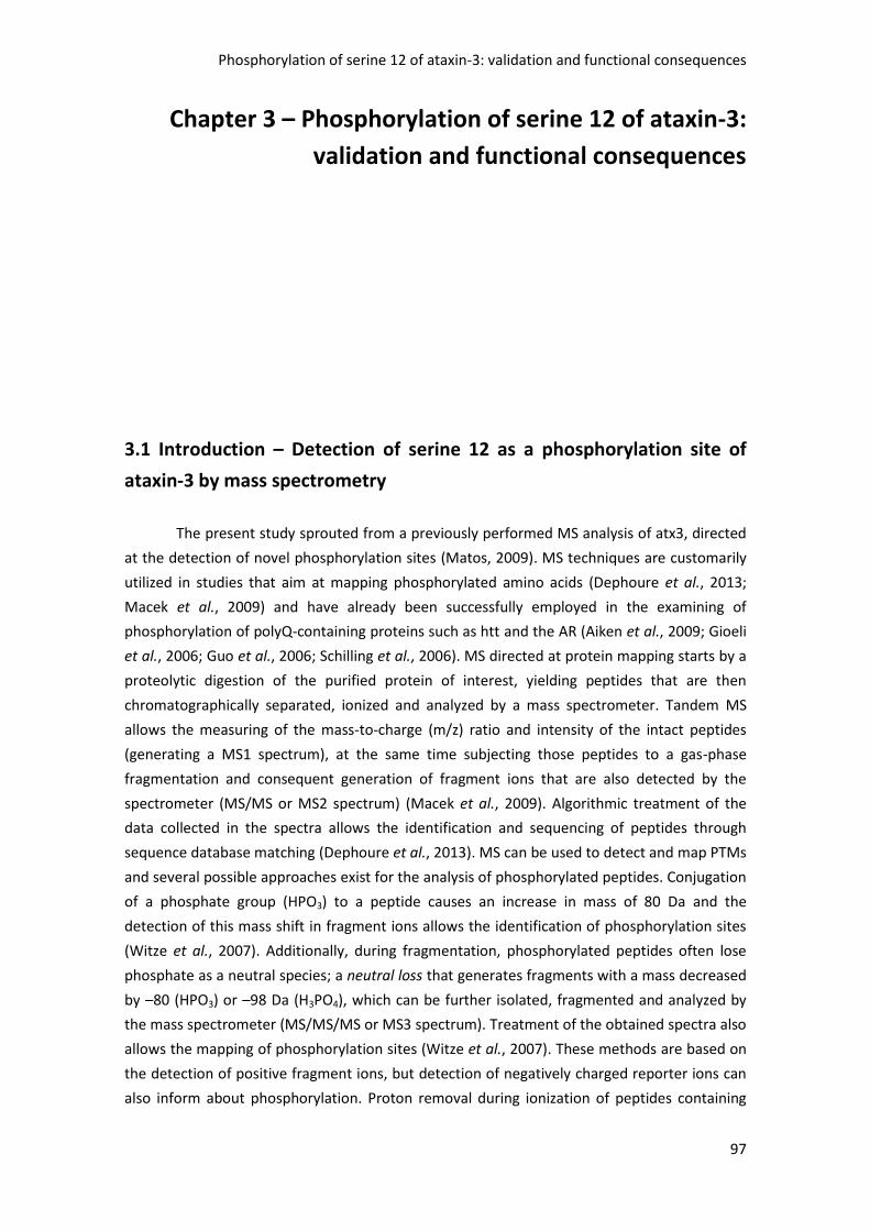
Phosphorylation of serine 12 of ataxin-3: validation and functional consequences
97
Chapter 3 – Phosphorylation of serine 12 of ataxin-3:
validation and functional consequences
3.1 Introduction – Detection of serine 12 as a phosphorylation site of
ataxin-3 by mass spectrometry
The present study sprouted from a previously performed MS analysis of atx3, directed
at the detection of novel phosphorylation sites (Matos, 2009). MS techniques are customarily
utilized in studies that aim at mapping phosphorylated amino acids (Dephoure et al., 2013;
Macek et al., 2009) and have already been successfully employed in the examining of
phosphorylation of polyQ-containing proteins such as htt and the AR (Aiken et al., 2009; Gioeli
et al., 2006; Guo et al., 2006; Schilling et al., 2006). MS directed at protein mapping starts by a
proteolytic digestion of the purified protein of interest, yielding peptides that are then
chromatographically separated, ionized and analyzed by a mass spectrometer. Tandem MS
allows the measuring of the mass-to-charge (m/z) ratio and intensity of the intact peptides
(generating a MS1 spectrum), at the same time subjecting those peptides to a gas-phase
fragmentation and consequent generation of fragment ions that are also detected by the
spectrometer (MS/MS or MS2 spectrum) (Macek et al., 2009). Algorithmic treatment of the
data collected in the spectra allows the identification and sequencing of peptides through
sequence database matching (Dephoure et al., 2013). MS can be used to detect and map PTMs
and several possible approaches exist for the analysis of phosphorylated peptides. Conjugation
of a phosphate group (HPO3) to a peptide causes an increase in mass of 80 Da and the
detection of this mass shift in fragment ions allows the identification of phosphorylation sites
(Witze et al., 2007). Additionally, during fragmentation, phosphorylated peptides often lose
phosphate as a neutral species; a neutral loss that generates fragments with a mass decreased
by –80 (HPO3) or –98 Da (H3PO4), which can be further isolated, fragmented and analyzed by
the mass spectrometer (MS/MS/MS or MS3 spectrum). Treatment of the obtained spectra also
allows the mapping of phosphorylation sites (Witze et al., 2007). These methods are based on
the detection of positive fragment ions, but detection of negatively charged reporter ions can
also inform about phosphorylation. Proton removal during ionization of peptides containing

Chapter 3
98
phosphorylated residues may yield phosphate-derived precursor ions (PO3-) of –79 Da that are
indicative of the presence of phosphorylated peptides, whose identity can be then determined
by positive ion MS2 (Macek et al., 2009; Witze et al., 2007).
In order to identify phosphorylated amino acids of atx3 that would be possibly relevant
for the functionality of the human protein, the samples subjected to MS consisted of GFP-
tagged human atx3 with 28Q (isoform MJD1a), transiently expressed in the human HEK 293FT
cell line. In order to enhance phosphorylation, cells were treated with the protein kinase C
(PKC) stimulator phorbol 12-myristate 13-acetate (PMA), or with sodium orthovanadate (SOV)
or okadaic acid (OA), two protein phosphatase inhibitors (Table IV in Chapter 2). GFP-atx3 28Q
was purified by immunoprecipitation with an anti-GFP antibody and by posterior
electrophoretic separation in polyacrylamide gel (Figure 10A); the bands corresponding to the
full-length fusion protein were excised and subjected to a tryptic digestion, and the resulting
GFP-atx3 28Q-derived peptides were separated by reverse-phase HPLC and directly
electrosprayed into the mass spectrometer. The analysis performed was directed at detecting
neutral losses of phosphate and obtain the MS3 spectra of the corresponding peptidic
fragment product. This analysis would allow the detection and sequencing of phosphorylated
peptides derived from human atx3.
The mass spectrometric examination of the GFP-atx3 28Q sample obtained from OA-
stimulated cells revealed the presence of one phosphorylated peptide: Q9-EG-pS12-
LCAQHCLNNLLQGEYFSPVELSSIAHQLDEEE-R45 (Figure 10B). Importantly, it identified for the
first time serine 12 of atx3 as a phosphorylated amino acid residue. The fact that S12 was
detected to be phosphorylated in HEK 293FT cells, under no pharmacological stimulus except
for the inhibition of endogenous phosphatases, indicates that this modification happens
normally under the cell culture conditions used and suggests that S12 phosphorylation may be
biologically relevant for human cells. The MS analysis did not yield any other phosphorylation
sites in this or in the other GFP-atx3 28Q samples, but given the stochastic nature of the MS
detection techniques and the variable results they yield, this observation cannot rule out the
possible phosphorylation of other atx3 amino acid residues, such as the ones previously
described (Fei et al., 2007; Mueller et al., 2009).
3.2 Detection of phosphorylated ataxin-3 in cell lines and neurons
Generation of the phospho-specific antibody anti-Patx3
Mass spectrometric detection of phosphorylated S12 in GFP-atx3 samples obtained
from transfected cells is a strong indication that atx3 is normally phosphorylated at that site,
under standard culture conditions. Nonetheless, given the originality of this observation, it
would be important to confirm the modification using other techniques. The selection of such
techniques could also be of radical significance in directing forthcoming experimental
approaches aiming at understanding the biologic relevance of S12 phosphorylation. Production

Phosphorylation of serine 12 of ataxin-3: validation and functional consequences
99
Figure 10: MS detection of S12 as a phosphorylation site of atx3. A HEK 293FT cells were transiently
transfected with GFP-atx3 28Q and expression was left to occur for 24 h, during which time period cells
were stimulated with phorbol 12-myristate 13-acetate (PMA), sodium orthovanadate (SOV) or okadaic
acid (OA). GFP-atx3 28Q was then purified by immunoprecipitation using a monoclonal anti-GFP
antibody and SDS-PAGE and the resulting gel was stained with Coomassie blue. Asterisk (*) represents
GFP-atx3 28Q bands (~75kDa) subjected to posterior tryptic digestion and MS analysis. Bands of ~50 kDa
and ~25 kDa are admitted to correspond to the heavy and light chains of the antibody (IgG). B LC-ES-MS
detected one phosphorylated peptide in the protein sample prepared after OA stimulation and mapped
the phosphorylation site to S12. Human atx3 isoform MJD1a amino acid sequence (NCBI accession
number: AAB33571.1) is represented with the peptides covered by the MS analysis colored in red
of an antibody that specifically detects the phosphorylated S12 residue of atx3 presented itself
as a promising approach, considering that such a tool would allow not only the confirmation of
the results of the MS analysis by detecting atx3 phosphorylation at S12 in transfected
mammalian cell lines, but would also permit expansion on that observation.

Chapter 3
100
Generation of the antibody recognizing atx3 phosphorylated at S12 (anti-Patx3) was
ordered to Eurogentec (Seraing, Belgium), a company specialized in manufacturing antibodies
recognizing proteins with modified amino acid residues. The antibody was raised in rabbits,
immunized against a peptide containing phosphorylated S12 and designed by the company
based on atx3 amino acid sequence: KQEG-S(PO3H2)-LCAQHCLN (Figure 11). The peptide shares
no significant homology with any other human, African green monkey, rat or mouse proteins,
according to Basic Local Alignment Search Tool (BLAST) analysis (NCBI, USA). This fact
decreased the probability of eventual unspecific reactivity with other targets. The generated
polyclonal antibodies were purified so that only the fraction specifically recognizing the
phosphorylated atx3 peptide was provided.
Detection of serine 12 phosphorylation with the anti-Patx3 antibody
The first approach to employ the anti-Patx3 phospho-specific antibody in confirming
atx3 phosphorylation in mammalian cells was to probe extracts of COS-7 cells transfected with
GFP-atx3 28Q by Western blot. Usage of GFP, a high molecular weight tag (27 kDa), would
allow the distinction between the overexpressed GFP-atx3 and the possibly phosphorylated
endogenous atx3 in these cells. Recombinant 6His-TEVg-atx3 produced in Escherichia coli was
concurrently analyzed as a negative control, since it is widely accepted that the
phosphorylation mechanisms of eukaryotes are not conserved (or are only poorly conserved
and limited) in prokaryotes (Huang et al., 2012; Macek et al., 2009).
Western blot analysis of the COS-7 cell samples transfected with GFP-atx3 28Q
revealed two discrete bands, with a difference of molecular weight of about 25 KDa (Figure
12A). This difference in size is similar to the difference between endogenous atx3 and GFP-atx3,
leading to the assumption that the higher-weight band revealed by the anti-Patx3 corresponds
to GFP-atx3 (~65 kDa) and the lower one to endogenous atx3 (~40 kDa). Furthermore, the
higher band is not present in non-transfected cells, corroborating the idea that it corresponds
to GFP-atx3. The absence of any band in the lane corresponding to the bacteria-derived
protein indicated that the immunoreactivity observed is directed at phosphorylated species.
Along with COS-7 cells transfected and incubated under regular conditions, samples
from transfected cells stimulated with compounds aimed at increasing protein
phosphorylation levels were also analyzed by Western blot (Figure 12A). Two of these
compounds – phosphatase inhibitors OA and SOV – were used with the objective of causing a
general increase of phosphorylated species, so as to strengthen the phosphorylation signal.
The other two compounds are activators of specific kinases: forskolin (FSK) activates protein
kinase A (PKA), while PMA activates PKC. A possible increase in the phosphorylated atx3 signal
elicited by one of them could be indicative of the involvement of the respective kinase in the
pathways responsible for phosphorylating S12 of atx3. However, none of the stimuli was able
to visibly increase the amount of phosphorylated GFP-atx3 or endogenous atx3, in COS-7 cells.

Phosphorylation of serine 12 of ataxin-3: validation and functional consequences
101
Figure 11: Amino acid sequences recognized by the antibodies used to detect atx3. The commercial mouse monoclonal antibody (clone 1H9) used to detect total atx3 recognizes an epitope localized in the vicinity of UIM1. The polyclonal phospho-specific antibody generated in this study was raised by immunizing rabbits of a peptide containing phosphorylated S12. The figure uses the same domain annotations as figure 3.
Western blot analysis of cells expressing expanded GFP-atx3 84Q also revealed two
bands, admittedly corresponding to the overexpressed GFP-atx3 and the endogenous protein
(Figure 12B). The 10 kDa molecular weight difference between GFP-atx3 28Q and GFP-atx3
84Q (~75 kDa) is in agreement with what is expected due to the difference in the number of
glutamine residues. GFP-atx3 mutants where S12 had been substituted by an aspartate residue
were also transfected into COS-7 and analyzed through the same procedures (Figure 12A and
12B). This kind of mutation is commonly used to mimic phosphorylation of serine residues, as
will be discussed in section 3.4. In agreement with what has been observed for phospho-
specific antibodies targeting other phosphoproteins (Li et al., 2010), the phosphomimetic
mutant of atx3 was recognized by the anti-Patx3.
The relative difference in molecular sizes explained above, along with the absence of
higher weight bands in the non-transfected cell samples, serves as good proof that the
phospho-specific antibody detects atx3. However, the sizes of the species revealed by the anti-
Patx3 antibody are lower than what is expected for GFP-atx3 28Q and 84Q and endogenous
atx3. Reprobing with the anti-atx3 antibody revealed bands of higher molecular weight: a ~75
kDa band for GFP-atx3 28Q and a ~47 kDa band corresponding to endogenous atx3 (Figure
12A). Nonetheless, given the evidence presented above, the bands observed with the anti-
Patx3 are admittedly atx3- and GFP-atx3-derived products. The discrepancies may be
explained by the fact that the anti-Patx3 antibody is detecting only fragments of GFP-atx3 and
endogenous atx3. Proteolytic cleavage of atx3 has been repeatedly studied and reports by
several authors have described a variety of cleavage products, deriving from endogenous atx3
(Berke et al., 2004) as well as from transfected expanded and non-expanded atx3, including in
COS-7 cells (Pozzi et al., 2008) (see section 3.4). The fact that only the putative fragments are
detected as being phosphorylated would however indicate that either atx3 is phosphorylated
at S12 only after being cleaved into the products of the observed sizes, or that phosphorylation
at S12 leads to cleavage.

Chapter 3
102
Figure 12: Phospho-specific anti-Patx3 antibody detects GFP-atx3 28Q and endogenous atx3-derived species in COS-7 cells. A COS-7 cells were transiently transfected with GFP-atx3 and stimulated with SOV (5 mM, 30 min), OA (20 mM, 16 h), FSK (10 µM, 20 min) or PMA (200 mM, 20 min) in order to enhance protein phosphorylation. Western blot analysis with the anti-Patx3 antibody, directed at the detection of phosphorylated S12 of atx3, revealed two protein bands: one corresponding to GFP-atx3 28Q (~65 kDa) and the other to endogenous COS-7 atx3 (~40 kDa). None of the chemical stimuli increased the signal revealed by the anti-Patx3. Non-transfected cells yielded just one band corresponding to endogenous atx3, and no protein band was detected in purified recombinant 6His-TEVg-atx3, produced in E. coli. Reprobing with the commercial anti-atx3 1H9 antibody labelled proteins of higher molecular weight, indicating that the species observed with the anti-Patx3 probably correspond to atx3-derived fragments (GFP-atx3 28Q: ~75 kDa; endogenous atx3 and 6His-TEVg-atx3: ~48 kDa). B In samples expressing GFP-atx3 84Q, the anti-Patx3 detected a higher molecular weight band corresponding to an expanded protein-derived fragment (~75 kDa). The phospho-specific antibody also detected species derived from phosphomimetic mutants GFP-atx3 28Q S12D and GFP-atx3 84Q S12D. COS-7 cell extracts were prepared in the presence of phosphatase inhibitors. 35 µg of each COS-7 sample and 30 µg of purified 6His-TEVg-atx3 were analyzed in these experiments.

Phosphorylation of serine 12 of ataxin-3: validation and functional consequences
103
Confirming phosphorylation – Lambda phosphatase experiments
In following Western blot experiments the bands revealed by the anti-Patx3 antibody
in COS-7 cells presented a different profile, with the band of highest intensity migrating at ~35
kDa. In order to confirm the identity of that band as a phosphorylated species, cell extracts
from non-transfected cells were prepared without any phosphatase inhibitors, and digested
with lambda phosphatase, an enzyme able to remove phosphate groups from serine and other
amino acid residues (Zhuo et al., 1993). Reaction with lambda phosphatase led to the
disappearance of the observed band, confirming that it in fact corresponds to a
phosphorylated protein (Figure 13A). Another band, fainter and of lower molecular weight
(~30 kDa), also disappeared with the treatment. Stimulation with staurosporine (STS), a broad-
spectrum inhibitor of protein kinases, had no effect in the observed bands.
In another experiment, COS-7 cells were treated with a cocktail of phosphatase
inhibitors (AO, SOV and sodium fluoride – NaF) and the anti-Patx3 immunoreactivity was
compared in extracts from treated and control cells, the latter prepared in the absence of any
phosphatase inhibitors (Figure 13B). Similarly to what was observed when using the lambda
phosphatase assay, this procedure yielded two protein bands of high intensity in the
phosphatase inhibitors-treated COS-7 cells, which were absent in non-stimulated samples.
Experiments with HeLa cell extracts also yielded two bands of similar sizes – that were
diminished or absent in the non-stimulated samples (Figure 13C).
Confirming ataxin-3 detection – Ataxin-3 silencing strategies
The phospho-specific antibody against phosphorylated S12 atx3 apparently labels an
atx3 fragment. If this molecular tool reliably detects the levels of endogenous S12-atx3
phosphorylation in different experimental conditions, it is very useful for the future
characterization of the importance of this PTM. No protein band in the Western blots labelled
with anti-atx3 antibody corresponds clearly to the bands observed with the phospho-specific
antibody, but the antibody against total atx3 used in this study targets an epitope (M221-LD-
E224; Goti et al., 2004) in the vicinity of UIM1 sequence, and may therefore be unable to
detect N-terminal atx-3 fragments that are detected by the anti-Patx3 antibody (Figure 11).
Nevertheless, to validate the specificity of the phospho-specific antibody, it is necessary to
confirm that the fragments observed do in fact result from atx3.
In order to clarify the identity of the lower molecular weight species detected by the
anti-Patx3 antibody we used an atx3 knockdown strategy employing shRNAs. In these
experiments, rat primary cortical neuron cultures were used instead of the above cell lines, for
two reasons. First, significantly knocking down atx3 in cell lines proved difficult, probably
because the continuous cell division assures a continuous production of cells that didn’t take
the shRNAs. Secondly, considering that MJD only affects neurons, it would be important to
expand on the observations made in cell lines and definitely prove that atx3 S12
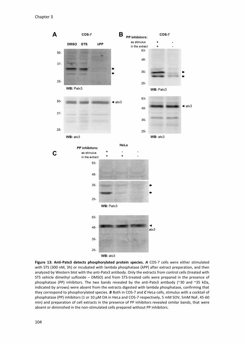
Chapter 3
104
Figure 13: Anti-Patx3 detects phosphorylated protein species. A COS-7 cells were either stimulated with STS (300 nM, 3h) or incubated with lambda phosphatase (λPP) after extract preparation, and then analyzed by Western blot with the anti-Patx3 antibody. Only the extracts from control cells (treated with STS vehicle dimethyl sulfoxide – DMSO) and from STS-treated cells were prepared in the presence of phosphatase (PP) inhibitors. The two bands revealed by the anti-Patx3 antibody (~30 and ~35 kDa, indicated by arrows) were absent from the extracts digested with lambda phosphatase, confirming that they correspond to phosphorylated species. B Both in COS-7 and C HeLa cells, stimulus with a cocktail of phosphatase (PP) inhibitors (1 or 10 µM OA in HeLa and COS-7 respectively, 5 mM SOV, 5mM NaF, 45-60 min) and preparation of cell extracts in the presence of PP inhibitors revealed similar bands, that were absent or diminished in the non-stimulated cells prepared without PP inhibitors.

Phosphorylation of serine 12 of ataxin-3: validation and functional consequences
105
phosphorylation occurs in that cell type. The shRNAs (shRNA-atx3), generated and
characterized by Alves and coworkers (2010) were delivered through lentiviral vectors and the
infected neurons were stimulated as above and analyzed by Western blot with the anti-Patx3
antibody. The bands detected by the antibody are absent from the cells where atx3 was
silenced, confirming their identity as endogenous atx3-derived species (Figure 14). Their size is
lower than that of the bands observed in the cell line samples, but it should be noted that the
endogenous rat atx3 has a lower molecular weight (~43 kDa) than the protein from the cell
lines (~48 kDa) (Berke et al., 2004).
Taken together, the above observations are strong proof that the antibody is detecting
phosphorylated species derived from atx3. It is therefore safe to assume that S12 of atx3 – or
of proteolysis-derived atx3 products – is phosphorylated in mammalian cell lines (including
human-derived cells) and, importantly, in neurons.
3.3 Ataxin-3 structure analysis and functional predictions
Having identified S12 of atx3 as a phosphorylation site, we focused on investigating
what are the possible functional consequences of this PTM. Phosphorylation is a highly
dynamic cellular mechanism capable of modulating different properties of a protein, including
enzymatic activity, intermolecular interactions, turnover and subcellular localization. In the
case of atx3, modification of S12 could assumably regulate at least a part of them.
A first analysis of atx3 amino acid sequence and domain structure reveals that S12
localizes in the JD (amino acids 8-168; Almeida et al., 2013), the catalytic domain of atx3
positioned at the N-terminal part of the protein (Figure 15C). Observing the tertiary NMR
structure of the JD of atx3 (Nicastro et al., 2005; PDB code: 1YZB), it is noticeable that S12
localizes on the surface of this domain and is consequently accessible for phosphate
conjugation (excluding the still undisclosed contribution of the C-terminal tail to atx3 tertiary
structure). Importantly, S12 is in close proximity to the cysteine residue of the catalytic site,
C14, but is also in the vicinity of the other amino acids of the catalytic triad, H119 and N134,
and nearby Q9, another amino acid important for the enzymatic activity (Figure 15A and B).
S12 is located in the joint region between the globular catalytic subdomain and the flexible
helical hairpin predicted to play a role in enzymatic target recognition (Komander et al., 2009;
Nicastro et al., 2009). According to the NMR structure data describing the Ub-binding sites
present at the surface of the JD, S12 is localized in close proximity to the region where the C-
terminus of the Ub molecule interacting with the catalytic cleft (site 1) is positioned upon
binding (Figure 15D and E) (Nicastro et al., 2009, PDB code: 2JRI).
Considering the proximity of S12 to elements related to atx3 enzymatic activity, it is
logical to speculate that phosphorylation at this residue could affect atx3 DUB activity.
Conjugation of a phosphate group to S12 might affect the conformation of the surrounding
amino acids, as a consequence of its volume and/or negative charge. Changes in the
conformation of the active site amino acids or alterations that could affect substrate binding

Chapter 3
106
Figure 14: Anti-Patx3 detects N-terminal fragments of atx3 in rat cortical neurons. Extracts from 14 DIV mice cortical neuron cultures were prepared with or without PP inhibitors and analyzed by Western blot. Probing with the anti-atx3, which targets an epitope in the vicinity of UIM1, revealed one band corresponding to endogenous rat atx3 (~43 kDa), while the anti-Patx3 antibody, directed at the N-terminal part of the atx3 antibody, revealed two bands of lower molecular weight (~25 and ~30 kDa, indicated by arrows). Neurons infected at 8 DIV with lentiviral particles encoding shRNAs targeting atx3, stimulated with a cocktail of PP phosphatase inhibitors (0,5 µM OA, 5 mM SOV, 5mM NaF, 45-60 min) and sampled in the presence of PP inhibitors yielded no similar bands. Actin was labelled as a loading control.
might result in changes in the proteolytic activity (Huang et al., 2012; Hunter, 2007; Renatus
and Farady, 2012; Tarrant and Cole, 2009). Importantly, a previously described PTM of atx3 –
ubiquitination at K117, localized in the JD, in the proximity of the catalytic site – was shown to
increase its DUB activity (Todi et al., 2010).
Other evidence suggesting the importance of the amino acid at position 12 in the
context of the catalytic activity of the JD may be inferred from the analysis of other JD-
containing proteins (Amerik and Hochstrasser, 2004; Komander et al., 2009; Reyes-Turcu et al.,
2009; Tzvetkov and Breuer, 2007). Comparing the amino acid sequences from atx3 homologs
in humans and other vertebrates with that of the Josephin-1 and -2 (which are mainly
constituted by the JD), it is interesting to note that, at the position corresponding to S12,
Josephin-1 and -2 have a negatively charged residue – glutamate (Figure 16). On the other

Phosphorylation of serine 12 of ataxin-3: validation and functional consequences
107
Figure 15: S12 is localized in the catalytic domain of atx3. A S12 (in yellow) is localized in the joint region between the two subdomains of the JD, B in close proximity to the amino acids of the catalytic triad and to glutamine 9 (PDB code: 1YZB). C S12 is accessible at the surface of the isolated JD. D, E Binding of a Ub molecule to the Ub-binding site 1 of the JD positions the C-terminal region of Ub close to S12 (PDB code: 2JRI).
hand, the human atx3L, which shares a high degree of homology with atx3, has an aromatic
phenylalanine at that position. The activity of the JDs isolated from human atx3, atx3L protein,
Josephin-1 and Josephin-2 has been shown to be markedly different, when tested against
several different Ub model substrates, in vitro (Weeks et al., 2011). The variations observed
may be attributed to other sequence or structural differences between the protein JDs, but it

Chapter 3
108
Figure 16: S12 is conserved in vertebrate atx3 but is substituted by a phenylalanine residue in atx3L and by a glutamate residue in Josephin-1 and -2. The figure represents the alignment of the JD of several vertebrate Josephin DUBs: human, mouse, rat and chick atx3; human atx3L; human, mouse, rat and bovine Josephin-1; and human and mouse Josephin-2. Protein sequences were obtained from the Uniprot database (codes are represented in brackets). Alignment was performed with Crustal Omega and prepared with ESPript 3.0, using the first 180 residues of atx3 and atx3L and the whole sequence of Josephin-1 and -2. Strictly conserved residues are represented by white letters in a black background and highly similar residues (global score: 0,7) by bold letters in framed columns. Arrowheads point the conserved residues of the catalytic triad (red), points the conserved glutamine residue next to the catalytic site (purple) and the position of S12 in atx3 (yellow).
is possible that the residue occupying the position of S12 plays an important role.
Notably, the crystal structure of the complex formed by Ub and the atx3L JD revealed
that F12, along with other aromatic amino acids, forms an hydrophobic enclosure around the
active center, through which the C-terminus of the Ub molecule threads (Weeks et al., 2011).
The position also appears to be suitable for interaction with the lysine side chain forming the
Phosphorylation of serine 12 of ataxin-3: validation and functional consequences
109
isopeptide bond. In fact, it has been reported that mutation of the S12 of the atx3 JD to a
phenylalanine increases proteolysis of Ub substrates, consistent with the proposed relevance
of this residue in determining activity. It is possible that a negative charge at amino acid
position 12 will possibly introduce steric hindrance leading to a rearrangement of the helical
hairpin, which is essential for the correct positioning of the R74 of the Ub substrate and
maintenance of the active site structure (Weeks et al., 2011).
Taking all these indications into consideration, we investigated the effect S12
phosphorylation has on atx3 enzymatic activity.
3.4 Generation of ataxin-3 phosphomutants
Taking into account that phosphorylation is a reversible, highly dynamic, modification,
it is believed that, at any given time point in a cell, only a fraction of the population of a
particular protein is phosphorylated (Seo and Lee, 2004). A common strategy used to study the
behavior and effects of a phosphoprotein is to constitutively mimic the conjugation of the
phosphate groups, by substituting some or all phosphorylatable residues of the protein with
acidic amino acids (Mueller et al., 2009; Pearlman et al., 2011; Thorsness and Koshland, 1987).
Serines are usually mutated to aspartate residues, though sometimes glutamate is used
instead. The reason for this is that conjugation of a phosphate group to the hydroxyl moiety of
a serine increases the volume of its side chain and renders it negatively charged; aspartate
shows relative chemical similarity to phosphoserine, mimicking both features to some degree
(Figure 17). The biochemical and biological behavior of these phosphomimetic mutants is
admitted to replicate (to some extent) that of the original protein, as if it was continuously
phosphorylated (Dephoure et al., 2013; Pearlman et al., 2011; Tarrant and Cole, 2009).
Simultaneously, experiments commonly include another kind of mutant of the
phosphorylation site(s) – the phospho-null, or phospho-dead, mutants. These mutants mimic
proteins so as to resemble them in a permanent non-phosphorylated state. In the case of
phosphorylated serines, this is achieved by substituting them with alanine residues, since the
two amino acids have comparable characteristics, but alanine lacks the hydroxyl group with
which the phosphate conjugates (Dephoure et al., 2013; Mueller et al., 2009; Pearlman et al.,
2011; Tarrant and Cole, 2009).
Phosphomimetic and phospho-null mutants of the atx3 constructs used in the present
study were generated through site-directed mutagenesis, by substituting S12 with an
aspartate (S12D) or an alanine residue (S12A), respectively. These mutants were used
throughout the several experiments aiming at understanding the functional effect of S12
phosphorylation, except for the experiments employing atx3 purified from E. coli, since the
non-mutated protein is considered non-phosphorylated (as explained in section 3.2).
Importantly, in this study the non-mutated protein is designated as wild-type (WT), regardless
of glutamine sequence size; WT thus describes the presence of serine at position 12 and not
the absence of polyQ expansion, as is the case of other works.

Chapter 3
110
Figure 17: Chemical structures of phospho-accepting amino acids and of amino acids used to model phosphorylation states. Eukaryotic proteins possess three amino acids with hydroxyl group-containing side chains able to conjugate with phosphate. Mimicking constitutive phosphorylation is commonly attempted by substituting them, through point mutations, with negatively charged acidic amino acids – aspartate or glutamate. Abrogating the ability to conjugate with phosphate is achieved by replacing the amino acids with alanine (or phenylalanine, in some studies of tyrosine phosphorylation). Amino acids are represented with neutralized charges and side chains are highlighted by yellow boxes.
3.5 Ataxin-3 phosphorylation at serine 12 affects its proteolytic
activity
In vitro activity assays with Ub-AMC
One method of studying atx3 proteolytic activity is to perform in vitro assays using Ub-
containing model substrates, by subjecting them to digestion with purified atx3 (Burnett et al.,
2003; Harris et al., 2010; Weeks et al., 2011). Ub-AMC is a compound commonly used when
studying DUB activity and has already been used in studies with atx3 (Burnett et al., 2003;
Harris et al., 2010; Love et al., 2007). Being a fluorogenic substrate, product formation

Phosphorylation of serine 12 of ataxin-3: validation and functional consequences
111
Figure 18: DUB activity assays with Ub-AMC. Ub-AMC is a fluorogenic model substrate which yields AMC upon proteolytic cleavage by DUBs. Recording the fluorescence emitted by AMC upon excitation (excitation – 380 nm; emission – 460 nm) allows a time-dependent assessment of product formation. The schematic representation uses atx3 as an example.
resulting from cleavage can be assessed by continuously recording the fluorescence emitted by
the AMC that is yielded during the reaction (Figure 18). This allows for a continuous and
sensitive detection of product formation and consequent evaluation of DUB activity.
With the objective of determining possible differences in DUB activity resulting from
atx3 phosphorylation at S12, recombinant 6His-atx3 WT and 6His-atx3 S12D were produced in
E. coli and purified in accordance to the procedure described by Gales and coworkers (2005).
Recombinant atx3 forms were incubated with Ub-AMC and fluorescence emitted by the AMC
fluorophore was measured throughout the course of the reaction. Observation of the
respective reaction curves reveals that the cleavage rate is higher for non-phosphorylated
6His-atx3 WT, comparing to the phosphomimetic 6His-atx3 S12D (Figure 19A). Moreover, the
initial reaction velocity, measured as the rate of product formation in the first 2 min (the
period while direct proportionality holds) significantly decreases to 43,6 ± 4,7 % (standard
error of the mean – SEM) of the initial velocity of the reaction catalyzed by 6His-atx3 WT, when
S12 is mutated (Figure 19B). Similar results were obtained with samples of atx3 containing a
different tag and prepared independently – 6His-TEVg-atx3 WT and S12D (Figure 19C).
As explained above, the proximity of S12 to the catalytic site strongly suggested that
phosphorylation of this residue would affect atx3 enzymatic activity. The observations made
with the Ub-AMC reaction support this idea, indicating that the phosphomimetic mutant of
atx3 is less effective at the cleavage of a monoUb substrate than the non-phosphorylated
protein.
In vitro activity assays with polyUb chains
Previous studies have shown that atx3 enzymatic activity in vitro is weak against
polyUb substrates with few (less than 4) monomers, leading to the idea that atx3 biological

Chapter 3
112
Figure 19: Mimicking atx3 phosphorylation at S12 decreases enzymatic activity against Ub-AMC. A Purified 6His-atx3 WT and 6His-atx3 S12D produced in E. coli were incubated (30 °C, for a maximum of 40 min) with Ub-AMC (0,5 µM) and product formation was assessed by measuring the fluorescence emission at 460 nm, during 380 nm excitation. Throughout the reaction, 6His-atx3 S12D shows a decreased enzymatic activity against Ub-AMC, comparing to 6His-atx3 WT. The reaction curve of initial product formation (0-120 s) is illustrated by one representative experiment; dots correspond to mean ± SEM fluorescence values (relative fluorescence units - RFU) of technical replicates (n = 3), after subtraction of the value of the negative control (buffer and Ub-AMC only, which showed no fluorescence increase during the assay). B The initial reaction velocity of 6His-atx3 S12D is decreased (to 43,6 ± 4,7 %) in relation to 6His-atx3 WT. Bars represent mean ± SEM of initial velocity values (RFU/min) of the replicates (n = 8) of 3 independent experiments, each of them normalized to the mean value of 6His-atx3 WT in the respective experiment. Student’s t-test: *** - P< 0,001. C Cleavage of Ub-AMC by 6His-TEVg-atx3 S12D is also reduced (initial velocity of 60,5 ± 11,1 %) in comparison to 6His-TEVg-atx3 WT (n = 10 replicates, from in 3 independent experiments). Student’s t-test: ** - P < 0,01.
function is more linked to polyUb chain editing than to complete disassembly of chains and
yielding of Ub monomers (Burnett and Pittman, 2005; Kuhlbrodt et al., 2011; Matos et al.,
2011; Winborn et al., 2008). For this reason, comparing to Ub-AMC, polyUb chains may be
better substitutes for the actual, yet unknown, biologic substrate(s) of atx3, in in vitro activity
assays. In these experiments, product formation is assessed through sampling of the reaction
mix at different time points, followed by Western blot analysis directed at the detection of Ub
Phosphorylation of serine 12 of ataxin-3: validation and functional consequences
113
species. Proteolytic cleavage leads to the formation of lower molecular weight species,
corresponding to polyUb chains of decreased number of monomers.
In order to evaluate the possible differences in the cleavage of polyUb chains between
non-phosphorylated atx3 and the phosphomimetic mutant, recombinant 6His-atx3 was
incubated with either K48- or K63-linked polyUb chains of 6 monomers and samples were
obtained at different time points, for a total period of 20 h. For both types of polyUb chains,
throughout the course of the experiment 6His-atx3 S12D led to a decreased formation of
lower molecular weight Ub species, comparing to 6His-atx3 WT (Figure 20). This was observed
for both K48- and K63-linked polyUb chains, though the difference is more prominent in the
reaction with the K63-linked chains. In fact, both 6His-atx3 WT and 6His-atx3 S12D are less
effective at cleaving K48-linked chains than chains of K63 linkage, an observation that is in
accordance with the atx3 cleavage preferences described by other authors (Harris et al., 2010;
Weeks et al., 2011; Winborn et al., 2008).
These results demonstrate that the presence of a negatively charged aspartate at
position 12 decreases the ability of atx3 to cleave polyUb chains of different linkage types.
Taken together, these in vitro activity assays strongly suggest that atx3 phosphorylation at S12
decreases its DUB activity. Observing that this effect extends to the cleavage of polyUb chains
is of particular interest, taking into account that K48- and K63-linked chains play many cellular
roles and are thus regarded as physiologically relevant (Weeks et al., 2011; Winborn et al.,
2008).
It is worthy of note that, throughout the course of the reaction, the levels of 6His-atx3
decrease, and that this effect is much more noticeable with the WT protein. Nonetheless, far
from contradicting the assumptions made, this observation gives further significance to the
explained differences in cleavage efficiency. Yet, it would be important to understand this
decrease in atx3 levels during an in vitro assay. Mauri and collaborators (2006) have described
that purified atx3 is subjected to slow autolytic fragmentation in vitro, a process that is
mediated by the cysteine residue of the active site (Pozzi et al., 2008). The “ability” of 6His-
atx3 S12D to better maintain its levels through the 20 h period of incubation could be
explained as resulting from lesser autolytic cleavage activity by this mutant. This could serve as
another testimony of the decrease on atx3 enzymatic activity that results from the
interference the negatively charged amino acid has on the active site. Another explanation for
the decrease on 6His-atx3 levels would be the occurrence of atx3 aggregation in vitro. Non-
expanded atx3 has repeatedly been shown to aggregate in vitro (Chow et al., 2004a; Ellisdon et
al., 2007; Gales et al., 2005; Saunders and Bottomley, 2009) and it could be the case that, in
these long-lasting activity assays, 6His-atx3 forms aggregates leading to a decrease in the
levels of its soluble monomeric form. This would imply that 6His-atx3 S12D has a lesser
tendency to aggregate than 6His-atx3 WT, under the conditions of this activity assay. The
current experiment does not allow for any definite conclusions whatsoever, as no atx3 bands
other than the ones expected for the full-length monomeric form were observed.

Chapter 3
114
Figure 20: Mimicking atx3 phosphorylation at S12 decreases enzymatic activity against polyUb chains. Purified 6His-atx3 WT and 6His-atx3 S12D produced in E. coli were incubated (37 °C) with A K48- or B K63-linked polyUb chains of six monomers (6Ub) and samples were collected at different time points (0, 2, 5 and 20 h) and analyzed by Western blot with an anti-Ub antibody, in order to assess product formation. Through the course of the reaction, cleavage of polyUb chains by 6His-atx3 leads to the formation of lower molecular weight Ub species, composed of a smaller number of monomers (Ub2-5). 6His-atx3 S12D elicits less polyUb cleavage than 6His-atx3 WT, against chains of either linkage type. The accompanying schematic representation of hexameric polyUb chains illustrates the fact that K63-linked chains are admitted to adopt a more extended conformation than chains of K48-linkage. C, D PolyUb chains incubated in the absence of 6His-atx3 showed no apparent degradation during the 20 h period of the reaction.
Ub-VS binding assays
The assays described strongly indicate that mimicking phosphorylation at S12 of atx3
results in a decrease of its DUB activity. It would be relevant to determine if the same
happened not only in vitro, but also in a cellular context. Still, testing that is problematic,
mainly because it is difficult to isolate the contribution of atx3 DUB activity from that of other,
possibly more efficient, DUBs (Chow et al., 2004b).
Labelling Ub in cell lysates prepared from HeLa cell cultures by Western blot renders a
protein smear corresponding to the several ubiquitinated proteins present in the cells (Figure

Phosphorylation of serine 12 of ataxin-3: validation and functional consequences
115
Figure 21: Phosphomutation of S12 does not alter the profile of ubiquitinated proteins in cells overexpressing atx3. HeLa cells were transfected with WT and phosphomutants S12D and S12A of both GFP-atx3 28Q and GFP-atx3 84Q and analyzed by Western blot with an anti-Ub antibody, in order to detect ubiquitinated proteins. Overexpression of atx3 leads to a decrease in intensity of the smear corresponding to the several ubiquitinated proteins present in HeLa cell lysates, having non-transfected cells as reference. The deubiquitination caused by GFP-atx3 expression occurs irrespectively of particular S12 mutations or polyQ length, considering that no blunt differences are observed between the samples of cells transfected with different GFP-atx3 forms. Atx3 was labelled as a control for the transfection procedure and actin as a loading control for the Western blot.
21). Cells transfected with GFP-atx3 constructs clearly show a decrease of ubiquitinated
species, admittedly due to the deubiquitination elicited by the overexpressed protein.
However, there is no apparent difference in the ubiquitinated species profiles between the WT
forms and the phosphomutants of GFP-atx3 with 28 or 84Q. There are several DUBs in
eukaryotic cells – approximately 79 are expressed in human cells (Komander et al., 2009;
Nijman et al., 2005) – and, consequently, it is not surprising that the relative contribution of
atx3 for overall cell deubiquitination is relatively modest and indistinguishable between the
several mutants used in this study. For the same reason, simple incubation of cell lysates with
model substrates such as those described above was not expected to be any more informative.

Chapter 3
116
Figure 22: Labelling DUBs with Ub-VS. Ub-VS conjugates with the cysteine residue of the active site of DUBs, forming adducts. These adducts can be detected by Western blot, through the characteristic molecular weight shift the conjugation of Ub-VS causes (~+10 kDa). The schematic representation uses atx3 as an example.
Ub-VS is a potent inhibitor of many cysteine protease DUBs, binding irreversibly to the
cysteine residue of the catalytic site and forming covalently conjugated adducts (Figure 22)
(Borodovsky et al., 2001; Reyes-Turcu et al., 2009; Wilkinson et al., 2005). It has been
repeatedly used to derivatize and consequently label several DUBs, including Josephin-1 and -2,
and such conjugation occurs only when DUBs are active (Borodovsky et al., 2001; Seki et al.,
2013). Since this compound has been used to successfully label DUBs in crude cell extracts
(Borodovsky et al., 2001; Wilkinson et al., 2005), we tried the analyze whether Ub-VS
conjugated with atx3 expressed in mammalian cell lines and check if there was any difference
in conjugation resulting from mutation of S12. The benefit of this approach is that it allows the
study of the behavior of atx3 independently of other DUBs.
Initially, we characterized the time-dependent formation of complexes between Ub-VS
and either non-expanded or expanded atx3. In order to do that, cell extracts prepared from
COS-7 cells transfected with GFP-atx3 28Q WT or GFP-atx3 84Q WT were incubated with Ub-VS
for a total period of 90 min. Protein samples were recovered at different time points and
analyzed by Western blot. Conjugation of Ub-VS to GFP-atx3 leads to the appearance of higher
molecular weight bands, corresponding to the resulting adducts (Figure 23A). Their intensity
increases with time, as opposed to that of the bands of the non-conjugated GFP-atx3. Non-
expanded and expanded GFP-atx3 show no apparent differences in their efficiency to
conjugate with Ub-VS throughout the assay, binding almost the totality of the inhibitor at the
end of the reaction period (Figure 23B).
Based on these results, we decided to simplify the assays with the S12
phosphomutants by analyzing the samples at just two different time points: 10 min, a period
of rapid increment in Ub-VS conjugation, and 45 min, at which time the maximum level of
conjugation was almost reached. Comparing the GFP-positive Western blot profile of the
samples transfected with GFP-atx3 WT, S12D and S12A (28Q) and incubated with Ub-VS, no
differences in conjugation with the inhibitor were detected (Figure 24). Conjugation of Ub-VS
to GFP-atx3 28Q thus appears to be independent from the substitution of S12 to aspartate or
alanine. These results indicate that the fraction of GFP-atx3 28Q that is active is equivalent
independently of mutation to a phosphomimetic or a phospho-null form. Importantly, this
should not be taken to mean that the enzymatic activity of the different forms is similar, as this

Phosphorylation of serine 12 of ataxin-3: validation and functional consequences
117
Figure 23: Conjugation of Ub-VS to atx3 occurs in a time-dependent manner. A COS-7 cells were transfected with GFP-atx3 28Q or GFP-atx3 84Q, lysated and incubated (RT) with Ub-VS. Samples were collected at different time points and analyzed by Western blot with an anti-GFP antibody, so as to detect GFP-atx3 and the adducts formed by Ub-VS conjugation. Through the course of incubation, there was an increase in intensity of the higher molecular weight species that correspond to the adducts (GFP-atx3-Ub-VS). B Western blot bands were densitometrically analyzed and the amount of conjugated GFP-atx3 at each time point was calculated as the percentage of the intensity of the GFP-atx3-Ub-VS bands relatively to the intensity of these and the unconjugated GFP-atx3 bands combined. The curve illustrates the time-dependent increase of Ub-VS conjugation to GFP-atx3, starting from a rapid increase in the first minutes of the reaction (2-15 min) but beginning to stabilize after ~45 min. The maximum amount of conjugation obtained was around 80 %. No apparent differences in Ub-VS conjugation exist between GFP-atx3 28Q and GFP-atx3 84Q.
assay does not provide information concerning the actual efficiency of the cleavage promoted
by the active site. In fact, studies made with other DUBs show that efficiency of Ub-VS
conjugation does not necessarily correlate with efficient proteolytic cleavage (Borodovsky et
al., 2001): otubain-1 is unable to bind Ub-VS but cleaves K48-linked chains in vitro (Wang et al.,
2009) and Ub C-terminal hydrolase 2 (USP2) is less sensitive to Ub-VS than Ub C-terminal
hydrolase 36 (USP21), but cleaves Ub-AMC with more efficiency (Rivkin et al., 2013).

Chapter 3
118
Figure 24: Phosphomutation of S12 does not alter atx3 conjugation to Ub-VS. COS-7 cells were transfected with WT or phosphomutated (S12D or S12A) forms of GFP-atx3 28Q, lysated and incubated (RT) with Ub-VS, for either A 10 min or B 45 min. Western blot analysis of the samples revealed that, for both incubation times, the amount of GFP-atx3 28Q that conjugated with Ub-VS (GFP-atx3-Ub-VS) is not affected by mutation of S12 residue.
These results suggest that mutation of S12 does not affect the binding of an Ub
derivative to the catalytic site, which indicates that the mutations of that residue do not
bluntly affect the conformation of the active site of the GFP-atx3 phosphomutants expressed
in cells. Attending to the observations made in vitro, this could indicate that phosphorylation
of S12 does not alter the binding of Ub substrates to C14, but affects the effectiveness of
substrate cleavage. However, the experiments performed cannot rule out the possibility that
the binding of the actual biologic Ub substrates differs between the non-phosphorylated and
the phosphorylated forms. Atx3 is known to show a preference for binding and cleaving
polyUb chains, thus limiting the conclusions made with a monoUb derivative (Burnett et al.,
2003; Mao et al., 2005; Winborn et al., 2008).
Chapter 4
Phosphorylation of serine 12 of ataxin-3:
consequences for polyglutamine-induced
toxicity


Phosphorylation of serine 12 of ataxin-3: consequences for polyglutamine-induced toxicity
121
Chapter 4 – Phosphorylation of serine 12 of ataxin-3:
consequences for polyglutamine-induced toxicity
4.1 Ataxin-3 phosphorylation at serine 12 prevents the decrease in
neuronal dendritic length caused by polyglutamine expansion
Phosphorylation of S12 was predicted to affect atx3 DUB activity based on the close
disposition of this amino acid residue to the catalytic site. The results obtained with the
phosphomimetic S12D mutant support this hypothesis. However, it would be important and
very relevant to test whether phosphorylation of S12 also affects events related to expanded
atx3 toxicity, such as cellular alterations with possible deleterious effects and aggregate
formation. Interference with the phosphorylation state of the protein may prove beneficial in
terms of disease progression and severity.
With the objective of characterizing the effect of S12 phosphorylation on expanded
atx3-induced toxicity, primary cultures of rat cortical neurons were favored over cell lines,
because it is believed that the cell toxicity mechanisms caused by polyQ-expansion depend
greatly on cell type and, in MJD, only neurons are targeted (Rüb et al., 2013; Takahashi et al.,
2010; Yamada et al., 2001; Zoghbi and Orr, 2000). Accordingly, any conclusions regarding
toxicity events that were based in experiments utilizing fibroblast-like cell lines would be
limited. Though MJD is chiefly described as a disease affecting the cerebellum and the
brainstem, increasing evidence has implicated the cerebral cortex in the pathogenic
mechanisms, contrary to the early idea that this region was mainly spared in MJD (Alves et al.,
2008b; Pedroso et al., 2013; Rüb et al., 2013; Yamada et al., 2001). Authors have described
reduced glucose metabolism (Soong et al., 1997; Taniwaki et al., 1997) and atrophy in the
cortex of MJD patients’ brain (D'Abreu et al., 2012; Lopes et al., 2013; Murata et al., 1998), as
well as the presence of polyQ NIs (Ishikawa et al., 2002; Yamada et al., 2001).
It is pertinent to investigate whether S12 phosphorylation of atx3 interferes with
neuromorphological changes resulting from polyQ expansion of the protein. In MJD, many
different neurodegeneration parameters have been studied in both patients and animal
models, including neuronal marker depletion, cell death and brain region atrophy. One

Chapter 4
122
Figure 25: Analysis of dendritic morphology. Imaging cultured neurons labeled with an appropriate fluorophore and virtually tracing their cell bodies and neurites allows the computational analysis of several morphological parameters. The total length of dendritic tracts can be determined by summing the length of each individual dendritic tract (represented in black). Sholl analysis consists in determining the number of times de dendritic tracts intersect equidistant circles (in red) centered on the cell body (in blue), thus allowing the appreciation of dendritic arborization. The current study used circles distancing 10 µm from each other.
possible output of cell toxicity in cortical neuron cultures is the observation of neuritic
degeneration, i. e., the loss of neuronal projections, such as dendrites. One important factor
that can be analyzed when comparing the dendritic morphology of neurons is the total length
of dendrites, expressed as the sum of the length of all the dendritic tracts of a cell.
Quantitative analysis of the complexity of dendritic arborization of an imaged neuron may be
achieved through Sholl analysis, a technique whereby equidistant circles of increasing radius
centered on the cell body are drawn and the number of intersections with dendrites is
determined (Figure 25). The resulting values are a function of both the number and length of
dendrites – an increased number of intersections corresponds to a larger number of dendritic
projections, at any given distance from the cell body.
First, in order to determine if expanded GFP-atx3 in fact affects the dendritic structure
of cortical neurons, cells transfected with GFP-atx3 28Q and GFP-atx3 84Q were imaged
through fluorescence microscopy (Figure 26A). Following the tracing of the respective cell
bodies and dendrites, the total length of dendritic tracts was compared between neurons
expressing the two constructs. GFP-atx3 84Q-expressing neurons had a lesser total extension
of dendrites, in line with the idea that polyQ expansion of atx3 leads to dendritic loss (Figure
26B). Sholl analysis of the neuronal tracing in each condition was then performed in order to
determine the number of crossings with circles distancing 10 µm from each other. Observation
of the resulting curves revealed that neurons expressing GFP-atx3 84Q WT have a withered
dendritic tree, with significantly less dendrites reaching 80 to 160 µm from the cell body
(Figure 26C). Furthermore, contrary to neurons transfected with GFP-atx3 28Q WT, GFP-atx3
84Q WT-expressing cells have no dendrites reaching more than 170 µm from the cell body.

Phosphorylation of serine 12 of ataxin-3: consequences for polyglutamine-induced toxicity
123
Figure 26: Pathogenic polyglutamine expansion of GFP-atx3 leads to dendrite tract loss and contraction in cortical neurons. A Cultured rat cortical neurons with 9-10 DIV were transfected with GFP-atx3 28Q WT or GFP-atx3 84Q WT, fixed 5 days later and imaged in the fluorescence microscope. Representative images of each condition are shown. B Neurons expressing GFP-atx3 84Q WT have a reduced total dendritic length, expressed by the sum of the length of all dendritic tracts in each particular neuron analyzed. Bars represent total dendritic length (mean ± SEM) of the neurons analyzed in each condition (n = 19-21, from two independent experiments), normalized to the mean total dendritic length of neurons transfected with GFP-atx3 28Q WT in each independent experiment. Values were compared using the student’s t-test: ** - P < 0,01. C Sholl analysis demonstrates a decrease of dendrites reaching more than 80 µm from the cell body. The curves represent the number of intersections (mean ± SEM, n = 19-21 neurons in each condition, from two independent experiments) as function of the distance from the cell body. Differences are statistically significative in the region distancing between 80 and 160 µm from the cell body. GFP-atx3 84Q WT-expressing neurons have no dendrites reaching 180 µm. The number of crossing at each distance was compared using the student’s t-test: * - P < 0,05; ** - P < 0,01.
This indicates that, in the cultured rat cortical neurons used in this study, expression of GFP-
atx3 with an expanded glutamine tract causes dendritic loss or shrinkage.

Chapter 4
124
Figure 27: Transfection of rat cortical neurons with GFP-atx3 phosphomutants. Cultured rat cortical neurons with 9-10 DIV were transfected with GFP-atx3 28Q WT, GFP-atx3 84Q WT or the respective S12D or S12A mutants and 5 days later whole cell lysates were analyzed by Western blot with an anti-GFP antibody. The differences in electrophoretic movement between GFP-atx3 28Q and GFP-atx3 84Q reflect the differences in molecular size resulting from the distinct number of glutamine residues.
Previous studies using neurons from transgenic Drosophila larvae have detected
dendritic abnormalities resulting from expanded atx3 expression (Lee et al., 2011), and a
severe impairment of dendritic arborization was also recently described to occur in the
Purkinje cells of a MJD mouse model (Konno et al., 2014). The experiments using the MJD
mouse model were performed in symptomatic animals expressing only a truncated fragment
of polyQ-expanded atx3 (known to be more toxic that the full-length protein; Goti et al., 2004;
Warrick et al., 1998; Yoshizawa et al., 2000) and comparison was made with wild-type
littermates. The current results constitute the first evidence that expanded atx3 causes an
observable dendritic loss/shrinkage in transfected mammalian neuron cultures, namely in
neurons isolated from the cortex, and represent compelling evidence that full-length atx3
causes dendritic loss or shortening as a consequence of polyQ expansion, in mammalian
neurons. These changes in the dendritic arborization may represent one pathologic
mechanism by which expanded atx3 disrupts neuronal structure and, consequently, function,
without (or before) inducing cell death.
In order to investigate if S12 phosphorylation affects the dendritic shrinkage caused by
GFP-atx3 84Q WT expression, cortical neurons were transfected with GFP-atx3 84Q S12D or
S12A (Figure 27) and total dendritic length was analyzed as above. The phospho-null mutant of
GFP-atx3 84Q exhibited similar effects to those of GFP-atx3 84Q WT but, interestingly, neurons
expressing GFP-atx3 84Q S12D had a significantly higher total dendritic length, showing no
differences relative to the total dendritic length of cells expressing GFP-atx3 28Q WT Figure
28A and B). Surprisingly, GFP-atx3 28Q S12A displayed an increased toxicity comparing to the
other non-expanded forms, leading to a decreased total length of dendrites. These results
suggest that phosphorylation of S12 protects against dendritic tract loss and that compromise
of this modification is enough to cause a loss of dendritic tracts.
Dendritic arborization was also evaluated by Sholl analysis, as above. Comparing the
curves obtained when analyzing the neurons transfected with GFP-atx3 84Q WT or its
phosphomutants, no striking differences were detected in the dendritic profile, namely in the
region distancing more than 80 µm from the cell body (Figure 29A and B). However, the

Phosphorylation of serine 12 of ataxin-3: consequences for polyglutamine-induced toxicity
125
Figure 28: Mimicking phosphorylation at S12 rescues dendritic tract loss caused by expanded atx3 expression. A Cultured rat cortical neurons with 9-10 DIV were transfected with GFP-atx3 28Q WT, GFP-atx3 84Q WT or the respective S12D or S12A mutants, fixed 5 days later and imaged in the fluorescence microscope. Representative images if neurons transfected with the phosphomutants are shown. B Transfection with GFP-atx3 84Q S12D elicits no differences comparing to the GFP-atx3 28Q WT control. Similarly to GFP-atx3 84Q WT, expression of phospho-null GFP-atx3 84Q S12A causes a reduction of total dendritic length. Bars represent total dendritic length (mean ± SEM) of the neurons analyzed in each condition (n = 16-21, from two independent experiments), normalized to the mean total dendritic length of neurons transfected with GFP-atx3 28Q WT in each independent experiment. The Mann-Whitney test was used to compare each condition with cells transfected with GFP-atx3 28Q WT: ** - P < 0,01; *** - P < 0,01. Comparison between other pairs of conditions was performed by two-way ANOVA, followed by Bonferroni post-hoc test: # - P < 0,05.
phosphomimetic mutant displays a slight tendency for having a larger number of dendrites at
almost every distance, while the phospho-null mutant shows the opposite behavior, even
producing a statistically significant decrease in the number of dendrites at 50 µm. Considering

Chapter 4
126
Figure 29: Mutating S12 does not hinder the dendritic shrinkage caused by expanded atx3 expression. Cultured rat cortical neurons transfected with GFP-atx3 28Q WT, GFP-atx3 84Q WT or the respective S12D or S12A mutants were imaged in the fluorescence microscope and subjected to Sholl analysis. A Mimicking S12 phosphorylation in GFP-atx3 84Q WT does not elicit striking differences regarding the way dendrites project from the cell body, though neurons transfected with GFP-atx3 84Q S12D tend to have more dendrites reaching every distance analyzed, albeit without statistical significance. B The phospho-null mutant of GFP-atx3 84Q WT shows the contrary tendency, which reaches statistical significance in the number of crossings at 50 µm. C, D The two phosphomutants of GFP-atx3 28Q display a tendency for decreasing the amount of dendrites reaching bigger distances from the cell body, limited in the case of GFP-atx3 28Q S12D, but expressive in the case of GFP-atx3 28Q S12A (especially in the region distancing between 120 and 160 µm from the cell body). The curves represent the number of intersections (mean ± SEM, n = 16-21 neurons in each condition, from two independent experiments) as function of the distance from the cell body. The number of crossing at each distance was compared between each condition pair using the Mann-Whitney test: * - P < 0,05; ** - P < 0,01; *** - P < 0,01.
the results obtained in the analysis of the total dendritic length, phosphorylation at S12 may
not counter the hampered dispersal of dendrites caused by polyQ expansion, but hinders the

Phosphorylation of serine 12 of ataxin-3: consequences for polyglutamine-induced toxicity
127
actual deletion of dendritic tracts. This effect is possibly expressed by the tendentially
increased number of interceptions detected in the Sholl analysis, when comparing GFP-atx3
S12D with its WT counterpart.
Comparison between the dendritic complexity of neurons expressing GFP-atx3 28Q WT
or its S12D and S12A forms revealed some propensity for lesser arborization in the neurons
transfected with the phosphomutants (Figure 29C and D). In fact, the only neurons presenting
dendrites at 190 µm from the cell body or at longer distances were the ones transfected with
GFP-atx3 28Q WT. While the effect is very mild in the case of GFP-atx3 28Q S12D, neurons
transfected with GFP-atx3 28Q S12A showed a noticeably shrunken dendritic tree starting at
120 µm from the cell body. It can be assumed that the phosphomutants of non-expanded GFP-
atx3, and especially GFP-atx3 28Q S12A, may have some associated toxicity, as had been
hinted by the total dendritic length results.
Taken together, the exploration of the two factors considered in the current analysis of
dendritic morphology – total dendritic length decrease and dendritic shrinkage, as inferred
from the Sholl Analysis curve – suggests that phosphorylation of S12 may be implicated in the
mechanisms linking atx3 to dendritic degeneration. GFP-atx3 28Q S12D shows no significant
evidence of inducing dendritic demise, comparing to GFP-atx3 28Q WT; on the other hand,
GFP-atx3 28Q S12A leads to a visible loss of dendritic dispersal and to a diminished length of
dendrites, revealing an increased neuronal toxicity by this non-phosphorylatable form. PolyQ
expansion causes a noticeable shrinkage of the dendritic tree in the neurons transfected with
any of the GFP-atx3 84Q constructs, but only the WT and S12A forms lead to an actual
decrease of the total dendritic length. The dendrites of neurons transfected with GFP-atx3 84Q
S12D have a total length comparable to that of the neurons transfected with GFP-28Q WT,
suggesting that phosphorylation of S12 may have a protective role against this particular
aspect of dendritic degeneration. All these differences caused by phosphomutation of atx3
indicate that, in a cellular context, S12 phosphorylation of expanded atx3 may hinder the
actual loss of dendrites, leading to a more contracted dendritic tree, while provoking no
decrease in the actual length of dendritic tracts.
4.2 Ataxin-3 phosphorylation at serine 12 prevents excitatory synapse
loss triggered by polyglutamine expansion
Having observed relevant effects of the phosphomimetic form of GFP-atx3 84Q in
protecting against decreased dendritic length, we tested whether the S12 phosphomutations
affect subtler characteristics of neuronal morphology, of no lesser biologic or pathogenic
relevance. Recently, investigation of proteinopathic neurodegenerative diseases has
frequently been moving its focus from the study of neuronal death mechanisms to the
characterization of the possible functional changes the pathogenic proteins cause in neurons.
It is believed that changes in neuronal functions may precede – or even cause – the actual loss
of neurons, and that some disease symptoms may arise from this type of defects. For this

Chapter 4
128
Figure 30: The glutamatergic synapse. In the presynaptic terminal, glutamate is accumulated in presynaptic vesicles through the action of transporter protein such as VGLUT1. Depolarization of the presynaptic membrane caused by an incoming action potential leads to the entry of calcium ions and the consequent rise in intracellular calcium concentration, activating cellular pathways that cause the vesicles to fuse with the plasma membrane. The neurotransmitter is released in the synaptic cleft, diffuses and binds to extracellular regions of glutamate receptors present in the postsynaptic membrane, leading to their activation and thus contributing to the depolarization of the post-synaptic neuron. Glutamate receptors are anchored to the underlying post-synaptic density, a protein-rich structure of the dendritic spine that contains the scaffolding protein PSD-95 with which glutamate receptors interact directly or indirectly.
reason, it is essential to evaluate if toxic proteins such as expanded polyQ-containing ones
cause neuronal changes with functional relevance.
Neurons of the rat cortex establish functional synapses when cultured under the
conditions used in this study (Louros et al., 2014). It is important to determine if expression of
atx3 with a pathogenic expansion affects their ability to establish and/or maintain synaptic
connections, and then evaluate the eventual differences caused by phosphomutation of S12.
This could serve not only as a measure of the health of the neurons, but also of the possible

Phosphorylation of serine 12 of ataxin-3: consequences for polyglutamine-induced toxicity
129
Figure 31: Effects of S12 phosphomutation on glutamatergic synapse markers of cortical neurons transfected with GFP-atx3. Low-density rat cortical neuron cultures with 9-10 DIV were transfected with GFP-atx3 28Q WT, GFP-atx3 84Q WT or the respective S12D or S12A mutants and fixed 5 days later. Glutamatergic synapse markers VGLUT1 (pre-synaptic) and PSD-95 (post-synaptic) were immunocytochemically labeled and dendritic tracts of GFP-atx3-expressing neurons were imaged. The figure shows representative dendritic sections evidencing VGLUT1 and PSD-95-positive puncta and the respective colocalization (merge).
compromise of their function, since synapses are the structures responsible for transmitting
signals between neurons.
Glutamate is the most prevalent excitatory neurotransmitter in the vertebrate brain. In
the human and rat cortices, the majority of neurons are glutamatergic (DeFelipe, 2011),
establishing synapses that use this amino acid as a means to propagate the nervous impulses
to the dendrites of contacting neurons. In this type of neurons, glutamate is accumulated
inside synaptic vesicles at the presynaptic region of an axon and, upon depolarization of the
plasma membrane caused by the arrival of an action potential, Ca2+ enters the cell and leads to
the fusion of those vesicles with the presynaptic membrane (Figure 30). Glutamate is thus
secreted into the synaptic cleft, diffuses and eventually binds to its receptors at the
postsynaptic membrane, contributing to the depolarization of the postsynaptic neuron (Popoli
et al., 2012).
We started by investigating if expression of GFP-atx3 84Q WT had a detectable effect
in the number of glutamatergic synapses present in cultured rat cortical neurons, comparing to
GFP-atx3 28Q WT. Immunocytochemical visualization of glutamatergic synapses can be
achieved through the labeling of pre- and postsynaptic protein markers. The vesicular
glutamate transporter subtype 1 (VGLUT1) is a protein associated with glutamate -
accumulating synaptic vesicles, playing a role in the transport and consequent packaging of
this neurotransmitter (Popoli et al., 2012). Being specifically localized in glutamate-releasing

Chapter 4
130
Figure 32: Polyglutamine expansion of GFP-atx3 causes a loss of functional glutamatergic synapses in cortical neurons. Comparing to GFP-atx3 28Q WT, expression of GFP-atx3 84Q WT in cultured cortical neurons causes a decrease in the intensity and area of A PSD-95 and B VGLUT1 puncta. The number of VGLUT1-positive accumulations also decreases significantly compared to control. C The number of functional glutamatergic synapses, defined as the instances of colocalization between the two synaptic markers, decreases in neurons transfected with GFP-atx3 84Q WT, comparing to cells expressing GFP-atx3 28Q WT. In every graph bars represent mean ± SEM of the values per dendritic section length (n = 30-32 neurons in each condition, from three independent experiments), normalized to the mean value of GFP-atx3 28Q WT in the respective experiment. Comparison between the two conditions used the Mann-Whitney test: ** - P < 0,01; ; *** - P < 0,001.
terminals, VGLUT1 was used as a presynaptic marker. In the postsynaptic region of excitatory
synapses, neurotransmitter receptors are clustered in protein-rich structures named
postsynaptic densities (PSDs), comprised also of many proteins of the cytoskeleton, proteins
acting in signal transduction and scaffolding proteins. PDS-95 is a scaffolding protein enriched
in the PSD of glutamatergic synapses (Hunt et al., 1996; Kim and Sheng, 2004), therefore
serving as a reliable postsynaptic marker.
Fluorescence microscopy observation of rat cortical neuron cultures labelled with anti-
PSD-95 and anti-VGLUT1 antibodies reveals a punctuate signal, as expected of synaptic
proteins, which are enriched in these discrete cell structures (Figure 30). Comparing to
neurons transfected with GFP-atx3 28Q WT, neurons expressing its expanded counterpart
show a remarkable decrease in the intensity and area of the PSD-95-positive puncta, indicative
of a possible interference of the polyQ-expanded protein with the accumulation of this
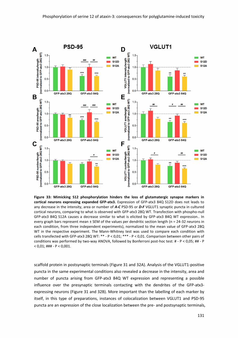
Phosphorylation of serine 12 of ataxin-3: consequences for polyglutamine-induced toxicity
131
Figure 33: Mimicking S12 phosphorylation hinders the loss of glutamatergic synapse markers in cortical neurons expressing expanded GFP-atx3. Expression of GFP-atx3 84Q S12D does not leads to any decrease in the intensity, area or number of A-C PSD-95 or D-F VGLUT1 synaptic puncta in cultured cortical neurons, comparing to what is observed with GFP-atx3 28Q WT. Transfection with phospho-null GFP-atx3 84Q S12A causes a decrease similar to what is elicited by GFP-atx3 84Q WT expression.. In every graph bars represent mean ± SEM of the values per dendritic section length (n = 24-32 neurons in each condition, from three independent experiments), normalized to the mean value of GFP-atx3 28Q WT in the respective experiment. The Mann-Whitney test was used to compare each condition with cells transfected with GFP-atx3 28Q WT: ** - P < 0,01; *** - P < 0,01. Comparison between other pairs of conditions was performed by two-way ANOVA, followed by Bonferroni post-hoc test: # - P < 0,05; ## - P < 0,01; ### - P < 0,001.
scaffold protein in postsynaptic terminals (Figure 31 and 32A). Analysis of the VGLUT1-positive
puncta in the same experimental conditions also revealed a decrease in the intensity, area and
number of puncta arising from GFP-atx3 84Q WT expression and representing a possible
influence over the presynaptic terminals contacting with the dendrites of the GFP-atx3-
expressing neurons (Figure 31 and 32B). More important than the labelling of each marker by
itself, in this type of preparations, instances of colocalization between VGLUT1 and PSD-95
puncta are an expression of the close localization between the pre- and postsynaptic terminals,

Chapter 4
132
Figure 34: Mimicking S12 phosphorylation hinders the loss of functional glutamatergic synapsis in cortical neurons expressing expanded GFP-atx3. The number of functional glutamatergic synapses, defined as the instances of colocalization between the two synaptic markers – PSD-95 and VGLUT1 –, does not decrease in neurons transfected with GFP-atx3 84Q S12D, comparing to cells expressing GFP-atx3 28Q WT. Expression of phopho-null GFP-atx3 84Q S12A causes a decrease similar to what is obtained by transfection with GFP-atx3 84Q WT. Bars represent mean ± SEM of the number of functional synapses per dendritic section length (n = 24-32 neurons in each condition, from three independent experiments), normalized to the mean value of GFP-atx3 28Q WT in the respective experiment. The Mann-Whitney test was used to compare each condition with cells transfected with GFP-atx3 28Q WT: ** - P < 0,01; *** - P < 0,01. Comparison between other pairs of conditions was performed by two-way ANOVA, followed by Bonferroni post-hoc test: # - P < 0,05; ## - P < 0,01.
admittedly representing the presence of functional glutamatergic synapses. Expression of GFP-
atx3 84Q WT in cortical neurons leads to a significant decrease in the number of glutamatergic
synapses, measured by the number of colocalizing VGLUT1 and PSD-95 puncta (Figure 31 and
32C).
All these observations constitute important and original evidence concerning the toxic
effects of expanded atx3 in cortical neurons. When expanded, atx3 leads to a decrease in the
signal of two synaptic markers and in the number of functional glutamatergic synapses, two
events that may be indicative not only of relevant particularities of cell demise, but that could
also be linked to possible nefarious effects in neuronal transmission. The results support the
idea of an involvement of synaptic dysfunction in the toxic mechanisms elicited by polyQ-
expanded atx3.
The effects of S12 phosphorylation in the observed changes caused by GFP-atx3 84Q
expression were once more addressed by comparison between the behavior of the GFP-atx3
WT forms and their phosphomutants (Figure 31). GFP-atx3 84Q S12A, the non-
phosphorylatable from, causes similar effects to the ones observed for GFP-atx3 84Q WT
(Figure 33A-F and 34). Comparing to GFP-atx3 28Q WT, the S12A expanded form leads to a
decrease in the intensity, area and number of puncta of both PSD-95 and VGLUT1, and also to
a reduced number of functional synapses. Contrastingly, GFP-atx3 S12D does not cause any
significant decrease in any of these parameters. In fact, overall the phosphomimetic mutation
reverts the effects provoked by GFP-atx3 84Q WT, leading to higher levels of synaptic marker
labelling and, importantly, to a number of glutamatergic synapses similar to that observed in
Phosphorylation of serine 12 of ataxin-3: consequences for polyglutamine-induced toxicity
133
neurons transfected with non-expanded atx3. Similarly to what was observed with the
experiments described above, the effect of GFP-atx3 28Q S12D is not different from that of
GFP-atx3 28Q WT, but the S12A mutant of the non-expanded form repeatedly shows a slight
tendency for the reduction of all the synaptic parameters analyzed.
The differential effects caused by mutation of S12 hint to the fact that phosphorylation
of this amino acid residue may play a role in protecting against the mechanisms by which
expanded atx3 leads to the observed synaptic dysfunction of cultured cortical neurons.
Phosphorylation of S12 appears to be important for the maintenance of the number of
functional glutamatergic synapses in cells expressing expanded atx3, suggesting that this PTM
constitutes a protective mechanism against the synaptotoxicity of the expanded protein. The
biological importance of atx3 as a synaptic regulator has not been explored, but may possibly
be related to an as of yet undisclosed function of this DUB.
4.3 Ataxin-3 phosphorylation at serine 12 affects its aggregation in
cortical neurons
Given the recognized role of aggregation in the toxicity pathways involved in polyQ
expansion diseases, we moved on to test whether the S12 phosphomutations affect atx3
tendency to aggregate. The other reports concerning atx3 phosphorylation described five
phosphorylation sites, distributed among the 3 UIMs, the regions of atx3 described to be
responsible for the selectivity in the interaction with PolyUb chains (Burnett et al., 2003; Fei et
al., 2007; Mao et al., 2005; Mueller et al., 2009; Schmitt et al., 2007; Winborn et al., 2008). In
cultured cells, modification of three of those sites to aspartate – S236, S340 and S352 – was
described to increase the nuclear localization of atx3, an important step leading to aggregation
and toxicity of the expanded protein (Bichelmeier et al., 2007; Macedo-Ribeiro et al., 2009;
Matos et al., 2011). Mimicking phosphorylation of residues S340 and S352 also increased the
stability and half-life of atx3 in cells. Pharmacological inhibition of CK2, an enzyme described as
being able to phosphorylate the UIM-containing C-terminal region of atx3, reduced the
amount of nuclear atx3 and decreased NI formation, possibly as a result of the two effects
described above (Bichelmeier et al., 2007; Breuer et al., 2010; Mueller et al., 2009).
Contrastingly, other authors described that preventing phosphorylation of S256 through a
phospho-null mutation to alanine enhanced its ability to aggregate in cells (Fei et al., 2007).
These observations demonstrate that interference with phosphorylation sites of atx3 may
affect its tendency to aggregate, though the actual effect – increase or decrease of aggregation
– differs depending on the phosphorylation sites in question, and possibly on the experimental
setting. It thus seemed pertinent to investigate whether phosphorylation of S12 has an effect
on the aggregation of atx3.
It has been repeatedly demonstrated that the atx3 JD is, by itself, prone to self-
assemble in vitro, playing a role in the early conformational changes leading to full-length atx3
Chapter 4
134
aggregation and modulating the aggregation of both expanded and non-expanded atx3
(Ellisdon et al., 2006; Gales et al., 2005; Masino et al., 2011; Masino et al., 2004; Saunders and
Bottomley, 2009). Conceivably, changes caused by PTMs of the JD, such as phosphorylation at
S12, could affect the dynamics of atx3 aggregation. As explained before (section 3.3),
according to the NMR structure of the complex formed between atx3 JD and two Ub moieties
(Nicastro et al., 2009; PDB code: 2JRI), S12 is in close proximity to the C-terminal part of the Ub
molecule that binds at site 1, which is localized in the cleft formed between the helical hairpin
and the globular subunit (Figure 15D and E). Interestingly, the two Ub binding sites identified
overlap with the regions described as taking part in JD self-assembly (Masino et al., 2011;
Nicastro et al., 2009). It has been hypothesized that atx3 aggregation is dependent upon the
molecular interactions it establishes and, in vitro, association of the JD with Ub protects
against aggregation (Fiumara et al., 2010; Masino et al., 2011; Song et al., 2010). It is therefore
plausible that phosphorylation at S12 may affect the interactions mediated by the Ub binding
sites of the JD, thus influencing atx3 aggregation. The studies of atx3 phosphorylation
mentioned above serve as testimony to the fact that interference with regions interacting with
Ub – the UIMs – affects aggregation of the protein.
In order to evaluate aggregation we again preferred the neuronal culture model over
cell lines, considering that aggregation is possibly linked to neuron-specific toxicity. Cortical
neurons were transfected with GFP-tagged WT or phosphomutated forms of atx3 with 28Q or
84Q, and the GFP fluorescent signal was visualized through fluorescence microscopy. Both
non-expanded and polyQ-expanded atx3 formed noticeable aggregates in cortical neurons, but
that was not the case for every cell, with many presenting a diffuse GFP-atx3 signal spread
over the whole neuron. Distribution of the aggregates varied between cells, being found
chiefly in the nucleus but sometimes also in the cytoplasm, or in both regions; in some rare
cases, aggregates were also observed in neurites. Nevertheless, no evident association was
established between any of the GFP-atx3 forms and the cellular localization of the
corresponding aggregates. The shape of the GFP-atx3 aggregates observed was generally
spherical or ovoid, but their size varied between cells (Figure 35A). Some cells presented just
minute accumulations of GFP-atx3, nonetheless considered as aggregates, whereas, in the
other extreme of the spectrum, other cells exhibited sizeable lumps of GFP-positive signal,
forming a structure akin to a net of aggregated GFP-atx3. The number of aggregates also
varied markedly, but there was no obvious relationship between aggregate size and/or
number and any of the GFP-atx3 forms that were transfected. The great diversity in aggregate
sizes, along with the large number of aggregates in some of the cells and the fact that they
distributed three-dimensionally, turned the simple quantification of the number of aggregates
unfeasible.
Objective comparison between the aggregation profiles generated by the different
GFP-atx3 forms was achieved through the determination of the number of cells that presented
aggregates, in the context of the overall population of transfected cells. Cells were counted as
having GFP-atx3 aggregation when they presented at least one noticeable instance of GFP-
positive accumulation, regardless of size; cells displaying only diffuse GFP signal were counted

Phosphorylation of serine 12 of ataxin-3: consequences for polyglutamine-induced toxicity
135
as having no aggregates (Figure 35A). Aggregation of GFP-atx3 in cortical neuron cultures was
thus expressed as the fraction of cells with aggregates, normalized to the values obtained with
transfection with GFP-atx3 84Q WT, in each experiment.
PolyQ expansion of atx3 is known to be the triggering factor for pathogenesis in MJD
and has been repeatedly described to be the cause of abnormal aggregation of polyQ
sequence-containing proteins (as discussed in section 1.5). In agreement with this premise,
non-expanded GFP-atx3 28Q WT displays a profoundly decreased tendency to aggregate,
when compared to the GFP-atx3 84Q WT (Figure 35B). As mentioned above, in this cortical
neuron-based assay even the non-expanded WT form with 28 glutamine residues formed
detectable aggregates, but results indicate that the 84 glutamine-containing WT protein
aggregates more. This observation serves as validation to this aggregation experiments, seeing
as it reflects the acknowledged association between the tendency to aggregate and the length
of the polyQ sequence.
Comparing aggregation between neurons transfected with GFP-atx3 84Q WT or its
phosphomutants S12D and S12A, it is possible to detect a significant decrease in the fraction of
neurons with aggregates, caused by the phospho-null mutation of S12 to alanine (Figure 35B).
GFP-atx3 84Q has a serine residue at position 12, available for phosphorylation by the cell
kinases, but alanine is not a subject for this PTM. This could indicate that, when polyQ-
expanded atx3 is not phosphorylated, its tendency to form detectable aggregates is diminished.
However, the phosphomimetic S12D mutant of GFP-atx3 84Q displayed a similar behavior,
although the tendential decrease does not reach statistical significance. This suggests that
phosphorylation of expanded atx3 at S12 decreases its tendency to aggregate and that the
non-phosphorylated S12 plays a role in the aggregation pathway.
Aggregation of GFP-atx3 28Q S12D is lower than what is observed for GFP-atx3 84Q
WT, but GFP-atx3 S12A appears to lead to higher levels of aggregation than its WT and S12D
counterparts. Considering that both phosphomutants of expanded atx3 apparently lead to
reduced aggregation, the effect of actual S12 phosphorylation of the pathogenic protein
remains indefinite, though blockade of this modification elicited the most evident decrease in
the tendency of GFP-atx3 84Q to form aggregates in cultured cortical neurons. However,
hindering phosphorylation of GFP-atx3 28Q appears to have the opposite effect, increasing the
amount of cells with aggregates. These observations suggest that the availability of S12 is an
important factor influencing this toxicity hallmark, in neurons. In a biological context, the
modifications caused by phosphorylation of S12 are very likely to influence whatever
aggregation mechanisms are mediated by this amino acid residue. Importantly, this toxic
outcome has parallels with the effects of GFP-atx3 S12A on neuronal morphology.
Finally, it is important to refer that, in cultures transfected with GFP-atx3 84Q WT but
contrary to the cells expressing other constructs, it was not uncommon to find cells containing
vast numbers of large aggregates that were not scored because of their unhealthy appearance
(Figure 35A). In other words, many neurons appeared to have died as a result of GFP-atx3 84Q
WT transfection (but not GFP-atx3 84Q S12D or S12A), possibly through mechanisms involving
aggregation. Consequently, the number of GFP-atx3 84Q WT-transfected cells displaying
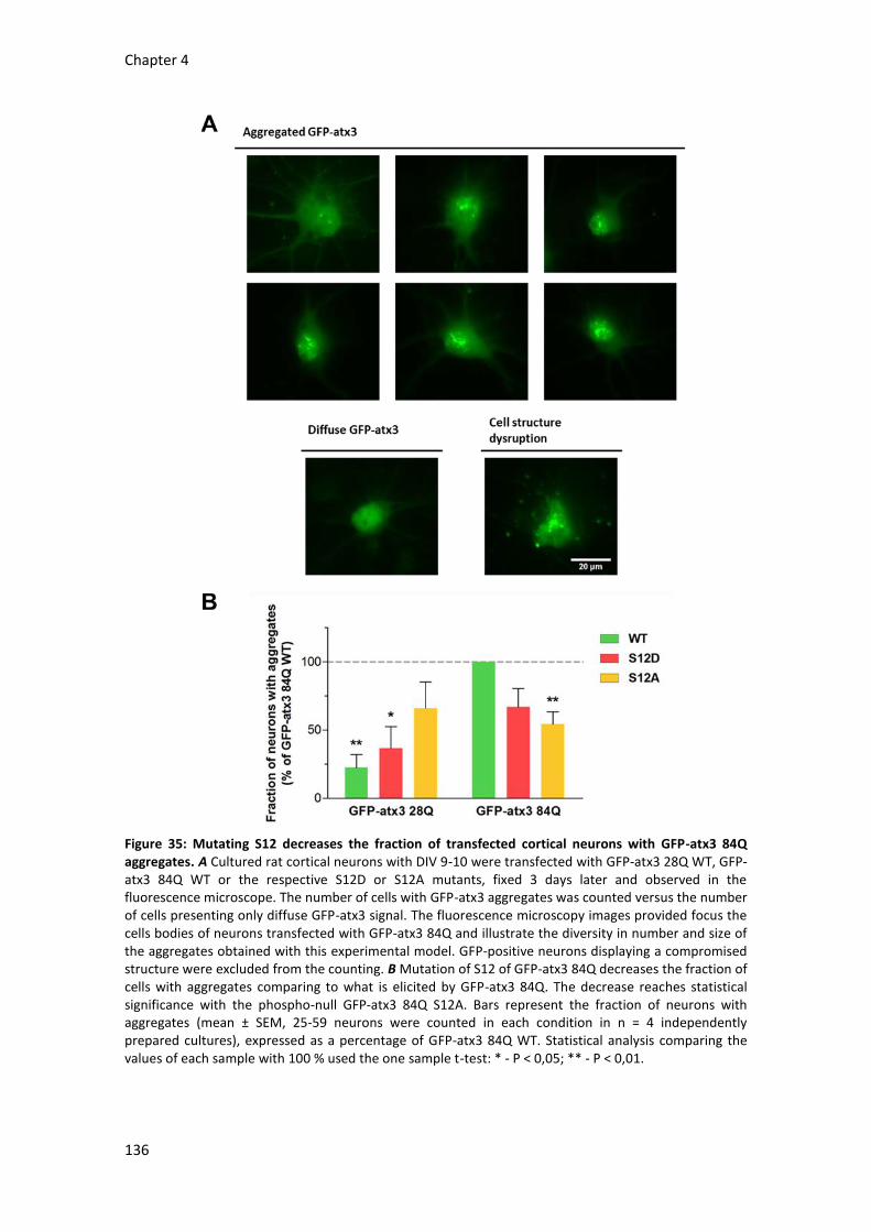
Chapter 4
136
Figure 35: Mutating S12 decreases the fraction of transfected cortical neurons with GFP-atx3 84Q aggregates. A Cultured rat cortical neurons with DIV 9-10 were transfected with GFP-atx3 28Q WT, GFP-atx3 84Q WT or the respective S12D or S12A mutants, fixed 3 days later and observed in the fluorescence microscope. The number of cells with GFP-atx3 aggregates was counted versus the number of cells presenting only diffuse GFP-atx3 signal. The fluorescence microscopy images provided focus the cells bodies of neurons transfected with GFP-atx3 84Q and illustrate the diversity in number and size of the aggregates obtained with this experimental model. GFP-positive neurons displaying a compromised structure were excluded from the counting. B Mutation of S12 of GFP-atx3 84Q decreases the fraction of cells with aggregates comparing to what is elicited by GFP-atx3 84Q. The decrease reaches statistical significance with the phospho-null GFP-atx3 84Q S12A. Bars represent the fraction of neurons with aggregates (mean ± SEM, 25-59 neurons were counted in each condition in n = 4 independently prepared cultures), expressed as a percentage of GFP-atx3 84Q WT. Statistical analysis comparing the values of each sample with 100 % used the one sample t-test: * - P < 0,05; ** - P < 0,01.

Phosphorylation of serine 12 of ataxin-3: consequences for polyglutamine-induced toxicity
137
aggregates may be underestimated; if that is the case, the differences observed in the
percentage of cells showing aggregates between the expanded GFP-atx3 84Q and the
phosphomutants-transfected cells should be even more marked. This serves as a testimony of
the great toxicity elicited by GFP-atx3 84Q WT, when compared with its phosphomutants and
the non-expanded constructs.
4.4 Phosphomutation of serine 12 reduces aggregation and
neurodegeneration in a lentiviral rat model of MJD
The above experiments characterize the effects of the phosphomimetic and phospho-
null mutations of S12 in several toxicity-related events caused by expression of expanded atx3
in cortical neurons. Considering the relevant differences that were detected in dendritic
arborization, synapse number and aggregation, it was appropriate to further investigate similar
effects in other, more complex, MJD-modeling systems. Accordingly, we explored the possible
changes caused by S12 phosphorylation on the pathogenic events occurring in an animal
model of MJD.
Since 1996 (Ikeda and Dikic, 2008), researchers have employed many different animal
models to the study of MJD. Models have been generated in Caenorhabditis elegans (Christie
et al., 2014; Teixeira-Castro et al., 2011), Drosophila (Warrick et al., 2005; Warrick et al., 1998),
mice (transgenic models are reviewed in Colomer Gould, 2012; Nóbrega et al., 2013) and rat
(Alves et al., 2008b), through the transgenic expression of either full-length human expanded
atx3 or a C-terminal fragment containing the polyQ sequence (Matos et al., 2011). The
available mammal models vary significantly in terms of the particular construct and isoform
used to generate them, the expression system employed, the number of glutamines of the
polyQ tract and the levels and distribution of the translated protein product (Colomer Gould,
2012; Matos et al., 2011). Nonetheless, overall they replicate many features with parallels in
the human disease, including inclusion formation in some regions of the CNS,
neurodegeneration (expressed as cell loss, morphological abnormalities or atrophy) and motor
symptoms, though varying in several particular aspects.
Using MJD animal models to evaluate the possible consequences of S12
phosphomutation of expanded atx3 required appropriate means to compare their effect with
that of its non-mutated counterpart. For this reason, the lentiviral rat model with striatal
pathology described by Alves and coworkers (2008a, 2008b, 2010) presented itself as an
advantageous experimental tool. The striatum has been increasingly implicated in MJD
pathogenesis, with reports describing decreased glucose metabolism (Taniwaki et al., 1997),
impaired dopaminergic transmission (Wullner et al., 2005; Yen et al., 2002) and the presence
of NIs (Alves et al., 2008b) in the striatum of patients. The rat model is generated by direct
delivery of viral particles encoding Myc-tagged full-length atx3 with 72 glutamines (isoform 1a)
to the striatum of adult animals by stereotaxic injection, leading to a much localized expression
of the transgene. Contrary to the expression of the correspondent non-expanded construct

Chapter 4
138
Figure 36: Strategy used to generate the lentiviral MJD rat model with striatal pathology and determine the outcome of atx3 S12 phosphomutations. Myc-atx3 72Q-encoding lentiviral particles were delivered billaterally into rat striata by stereotaxic injection. The left hemisphere striatum was injected with Myc-atx3 72Q WT and the right hemisphere with one of its respective phosphomutants: S12D or S12A. Four weeks after injection striatal slices were prepared and immunohistochemically analyzed in order to evaluate the Myc-atx3 72Q-derived lesions.
(27Q), transduction with Myc-atx3 72Q leads to the formation of NIs and to neuronal loss,
conveyed by the observation of cell death features and the depletion of neuronal markers
(Alves et al., 2008b). Because viral vectors encoding different proteins can be injected into the
striatal region of each of the brain’s hemispheres, comparison between the effects of two
forms of expanded atx3 – one of which may be a phosphomutant – can be performed in the
tissue of the same animal, increasing the significance of the observed differences. A very
similar approach has been used to assess differences in neuronal death caused by
phosphorylation of htt at S421 (Pardo et al., 2006). Another advantage of this model, when
compared with transgenic animals, is that expression of the phosphomutants starts only at the
adult phase. Phosphomutants of a protein mimic phosphorylation states in a constitutive way,
whereas actual phosphorylation is assumed to be dynamic and reversible. Expression of the
phosphomutated atx3 in a transgenic animal could implicate developmental effects that are
not within the scope of the current study, as the objective is to study effects of
phosphorylation in neurodegenerative features, such as those of the lentiviral rat model.
Experiments were planned so as to compare the toxic effects of Myc-atx3 72Q WT with
those of each of its S12 phosphomutants – phosphomimetic Myc-atx3 72Q S12D and non-
phosphorylatable Myc-atx3 72Q S12A. Accordingly, for every animal, lentiviral particles
encoding Myc-atx3 72Q WT were stereotaxically injected into the striatum of one of the
brain’s hemispheres (left), while the striatum of the contralateral hemisphere (right) was
injected with particles encoding either Myc-atx3 72Q S12D or Myc-atx3 72Q S12A (Figure 36).
Animals were sacrificed 4 weeks after injection, and coronal brain slices of the whole striatal
region were immunohistochemically processed and analyzed.

Phosphorylation of serine 12 of ataxin-3: consequences for polyglutamine-induced toxicity
139
Figure 37: Mutating S12 reduces the amount of atx3 aggregates in the striatum of a lentiviral MJD rat model. A, B Striatal slices of lentiviral MJD rat models were immunohistochemically probed with an anti-atx3 antibody and imaged in the optical microscope in order to detect Myc-atx3 72Q accumulations. Representative images of the peroxidase staining of striatal sections displaying aggregates are shown for each bilateral injection condition (Myc-atx3 72Q WT:Myc-atx3 72Q S12D or Myc-atx3 72Q WT:Myc-atx3 72Q S12A). C, D Comparing to the hemisphere expressing Myc-atx3 72Q WT, both phosphomutants lead to a tendential decrease in the number of aggregates in the striatum. The decrease is statistically significant in the case of the phospho-null mutant. Bars represent the number of atx3-positive accumulation in each hemisphere (mean ± SEM; n = 4 animals injected with Myc-atx3 72Q WT:Myc-atx3 72Q S12D; n = 6 animals injected with Myc-atx3 72Q S12A), as a percentage of the total number of aggregates in the two hemispheres. Estimation of the number of aggregates in the whole striatum of each hemisphere was based on the quantification of 7-12 equidistant striatal slices. Counting of the number of aggregates in each slice was performed in mosaics of images comprising the whole region with striatal aggregates. Statistical analysis used the Mann-Whitney test: * - P < 0,05.
In order to evaluate and compare the formation of aggregates elicited by each of Myc-
atx3 72Q forms, slices were labelled with an anti-atx3 antibody and the regions of the striatum
demonstrating atx3 aggregation were imaged and quantified (Alves et al., 2008a; Nascimento-
Ferreira et al., 2011). Anti-atx3 antibody labelling was considered to correspond to instances of
aggregation whenever a strong and compact signal was observed. Transduction of the rat
striatum with any of Myc-atx3 72Q forms led to the formation of detectable aggregates, in
agreement with what has been described for this animal model (Figure 37A and B). However,
mutation of S12 to alanine significantly decreased the amount of aggregates formed,

Chapter 4
140
Figure 38: Mutating S12 of atx3 reduces the lesion induced by expanded atx3 injection in a striatum of a lentiviral MJD rat model. A, B Striatal slices of lentiviral MJD rat models were immunohistochemically probed with an anti-DARPP-32 antibody and imaged in the optical microscope. Representative images of the peroxidase staining of striatal sections evidencing DARPP-32 immunoreactivity loss (limited by the interrupted line) are shown for each bilateral injection condition (Myc-atx3 72Q WT:Myc-atx3 72Q S12D or Myc-atx3 72Q WT:Myc-atx3 72Q S12A). C, D Comparing to the hemisphere expressing Myc-atx3 72Q WT, both phosphomutants lead to a statistically significant decrease in the volume of the region displaying DARPP-32 immunoreactivity depletion. Bars represent the volume of the DARPP-32-depleted region in each hemisphere (mean ± SEM; n = 4 animals injected with Myc-atx3 72Q WT:Myc-atx3 72Q S12D; n = 5 animals injected with Myc-atx3 72Q WT:Myc-atx3 72Q S12A), as a percentage of the total volume in the two hemispheres. The whole volume of the DARPP-32-depleted region was estimated based on the area of DARPP-32 depletion in 7-11 equidistant striatal slices. Statistical analysis used the Mann-Whitney test: * - P < 0,05; ** - P < 0,01.
comparing to Myc-atx3 72Q WT (Figure 37D). The phosphomimetic S12D mutant showed the
same tendency, though without statistical significance (Figure 37C). Phosphomutation of S12 is
consequently admitted to be able to decrease the propensity of expanded atx3 to form
aggregates.

Phosphorylation of serine 12 of ataxin-3: consequences for polyglutamine-induced toxicity
141
Examination of the severity of neurodegeneration caused by each Myc-atx3 72Q form
was performed by labelling the slices with an antibody detecting a neuronal marker:
dopamine- and cyclic AMP-regulated phosphoprotein of 32 kDa (DARPP-32). This protein is
associated with dopaminergic transmission both structurally and functionally, being enriched
in neurons receiving dopaminergic inputs and playing a role in the regulation of dopamine-
mediated neurotransmission (Fienberg et al., 1998; Ouimet et al., 1984). Because DARPP-32 is
found in the cytosol and is highly enriched in the striatum (Blom et al., 2013; Fienberg et al.,
1998), depletion arising from Myc-atx3 72Q WT expression in this region is interpreted as
resulting from the degeneration of striatal neurons (Alves et al., 2008a; Nascimento-Ferreira et
al., 2011; Pardo et al., 2006). Expression of Myc-atx3 72Q WT, S12D or S12A led to a localized
loss of anti-DARPP-32 antibody immunoreactivity, compatible with the toxicity expected of
atx3 with a pathogenic polyQ expansion (Figure 38A and B). Nonetheless, the volume of the
DARPP-32-depleted region was significantly reduced in the case of the injections with the
phosphomutants, comparing to Myc-atx3 72Q WT (Figure 38C and D). These observations
indicate that mutations of S12 of atx3 to alanine or aspartate decrease the degree of
neurodegeneration caused by expression of the expanded protein.
Previous studies with this lentiviral MJD rat model show that treatments targeting atx3
toxicity (protein silencing or autophagy activation) reduce both the amount of atx3 aggregates
and the extent of degeneration (Alves et al., 2008a; Alves et al., 2010; Alves et al., 2008b;
Nascimento-Ferreira et al., 2011). Accordingly, the two events are interpreted as being
different expressions of expanded atx3 toxicity, regardless of whether the aggregates are
actually harmful by themselves or in fact result from cellular mechanisms targeting other
deleterious species. Our results indicate that the Myc-atx3 72Q phosphomutants are less toxic
than the WT expanded protein with a serine at site 12, giving rise to smaller amounts of
aggregates and to less severe neuronal marker depletion in the striatum. Such markedly
similar behaviors between the two phosphomutants, with parallels in the aggregation
experiments performed in cortical neurons, is somewhat unexpected and hard to interpret in
terms of the actual effect of protein phosphorylation. Regardless, the observations made in
this MJD animal model strongly suggest that changes targeting S12 may constitute an
importantly mechanism modulating the toxicity of expanded atx3, with possible protective
outcomes.

Chapter 4
142

Final considerations
Chapter 5
Final considerations

Chapter 5

Final considerations
119
Chapter 5 – Final considerations
5.1 General discussion
The current study aimed at characterizing a novel PTM of atx3 – phosphorylation of
S12, a residue localized in the catalytic domain of the protein. Utilizing a phospho-specific
antibody specifically targeting phosphorylated S12, we were able to detect phosphorylated
species in mammalian cell lines, which were confirmed to correspond to atx3-derived
fragments in rat neurons. The results indicate that S12 of atx3 is phosphorylated in human
cells but the observations made in rat neurons are of special relevance, considering that MJD
specifically affects neuronal cells in human patients.
Having observed that S12 is localized in the vicinity of the catalytic site amino acids and
is especially close to C14, we predicted that phosphorylation of that residue could affect the
proteolytic activity of atx3. Using model substrates usually employed in the study of the
enzymatic activity of atx3 and other DUBs, we observed that when S12 is mutated to aspartate
in order to mimic the phosphorylated state, the activity of the protein decreases, in vitro. The
results obtained thus suggest that phosphorylation of S12 decreases the DUB activity of atx3.
The observation that this decrease is also detected when atx3 reacts with polyUb chains is of
particular physiologic relevance, considering that atx3 is believed to target polyubiquitinated
proteins in vivo.
The analysis of the possible influence of S12 phosphorylation on the toxicity of atx3
carrying a pathogenic expansion of glutamine residues was performed in cultured neurons,
which have been very rarely used in MJD studies, despite their relevance in comparison to the
more widely used cell lines. Transfection of cortical neurons with expanded GFP-tagged atx3
resulted in less arborized dendritic trees and in a decreased amount of functional
glutamatergic synapses, in comparison to neurons transfected with non-expanded GFP-atx3.
Remarkably, non-phosphorylatable expanded GFP-atx3 elicited similar effects, but the
phosphomimetic mutant limited the loss of total length of dendrites and the decrease of
functional synapse number. The contrasting effects of different phosphomutants of S12
suggest that the phosphorylation of this amino acid residue influences specific morphological
(and possibly functional) outcomes of expanded atx3 toxicity. Atx3 aggregation is recognized

Chapter 5
to constitute an important aspect of expanded atx3 toxicity and transfection of rat cortical
eurons with GFP- atx3 leads to the formation of GFP-positive aggregates. While expression of
expanded atx3 led to a higher amount of cells with aggregates than the non-expanded protein,
hindering phosphorylation of S12 by mutation of this residue to alanine decreased the fraction
of cells containing atx3 aggregates, indicating that interfering with S12 phosphorylation may
be important in mediating atx3 aggregation mechanisms.
Finally, we explored the effect of the S12 mutants of atx3 in the disease-related
characteristics of a MJD lentiviral rat model with striatal pathology. Injection of lentiviral
particles encoding expanded atx3 into the rat striatum leads to the formation of atx3-positive
aggregation bodies and to a localized loss of neuronal markers, reflecting demise of striatal
neurons. Mutation of S12 to aspartate or alanine decreases the number of aggregates formed
and the extent of neuronal loss, suggesting a protective effect that may be modulated in a
natural context by the phosphorylation of S12 of atx3.
The following segments will elaborate on these considerations and suggest additional
explanations for the results obtained.
Phosphorylation of ataxin-3 fragments
The mass spectrometric detection of S12 phosphorylation used a protein sample of
GFP-atx3, immunoprecipitated from HEK 293FT cells with an anti-GFP antibody, targeting the
N-terminally localized tag. The electrophoretic separation that followed allowed the
separation of the protein species contained in the immunoprecipitants, which, attending to
the Coomassie blue staining, consisted uniquely of full-length GFP-atx3 and IgG chains. Since
the band that was later excised and analyzed by MS presented a molecular weight consistent
with what was expected and routinely obtained with full-length GFP-atx3 28Q (~75 kDa), it is
admitted that, in HEK 293FT cells, full-length atx3 is phosphorylated at S12.
However, the phospho-specific anti-Patx3 antibody that was produced so as to
specifically recognize phosphorylation of atx3 at S12 did not recognize the full-length protein,
labelling only protein species of lower molecular weight than what was expected for
endogenous and GFP-tagged atx3. The lambda phosphatase assays and the experiments with
phosphatase inhibitors stimuli indicate that the protein products labelled by the anti-Patx3
correspond to phosphorylated protein, while the shRNA silencing of atx3 in cortical neurons
confirms that the observed species derive from atx3. Therefore, it is possible to conclude that
the anti-Patx3 labelled only atx3-derived fragments.
Considering that the anti-Patx3 antibody targets the N-terminal region of atx3, the
fragments must include N-terminal parts of the protein. As exposed in section 1.4, over the
years different authors have reported the observation of atx3-derived fragments of variable
molecular size, deriving from different regions. Fragments have been detected in diverse types
of cell cultures, resulting from endogenous or transfected atx3, in transgenic animal models
and in patients’ brain tissue (Goti et al., 2004). Particularly, in COS-7 cells, endogenous atx3
Final considerations
121
was shown to generate a caspase-derived fragment of about 28 kDa that was labelled by the
anti-atx3 antibody (Berke et al., 2004). In the same cell line, Pozzi and coworkers (2008)
described two fragments derived from transfected non-expanded (6 glutamines) murine atx3,
which were immunoreactive to an antibody directed at a N-terminal tag: one fragment with 37
and the other with 42 kDa. Upon digestion with calpain, human atx3 expressed in HEK 293FT
cells yields a ~30 kDa fragment that is labelled with the anti-atx3 1H9 antibody, nonetheless
described as an N-terminal fragment, seeing as polyQ sequence length does not alter the
molecular size of the fragment that is produced (Hübener et al., 2013). Simões and
collaborators (2012) detected a ~26 kDa fragment of human atx3 containing the N-terminus of
the protein, that also arises independently from polyQ repeat length and that was also labelled
by the monoclonal anti-atx3 antibody.
In our study, the endogenous atx3-derived fragments labelled by the anti-Patx3
antibody in COS-7 and HeLa cells had an apparent molecular weight of 30 and 35 kDa (and 40
kDa, in the first experiment described). The fragments detected in rat cortical neuron migrated
at an apparent molecular size of 30 and 22 kDa. It is possible that these fragments correspond
to the atx3-derived species described by other authors, but it is challenging to try to make any
definite correspondence, in part because of the uncertainty in the molecular weight estimation
based on the molecular weight standards used in Western blot techniques. Furthermore,
comparison with fragments yielded by overexpressed atx3 presents additional sources of
variability, since different studies employ different atx3 forms, derived from different animal
species, fused with diverse tags and with varying glutamine lengths. Studies delving into the
formation of atx3 fragments also employ different sampling techniques and frequently
different kinds of procedures that direct the activity of proteolytic enzymes; it is not surprising
that the observed fragments are consequently diverse. Nonetheless, the observation of N-
terminal fragments by other authors supports the plausibility of the detection of N-terminal
fragments with the anti-Patx3 antibody we generated.
The endogenous atx3 fragments described in COS-7 cells (28 kDa; Berke et al., 2004)
and mouse (30 kDa; Hübener et al., 2013) and the polyQ size-independent N-terminal
fragments derived from human atx3 (Simões et al., 2012) are all labelled by the commercial
1H9 anti-atx3 antibody, which binds a region in the vicinity of UIM1 (amino acids 221-224,
localized in the C-terminal tail of atx3). However, in our Western blot experiments, the anti-
atx3 antibody does not label any protein species that clearly correspond to the fragments
labelled by the phospho-specific antibody (but the anti-Patx3 signal disappears when atx3 is
silenced in neurons, proving the specificity of the immunoreactivity). This could mean that the
fragments we observe result from cleavage occurring N-terminally of amino acids 221-224.
Hübener and coworkers (2011; 2013) have suggested that the 30 kDa fragment they observed
derived from cleavage at position 260 and, in fact, a protein fragment composed by the first
259 amino acids of atx3 (which include the JD, UIM1 and part of UIM2) was calculated to have
a molecular size of ~30 kDa, for both mouse and human atx3. Though we observed a fragment
with this molecular size in COS-7 and HeLa cells and in cortical neurons, it would not be
expected that they included the amino-acids 221-224. Estimation of the molecular weight of a

Chapter 5
fragment formed by the 220 first amino acids of human, rat and mouse atx3 (the length
between the N-terminus of atx3 and the N-terminus of anti-atx3 epitope) using the same
calculation tools (Protein Molecular Weight Calculator – Science Gateway) yields a value of ~25
kD, which is compatible with the smaller (and most evident) fragment we observed in neuronal
cultures. If the remaining fragments we detected in our analyses in fact contain the 221-224
amino acid epitope, in could be the case that the anti-atx3 is not detecting them because they
are rare species, when compared to full-length endogenous atx3; the stronger
immunoreactivity derived from a higher proportion of full-length atx3 could mask the
fragments, and then render them undetectable in our experiments. In COS-7 cells, the anti-
atx3 antibody reveals several faint bands with lower molecular weight than the full-length
protein that could possibly correspond to the fragments.
In part, results actually suggest that some of the fragments may contain the polyQ
sequence and, by extension, amino acids 221-224. Only this explains why the phospho-specific
antibody detected fragments of GFP-atx3 28Q and GFP-atx3 84Q that differed in molecular size,
when the only difference between these two atx3 forms is the length of the polyQ region.
Interestingly, the estimated molecular size of a fragment containing the amino acids of atx3
comprised between the N-terminus and the end of the polyQ sequence is ~35 kDa for the
human protein (with 13Q) and ~34 kDa for both mouse (6Q) and rat (5Q) atx3; this matches
the size of one of the endogenous atx3-derived fragments detected in COS-7 and HeLa cells.
The molecular weight of atx3 varies depending on the number of repeated glutamines, and the
differences in electrophoretic movement detected between atx3 of different mammalian
species are largely attributed to differences in polyQ sequence length (Berke et al., 2004). In
our study, atx3 from COS-7 and HeLa cells lines displays about 50 kDa, while rat atx3 migrates
at about 43 kDa, meaning that atx3 from the cell lines shall have a larger number of glutamines.
The higher molecular weight of the fragments produced in the cell lines could result from the
fact that the endogenous protein is larger, and this would support the idea that at least the
fragment with ~35 kDa contains the polyQ sequence. Independently of the C-terminal
topology of the fragments observed, it is important to stress the point that our results indicate
that they include the N-terminal part of atx3.
Having detected phosphorylation at S12 in full-length atx3 through MS, it is somewhat
puzzling to observe that the phospho-specific antibody only recognizes fragments of atx3. One
simple explanation would be that, contrary to HEK 239FT cells, in COS-7 cells phosphorylation
of S12 targets only atx3 fragments. However, if we would consider that phosphorylation occurs
similarly in both cell lines and in the other cells used in this study as well, another explanation
would be that, although the full-length protein is phosphorylated at S12, the fraction of
modified protein is low, compared to the fraction of fragments that are phosphorylated; the
anti-Patx3 antibody would yield a stronger signal when labelling the fragments, ultimately
masking the full-length protein signal. This possibility raises the hypothesis that
phosphorylation at S12 may be intimately linked to the proteolytic events generating the
fragments we detected. Either atx3 is preferentially phosphorylated at S12 upon cleavage, or
phosphorylation of this residue eventually leads to proteolytic cleavage of atx3; the two

Final considerations
123
hypotheses would help explain why the antibody only detected fragments as being
phosphorylated. Considering that even endogenous atx3 is known – and here further
demonstrated – to yield cleavage product, these events could have possible ties with atx3
function. In fact, increasing evidence demonstrates that there is frequent interplay between
phosphorylation events and proteolytic cleavage (Renatus and Farady, 2012).
One study exploring phosphorylation of htt at S13 employed a phospho-specific
antibody that also labelled htt fragments derived from the endogenous protein (Thompson et
al., 2009). Interestingly – and with parallels with our own observations – in some experiments
with cultured cells the antibody did not detect full-length endogenous htt (or gave only a very
faint sign) unless IKK (the kinase responsible for the modification) was overexpressed or
pharmacologically stimulated. At the same time, however, phosphorylated htt fragments could
be detected. Taking also into account that IKK activation leads to proteolysis of htt (Khoshnan
et al., 2009), it was suggested that phosphorylation of htt could mediate the cleavage of this
protein (Thompson et al., 2009).
Phosphorylation of serine 12 and atx3 deubiquitinase activity
Analysis of the tertiary structure of the JD and the localization of S12 close to the
catalytic site led to the prediction that modification of this residue by phosphorylation could
affect the enzymatic activity of atx3. The in vitro assays performed with the S12D
phosphomimetic mutant of atx3 confirmed this assumption, suggesting that phosphorylation
at S12 reduces the enzymatic activity of the protein. In addition, the results obtained when
incubating the protein with polyUb chains indicate that the modification does not alter the
preference of atx3 for chains of particular linkage type, considering that the decrease in
product formation was observed for both K48- and K63-linked chains. These results, along with
the fact that mutating S12 to aspartate or alanine does not alter Ub-VS binding to the C14,
suggest that phosphorylation may thus produce a decrease in the efficiency of ataxin-3 for
cleaving isopeptide bonds formed by Ub.
Structural studies show that, contrary to several other DUBs, atx3 is synthetized in an
active form and substrate binding does not conduct to major structural rearrangements
(Nicastro et al., 2009; Nicastro et al., 2005). The fact that purified atx3 displays DUB activity in
vitro is in agreement with this idea. The effects we observe when mutating S12 to aspartate
can either result from alterations in conformation, possibly of the amino acids of the catalytic
site, or derive from changes in the interaction with the Ub substrates. Phosphorylation is
generally believed to mediate activity changes through this type of modifications. It is
tempting to assume that, given the localization of S12 in the JD, conjugation of a phosphate
group could alter, even if only subtly, the conformation of the amino acids of the active site,
hindering the proteolysis mechanism they catalyze. Todi and coworkers (2010), who observed
an increase in atx3 DUB activity when atx3 is ubiquitinated at K177 (a residue localized in the
surface of the JD that is opposite to the catalytic cleft), suggest that the changes they observe

Chapter 5
could be due to an increased exposure of the catalytic site or to an altered position of the
catalytic residues and consequent changes in isopeptide bond attack.
Interestingly, however, observations with many proteases suggest that changes in
activity caused by phosphorylation frequently involve alterations in substrate binding, rather
than modifications of the catalytic residues (Renatus and Farady, 2012). Weeks and coworkers
(2011) suggested that changes in the efficiency of atx3 activity by mutation of S12 (in their
study to phenylalanine) could result from an alteration of atx3 affinity for Ub. The same
authors postulated that S12 of atx3 is appropriately positioned to interact with the side chain
of the lysine residue forming the isopeptide bond, and if this is true, the negative charge of
aspartate – and admittedly of a phosphorylated serine – could cause changes in that
interaction. Phosphorylation of DUBA at S177 activates the enzyme, but the modification was
shown to not interact with the active site amino acids; instead, it was suggested to stabilize the
structural core of the enzyme and the phosphorylated serine was demonstrated to be able to
directly interact with the C-terminal region of Ub (Huang et al., 2012). Though DUBA is
catalytically inactive when non-phosphorylated and requires interaction with the Ub to acquire
a catalytically competent conformation, this example demonstrates that phosphorylation
targeting residues able to interact with Ub C-terminus may mediate changes in DUB activity.
Phosphorylation of caspase-6 at S257 or substitution of this residue to aspartate has been
shown to inactivate proteolysis by this enzyme (Velazquez-Delgado and Hardy, 2012).
Structural studies of the phosphomimetic mutant suggested that the steric bulk introduced by
phosphorylation causes structural alterations that, while having limited impact on the catalytic
residues, deform the substrate recognition site (Renatus and Farady, 2012; Velazquez-Delgado
and Hardy, 2012). These results support the idea that the reduced DUB activity we observe
with the phosphomimetic atx3 mutant may derive from changes in Ub interaction.
Whether S12 phosphorylation affects the conformation of the active site amino acids
or the interaction of the JD with Ub, apparently it does not alter binding of Ub to C14 of atx3,
attending to the Ub-VS assays performed. It may thus decrease atx3 enzymatic activity by
hindering the following steps of isopeptide bond cleavage, either by affecting the dynamics of
the interaction of the amino acids playing a role in cleavage, or by altering the binding and
positioning of Ub at other levels. S12 is localized close to the JD Ub-binding site 1, whose
integrity is essential for Ub substrate cleavage; the presence of a negative charge may alter the
docking of Ub and the way Ub is positioned. Additionally, S12 position may influence the
mobility of the helical hairpin of the JD, which has been shown to undergo conformational
changes when binding Ub and to interact with this protein (Nicastro et al., 2009; Weeks et al.,
2011). The helical hairpin has been suggested to be implicated in determining the specificity of
the JD and in stabilizing substrates (Nicastro et al., 2009; Nicastro et al., 2005).
The biological significance of the activity decrease we observe remains to be further
investigated. The comparative activity of DUBs, and in particular of DUBs of the Josephin
family, is markedly different depending on the substrate that is used; for instance, while the JD
of Josephin-2 is better at cleaving Ub-AMC and K63-linked polyUb chains than atx3 JD, it shows
less activity than atx3 JD against 48-linked chains (Weeks et al., 2011). More than illustrating

Final considerations
125
how closely-related proteins show different substrate preferences, these observation
demonstrate that the conclusions regarding DUB activity are always dependent on the
substrates that are used. Our results strongly indicate that phosphorylation causes a decrease
in activity, but in order to truly comprehend the functional outcomes of these changes in
activity, it would be important to determine how phosphorylation affected deubiquitination of
particular endogenous proteins. These ubiquitinated substrates remain, however, illusive, and
it is thus hard to predict any cellular changes resulting from atx3 decreased activity. Mutation
of C14 to an alanine renders atx3 inactive and leads to the accumulation of ubiquitinated
proteins of various molecular sizes (Berke et al., 2005). It would be expected that the decrease
in activity caused by phosphorylation of S12 also contributes to a lesser deubiquitination of the
ubiquitinated substrates of atx3. We could not detect any clear difference between the
ubiquitinated protein profile after transfecting HeLa cells with GFP-atx3 or its S12D
phosphomutants, but it is admissible that such procedure is not sufficient to observe marked
differences. For example, the accumulation of polyubiquitinated proteins upon C14A mutation
that Berke and coworkers (2005) observed in Western blots resulted from experiments that
made also use of the overexpression of Ub. The accumulation of endogenous Ub signal was
only reported in immunocytochemistry experiments. Ideally, the effects of atx3 S12
phosphorylation would be evaluated in the ubiquitination profile of a particular protein known
to be endogenously deubiquitinated by atx3, but candidates are still scant; possibly the E3
ligases known to interact with atx3 (CHIP and parkin) would be important starting points (Todi
et al., 2010; Winborn et al., 2008).
A decreased of deubiquitination of atx3 substrates may have diverse cellular
consequences, considering the versatility of Ub signals. Phosphorylation of S12 may modulate
the turnover of designated proteins degraded through the UPP or the ERAD, interfere with
transcription regulation or affect any other cellular function with which atx3 is involved
through its Ub chain editing activity. Particularly, complete inactivation of atx3 DUB activity by
the C14A mutation was reported to stabilize ERAD substrates (Wang et al., 2006) and to
decrease atx3-mediated transcriptional repression (Evert et al., 2006a). It could be argued that,
according to our results with the phosphomutants using Ub-AMC, S12 phosphorylation only
decreases the enzymatic activity by ~40-60 % and so the cellular outcomes of this activity
change would be limited. However, ubiquitination is a very dynamic process, and a particular
Ub signal is the result of a complex interplay between ubiquitination and deubiquitination,
possibly involving different cycles of Ub conjugation and removal (Marfany and Denuc, 2008;
Nguyen et al., 2014; Zhang et al., 2013). Even discrete changes in the rate of atx3 activity could
have a significant impact on the formation of Ub signals and the time different Ub
arrangements dwell in particular proteins. Furthermore, considering that DUBs may act at
different points of the pathways using Ub signals and admitting that there exists a subset of
substrates that are preferentially deubiquitinated by atx3, even a slight activity decrease may
elicit downstream alterations that critically affect the functioning of particular cellular systems.
Finally, taking into account that the enzymatic activity of atx3 is normally low in vitro possibly
as a result from the fact that the substrates used do not appropriately reflect the endogenous

Chapter 5
substrates of atx3, it is possible that phosphorylation of atx3 produces effects on the
deubiquitination of physiological substrates that are different than the ones we observe with
the Ub models: the decrease in activity might even be more pronounced, or maybe
phosphorylation affects the preference for particular ubiquitinated substrates. Whatever the
case, phosphorylation at S12 of atx3 may constitute a regulation point of different cellular
pathways, by interfering with ubiquitination and with the downstream interpretation of Ub
signals.
The comparison between the amino acid sequences of the four DUBs of the Josephin
family raises an interesting hypothesis regarding the evolutionary importance of S12
phosphorylation. The residue at the position equivalent to S12 is a negatively charged
glutamate, for Josephin-1 and Josephin-2, and an aromatic phenylalanine, in atx3L. According
to the observation made by Weeks and collaborators (2011) using the isolated JDs of these
proteins, regardless of the model substrate used (Ub-AMC, Ub-6His, K48-linked polyUb chains
or K63-linked polyUb chains), the enzymatic activity of atx3 is higher than that of Josephin-1,
and lower than the activity of atx3L (as detailed above, however, the comparison between the
activity of atx3 JD and Josephin-2 JD is not so straightforward). The same authors reported that
mutation of S12 to phenylalanine increases atx3 activity, while the current study demonstrates
that mutation to a negatively charged aspartate decreases it. Though atx3L, and specially
Josephin-1, display many other differences besides the residue at that particular position, it is
possible that it plays a part in determining cleavage efficiency. It is thus conceivable that, in the
case of atx3, evolution favored the maintenance of an amino acid – serine – that can be
reversibly turned negative or neutral as a result of upstream regulatory pathways. Results from
a recent study employing comparative genomics (Pearlman et al., 2011) suggest that this
evolutionary “trick”, whereby phosphorylation sites arise from aspartate or glutamate residues,
is highly likely to have occurred in some phosphorylated proteins (5% of the sites analyzed).
Ataxin-3 in the cerebral cortex
The current work represents the first effort at exploring the effects of atx3
phosphorylation in MJD-related toxicity events using neuronal cells and in vivo animal models.
This approach frankly increases the relevance of the results obtained, considering that MJD
specifically targets neuronal cells. Nonetheless, the preference for cortical neuron cultures is
possibly not as obvious, since the cerebral cortex is commonly believed to be spared in MJD.
This idea is nowadays, questionable. As explained in section 4.1, studies have shown that the
cortex of MJD patients is sometimes atrophic (D'Abreu et al., 2012; Lopes et al., 2013; Murata
et al., 1998). Classically, however, studies have failed to detect changes in the cortex, and even
when polyQ-containing inclusions have been observed (Ishikawa et al., 2002; Yamada et al.,
2001), this fact has been related to the concept that inclusion do not play a part in the toxicity
mechanisms. Alves and coworkers (2008b) have observed that the injection of lentiviral
vectors encoding expanded atx3 into the rat cortex leads to inclusion formation, but to no

Final considerations
127
detectable loss of neurons. Regardless of the questionable importance of the visible inclusions
in the pathogenesis mechanisms, they serve as disease markers. Though the cortical neurons
are not frequently found to visibly degenerate, it is possible that they are dysfunctional in MJD;
the presence of inclusions may reflect this dysfunction, even if it is not causatively involved
(Schöls et al., 2004). Purkinje cells, which are also usually considered to be spared by expanded
atx3-induced degeneration, have been shown to be dysfunctional in a symptomatic animal
model with ubiquitous atx3 expression (Shakkottai et al., 2011). The cortex of MJD patients has
been described to display impaired glucose metabolism (Soong et al., 1997; Taniwaki et al.,
1997), which may be indicative of dysfunction, and including in presymptomatic disease gene
carriers, demonstrating that early functional changes may precede the harsher compromise of
neuronal structure (Soong and Liu, 1998). Furthermore, Rüb and collaborators (2013) recently
postulated that the pathophysiologic involvement of the cerebellothalamocortical motor loop
in MJD involves Betz pyramidal cells of the primary motor cortex. Remarkably, atx3 has been
described to be highly expressed in the cortex; in the human brain, expression of atx3 is higher
in the cortex than in the cerebellum (Trottier et al., 1998). Additionally, in the motor cortex,
atx3 is specially enriched in pyramidal neurons, the most abundant neuronal subtype of the
cortex. These observations support the idea that atx3 function may be particularly important
in the cortex.
Assuming that the polyQ expansion of atx3 is deleterious for cortical neurons in MJD
patients, even when no visible neuronal loss is registered, the current study represents an
important first insight into the mechanisms of neuronal dysfunction that may be at work. We
observed that expression of expanded atx3 reduces the branching and the total extension of
dendrites, and decreases the number of functional glutamatergic synapses. In the brain,
similar changes might have a negative impact in the establishment/maintenance of the
neuronal network, possibly leading to dysfunction. It is worthy to contemplate the possibility
that these putative disease outcomes are not limited to the cortex, and take part in the
neurodegeneration-related changes occurring in other regions of the brain, such as the
cerebellum, the pons or the striatum. These neuron-specific alterations may help explain why
neurons are specifically targeted in MJD.
The changes in neuronal structure caused by expanded atx3 expression can result from
different effects. First, as atx3 has been related to transcriptional regulation and cytoskeletal
organization, changes caused by polyQ expansion could affect the expression and correct
organization of proteins responsible for the development and maintenance of neurites. In C.
elegans, knocking out atx3 was shown to alter the expression of diverse genes related to cell
structure, mobility and development (Rodrigues et al., 2007). In HeLa cells, silencing atx3
disorganizes the cytoskeleton, even without changing the levels of tubulin, actin or vimentin,
and decreases the levels of cell adhesion molecules (Rodrigues et al., 2010). The effects we
observe in relation to the synaptic marker accumulations can also be explained by changes in
transcription and in cytoskeletal structure. Chou and collaborators (2008) have shown a
decrease in the genetic expression of several synaptic proteins in the cerebellum of transgenic
mice. One of the genes whose expression was affected was VGLUT2, though no changes were

Chapter 5
reported regarding the actual protein levels. The role of the cytoskeleton in formation and
maintaining of synapses is also critical, considering that synaptic proteins are usually organized
in cytoskeletal specializations such as the PSDs and that the contact between pre- and
postsynaptic terminal is mediated by proteins that are intrinsically associated with the
cytoskeleton. In our experiments, VGLUT1 was labelled in presynaptic regions, in neurons that
were not expressing GFP-atx3; the observed effects may be due to alterations in the regulation
of contact proteins that alter the establishment of neuronal contacts. The phenotype caused
by expanded atx3 expression could also be underlined by alterations in protein quality control
systems caused by expansion of atx3: an increased degradation of important players may have
an impact in both dendrite development and maintenance and in synapse structure and
functionality. Furthermore, the synaptic defects may also be caused by impaired delivery of
protein to the synapse, either through possible blockade of protein transport along neurites
caused by atx3 aggregation, or by alterations in protein sorting resulting from atx3 function in
the ERAD and/or in the editing of K63-linked polyUb chains. Recently, in transfected cell lines,
the JD of Josephin-1 was found to associate with the plasma membrane and to affect its
dynamics (Seki et al., 2013). The protein was further observed to enhance endocytosis in an
activity-dependent manner, supporting the idea that DUBs of the Josephin family may regulate
the levels of proteins expressed at the plasma membrane.
The aggregation of expanded atx3 may be responsible for the dysfunctionality of the
protein upon expansion, possibly by abrogating physiological interactions that are important
for normal functions or by mediating deleterious interactions that sequester proteins from the
systems with which they are normally involved. This setup could explain effects at the different
levels such as transcriptional regulation, cytoskeletal organization, intracellular transport and
degradation. This is not to say that the visible aggregates we detect are the actual toxic species,
considering that it is commonly believed that small oligomeric species are the ones linking
aggregation to toxicity (Takahashi et al., 2010). Moreover, is also plausible that even
monomeric expanded atx3 mediates some of these possible pathogenic pathways.
PolyQ-expansion and phosphorylation of serine 12 as determinants of neuronal
morphology
The results we obtained suggest that the alterations in dendritic morphology and in
synapse number may not just be due to polyQ toxicity mechanisms, but be instead (or at least
partially) related to a possible function of atx3 in maintaining dendrites and synapses.
Throughout the experiments characterizing the morphology of transfected cortical neurons,
the phospho-null mutant of non-expanded atx3 leads to a tendential impairment of the
structures analyzed. Cells transfected with phosphomimetic non-expanded atx3 however, are
similar to the cells transfected with WT non-expanded atx3. One possible explanation for these
results is that atx3 function is important in the development and/or maintenance of dendritic
projections and synapses, but this function is dependent on the phosphorylation state of S12:

Final considerations
129
the WT, with serine at position 12, can be phosphorylated, and thus acts similarly to the
phosphomimetic S12D mutant; conversely, non-phosphorylatable atx3 S12A does not elicit the
same effects, resulting in less dendrites and a decreased number of synapses. The effects
observed were not always statistically significative, but the repeated tendency in different
types of analyses is nonetheless striking. Another possible hint into the fact that the
morphological phenotype produced by polyQ expansion is related to atx3 function is that it
can be partially prevented by mimicking phosphorylation.
If S12 phosphorylation is important in some atx3-mediated regulation of cell structure,
then why does polyQ-expanded atx3 with serine at position 12 elicit the same phenotype as
the phospho-null mutant? If the protein can be phosphorylated, it would be expected to
behave as the phosphomimetic mutant of the expanded form. It is interesting to notice that
while the effects of non-expanded WT atx3 are similar to those of its S12D mutant, the effects
of the WT expanded form are more comparable to those of its S12A mutant. This could mean
that polyQ expansion of atx3 hinders its phosphorylation, thus affecting the possible
mechanisms responsible for the maintenance of neuronal structure. Mimicking
phosphorylation in the expanded protein, however, reverts the defects. It is still unknown how
the C-terminal tail of atx3 is organized is relation to the JD, but it is possible that, when the
polyQ region is expanded, the C-terminal tail blocks accessibility to S12, hindering its
phosphorylation. The results obtained are insufficient to support this hypothesis, but it is a
possibility worthy of further investigation.
The effects of S12 phosphorylation may be a result of its effect on atx3 DUB activity,
which we demonstrated to be decreased in vitro. In accordance to what was stated above, we
would expect that, in cortical neurons, non-expanded WT atx3 and its S12D would be less
active than the S12A form, allowing the maintenance of Ub signals that are important for the
pathways regulating the maintenance of dendrites and synapses. In the expanded forms we
tested, the activity of S12D atx3 would be maintained low, but would increase in the WT – if in
fact the polyQ expansion hindered phosphorylation – to levels similar to those of S12A, thus
explaining the similar phenotype. It is interesting to note that, when the DUB activity is
completely inhibited in non-expanded atx3, the impact in atx3-mediated transcriptional
regulation is rather modest; however, when the protein is expanded, that inhibition is
sufficient to counter the aberrant expression caused by expanded atx3 (Evert et al., 2006a). If
in fact polyQ expansion of atx3 would hinder the phosphorylation of this protein, it could
represent the loss of an endogenous mechanism responsible for keeping atx3 activity in check,
leading to transcriptional aberration. The changes in activity caused by S12 phosphorylation
may nonetheless be related to the other possible mechanisms mentioned in the previous
section. PSD-95 is known to be degraded by the proteasome as a consequence of
polyubiquitination (Colledge et al., 2003); given the role proposed for atx3 as an editor of
atypical polyUb chains, the increased activity of non-phosphorylated atx3 could enhance
editing of mixed linkage polyUb chains bound to PSD-95, yielding pure K48 that could target it
for degradation, thus reducing its levels.

Chapter 5
Another possibility that cannot be excluded is the conceivable interference of S12
phosphorylation with atx3 properties other than its enzymatic activity. S12 may mediate
protein-protein contacts that occur specifically when the residue is phosphorylated, and these
functional interactions are possibly responsible for the development and/or maintenance of
dendrites and synapses. Consequently, the changes observed when S12 is mutated to alanine
could be due to alterations of the way atx3 binds to its protein partners, leading to deleterious
effects. The effects of WT expanded atx3 could again be explained by a hindered
phosphorylation: when the protein is not expanded it is phosphorylated normally and is thus
able to interact regularly, but when expanded phosphorylation is hindered and so are the
interactions atx3 establishes. S12 phosphorylation may interfere with the binding of known
molecular partners of atx3, especially the ones that interact directly with the JD, such as
NEDD8 (Ferro et al., 2007), tubulin (Mazzucchelli et al., 2009), HHR23A/B (Nicastro et al., 2005)
and parkin (Durcan et al., 2012; Durcan et al., 2011). Changes in the interaction between atx3
and tubulin might produce effects in the organization of the cytoskeleton or in intracellular
protein transport. Interference with HHR23A/B could be related to changes in protein
degradation, while atx3 interaction with parkin and the E2s that participate in parkin
ubiquitination could affect the generation of ubiquitination signals. In none of the others DUBs
whose enzymatic activity was described to be diminished by phosphorylation, the modified
residue is localized next to the catalytic site; interestingly the phosphorylated residue in CYLD –
S418 – is localized in the region admitted to regulate the interaction with its endogenous
substrate TARF2, exemplifying how the modification of a residue in a region mediating the
interactions can affect deubiquitination of a physiologic ubiquitinated substrate (Riley et al.,
2013). Finally, an interactomics study of several proteins involved in hereditary ataxias
suggested that atx3 may interact with protein interacting with C kinase (PICK1), which is
involved in the organization of membrane proteins, such as glutamate receptors and ion
channels (Lim et al., 2006). This type of interaction may impart changes at the level of the
synaptic expression of proteins. Interestingly, silencing of PICK in Drosophila expressing a
truncated form of expanded atx3 has been shown to have a protective effect, preventing the
degeneration caused by the polyQ-expanded fragment (McHurk 2012). Ubiquilin-1,
demonstrated to interact with atx3 (albeit through undisclosed mechanisms), is known to
directly interact with GABARs, interfere with their assembly in the endoplasmic reticulum and
to mediate their insertion in the plasma membrane (Saliba et al., 2008). Indeed, it would be
relevant to test whether expanded atx3 affects inhibitory synapses, and whether
phosphorylation of S12 is implicated.
Phosphorylation of serine 12 as a protector against expanded ataxin-3-induced toxicity
In the biological setting of a cell, atx3 always has a serine at site 12. According to our
results, this serine residue is a target for phosphorylation, and considering that this PTM is
reversible and dynamic, it is assumed that only a fraction of the total population of atx3 is

Final considerations
131
actually phosphorylated at any given time point. Phosphomimetic S12D atx3 mimics the
protein in a permanent phosphorylated state, as if 100% of the atx3 population was
phosphorylated at all time, while the phospho-null S12A atx3 cannot be phosphorylated at any
moment. The behavior of WT atx3 would be expected to be something in between that of the
two phosphomutants: if the cellular population of WT atx3 is highly phosphorylated, it would
be expectable that its behavior would be close to that of the S12D mutant; conversely, its
behavior would be more similar to that of the S12A, if the population of phosphorylated atx3
was limited. This appears to be the case in our experiments exploring dendritic morphology
and synapse number, even suggesting that expanded atx3 may by less phosphorylated than
the non-expanded form, considering that the effects of the transfection with expanded atx3
are more similar to those of its phospho-null mutant. Considering the impairment of dendritic
morphology and loss of synapses a result of a toxic action of atx3 elicited by polyQ expansion,
results support the idea that S12 phosphorylation has a protective effect.
However, contrary to the notion that the behavior of the naturally-occurring protein
lies somewhere between that of its phospho-null and phosphomimetic mutants, the analysis
of the fraction of cells with GFP-atx3 aggregates in cortical neuron cultures and the results with
the lentiviral rat model indicate that, independently of the actual amino acid residue into
which serine is mutated, the toxicity of expanded atx3 diminishes. In both cases, aggregates
are clearly a mark of toxicity: in cortical neurons, the percentage of cells with aggregates is
significantly higher in cells expressing GFP-atx3 84Q, when compared to cells transfected with
GFP-atx3 28Q; in the lentiviral rat model, not only do we and others observe that an increased
number of aggregates is associated with larger depletion of a neuronal marker (Alves et al.,
2008a; Nascimento-Ferreira et al., 2011), but the authors of the model also demonstrate that
expression of non-expanded atx3 does not lead to the aggregation elicited by expanded atx3
expression (Alves et al., 2008b). While the phospho-null mutation is the only to significantly
decrease aggregation of expanded atx3 (although the phosphomimetic mutant shows a clear
trend to reduced aggregation), in the neuronal cultures and animal model, both mutants
present a reduced neuronal toxicity in the striatum, as observed by the lesser depletion of
DARPP-32.
Seeing things from a different angle, it could be argued that the presence of an
unchanged S12 may be a factor leading to toxicity; the phosphorylation of S12, as a biologic
mechanism actually turning it into a different chemical entity, could have an effect in “turning
it off”. In a cellular context, S1S of expanded atx3 may contribute to pathways turning the
protein more toxic, like misfolding, cleavage, or aggregation, through mechanisms involving its
lateral chain. Phosphorylation would alter the serine’s side chain, limiting those toxic functions,
in accordance to what is observed for the phosphomimetic mutant. According to this
hypothesis, the alanine at position 12 in the phospho-null mutant would not be able to
participate in the toxic pathways involving S12, because of the differences between the two
amino acids (namely their side chains). This would explain the similar behaviors between the
two phosphomutants.

Chapter 5
One way to conciliate the opposing effects of the mutants in the neuromorphology
experiments with their similar effect regarding toxicity is that, although the toxicity-related
characteristics (aggregation and DARPP-32 depletion) we analyzed are mediated by a non-
phosphorylated serine, the “morphological effects” are indeed a functional consequence of
phosphorylation, and so the alanine mutant does not produce them, contrary to the mutant to
aspartate. When describing the effects htt phosphorylation at T3, Aiken and coworkers (2009)
encountered apparent discrepancies with parallels with our observations. Though the
phosphomimetic mutant of T3 led to increased aggregation of an htt fragment in striatal
precursor cells and in a Drosophila model, the respective phospho-null mutant decreased
aggregation. However, both mutations were neuroprotective in the animal model, reducing
lethality and neurodegeneration. This led the authors to suggest two explanations: either
there was another modification of T3, such as glycosylation, that could be responsible for the
toxicity of the expanded htt fragment, or T3 displays an “intrinsic property” that leads to
toxicity (Aiken et al., 2009). The rescue they observed when mutating T3 could be due to
distinct mechanisms mediated by each mutation, seeing as the mutation to aspartate (which
elicited the greatest neurodegeneration rescue) increases aggregation, while the mutation to
alanine reduces it. Interestingly, it was also determined that increased number of glutamines
correlate with decreased phosphorylation of T3. In the case of the phosphorylation of S12, we
suggest that both the S12D and the S12A mutations may interfere with the putative toxic
mechanism mediated by S12, hindering aggregation and cell loss. However, phosphorylation,
as observed by the behavior of the phosphomimetic mutant, contributes to other functional
pathways with which atx3 may be involved and that are admittedly important in maintaining
neuronal function, such as the maintenance of dendrites and functional synapses. These
effects may be mediated by the reduced DUB activity of the phosphorylated protein, or by the
putative interaction it influences.
Intriguingly, on par with the tendency of the S12A mutant of non-expanded atx3 to
decrease synaptic markers and dendritic arborization and length in cortical neurons, results
indicate that the protein has an increased tendency to aggregate as well. Possibly S12 not only
mediates toxicity when the protein is expanded, by is also important in keeping atx3 in a non-
toxic state when the protein is not expanded. A hypothesis that would explain why the two
phosphomutants appear to be protective against expanded atx3-induced toxicity, while the
phospho-null mutant of the non-expanded protein appears to be itself toxic, could make use of
the growing concept that aberrant interactions underlie the toxicity of pathogenic polyQ-
containing protein (Williams and Paulson, 2008). S12 of atx3 may be critical for the mediation
of important protein-protein interactions established by atx3, in part by its ability to be
phosphorylated and modulate interactions in that state. That would explain why the
phosphomimetic mutant displays a similar behavior to that of the WT protein; in the phospho-
null mutant, S12 is not there to be phosphorylated or to mediate conceivable interactions that
depend on its non-phosphorylated state. When atx3 is expanded, it interacts aberrantly: with
other atx3 molecules, leading to aggregation; with physiological molecular partners, leading to
abnormalities in its normal cellular functions; or with new partners with which atx3 interacts

Final considerations
133
only when expanded, possibly capturing important elements of cellular systems that thus
become dysfunctional. Possibly some physiologic interactions are actually abrogated, similarly
leading to impairment of atx3 normal functions. An alanine at position 12 may inhibit some of
the aberrant interactions, eliciting the protective effect observed and reducing aggregation. An
aspartate at that position could rescue a subset of the normal interactions, thus producing a
protective effect, albeit not as strong. Interestingly, the maintenance of functional interactions
with Ub has been shown to be protective against atx3 aggregation (Masino et al., 2011).
Considering the possibility that the similar outcomes of the two mutations are the result of a
similar “net” interference with molecular interactions, it is also worthy to note that the
detection of analogous functional effects elicited by a phosphomimetic and a phospho-null
mutant of a particular phosphorylation site have been observed with other proteins; for
example, in the collapsing response mediator protein-2 (CRMP-2), both phosphomutants of
S522 elicited similar outcomes in a downstream calcium channel, decreasing calcium influx
(Brittain et al., 2012). The effects of S12 in the dendritic morphology would be specific of
interactions mediated by the phosphoserine, and are thus only replicated by the aspartate
mutant.
Lastly, an additional comment must be added regarding the usage of phosphomimetic
mutants to study the behavior of phosphorylated proteins. Mutation of phosphorylatable
residues to negatively charged residues is a widespread strategy employed to mimic
constitutively phosphorylated amino acids, as explained in section 3.4 and repeatedly
exemplified in section 1.7. Nonetheless, there are limitations to their usage, and examples
where they fail to mimic a phosphorylated state. For example, the triggering of the enzymatic
activity of DUBA by S177 phosphorylation could not be replicated by substituting S177 with an
aspartate or a glutamate residue (Huang et al., 2012). The limitations of phosphomimetic
mutants may be explained by several factors. First, mutations to alanine or glutamate
constitutively introduce a negative charge on the protein that, in spite of representing a great
experimental advantage, is in contradiction with the reversible nature of phosphorylation.
Proteins are phosphorylated after being synthetized and acquiring the correct folding, and
consequently the extemporaneous negative charge may interfere with the folding of the
protein. Additionally, phosphoamino acids are not completely similar to any natural amino
acid: the negative charge introduced by an aspartate or a glutamate residue (–1) is not the
same as that of a phosphorylated residue (–1,5 or –2), at physiologic pH, and the size of the
ionic shell produced by a conjugated phosphate is also different (Dephoure et al., 2013; Hunter,
2012; Pearlman et al., 2011). Thus, phosphomimetic amino acids may not be able to replicate
particular molecular interactions. It has been suggested that substituting a single
phosphorylatable amino acid with two neighboring acidic amino acids may produce better
phosphomimetics. In our study, it is difficult to ascertain how well our phosphomutants
replicate the phosphorylated state of atx3. The phospho-specific antibody was able to
recognize the phosphomimetic mutants, suggesting that their denatured structure is
sufficiently similar to that of the phosphoserine-containing peptide that was used to produce
and purify the anti-Patx3 antibody. The fact that they produce effects that are markedly
Chapter 5
distinct from the WT protein indicates that our mutations were successful in producing
changes in the properties of the protein, that possibly reflect those of phosphorylated atx3.
Many of the hypotheses raised by the results obtained in this study require additional
testing and further delving into the consequences of phosphorylation of S12 of atx3. The
drawing of a clearer picture of the effects of this PTM entails the exploration of the structural
effects of phosphate conjugation to S12, the investigation of possible effects in protein
properties other than activity and a more particularized analysis of the cellular systems
involved in the changes observed. Nonetheless, the current results indicate that interference
with the native state of S12 decreases the toxicity of atx3. In a natural context, a way to
interfere with S12 would be by phosphorylating this residue. Considering the effects of the
phosphomimetic mutant in the rescue of the morphology of the dendritic tree, the number of
synapses and the depletion of DARPP-32-positive signal, we suggest that phosphorylation of
atx3 may be protective against the toxicity of expanded atx3.
5.2 Future perspectives
A better characterization of the effects of S12 phosphorylation will benefit from
additional experiments aimed at unveiling factors that relate the levels of phosphorylated atx3
with MJD pathogenesis. The phospho-specific antibody that we produced will be useful in
detecting phosphorylation in MJD animal models or in MJD patients’ brain, allowing the
comparison between the S12 phosphorylation state between disease and healthy conditions.
This could help determine if atx3 polyQ expansion affects phosphorylation and possibly
confirm some of the hypotheses presented above. Furthermore, it will be very important to
determine if atx3 phosphorylation levels correlate with the regional vulnerability in MJD. If S12
phosphorylation is protective against atx3 toxicity, lower levels of phosphorylation in particular
brain regions could be related to the toxicity outcomes observed.
Mutating S12 to aspartate elicited a significant effect in atx3 DUB activating, leading to
reduction of the cleavage of Ub substrates. The current study used atx3 WT produced in E. coli
as the non-phosphorylated control, but considering that other effects were analyzed also
through the lens of the phospho-null mutant to alanine, it will be important to check if the
same mutant, produced and purified under the same conditions, affects the DUB activity of
atx3 in any way. We predict the cleavage efficiency of the S12A mutant will be similar to that
of atx3 WT or possibly higher, attending to the effects produced when S12 is mutated to
phenylalanine (Weeks et al., 2011). Moreover, phosphorylation of S12 may affect other
properties of atx3, such as its subcellular localization (including nuclear shuttling), its turnover,
and, importantly, its molecular interactions. A better comprehension of the biologic
importance of this PTM will require the characterization of the effect of phosphorylation on
these parameters. Comparison between atx3 WT and its S12 phosphomutants may be
informative, and it will also be substantial to observe the possible differences that arise in the
presence polyQ expansion. Preliminary studies applying nuclear fractionation protocols to
Final considerations
135
transfected cells lines yielded no definitive conclusions regarding the enrichment of any atx3
form in the nucleus, but it could be important to make a similar characterization in neurons.
Our microscopic observations showed no visible effect of the mutants in atx3 subcellular
localization, but a biochemical characterization is possibly bound to express tendencies more
objectively. Experiments employing fluorescence recovery after photobleaching (FRAP)
performed in transfected COS-7 cells revealed no striking differences in the mobility of GFP-
atx3 between the WT protein and the respective phosphomutants, but further investigation
will be necessary to appreciate this property. The interference of the phosphomutants in atx3
interactions or turnover are yet to be investigated. Additionally, it will be important to use
other strategies to evaluate the aggregation of the phosphomutants. In vitro studies (Gales et
al., 2005) could be helpful in appreciating the intrinsic tendency of the proteins to aggregate,
in the absence of other cellular players that may possibly influence this process. Furthermore,
it will be important to determine if the different phosphomutations, apart from affecting the
number of cells that present aggregation, also produce alterations in the number, size and
subscellular localization of aggregates, that could not be evaluated in the current study. For
this task, it will crucial to analyze the aggregates three-dimensionally, so that their volume,
shape and localization can be correctly appreciated.
Understanding the mechanisms underlying the morphological changes that expanded
atx3 causes in cortical neurons may yield significant contributions to the comprehension of the
pathogenesis of MJD. The alterations we observed may possibly lead to neuronal dysfunction,
representing toxicity mechanisms that have not been appropriately contemplated so far.
Importantly, the analysis of dendritic morphology and synaptic number and structure could
also be performed in cells derived from other brain regions such as the cerebellum, possibly
derived from MJD animal models, thus excluding the need for transfection. It will be important
to determine if the changes caused by expanded atx3 and the countering effect of S12
phosphorylation are due to an action at the level of transcription regulation, secretory
pathway regulation and sorting, axonal transport, membrane trafficking or protein
degradation, and what are the molecular targets of these actions.
The current results are not sufficient to ascertain if the effects in neurons are a
consequence of the activity changes produced by phosphomutation. Considering that the
phosphomimetic mutant decreases atx3 enzymatic activity, it would be interesting to test the
effect of the expression of the inactive C14A mutant, using atx3 with polyQ tracts of different
length. Admittedly, complete abrogation of DUB activity may produce different effects than a
decrease in the rate of cleavage, but these studies could elucidate if the phenotypes we
observe are linked with atx3 DUB activity or other properties that are also regulated by S12
phosphorylation.
A promising approach to further explore the effects of S12 phosphorylation in vivo,
possibly beyond the influence on aggregate formation and neuronal depletion, could make use
of transgenic animals expressing atx3 with S12 mutated to aspartate. In this respect, it would
be interesting to utilize a recently developed strategy for generating knock-in mouse models
expressing human expanded atx3 (Figiel et al., 2014). Expression of expanded atx3 S12D using

Chapter 5
Table V: Kinases putatively responsible for phosphorylation of S12 of atx3
Kinase Score
Protein kinase A PKA 0,57
DNA-dependent protein kinase DNA PK 0,48
Cell division cycle 2 kinase Cdc2 0,46
Glycogen synthase kinase 3 GSK3 0,46
Calcium/calmodulin-dependent protein kinase 2 CaMK2 0,42
Casein kinase 1 CK1 0,38
Casein kinase 2 CK2 0,35
Protein kinase C PKC 0,32
Ribosomal s6 kinase RSK 0,28
p38 mitogen-activated protein kinase p38MAPK 0,28
Protein kinase ataxia-telangiectasia mutated ATM 0,26
Protein kinase G PKG 0,26
Cyclin-dependent kinase 5 Cdk5 0,14
Protein kinase B PKB 0,08
Prediction was made with NetPhosk 1.0 (Blom et al., 2004) using the amino acid sequence of human
atx3 isoform MJD1a (NCBI accession number: AAB33571.1). Score values (0,00-1,00) define how well the
amino acid sequence containing the phosphate-accepting residue conforms with the sequence
preferences determined for each kinase
the same approach could yield enlightening outputs in terms of the outcomes of
phosphorylation in behavioral phenotypes and other MJD neurodegenerative features. Cell
culture models derived from these mice could help understand the molecular mechanisms and
cellular pathways with which S12 phosphorylation interferes.
Finally, unveiling the kinase (or kinases) that mediates S12 phosphorylation will be
decisive for the future outcomes of the study of this PTM. One way to tackle this issue would
be to employ in vitro phosphorylation assays, using purified atx3 and particular kinases that
could putatively target S12. The selection of these kinases can be made based on the
comparison between their preferred consensus sequences and the amino acid sequence in
which S12 is inserted (Table V). Detection of phosphorylation could make use of radiolabelled
phosphate and the determination of phosphate conjugation specifically to S12 could be
achieved by comparing the results obtained between atx3 WT and atx3 with a mutated S12.
Alternatively, detection of S12 phosphorylation could also take advantage of the phospho-
specific antibody generated. Knowing the enzyme(s) that could be involved in S12
phosphorylation in vitro would then help design additional experimental approaches.
Modifying the activity of kinases (either through pharmacological stimuli of by regulating the
levels of the enzymes) and comparing the effects produced on atx3 properties could allow the
evaluation of the effects of S12 phosphorylation without the need to nelly on phosphomimetic
mutants. Modulating the phosphorylation state of atx3 by interfering with the kinases involved
could also allow the analysis of the effects in neuronal cultures derived from MJD models.

Final considerations
137
Importantly, in some experiments with a phospho-specific antibody recognizing
phosphorylated 421 of htt, satisfactory detection of the htt could only be achieved after
stimulating or overexpressing Akt, which is one the kinases responsible for that modification
(Humbert 2002). It is possible that a similar approach will help detect phosphorylation of full-
length atx3 in the culture models used in our study.
5.3 Conclusion
The current study demonstrated that S12 of atx3 is phosphorylated in mammalian cell
lines and in cultured cortical neurons, where it chiefly targets atx3-derived fragments.
Importantly, this is the first study to show that atx3 is phosphorylated in neuronal cells,
suggesting that phosphorylation of S12 may participate in regulatory mechanisms occurring in
neurons. The presence of a phosphomimetic aspartate at position 12 was shown to decrease
atx3 DUB activity, a change that may interfere with the pathways with which atx3 is
functionally involved.
A morphological analysis of neurons expressing polyQ-expanded atx3 suggested that
the pathogenic pathways involved in MJD may include neuronal dysfunction derived from an
impaired establishment of neuronal networks, resulting from poor dendritic arborization and
loss of functional synapses. These changes are possibly related to yet undescribed pathogenic
alterations that occur in the cortex of MJD patients even without significative neuronal loss,
but may take place in neurons from other brain regions as well, representing a wither
neuronal-specific mechanism of cellular dysfunction. The cortical cell model generated in this
study may be useful in evaluating the impact of different factors putatively affecting atx3
toxicity in neurons. Importantly, mimicking phosphorylation of expanded atx3 reverts the
phenotype observed, indicating that this novel PTM mediates a protective mechanism against
the neuromorphological alterations. In lentiviral rat MJD models with striatal pathology,
mutations of S12 also decreased neuronal toxicity, indicating that interference with S12 in vivo,
such as that resulting from phosphorylation, is a possible means of modulating the toxicity of
expanded atx3.
Further studies will be necessary to determine if S12 phosphorylation is a promising
target for therapeutic strategies aiming at MJD treatment.


139
References
Aiken, C.T., Steffan, J.S., Guerrero, C.M., Khashwji, H., Lukacsovich, T., Simmons, D., Purcell, J.M., Menhaji, K., Zhu, Y.Z., Green, K., Laferla, F., Huang, L., Thompson, L.M., Marsh, J.L., 2009. Phosphorylation of threonine 3: implications for Huntingtin aggregation and neurotoxicity. J Biol Chem 284, 29427-29436.
Albrecht, M., Golatta, M., Wullner, U., Lengauer, T., 2004. Structural and functional analysis of ataxin-2 and ataxin-3. Eur J Biochem 271, 3155-3170.
Albrecht, M., Hoffmann, D., Evert, B.O., Schmitt, I., Wullner, U., Lengauer, T., 2003. Structural modeling of ataxin-3 reveals distant homology to adaptins. Proteins 50, 355-370.
Almeida, B., Fernandes, S., Abreu, I.A., Macedo-Ribeiro, S., 2013. Trinucleotide repeats: a structural perspective. Frontiers in neurology 4, 76.
Alves, S., Nascimento-Ferreira, I., Auregan, G., Hassig, R., Dufour, N., Brouillet, E., Pedroso de Lima, M.C., Hantraye, P., Pereira de Almeida, L., Deglon, N., 2008a. Allele-specific RNA silencing of mutant ataxin-3 mediates neuroprotection in a rat model of Machado-Joseph disease. PLoS One 3, e3341.
Alves, S., Nascimento-Ferreira, I., Dufour, N., Hassig, R., Auregan, G., Nobrega, C., Brouillet, E., Hantraye, P., Pedroso de Lima, M.C., Deglon, N., de Almeida, L.P., 2010. Silencing ataxin-3 mitigates degeneration in a rat model of Machado-Joseph disease: no role for wild-type ataxin-3? Hum Mol Genet 19, 2380-2394.
Alves, S., Regulier, E., Nascimento-Ferreira, I., Hassig, R., Dufour, N., Koeppen, A., Carvalho, A.L., Simoes, S., de Lima, M.C., Brouillet, E., Gould, V.C., Deglon, N., de Almeida, L.P., 2008b. Striatal and nigral pathology in a lentiviral rat model of Machado-Joseph disease. Hum Mol Genet 17, 2071-2083.
Amerik, A.Y., Hochstrasser, M., 2004. Mechanism and function of deubiquitinating enzymes. Biochim Biophys Acta 1695, 189-207.
Amm, I., Sommer, T., Wolf, D.H., 2014. Protein quality control and elimination of protein waste: the role of the ubiquitin-proteasome system. Biochim Biophys Acta 1843, 182-196.
Anne, S.L., Saudou, F., Humbert, S., 2007. Phosphorylation of huntingtin by cyclin-dependent kinase 5 is induced by DNA damage and regulates wild-type and mutant huntingtin toxicity in neurons. J Neurosci 27, 7318-7328.
Antony, P.M., Mantele, S., Mollenkopf, P., Boy, J., Kehlenbach, R.H., Riess, O., Schmidt, T., 2009. Identification and functional dissection of localization signals within ataxin-3. Neurobiol Dis 36, 280-292.
Araujo, J., Breuer, P., Dieringer, S., Krauss, S., Dorn, S., Zimmermann, K., Pfeifer, A., Klockgether, T., Wuellner, U., Evert, B.O., 2011. FOXO4-dependent upregulation of superoxide dismutase-2 in response to oxidative stress is impaired in spinocerebellar ataxia type 3. Hum Mol Genet 20, 2928-2941.

140
Arrasate, M., Mitra, S., Schweitzer, E.S., Segal, M.R., Finkbeiner, S., 2004. Inclusion body formation reduces levels of mutant huntingtin and the risk of neuronal death. Nature 431, 805-810.
Ballif, B.A., Cao, Z., Schwartz, D., Carraway, K.L., 3rd, Gygi, S.P., 2006. Identification of 14-3-3epsilon substrates from embryonic murine brain. Journal of proteome research 5, 2372-2379.
Bauer, P.O., Nukina, N., 2009. The pathogenic mechanisms of polyglutamine diseases and current therapeutic strategies. J Neurochem 110, 1737-1765.
Baylies, M.K., Michelson, A.M., 2001. Invertebrate myogenesis: looking back to the future of muscle development. Curr Opin Genet Dev 11, 431-439.
Berke, S.J., Chai, Y., Marrs, G.L., Wen, H., Paulson, H.L., 2005. Defining the role of ubiquitin-interacting motifs in the polyglutamine disease protein, ataxin-3. J Biol Chem 280, 32026-32034.
Berke, S.J., Schmied, F.A., Brunt, E.R., Ellerby, L.M., Paulson, H.L., 2004. Caspase-mediated proteolysis of the polyglutamine disease protein ataxin-3. J Neurochem 89, 908-918.
Berzofsky, J.A., Berkower, I.J. 1993. Immunogenicity and Antigen Structure. In: Fundamental Immunology. pp. 235-282. Ed. W.E. Paul. Raven Press: New York.
Bettencourt, C., Fialho, R.N., Santos, C., Montiel, R., Bruges-Armas, J., Maciel, P., Lima, M., 2008. Segregation distortion of wild-type alleles at the Machado-Joseph disease locus: a study in normal families from the Azores islands (Portugal). J Hum Genet 53, 333-339.
Bettencourt, C., Lima, M., 2011. Machado-Joseph Disease: from first descriptions to new perspectives. Orphanet journal of rare diseases 6, 35.
Bettencourt, C., Santos, C., Montiel, R., Costa Mdo, C., Cruz-Morales, P., Santos, L.R., Simoes, N., Kay, T., Vasconcelos, J., Maciel, P., Lima, M., 2009. Increased transcript diversity: novel splicing variants of Machado-Joseph disease gene (ATXN3). Neurogenetics 11, 193-202.
Bevivino, A.E., Loll, P.J., 2001. An expanded glutamine repeat destabilizes native ataxin-3 structure and mediates formation of parallel beta -fibrils. Proc Natl Acad Sci U S A 98, 11955-11960.
Bichelmeier, U., Schmidt, T., Hubener, J., Boy, J., Ruttiger, L., Habig, K., Poths, S., Bonin, M., Knipper, M., Schmidt, W.J., Wilbertz, J., Wolburg, H., Laccone, F., Riess, O., 2007. Nuclear localization of ataxin-3 is required for the manifestation of symptoms in SCA3: in vivo evidence. J Neurosci 27, 7418-7428.
Blom, H., Ronnlund, D., Scott, L., Westin, L., Widengren, J., Aperia, A., Brismar, H., 2013. Spatial distribution of DARPP-32 in dendritic spines. PLoS One 8, e75155.
Blom, N., Sicheritz-Ponten, T., Gupta, R., Gammeltoft, S., Brunak, S., 2004. Prediction of post-translational glycosylation and phosphorylation of proteins from the amino acid sequence. Proteomics 4, 1633-1649.

141
Boeddrich, A., Gaumer, S., Haacke, A., Tzvetkov, N., Albrecht, M., Evert, B.O., Muller, E.C., Lurz, R., Breuer, P., Schugardt, N., Plassmann, S., Xu, K., Warrick, J.M., Suopanki, J., Wullner, U., Frank, R., Hartl, U.F., Bonini, N.M., Wanker, E.E., 2006. An arginine/lysine-rich motif is crucial for VCP/p97-mediated modulation of ataxin-3 fibrillogenesis. EMBO J 25, 1547-1558.
Borodovsky, A., Kessler, B.M., Casagrande, R., Overkleeft, H.S., Wilkinson, K.D., Ploegh, H.L., 2001. A novel active site-directed probe specific for deubiquitylating enzymes reveals proteasome association of USP14. EMBO J 20, 5187-5196.
Boutell, C., Canning, M., Orr, A., Everett, R.D., 2005. Reciprocal activities between herpes simplex virus type 1 regulatory protein ICP0, a ubiquitin E3 ligase, and ubiquitin-specific protease USP7. Journal of virology 79, 12342-12354.
Boy, J., Schmidt, T., Wolburg, H., Mack, A., Nuber, S., Bottcher, M., Schmitt, I., Holzmann, C., Zimmermann, F., Servadio, A., Riess, O., 2009. Reversibility of symptoms in a conditional mouse model of spinocerebellar ataxia type 3. Hum Mol Genet 18, 4282-4295.
Bredesen, D.E., Rao, R.V., Mehlen, P., 2006. Cell death in the nervous system. Nature 443, 796-802.
Breuer, P., Haacke, A., Evert, B.O., Wullner, U., 2010. Nuclear aggregation of polyglutamine-expanded ataxin-3: fragments escape the cytoplasmic quality control. J Biol Chem 285, 6532-6537.
Brittain, J.M., Wang, Y., Eruvwetere, O., Khanna, R., 2012. Cdk5-mediated phosphorylation of CRMP-2 enhances its interaction with CaV2.2. FEBS Lett 586, 3813-3818.
Bryson-Richardson, R.J., Currie, P.D., 2008. The genetics of vertebrate myogenesis. Nat Rev Genet 9, 632-646.
Burk, K., Globas, C., Bosch, S., Klockgether, T., Zuhlke, C., Daum, I., Dichgans, J., 2003. Cognitive deficits in spinocerebellar ataxia type 1, 2, and 3. Journal of neurology 250, 207-211.
Burnett, B., Li, F., Pittman, R.N., 2003. The polyglutamine neurodegenerative protein ataxin-3 binds polyubiquitylated proteins and has ubiquitin protease activity. Hum Mol Genet 12, 3195-3205.
Burnett, B.G., Pittman, R.N., 2005. The polyglutamine neurodegenerative protein ataxin 3 regulates aggresome formation. Proc Natl Acad Sci U S A 102, 4330-4335.
Campbell, D.S., Holt, C.E., 2001. Chemotropic responses of retinal growth cones mediated by rapid local protein synthesis and degradation. Neuron 32, 1013-1026.
Cancel, G., Gourfinkel-An, I., Stevanin, G., Didierjean, O., Abbas, N., Hirsch, E., Agid, Y., Brice, A., 1998. Somatic mosaicism of the CAG repeat expansion in spinocerebellar ataxia type 3/Machado-Joseph disease. Human mutation 11, 23-27.
Cartier, A.E., Djakovic, S.N., Salehi, A., Wilson, S.M., Masliah, E., Patrick, G.N., 2009. Regulation of synaptic structure by ubiquitin C-terminal hydrolase L1. J Neurosci 29, 7857-7868.

142
Carvalho, D.R., La Rocque-Ferreira, A., Rizzo, I.M., Imamura, E.U., Speck-Martins, C.E., 2008. Homozygosity enhances severity in spinocerebellar ataxia type 3. Pediatric neurology 38, 296-299.
Cemal, C.K., Carroll, C.J., Lawrence, L., Lowrie, M.B., Ruddle, P., Al-Mahdawi, S., King, R.H., Pook, M.A., Huxley, C., Chamberlain, S., 2002. YAC transgenic mice carrying pathological alleles of the MJD1 locus exhibit a mild and slowly progressive cerebellar deficit. Hum Mol Genet 11, 1075-1094.
Chai, Y., Berke, S.S., Cohen, R.E., Paulson, H.L., 2004. Poly-ubiquitin binding by the polyglutamine disease protein ataxin-3 links its normal function to protein surveillance pathways. J Biol Chem 279, 3605-3611.
Chai, Y., Koppenhafer, S.L., Bonini, N.M., Paulson, H.L., 1999a. Analysis of the role of heat shock protein (Hsp) molecular chaperones in polyglutamine disease. J Neurosci 19, 10338-10347.
Chai, Y., Koppenhafer, S.L., Shoesmith, S.J., Perez, M.K., Paulson, H.L., 1999b. Evidence for proteasome involvement in polyglutamine disease: localization to nuclear inclusions in SCA3/MJD and suppression of polyglutamine aggregation in vitro. Hum Mol Genet 8, 673-682.
Chai, Y., Shao, J., Miller, V.M., Williams, A., Paulson, H.L., 2002. Live-cell imaging reveals divergent intracellular dynamics of polyglutamine disease proteins and supports a sequestration model of pathogenesis. Proc Natl Acad Sci U S A 99, 9310-9315.
Chen, F., Sugiura, Y., Myers, K.G., Liu, Y., Lin, W., 2010. Ubiquitin carboxyl-terminal hydrolase L1 is required for maintaining the structure and function of the neuromuscular junction. Proc Natl Acad Sci U S A 107, 1636-1641.
Chen, H., Polo, S., Di Fiore, P.P., De Camilli, P.V., 2003a. Rapid Ca2+-dependent decrease of protein ubiquitination at synapses. Proc Natl Acad Sci U S A 100, 14908-14913.
Chen, H.K., Fernandez-Funez, P., Acevedo, S.F., Lam, Y.C., Kaytor, M.D., Fernandez, M.H., Aitken, A., Skoulakis, E.M., Orr, H.T., Botas, J., Zoghbi, H.Y., 2003b. Interaction of Akt-phosphorylated ataxin-1 with 14-3-3 mediates neurodegeneration in spinocerebellar ataxia type 1. Cell 113, 457-468.
Chen, P.C., Qin, L.N., Li, X.M., Walters, B.J., Wilson, J.A., Mei, L., Wilson, S.M., 2009. The proteasome-associated deubiquitinating enzyme Usp14 is essential for the maintenance of synaptic ubiquitin levels and the development of neuromuscular junctions. J Neurosci 29, 10909-10919.
Chen, S., Berthelier, V., Hamilton, J.B., O'Nuallain, B., Wetzel, R., 2002. Amyloid-like features of polyglutamine aggregates and their assembly kinetics. Biochemistry 41, 7391-7399.
Chen, X., Tang, T.S., Tu, H., Nelson, O., Pook, M., Hammer, R., Nukina, N., Bezprozvanny, I., 2008. Deranged calcium signaling and neurodegeneration in spinocerebellar ataxia type 3. J Neurosci 28, 12713-12724.
Cholay, M., Reverdy, C., Benarous, R., Colland, F., Daviet, L., 2010. Functional interaction between the ubiquitin-specific protease 25 and the SYK tyrosine kinase. Experimental cell research 316, 667-675.

143
Chou, A.H., Chen, S.Y., Yeh, T.H., Weng, Y.H., Wang, H.L., 2011. HDAC inhibitor sodium butyrate reverses transcriptional downregulation and ameliorates ataxic symptoms in a transgenic mouse model of SCA3. Neurobiol Dis 41, 481-488.
Chou, A.H., Yeh, T.H., Kuo, Y.L., Kao, Y.C., Jou, M.J., Hsu, C.Y., Tsai, S.R., Kakizuka, A., Wang, H.L., 2006. Polyglutamine-expanded ataxin-3 activates mitochondrial apoptotic pathway by upregulating Bax and downregulating Bcl-xL. Neurobiol Dis 21, 333-345.
Chou, A.H., Yeh, T.H., Ouyang, P., Chen, Y.L., Chen, S.Y., Wang, H.L., 2008. Polyglutamine-expanded ataxin-3 causes cerebellar dysfunction of SCA3 transgenic mice by inducing transcriptional dysregulation. Neurobiol Dis 31, 89-101.
Chow, M.K., Ellisdon, A.M., Cabrita, L.D., Bottomley, S.P., 2004a. Polyglutamine expansion in ataxin-3 does not affect protein stability: implications for misfolding and disease. J Biol Chem 279, 47643-47651.
Chow, M.K., Mackay, J.P., Whisstock, J.C., Scanlon, M.J., Bottomley, S.P., 2004b. Structural and functional analysis of the Josephin domain of the polyglutamine protein ataxin-3. Biochem Biophys Res Commun 322, 387-394.
Chow, M.K., Paulson, H.L., Bottomley, S.P., 2004c. Destabilization of a non-pathological variant of ataxin-3 results in fibrillogenesis via a partially folded intermediate: a model for misfolding in polyglutamine disease. J Mol Biol 335, 333-341.
Christie, N.T., Lee, A.L., Fay, H.G., Gray, A.A., Kikis, E.A., 2014. Novel polyglutamine model uncouples proteotoxicity from aging. PLoS One 9, e96835.
Clague, M.J., Urbe, S., 2006. Endocytosis: the DUB version. Trends in cell biology 16, 551-559.
Cohen, P., 2002. The origins of protein phosphorylation. Nat Cell Biol 4, E127-130.
Colin, E., Zala, D., Liot, G., Rangone, H., Borrell-Pages, M., Li, X.J., Saudou, F., Humbert, S., 2008. Huntingtin phosphorylation acts as a molecular switch for anterograde/retrograde transport in neurons. EMBO J 27, 2124-2134.
Colledge, M., Snyder, E.M., Crozier, R.A., Soderling, J.A., Jin, Y., Langeberg, L.K., Lu, H., Bear, M.F., Scott, J.D., 2003. Ubiquitination regulates PSD-95 degradation and AMPA receptor surface expression. Neuron 40, 595-607.
Colomer Gould, V.F., 2012. Mouse models of spinocerebellar ataxia type 3 (Machado-Joseph disease). Neurotherapeutics : the journal of the American Society for Experimental NeuroTherapeutics 9, 285-296.
Costa, M.C., Gomes-da-Silva, J., Miranda, C.J., Sequeiros, J., Santos, M.M., Maciel, P., 2004. Genomic structure, promoter activity, and developmental expression of the mouse homologue of the Machado-Joseph disease (MJD) gene. Genomics 84, 361-373.
Coutinho, P., Ruano, L., Loureiro, J.L., Cruz, V.T., Barros, J., Tuna, A., Barbot, C., Guimaraes, J., Alonso, I., Silveira, I., Sequeiros, J., Marques Neves, J., Serrano, P., Silva, M.C., 2013. Hereditary ataxia and spastic paraplegia in Portugal: a population-based prevalence study. JAMA neurology 70, 746-755.

144
Crimmins, S., Jin, Y., Wheeler, C., Huffman, A.K., Chapman, C., Dobrunz, L.E., Levey, A., Roth, K.A., Wilson, J.A., Wilson, S.M., 2006. Transgenic rescue of ataxia mice with neuronal-specific expression of ubiquitin-specific protease 14. J Neurosci 26, 11423-11431.
Cummings, C.J., Zoghbi, H.Y., 2000. Fourteen and counting: unraveling trinucleotide repeat diseases. Hum Mol Genet 9, 909-916.
D'Abreu, A., Franca, M.C., Jr., Yasuda, C.L., Campos, B.A., Lopes-Cendes, I., Cendes, F., 2012. Neocortical atrophy in Machado-Joseph disease: a longitudinal neuroimaging study. Journal of neuroimaging : official journal of the American Society of Neuroimaging 22, 285-291.
de Almeida, L.P., Ross, C.A., Zala, D., Aebischer, P., Deglon, N., 2002. Lentiviral-mediated delivery of mutant huntingtin in the striatum of rats induces a selective neuropathology modulated by polyglutamine repeat size, huntingtin expression levels, and protein length. J Neurosci 22, 3473-3483.
DeFelipe, J., 2011. The evolution of the brain, the human nature of cortical circuits, and intellectual creativity. Frontiers in neuroanatomy 5, 29.
Demuro, A., Mina, E., Kayed, R., Milton, S.C., Parker, I., Glabe, C.G., 2005. Calcium dysregulation and membrane disruption as a ubiquitous neurotoxic mechanism of soluble amyloid oligomers. J Biol Chem 280, 17294-17300.
Denuc, A., Bosch-Comas, A., Gonzalez-Duarte, R., Marfany, G., 2009. The UBA-UIM domains of the USP25 regulate the enzyme ubiquitination state and modulate substrate recognition. PLoS One 4, e5571.
Dephoure, N., Gould, K.L., Gygi, S.P., Kellogg, D.R., 2013. Mapping and analysis of phosphorylation sites: a quick guide for cell biologists. Molecular biology of the cell 24, 535-542.
DiAntonio, A., Haghighi, A.P., Portman, S.L., Lee, J.D., Amaranto, A.M., Goodman, C.S., 2001. Ubiquitination-dependent mechanisms regulate synaptic growth and function. Nature 412, 449-452.
do Carmo Costa, M., Bajanca, F., Rodrigues, A.J., Tome, R.J., Corthals, G., Macedo-Ribeiro, S., Paulson, H.L., Logarinho, E., Maciel, P., 2010. Ataxin-3 plays a role in mouse myogenic differentiation through regulation of integrin subunit levels. PLoS One 5, e11728.
Donaldson, K.M., Li, W., Ching, K.A., Batalov, S., Tsai, C.C., Joazeiro, C.A., 2003. Ubiquitin-mediated sequestration of normal cellular proteins into polyglutamine aggregates. Proc Natl Acad Sci U S A 100, 8892-8897.
Dorval, V., Fraser, P.E., 2006. Small ubiquitin-like modifier (SUMO) modification of natively unfolded proteins tau and alpha-synuclein. J Biol Chem 281, 9919-9924.
Doss-Pepe, E.W., Stenroos, E.S., Johnson, W.G., Madura, K., 2003. Ataxin-3 interactions with rad23 and valosin-containing protein and its associations with ubiquitin chains and the proteasome are consistent with a role in ubiquitin-mediated proteolysis. Mol Cell Biol 23, 6469-6483.
145
Durcan, T.M., Fon, E.A., 2013. Ataxin-3 and its e3 partners: implications for machado-joseph disease. Frontiers in neurology 4, 46.
Durcan, T.M., Kontogiannea, M., Bedard, N., Wing, S.S., Fon, E.A., 2012. Ataxin-3 deubiquitination is coupled to Parkin ubiquitination via E2 ubiquitin-conjugating enzyme. J Biol Chem 287, 531-541.
Durcan, T.M., Kontogiannea, M., Thorarinsdottir, T., Fallon, L., Williams, A.J., Djarmati, A., Fantaneanu, T., Paulson, H.L., Fon, E.A., 2011. The Machado-Joseph disease-associated mutant form of ataxin-3 regulates parkin ubiquitination and stability. Hum Mol Genet 20, 141-154.
Durr, A., 2010. Autosomal dominant cerebellar ataxias: polyglutamine expansions and beyond. Lancet Neurol 9, 885-894.
Edelmann, M.J., Kramer, H.B., Altun, M., Kessler, B.M., 2010. Post-translational modification of the deubiquitinating enzyme otubain 1 modulates active RhoA levels and susceptibility to Yersinia invasion. FEBS J 277, 2515-2530.
Ehrnhoefer, D.E., Sutton, L., Hayden, M.R., 2011. Small changes, big impact: posttranslational modifications and function of huntingtin in Huntington disease. The Neuroscientist : a review journal bringing neurobiology, neurology and psychiatry 17, 475-492.
Eletr, Z.M., Wilkinson, K.D., 2014. Regulation of proteolysis by human deubiquitinating enzymes. Biochim Biophys Acta 1843, 114-128.
Ellisdon, A.M., Pearce, M.C., Bottomley, S.P., 2007. Mechanisms of ataxin-3 misfolding and fibril formation: kinetic analysis of a disease-associated polyglutamine protein. J Mol Biol 368, 595-605.
Ellisdon, A.M., Thomas, B., Bottomley, S.P., 2006. The two-stage pathway of ataxin-3 fibrillogenesis involves a polyglutamine-independent step. J Biol Chem 281, 16888-16896.
Emamian, E.S., Kaytor, M.D., Duvick, L.A., Zu, T., Tousey, S.K., Zoghbi, H.Y., Clark, H.B., Orr, H.T., 2003. Serine 776 of ataxin-1 is critical for polyglutamine-induced disease in SCA1 transgenic mice. Neuron 38, 375-387.
Evers, M.M., Toonen, L.J., van Roon-Mom, W.M., 2014. Ataxin-3 protein and RNA toxicity in spinocerebellar ataxia type 3: current insights and emerging therapeutic strategies. Molecular neurobiology 49, 1513-1531.
Evert, B.O., Araujo, J., Vieira-Saecker, A.M., de Vos, R.A., Harendza, S., Klockgether, T., Wullner, U., 2006a. Ataxin-3 represses transcription via chromatin binding, interaction with histone deacetylase 3, and histone deacetylation. J Neurosci 26, 11474-11486.
Evert, B.O., Schelhaas, J., Fleischer, H., de Vos, R.A., Brunt, E.R., Stenzel, W., Klockgether, T., Wullner, U., 2006b. Neuronal intranuclear inclusions, dysregulation of cytokine expression and cell death in spinocerebellar ataxia type 3. Clin Neuropathol 25, 272-281.
Evert, B.O., Vogt, I.R., Kindermann, C., Ozimek, L., de Vos, R.A., Brunt, E.R., Schmitt, I., Klockgether, T., Wullner, U., 2001. Inflammatory genes are upregulated in expanded

146
ataxin-3-expressing cell lines and spinocerebellar ataxia type 3 brains. J Neurosci 21, 5389-5396.
Evert, B.O., Vogt, I.R., Vieira-Saecker, A.M., Ozimek, L., de Vos, R.A., Brunt, E.R., Klockgether, T., Wullner, U., 2003. Gene expression profiling in ataxin-3 expressing cell lines reveals distinct effects of normal and mutant ataxin-3. J Neuropathol Exp Neurol 62, 1006-1018.
Fan, M.M., Raymond, L.A., 2007. N-methyl-D-aspartate (NMDA) receptor function and excitotoxicity in Huntington's disease. Prog Neurobiol 81, 272-293.
Fang, S., Weissman, A.M., 2004. A field guide to ubiquitylation. Cellular and molecular life sciences : CMLS 61, 1546-1561.
Fei, E., Jia, N., Zhang, T., Ma, X., Wang, H., Liu, C., Zhang, W., Ding, L., Nukina, N., Wang, G., 2007. Phosphorylation of ataxin-3 by glycogen synthase kinase 3beta at serine 256 regulates the aggregation of ataxin-3. Biochem Biophys Res Commun 357, 487-492.
Ferrigno, P., Silver, P.A., 2000. Polyglutamine expansions: proteolysis, chaperones, and the dangers of promiscuity. Neuron 26, 9-12.
Ferro, A., Carvalho, A.L., Teixeira-Castro, A., Almeida, C., Tome, R.J., Cortes, L., Rodrigues, A.J., Logarinho, E., Sequeiros, J., Macedo-Ribeiro, S., Maciel, P., 2007. NEDD8: a new ataxin-3 interactor. Biochim Biophys Acta 1773, 1619-1627.
Fienberg, A.A., Hiroi, N., Mermelstein, P.G., Song, W., Snyder, G.L., Nishi, A., Cheramy, A., O'Callaghan, J.P., Miller, D.B., Cole, D.G., Corbett, R., Haile, C.N., Cooper, D.C., Onn, S.P., Grace, A.A., Ouimet, C.C., White, F.J., Hyman, S.E., Surmeier, D.J., Girault, J., Nestler, E.J., Greengard, P., 1998. DARPP-32: regulator of the efficacy of dopaminergic neurotransmission. Science 281, 838-842.
Figiel, M., Switonski, P.M., Krzyzosiak, W.J. (2014) Humanized ataxin-3 knock-in mouse recapitulates the genetic features and SCA3/MJD pathogenesis of neurons and glia. In: 9th FENS Forum of Neuroscience: Milan, Italy.
Fiumara, F., Fioriti, L., Kandel, E.R., Hendrickson, W.A., 2010. Essential role of coiled coils for aggregation and activity of Q/N-rich prions and PolyQ proteins. Cell 143, 1121-1135.
Fujigasaki, H., Uchihara, T., Koyano, S., Iwabuchi, K., Yagishita, S., Makifuchi, T., Nakamura, A., Ishida, K., Toru, S., Hirai, S., Ishikawa, K., Tanabe, T., Mizusawa, H., 2000. Ataxin-3 is translocated into the nucleus for the formation of intranuclear inclusions in normal and Machado-Joseph disease brains. Exp Neurol 165, 248-256.
Fujigasaki, H., Uchihara, T., Takahashi, J., Matsushita, H., Nakamura, A., Koyano, S., Iwabuchi, K., Hirai, S., Mizusawa, H., 2001. Preferential recruitment of ataxin-3 independent of expanded polyglutamine: an immunohistochemical study on Marinesco bodies. J Neurol Neurosurg Psychiatry 71, 518-520.
Funderburk, S.F., Shatkina, L., Mink, S., Weis, Q., Weg-Remers, S., Cato, A.C., 2009. Specific N-terminal mutations in the human androgen receptor induce cytotoxicity. Neurobiology of aging 30, 1851-1864.

147
Gafni, J., Hermel, E., Young, J.E., Wellington, C.L., Hayden, M.R., Ellerby, L.M., 2004. Inhibition of calpain cleavage of huntingtin reduces toxicity: accumulation of calpain/caspase fragments in the nucleus. J Biol Chem 279, 20211-20220.
Gales, L., Cortes, L., Almeida, C., Melo, C.V., Costa, M.C., Maciel, P., Clarke, D.T., Damas, A.M., Macedo-Ribeiro, S., 2005. Towards a structural understanding of the fibrillization pathway in Machado-Joseph's disease: trapping early oligomers of non-expanded ataxin-3. J Mol Biol 353, 642-654.
Gaspar, C., Lopes-Cendes, I., Hayes, S., Goto, J., Arvidsson, K., Dias, A., Silveira, I., Maciel, P., Coutinho, P., Lima, M., Zhou, Y.X., Soong, B.W., Watanabe, M., Giunti, P., Stevanin, G., Riess, O., Sasaki, H., Hsieh, M., Nicholson, G.A., Brunt, E., Higgins, J.J., Lauritzen, M., Tranebjaerg, L., Volpini, V., Wood, N., Ranum, L., Tsuji, S., Brice, A., Sequeiros, J., Rouleau, G.A., 2001. Ancestral origins of the Machado-Joseph disease mutation: a worldwide haplotype study. Am J Hum Genet 68, 523-528.
Gatchel, J.R., Zoghbi, H.Y., 2005. Diseases of unstable repeat expansion: mechanisms and common principles. Nat Rev Genet 6, 743-755.
Gioeli, D., Black, B.E., Gordon, V., Spencer, A., Kesler, C.T., Eblen, S.T., Paschal, B.M., Weber, M.J., 2006. Stress kinase signaling regulates androgen receptor phosphorylation, transcription, and localization. Molecular endocrinology 20, 503-515.
Goellner, G.M., Rechsteiner, M., 2003. Are Huntington's and polyglutamine-based ataxias proteasome storage diseases? Int J Biochem Cell Biol 35, 562-571.
Gomes, A.R., Cunha, P., Nuriya, M., Faro, C.J., Huganir, R.L., Pires, E.V., Carvalho, A.L., Duarte, C.B., 2004. Metabotropic glutamate and dopamine receptors co-regulate AMPA receptor activity through PKA in cultured chick retinal neurones: effect on GluR4 phosphorylation and surface expression. J Neurochem 90, 673-682.
Goti, D., Katzen, S.M., Mez, J., Kurtis, N., Kiluk, J., Ben-Haiem, L., Jenkins, N.A., Copeland, N.G., Kakizuka, A., Sharp, A.H., Ross, C.A., Mouton, P.R., Colomer, V., 2004. A mutant ataxin-3 putative-cleavage fragment in brains of Machado-Joseph disease patients and transgenic mice is cytotoxic above a critical concentration. J Neurosci 24, 10266-10279.
Goto, J., Watanabe, M., Ichikawa, Y., Yee, S.B., Ihara, N., Endo, K., Igarashi, S., Takiyama, Y., Gaspar, C., Maciel, P., Tsuji, S., Rouleau, G.A., Kanazawa, I., 1997. Machado-Joseph disease gene products carrying different carboxyl termini. Neurosci Res 28, 373-377.
Gu, X., Greiner, E.R., Mishra, R., Kodali, R., Osmand, A., Finkbeiner, S., Steffan, J.S., Thompson, L.M., Wetzel, R., Yang, X.W., 2009. Serines 13 and 16 are critical determinants of full-length human mutant huntingtin induced disease pathogenesis in HD mice. Neuron 64, 828-840.
Gunawardena, S., Her, L.S., Brusch, R.G., Laymon, R.A., Niesman, I.R., Gordesky-Gold, B., Sintasath, L., Bonini, N.M., Goldstein, L.S., 2003. Disruption of axonal transport by loss of huntingtin or expression of pathogenic polyQ proteins in Drosophila. Neuron 40, 25-40.
Guo, Z., Dai, B., Jiang, T., Xu, K., Xie, Y., Kim, O., Nesheiwat, I., Kong, X., Melamed, J., Handratta, V.D., Njar, V.C., Brodie, A.M., Yu, L.R., Veenstra, T.D., Chen, H., Qiu, Y., 2006.

148
Regulation of androgen receptor activity by tyrosine phosphorylation. Cancer cell 10, 309-319.
Haacke, A., Broadley, S.A., Boteva, R., Tzvetkov, N., Hartl, F.U., Breuer, P., 2006. Proteolytic cleavage of polyglutamine-expanded ataxin-3 is critical for aggregation and sequestration of non-expanded ataxin-3. Hum Mol Genet 15, 555-568.
Haacke, A., Hartl, F.U., Breuer, P., 2007. Calpain inhibition is sufficient to suppress aggregation of polyglutamine-expanded ataxin-3. J Biol Chem 282, 18851-18856.
Hamilton, A.M., Zito, K., 2013. Breaking it down: the ubiquitin proteasome system in neuronal morphogenesis. Neural plasticity 2013, 196848.
Hands, S., Sinadinos, C., Wyttenbach, A., 2008. Polyglutamine gene function and dysfunction in the ageing brain. Biochim Biophys Acta 1779, 507-521.
Harris, G.M., Dodelzon, K., Gong, L., Gonzalez-Alegre, P., Paulson, H.L., 2010. Splice isoforms of the polyglutamine disease protein ataxin-3 exhibit similar enzymatic yet different aggregation properties. PLoS One 5, e13695.
Havel, L.S., Wang, C.E., Wade, B., Huang, B., Li, S., Li, X.J., 2011. Preferential accumulation of N-terminal mutant huntingtin in the nuclei of striatal neurons is regulated by phosphorylation. Hum Mol Genet 20, 1424-1437.
Hayashi, M., Kobayashi, K., Furuta, H., 2003. Immunohistochemical study of neuronal intranuclear and cytoplasmic inclusions in Machado-Joseph disease. Psychiatry and clinical neurosciences 57, 205-213.
Hegde, A.N., Inokuchi, K., Pei, W., Casadio, A., Ghirardi, M., Chain, D.G., Martin, K.C., Kandel, E.R., Schwartz, J.H., 1997. Ubiquitin C-terminal hydrolase is an immediate-early gene essential for long-term facilitation in Aplysia. Cell 89, 115-126.
Heir, R., Ablasou, C., Dumontier, E., Elliott, M., Fagotto-Kaufmann, C., Bedford, F.K., 2006. The UBL domain of PLIC-1 regulates aggresome formation. EMBO reports 7, 1252-1258.
Higashiyama, H., Hirose, F., Yamaguchi, M., Inoue, Y.H., Fujikake, N., Matsukage, A., Kakizuka, A., 2002. Identification of ter94, Drosophila VCP, as a modulator of polyglutamine-induced neurodegeneration. Cell Death Differ 9, 264-273.
Hirabayashi, M., Inoue, K., Tanaka, K., Nakadate, K., Ohsawa, Y., Kamei, Y., Popiel, A.H., Sinohara, A., Iwamatsu, A., Kimura, Y., Uchiyama, Y., Hori, S., Kakizuka, A., 2001. VCP/p97 in abnormal protein aggregates, cytoplasmic vacuoles, and cell death, phenotypes relevant to neurodegeneration. Cell Death Differ 8, 977-984.
Hirsch, C., Gauss, R., Horn, S.C., Neuber, O., Sommer, T., 2009. The ubiquitylation machinery of the endoplasmic reticulum. Nature 458, 453-460.
Huang, O.W., Ma, X., Yin, J., Flinders, J., Maurer, T., Kayagaki, N., Phung, Q., Bosanac, I., Arnott, D., Dixit, V.M., Hymowitz, S.G., Starovasnik, M.A., Cochran, A.G., 2012. Phosphorylation-dependent activity of the deubiquitinase DUBA. Nat Struct Mol Biol 19, 171-175.

149
Hübener, J., Riess, O., 2010. Polyglutamine-induced neurodegeneration in SCA3 is not mitigated by non-expanded ataxin-3: conclusions from double-transgenic mouse models. Neurobiol Dis 38, 116-124.
Hübener, J., Vauti, F., Funke, C., Wolburg, H., Ye, Y., Schmidt, T., Wolburg-Buchholz, K., Schmitt, I., Gardyan, A., Driessen, S., Arnold, H.H., Nguyen, H.P., Riess, O., 2011. N-terminal ataxin-3 causes neurological symptoms with inclusions, endoplasmic reticulum stress and ribosomal dislocation. Brain 134, 1925-1942.
Hübener, J., Weber, J.J., Richter, C., Honold, L., Weiss, A., Murad, F., Breuer, P., Wullner, U., Bellstedt, P., Paquet-Durand, F., Takano, J., Saido, T.C., Riess, O., Nguyen, H.P., 2013. Calpain-mediated ataxin-3 cleavage in the molecular pathogenesis of spinocerebellar ataxia type 3 (SCA3). Hum Mol Genet 22, 508-518.
Humbert, S., Bryson, E.A., Cordelieres, F.P., Connors, N.C., Datta, S.R., Finkbeiner, S., Greenberg, M.E., Saudou, F., 2002. The IGF-1/Akt pathway is neuroprotective in Huntington's disease and involves Huntingtin phosphorylation by Akt. Developmental cell 2, 831-837.
Hunt, C.A., Schenker, L.J., Kennedy, M.B., 1996. PSD-95 is associated with the postsynaptic density and not with the presynaptic membrane at forebrain synapses. J Neurosci 16, 1380-1388.
Hunter, T., 2007. The age of crosstalk: phosphorylation, ubiquitination, and beyond. Mol Cell 28, 730-738.
Hunter, T., 2012. Why nature chose phosphate to modify proteins. Philos Trans R Soc Lond B Biol Sci 367, 2513-2516.
Huntley, M.A., Golding, G.B., 2004. Neurological proteins are not enriched for repetitive sequences. Genetics 166, 1141-1154.
Hutti, J.E., Turk, B.E., Asara, J.M., Ma, A., Cantley, L.C., Abbott, D.W., 2007. IkappaB kinase beta phosphorylates the K63 deubiquitinase A20 to cause feedback inhibition of the NF-kappaB pathway. Mol Cell Biol 27, 7451-7461.
Ichikawa, Y., Goto, J., Hattori, M., Toyoda, A., Ishii, K., Jeong, S.Y., Hashida, H., Masuda, N., Ogata, K., Kasai, F., Hirai, M., Maciel, P., Rouleau, G.A., Sakaki, Y., Kanazawa, I., 2001. The genomic structure and expression of MJD, the Machado-Joseph disease gene. J Hum Genet 46, 413-422.
Ikeda, F., Dikic, I., 2008. Atypical ubiquitin chains: new molecular signals. 'Protein Modifications: Beyond the Usual Suspects' review series. EMBO reports 9, 536-542.
Ikeda, H., Yamaguchi, M., Sugai, S., Aze, Y., Narumiya, S., Kakizuka, A., 1996. Expanded polyglutamine in the Machado-Joseph disease protein induces cell death in vitro and in vivo. Nat Genet 13, 196-202.
Ishikawa, A., Yamada, M., Makino, K., Aida, I., Idezuka, J., Ikeuchi, T., Soma, Y., Takahashi, H., Tsuji, S., 2002. Dementia and delirium in 4 patients with Machado-Joseph disease. Arch Neurol 59, 1804-1808.

150
Ito, Y., Tanaka, F., Yamamoto, M., Doyu, M., Nagamatsu, M., Riku, S., Mitsuma, T., Sobue, G., 1998. Somatic mosaicism of the expanded CAG trinucleotide repeat in mRNAs for the responsible gene of Machado-Joseph disease (MJD), dentatorubral-pallidoluysian atrophy (DRPLA), and spinal and bulbar muscular atrophy (SBMA). Neurochem Res 23, 25-32.
Jana, N.R., Dikshit, P., Goswami, A., Kotliarova, S., Murata, S., Tanaka, K., Nukina, N., 2005. Co-chaperone CHIP associates with expanded polyglutamine protein and promotes their degradation by proteasomes. J Biol Chem 280, 11635-11640.
Jana, N.R., Nukina, N., 2004. Misfolding promotes the ubiquitination of polyglutamine-expanded ataxin-3, the defective gene product in SCA3/MJD. Neurotox Res 6, 523-533.
Jellinger, K.A., 2009. Recent advances in our understanding of neurodegeneration. Journal of neural transmission 116, 1111-1162.
Jennekens, F.G., 2014. A short history of the notion of neurodegenerative disease. J Hist Neurosci 23, 85-94.
Jeub, M., Herbst, M., Spauschus, A., Fleischer, H., Klockgether, T., Wuellner, U., Evert, B.O., 2006. Potassium channel dysfunction and depolarized resting membrane potential in a cell model of SCA3. Exp Neurol 201, 182-192.
Jiang, M., Deng, L., Chen, G., 2004. High Ca(2+)-phosphate transfection efficiency enables single neuron gene analysis. Gene therapy 11, 1303-1311.
Jung, J., Xu, K., Lessing, D., Bonini, N.M., 2009. Preventing Ataxin-3 protein cleavage mitigates degeneration in a Drosophila model of SCA3. Hum Mol Genet 18, 4843-4852.
Kawaguchi, Y., Okamoto, T., Taniwaki, M., Aizawa, M., Inoue, M., Katayama, S., Kawakami, H., Nakamura, S., Nishimura, M., Akiguchi, I., et al., 1994. CAG expansions in a novel gene for Machado-Joseph disease at chromosome 14q32.1. Nat Genet 8, 221-228.
Kayed, R., Head, E., Thompson, J.L., McIntire, T.M., Milton, S.C., Cotman, C.W., Glabe, C.G., 2003. Common structure of soluble amyloid oligomers implies common mechanism of pathogenesis. Science 300, 486-489.
Kayed, R., Sokolov, Y., Edmonds, B., McIntire, T.M., Milton, S.C., Hall, J.E., Glabe, C.G., 2004. Permeabilization of lipid bilayers is a common conformation-dependent activity of soluble amyloid oligomers in protein misfolding diseases. J Biol Chem 279, 46363-46366.
Kazachkova, N., Raposo, M., Montiel, R., Cymbron, T., Bettencourt, C., Silva-Fernandes, A., Silva, S., Maciel, P., Lima, M., 2013. Patterns of mitochondrial DNA damage in blood and brain tissues of a transgenic mouse model of Machado-Joseph disease. Neuro-degenerative diseases 11, 206-214.
Kessler, B.M., Edelmann, M.J., 2011. PTMs in conversation: activity and function of deubiquitinating enzymes regulated via post-translational modifications. Cell biochemistry and biophysics 60, 21-38.
Khoshnan, A., Ko, J., Tescu, S., Brundin, P., Patterson, P.H., 2009. IKKalpha and IKKbeta regulation of DNA damage-induced cleavage of huntingtin. PLoS One 4, e5768.

151
Kieling, C., Prestes, P.R., Saraiva-Pereira, M.L., Jardim, L.B., 2007. Survival estimates for patients with Machado-Joseph disease (SCA3). Clinical genetics 72, 543-545.
Kim, E., Sheng, M., 2004. PDZ domain proteins of synapses. Nat Rev Neurosci 5, 771-781.
Kim, M.S., Kim, Y.K., Kim, Y.S., Seong, M., Choi, J.K., Baek, K.H., 2005. Deubiquitinating enzyme USP36 contains the PEST motif and is polyubiquitinated. Biochem Biophys Res Commun 330, 797-804.
Kim, M.W., Chelliah, Y., Kim, S.W., Otwinowski, Z., Bezprozvanny, I., 2009. Secondary structure of Huntingtin amino-terminal region. Structure 17, 1205-1212.
Kirkwood, T.B., 2008. A systematic look at an old problem. Nature 451, 644-647.
Koch, P., Breuer, P., Peitz, M., Jungverdorben, J., Kesavan, J., Poppe, D., Doerr, J., Ladewig, J., Mertens, J., Tuting, T., Hoffmann, P., Klockgether, T., Evert, B.O., Wullner, U., Brustle, O., 2011. Excitation-induced ataxin-3 aggregation in neurons from patients with Machado-Joseph disease. Nature 480, 543-546.
Komander, D., Clague, M.J., Urbe, S., 2009. Breaking the chains: structure and function of the deubiquitinases. Nat Rev Mol Cell Biol 10, 550-563.
Konno, A., Shuvaev, A.N., Miyake, N., Miyake, K., Iizuka, A., Matsuura, S., Huda, F., Nakamura, K., Yanagi, S., Shimada, T., Hirai, H., 2014. Mutant ataxin-3 with an abnormally expanded polyglutamine chain disrupts dendritic development and metabotropic glutamate receptor signaling in mouse cerebellar Purkinje cells. Cerebellum 13, 29-41.
Kopito, R.R., 2000. Aggresomes, inclusion bodies and protein aggregation. Trends in cell biology 10, 524-530.
Kowalski, J.R., Juo, P., 2012. The role of deubiquitinating enzymes in synaptic function and nervous system diseases. Neural plasticity 2012, 892749.
Kuhlbrodt, K., Janiesch, P.C., Kevei, E., Segref, A., Barikbin, R., Hoppe, T., 2011. The Machado-Joseph disease deubiquitylase ATX-3 couples longevity and proteostasis. Nat Cell Biol 13, 273-281.
La Spada, A.R., Taylor, J.P., 2003. Polyglutamines placed into context. Neuron 38, 681-684.
La Spada, A.R., Taylor, J.P., 2010. Repeat expansion disease: progress and puzzles in disease pathogenesis. Nat Rev Genet 11, 247-258.
Laço, M.N., Cortes, L., Travis, S.M., Paulson, H.L., Rego, A.C., 2012a. Valosin-containing protein (VCP/p97) is an activator of wild-type ataxin-3. PLoS One 7, e43563.
Laço, M.N., Oliveira, C.R., Paulson, H.L., Rego, A.C., 2012b. Compromised mitochondrial complex II in models of Machado-Joseph disease. Biochim Biophys Acta 1822, 139-149.
LaFevre-Bernt, M.A., Ellerby, L.M., 2003. Kennedy's disease. Phosphorylation of the polyglutamine-expanded form of androgen receptor regulates its cleavage by caspase-3 and enhances cell death. J Biol Chem 278, 34918-34924.
152
Lang, A.E., Rogaeva, E.A., Tsuda, T., Hutterer, J., St George-Hyslop, P., 1994. Homozygous inheritance of the Machado-Joseph disease gene. Ann Neurol 36, 443-447.
Lappe-Siefke, C., Loebrich, S., Hevers, W., Waidmann, O.B., Schweizer, M., Fehr, S., Fritschy, J.M., Dikic, I., Eilers, J., Wilson, S.M., Kneussel, M., 2009. The ataxia (axJ) mutation causes abnormal GABAA receptor turnover in mice. PLoS genetics 5, e1000631.
Lee, M.Y., Ajjappala, B.S., Kim, M.S., Oh, Y.K., Baek, K.H., 2008. DUB-1, a fate determinant of dynein heavy chain in B-lymphocytes, is regulated by the ubiquitin-proteasome pathway. Journal of cellular biochemistry 105, 1420-1429.
Lee, S.B., Bagley, J.A., Lee, H.Y., Jan, L.Y., Jan, Y.N., 2011. Pathogenic polyglutamine proteins cause dendrite defects associated with specific actin cytoskeletal alterations in Drosophila. Proc Natl Acad Sci U S A 108, 16795-16800.
Lerer, I., Merims, D., Abeliovich, D., Zlotogora, J., Gadoth, N., 1996. Machado-Joseph disease: correlation between the clinical features, the CAG repeat length and homozygosity for the mutation. European journal of human genetics : EJHG 4, 3-7.
Leroy, E., Boyer, R., Auburger, G., Leube, B., Ulm, G., Mezey, E., Harta, G., Brownstein, M.J., Jonnalagada, S., Chernova, T., Dehejia, A., Lavedan, C., Gasser, T., Steinbach, P.J., Wilkinson, K.D., Polymeropoulos, M.H., 1998. The ubiquitin pathway in Parkinson's disease. Nature 395, 451-452.
Li, F., Macfarlan, T., Pittman, R.N., Chakravarti, D., 2002. Ataxin-3 is a histone-binding protein with two independent transcriptional corepressor activities. J Biol Chem 277, 45004-45012.
Li, L.B., Yu, Z., Teng, X., Bonini, N.M., 2008. RNA toxicity is a component of ataxin-3 degeneration in Drosophila. Nature 453, 1107-1111.
Li, W., Tu, D., Brunger, A.T., Ye, Y., 2007. A ubiquitin ligase transfers preformed polyubiquitin chains from a conjugating enzyme to a substrate. Nature 446, 333-337.
Li, X., Moore, D.J., Xiong, Y., Dawson, T.M., Dawson, V.L., 2010. Reevaluation of phosphorylation sites in the Parkinson disease-associated leucine-rich repeat kinase 2. J Biol Chem 285, 29569-29576.
Lim, J., Hao, T., Shaw, C., Patel, A.J., Szabo, G., Rual, J.F., Fisk, C.J., Li, N., Smolyar, A., Hill, D.E., Barabasi, A.L., Vidal, M., Zoghbi, H.Y., 2006. A protein-protein interaction network for human inherited ataxias and disorders of Purkinje cell degeneration. Cell 125, 801-814.
Lobo, A.C., Gomes, J.R., Catarino, T., Mele, M., Fernandez, P., Inacio, A.R., Bahr, B.A., Santos, A.E., Wieloch, T., Carvalho, A.L., Duarte, C.B., 2011. Cleavage of the vesicular glutamate transporters under excitotoxic conditions. Neurobiol Dis 44, 292-303.
Lopes-Cendes, I., Maciel, P., Kish, S., Gaspar, C., Robitaille, Y., Clark, H.B., Koeppen, A.H., Nance, M., Schut, L., Silveira, I., Coutinho, P., Sequeiros, J., Rouleau, G.A., 1996. Somatic mosaicism in the central nervous system in spinocerebellar ataxia type 1 and Machado-Joseph disease. Ann Neurol 40, 199-206.
Lopes, T.M., D'Abreu, A., Franca, M.C., Jr., Yasuda, C.L., Betting, L.E., Samara, A.B., Castellano, G., Somazz, J.C., Balthazar, M.L., Lopes-Cendes, I., Cendes, F., 2013. Widespread
153
neuronal damage and cognitive dysfunction in spinocerebellar ataxia type 3. Journal of neurology 260, 2370-2379.
Louros, S.R., Hooks, B.M., Litvina, L., Carvalho, A.L., Chen, C., 2014. A role for stargazin in experience-dependent plasticity. Cell reports 7, 1614-1625.
Love, K.R., Catic, A., Schlieker, C., Ploegh, H.L., 2007. Mechanisms, biology and inhibitors of deubiquitinating enzymes. Nature chemical biology 3, 697-705.
Luo, S., Vacher, C., Davies, J.E., Rubinsztein, D.C., 2005. Cdk5 phosphorylation of huntingtin reduces its cleavage by caspases: implications for mutant huntingtin toxicity. J Cell Biol 169, 647-656.
Macedo-Ribeiro, S., Cortes, L., Maciel, P., Carvalho, A.L., 2009. Nucleocytoplasmic shuttling activity of ataxin-3. PLoS One 4, e5834.
Macek, B., Mann, M., Olsen, J.V., 2009. Global and site-specific quantitative phosphoproteomics: principles and applications. Annual review of pharmacology and toxicology 49, 199-221.
Maciel, P., Costa, M.C., Ferro, A., Rousseau, M., Santos, C.S., Gaspar, C., Barros, J., Rouleau, G.A., Coutinho, P., Sequeiros, J., 2001. Improvement in the molecular diagnosis of Machado-Joseph disease. Arch Neurol 58, 1821-1827.
Maciel, P., Gaspar, C., DeStefano, A.L., Silveira, I., Coutinho, P., Radvany, J., Dawson, D.M., Sudarsky, L., Guimaraes, J., Loureiro, J.E., et al., 1995. Correlation between CAG repeat length and clinical features in Machado-Joseph disease. Am J Hum Genet 57, 54-61.
Mao, Y., Senic-Matuglia, F., Di Fiore, P.P., Polo, S., Hodsdon, M.E., De Camilli, P., 2005. Deubiquitinating function of ataxin-3: insights from the solution structure of the Josephin domain. Proc Natl Acad Sci U S A 102, 12700-12705.
Marfany, G., Denuc, A., 2008. To ubiquitinate or to deubiquitinate: it all depends on the partners. Biochemical Society transactions 36, 833-838.
Markossian, K.A., Kurganov, B.I., 2004. Protein folding, misfolding, and aggregation. Formation of inclusion bodies and aggresomes. Biochemistry (Mosc) 69, 971-984.
Martin, K.H., Jeffery, E.D., Grigera, P.R., Shabanowitz, J., Hunt, D.F., Parsons, J.T., 2006. Cortactin phosphorylation sites mapped by mass spectrometry. Journal of cell science 119, 2851-2853.
Martins, S., Calafell, F., Gaspar, C., Wong, V.C., Silveira, I., Nicholson, G.A., Brunt, E.R., Tranebjaerg, L., Stevanin, G., Hsieh, M., Soong, B.W., Loureiro, L., Durr, A., Tsuji, S., Watanabe, M., Jardim, L.B., Giunti, P., Riess, O., Ranum, L.P., Brice, A., Rouleau, G.A., Coutinho, P., Amorim, A., Sequeiros, J., 2007. Asian origin for the worldwide-spread mutational event in Machado-Joseph disease. Arch Neurol 64, 1502-1508.
Martins, S., Soong, B.W., Wong, V.C., Giunti, P., Stevanin, G., Ranum, L.P., Sasaki, H., Riess, O., Tsuji, S., Coutinho, P., Amorim, A., Sequeiros, J., Nicholson, G.A., 2012. Mutational origin of Machado-Joseph disease in the Australian Aboriginal communities of Groote Eylandt and Yirrkala. Arch Neurol 69, 746-751.

154
Maruyama, H., Nakamura, S., Matsuyama, Z., Sakai, T., Doyu, M., Sobue, G., Seto, M., Tsujihata, M., Oh-i, T., Nishio, T., et al., 1995. Molecular features of the CAG repeats and clinical manifestation of Machado-Joseph disease. Hum Mol Genet 4, 807-812.
Masino, L., Musi, V., Menon, R.P., Fusi, P., Kelly, G., Frenkiel, T.A., Trottier, Y., Pastore, A., 2003. Domain architecture of the polyglutamine protein ataxin-3: a globular domain followed by a flexible tail. FEBS Lett 549, 21-25.
Masino, L., Nicastro, G., Calder, L., Vendruscolo, M., A., P., 2011. Functional interactions as a survival strategy against abnormal aggregation. FASEB J 25, 45-54.
Masino, L., Nicastro, G., Menon, R.P., Dal Piaz, F., Calder, L., Pastore, A., 2004. Characterization of the structure and the amyloidogenic properties of the Josephin domain of the polyglutamine-containing protein ataxin-3. J Mol Biol 344, 1021-1035.
Matos, C.A. (2009) Post-Translational Modifications of Ataxin-3, the Protein Involved in Machado-Joseph Disease: Evidences for Phosphorylation and Sumoylation. In: Departamento de Zoologia, Faculdade de Ciências e Tecnologia. Universidade de Coimbra: Coimbra.
Matos, C.A., de Macedo-Ribeiro, S., Carvalho, A.L., 2011. Polyglutamine diseases: the special case of ataxin-3 and Machado-Joseph disease. Prog Neurobiol 95, 26-48.
Matsumoto, M., Yada, M., Hatakeyama, S., Ishimoto, H., Tanimura, T., Tsuji, S., Kakizuka, A., Kitagawa, M., Nakayama, K.I., 2004. Molecular clearance of ataxin-3 is regulated by a mammalian E4. EMBO J 23, 659-669.
Mauri, P.L., Riva, M., Ambu, D., De Palma, A., Secundo, F., Benazzi, L., Valtorta, M., Tortora, P., Fusi, P., 2006. Ataxin-3 is subject to autolytic cleavage. FEBS J 273, 4277-4286.
Mayer, U., 2003. Integrins: redundant or important players in skeletal muscle? J Biol Chem 278, 14587-14590.
Mazzucchelli, S., De Palma, A., Riva, M., D'Urzo, A., Pozzi, C., Pastori, V., Comelli, F., Fusi, P., Vanoni, M., Tortora, P., Mauri, P., Regonesi, M.E., 2009. Proteomic and biochemical analyses unveil tight interaction of ataxin-3 with tubulin. Int J Biochem Cell Biol 41, 2485-2492.
McCampbell, A., Taylor, J.P., Taye, A.A., Robitschek, J., Li, M., Walcott, J., Merry, D., Chai, Y., Paulson, H., Sobue, G., Fischbeck, K.H., 2000. CREB-binding protein sequestration by expanded polyglutamine. Hum Mol Genet 9, 2197-2202.
Meray, R.K., Lansbury, P.T., Jr., 2007. Reversible monoubiquitination regulates the Parkinson disease-associated ubiquitin hydrolase UCH-L1. J Biol Chem 282, 10567-10575.
Metzler, M., Gan, L., Mazarei, G., Graham, R.K., Liu, L., Bissada, N., Lu, G., Leavitt, B.R., Hayden, M.R., 2010. Phosphorylation of huntingtin at Ser421 in YAC128 neurons is associated with protection of YAC128 neurons from NMDA-mediated excitotoxicity and is modulated by PP1 and PP2A. J Neurosci 30, 14318-14329.
Meulmeester, E., Kunze, M., Hsiao, H.H., Urlaub, H., Melchior, F., 2008. Mechanism and consequences for paralog-specific sumoylation of ubiquitin-specific protease 25. Mol Cell 30, 610-619.

155
Meusser, B., Hirsch, C., Jarosch, E., Sommer, T., 2005. ERAD: the long road to destruction. Nat Cell Biol 7, 766-772.
Miller, S.L., Malotky, E., O'Bryan, J.P., 2004. Analysis of the role of ubiquitin-interacting motifs in ubiquitin binding and ubiquitylation. J Biol Chem 279, 33528-33537.
Miller, V.M., Nelson, R.F., Gouvion, C.M., Williams, A., Rodriguez-Lebron, E., Harper, S.Q., Davidson, B.L., Rebagliati, M.R., Paulson, H.L., 2005. CHIP suppresses polyglutamine aggregation and toxicity in vitro and in vivo. J Neurosci 25, 9152-9161.
Mishra, R., Hoop, C.L., Kodali, R., Sahoo, B., van der Wel, P.C., Wetzel, R., 2012. Serine phosphorylation suppresses huntingtin amyloid accumulation by altering protein aggregation properties. J Mol Biol 424, 1-14.
Mizuno, E., Kitamura, N., Komada, M., 2007. 14-3-3-dependent inhibition of the deubiquitinating activity of UBPY and its cancellation in the M phase. Experimental cell research 313, 3624-3634.
Mori, F., Nishie, M., Piao, Y.S., Kito, K., Kamitani, T., Takahashi, H., Wakabayashi, K., 2005. Accumulation of NEDD8 in neuronal and glial inclusions of neurodegenerative disorders. Neuropathol Appl Neurobiol 31, 53-61.
Muchowski, P.J., Schaffar, G., Sittler, A., Wanker, E.E., Hayer-Hartl, M.K., Hartl, F.U., 2000. Hsp70 and hsp40 chaperones can inhibit self-assembly of polyglutamine proteins into amyloid-like fibrils. Proc Natl Acad Sci U S A 97, 7841-7846.
Muchowski, P.J., Wacker, J.L., 2005. Modulation of neurodegeneration by molecular chaperones. Nat Rev Neurosci 6, 11-22.
Mueller, T., Breuer, P., Schmitt, I., Walter, J., Evert, B.O., Wullner, U., 2009. CK2-dependent phosphorylation determines cellular localization and stability of ataxin-3. Hum Mol Genet 18, 3334-3343.
Mukhopadhyay, D., Riezman, H., 2007. Proteasome-independent functions of ubiquitin in endocytosis and signaling. Science 315, 201-205.
Murata, Y., Yamaguchi, S., Kawakami, H., Imon, Y., Maruyama, H., Sakai, T., Kazuta, T., Ohtake, T., Nishimura, M., Saida, T., Chiba, S., Oh-i, T., Nakamura, S., 1998. Characteristic magnetic resonance imaging findings in Machado-Joseph disease. Arch Neurol 55, 33-37.
Nagai, Y., Inui, T., Popiel, H.A., Fujikake, N., Hasegawa, K., Urade, Y., Goto, Y., Naiki, H., Toda, T., 2007. A toxic monomeric conformer of the polyglutamine protein. Nat Struct Mol Biol 14, 332-340.
Nakano, K.K., Dawson, D.M., Spence, A., 1972. Machado disease. A hereditary ataxia in Portuguese emigrants to Massachusetts. Neurology 22, 49-55.
Nascimento-Ferreira, I., Santos-Ferreira, T., Sousa-Ferreira, L., Auregan, G., Onofre, I., Alves, S., Dufour, N., Colomer Gould, V.F., Koeppen, A., Deglon, N., Pereira de Almeida, L., 2011. Overexpression of the autophagic beclin-1 protein clears mutant ataxin-3 and alleviates Machado-Joseph disease. Brain 134, 1400-1415.

156
Nguyen, L.K., Dobrzynski, M., Fey, D., Kholodenko, B.N., 2014. Polyubiquitin chain assembly and organization determine the dynamics of protein activation and degradation. Frontiers in physiology 5, 4.
Nicastro, G., Masino, L., Esposito, V., Menon, R.P., De Simone, A., Fraternali, F., Pastore, A., 2009. Josephin domain of ataxin-3 contains two distinct ubiquitin-binding sites. Biopolymers 91, 1203-1214.
Nicastro, G., Menon, R.P., Masino, L., Knowles, P.P., McDonald, N.Q., Pastore, A., 2005. The solution structure of the Josephin domain of ataxin-3: structural determinants for molecular recognition. Proc Natl Acad Sci U S A 102, 10493-10498.
Nicastro, G., Todi, S.V., Karaca, E., Bonvin, A.M., Paulson, H.L., Pastore, A., 2010. Understanding the role of the Josephin domain in the PolyUb binding and cleavage properties of ataxin-3. PLoS One 5, e12430.
Nijman, S.M., Luna-Vargas, M.P., Velds, A., Brummelkamp, T.R., Dirac, A.M., Sixma, T.K., Bernards, R., 2005. A genomic and functional inventory of deubiquitinating enzymes. Cell 123, 773-786.
Nishiyama, K., Murayama, S., Goto, J., Watanabe, M., Hashida, H., Katayama, S., Nomura, Y., Nakamura, S., Kanazawa, I., 1996. Regional and cellular expression of the Machado-Joseph disease gene in brains of normal and affected individuals. Ann Neurol 40, 776-781.
Nóbrega, C., Nascimento-Ferreira, I., Onofre, I., Albuquerque, D., Conceicao, M., Deglon, N., de Almeida, L.P., 2013. Overexpression of mutant ataxin-3 in mouse cerebellum induces ataxia and cerebellar neuropathology. Cerebellum 12, 441-455.
Okamura-Oho, Y., Miyashita, T., Nagao, K., Shima, S., Ogata, Y., Katada, T., Nishina, H., Yamada, M., 2003. Dentatorubral-pallidoluysian atrophy protein is phosphorylated by c-Jun NH2-terminal kinase. Hum Mol Genet 12, 1535-1542.
Ordway, J.M., Cearley, J.A., Detloff, P.J., 1999. CAG-polyglutamine-repeat mutations: independence from gene context. Philos Trans R Soc Lond B Biol Sci 354, 1083-1088.
Osaka, H., Wang, Y.L., Takada, K., Takizawa, S., Setsuie, R., Li, H., Sato, Y., Nishikawa, K., Sun, Y.J., Sakurai, M., Harada, T., Hara, Y., Kimura, I., Chiba, S., Namikawa, K., Kiyama, H., Noda, M., Aoki, S., Wada, K., 2003. Ubiquitin carboxy-terminal hydrolase L1 binds to and stabilizes monoubiquitin in neuron. Hum Mol Genet 12, 1945-1958.
Ouimet, C.C., Miller, P.E., Hemmings, H.C., Jr., Walaas, S.I., Greengard, P., 1984. DARPP-32, a dopamine- and adenosine 3':5'-monophosphate-regulated phosphoprotein enriched in dopamine-innervated brain regions. III. Immunocytochemical localization. J Neurosci 4, 111-124.
Ouyang, H., Ali, Y.O., Ravichandran, M., Dong, A., Qiu, W., MacKenzie, F., Dhe-Paganon, S., Arrowsmith, C.H., Zhai, R.G., 2012. Protein aggregates are recruited to aggresome by histone deacetylase 6 via unanchored ubiquitin C termini. J Biol Chem 287, 2317-2327.
Pak, D.T., Sheng, M., 2003. Targeted protein degradation and synapse remodeling by an inducible protein kinase. Science 302, 1368-1373.

157
Palazzolo, I., Burnett, B.G., Young, J.E., Brenne, P.L., La Spada, A.R., Fischbeck, K.H., Howell, B.W., Pennuto, M., 2007. Akt blocks ligand binding and protects against expanded polyglutamine androgen receptor toxicity. Hum Mol Genet 16, 1593-1603.
Palazzolo, I., Stack, C., Kong, L., Musaro, A., Adachi, H., Katsuno, M., Sobue, G., Taylor, J.P., Sumner, C.J., Fischbeck, K.H., Pennuto, M., 2009. Overexpression of IGF-1 in muscle attenuates disease in a mouse model of spinal and bulbar muscular atrophy. Neuron 63, 316-328.
Palop, J.J., Chin, J., Mucke, L., 2006. A network dysfunction perspective on neurodegenerative diseases. Nature 443, 768-773.
Pardo, R., Colin, E., Regulier, E., Aebischer, P., Deglon, N., Humbert, S., Saudou, F., 2006. Inhibition of calcineurin by FK506 protects against polyglutamine-huntingtin toxicity through an increase of huntingtin phosphorylation at S421. J Neurosci 26, 1635-1645.
Patrick, G.N., Bingol, B., Weld, H.A., Schuman, E.M., 2003. Ubiquitin-mediated proteasome activity is required for agonist-induced endocytosis of GluRs. Current biology : CB 13, 2073-2081.
Paulson, H.L., Das, S.S., Crino, P.B., Perez, M.K., Patel, S.C., Gotsdiner, D., Fischbeck, K.H., Pittman, R.N., 1997a. Machado-Joseph disease gene product is a cytoplasmic protein widely expressed in brain. Ann Neurol 41, 453-462.
Paulson, H.L., Perez, M.K., Trottier, Y., Trojanowski, J.Q., Subramony, S.H., Das, S.S., Vig, P., Mandel, J.L., Fischbeck, K.H., Pittman, R.N., 1997b. Intranuclear inclusions of expanded polyglutamine protein in spinocerebellar ataxia type 3. Neuron 19, 333-344.
Pawson, T., Scott, J.D., 2005. Protein phosphorylation in signaling--50 years and counting. Trends in biochemical sciences 30, 286-290.
Pearlman, S.M., Serber, Z., Ferrell, J.E., Jr., 2011. A mechanism for the evolution of phosphorylation sites. Cell 147, 934-946.
Pedroso, J.L., Franca, M.C., Jr., Braga-Neto, P., D'Abreu, A., Saraiva-Pereira, M.L., Saute, J.A., Teive, H.A., Caramelli, P., Jardim, L.B., Lopes-Cendes, I., Barsottini, O.G., 2013. Nonmotor and extracerebellar features in Machado-Joseph disease: a review. Mov Disord 28, 1200-1208.
Pennuto, M., Palazzolo, I., Poletti, A., 2009. Post-translational modifications of expanded polyglutamine proteins: impact on neurotoxicity. Hum Mol Genet 18, R40-47.
Perez, M.K., Paulson, H.L., Pendse, S.J., Saionz, S.J., Bonini, N.M., Pittman, R.N., 1998. Recruitment and the role of nuclear localization in polyglutamine-mediated aggregation. J Cell Biol 143, 1457-1470.
Perez, M.K., Paulson, H.L., Pittman, R.N., 1999. Ataxin-3 with an altered conformation that exposes the polyglutamine domain is associated with the nuclear matrix. Hum Mol Genet 8, 2377-2385.
Pickart, C.M., Eddins, M.J., 2004. Ubiquitin: structures, functions, mechanisms. Biochim Biophys Acta 1695, 55-72.

158
Pickart, C.M., Fushman, D., 2004. Polyubiquitin chains: polymeric protein signals. Current opinion in chemical biology 8, 610-616.
Popoli, M., Yan, Z., McEwen, B.S., Sanacora, G., 2012. The stressed synapse: the impact of stress and glucocorticoids on glutamate transmission. Nat Rev Neurosci 13, 22-37.
Pountney, D.L., Huang, Y., Burns, R.J., Haan, E., Thompson, P.D., Blumbergs, P.C., Gai, W.P., 2003. SUMO-1 marks the nuclear inclusions in familial neuronal intranuclear inclusion disease. Exp Neurol 184, 436-446.
Pozzi, C., Valtorta, M., Tedeschi, G., Galbusera, E., Pastori, V., Bigi, A., Nonnis, S., Grassi, E., Fusi, P., 2008. Study of subcellular localization and proteolysis of ataxin-3. Neurobiol Dis 30, 190-200.
Rangone, H., Poizat, G., Troncoso, J., Ross, C.A., MacDonald, M.E., Saudou, F., Humbert, S., 2004. The serum- and glucocorticoid-induced kinase SGK inhibits mutant huntingtin-induced toxicity by phosphorylating serine 421 of huntingtin. The European journal of neuroscience 19, 273-279.
Reiley, W., Zhang, M., Wu, X., Granger, E., Sun, S.C., 2005. Regulation of the deubiquitinating enzyme CYLD by IkappaB kinase gamma-dependent phosphorylation. Mol Cell Biol 25, 3886-3895.
Reina, C.P., Zhong, X., Pittman, R.N., 2010. Proteotoxic stress increases nuclear localization of ataxin-3. Hum Mol Genet 19, 235-249.
Renatus, M., Farady, C.J., 2012. Phosphorylation meets proteolysis. Structure 20, 570-571.
Reyes-Turcu, F.E., Ventii, K.H., Wilkinson, K.D., 2009. Regulation and cellular roles of ubiquitin-specific deubiquitinating enzymes. Annu Rev Biochem 78, 363-397.
Reyes-Turcu, F.E., Wilkinson, K.D., 2009. Polyubiquitin binding and disassembly by deubiquitinating enzymes. Chem Rev 109, 1495-1508.
Rieser, E., Cordier, S.M., Walczak, H., 2013. Linear ubiquitination: a newly discovered regulator of cell signalling. Trends in biochemical sciences 38, 94-102.
Riess, O., Rüb, U., Pastore, A., Bauer, P., Schöls, L. (2008) SCA3: neurological features, pathogenesis and animal models. In: Cerebellum. pp. 125-137.
Riley, B.E., Lougheed, J.C., Callaway, K., Velasquez, M., Brecht, E., Nguyen, L., Shaler, T., Walker, D., Yang, Y., Regnstrom, K., Diep, L., Zhang, Z., Chiou, S., Bova, M., Artis, D.R., Yao, N., Baker, J., Yednock, T., Johnston, J.A., 2013. Structure and function of Parkin E3 ubiquitin ligase reveals aspects of RING and HECT ligases. Nature communications 4, 1982.
Rivkin, E., Almeida, S.M., Ceccarelli, D.F., Juang, Y.C., MacLean, T.A., Srikumar, T., Huang, H., Dunham, W.H., Fukumura, R., Xie, G., Gondo, Y., Raught, B., Gingras, A.C., Sicheri, F., Cordes, S.P., 2013. The linear ubiquitin-specific deubiquitinase gumby regulates angiogenesis. Nature 498, 318-324.
Robert, X., Gouet, P., 2014. Deciphering key features in protein structures with the new ENDscript server. Nucleic acids research 42, W320-324.

159
Robertson, A.L., Bottomley, S.P., 2010. Towards the treatment of polyglutamine diseases: the modulatory role of protein context. Curr Med Chem 17, 3058-3068.
Rodrigues, A.J., Coppola, G., Santos, C., Costa Mdo, C., Ailion, M., Sequeiros, J., Geschwind, D.H., Maciel, P., 2007. Functional genomics and biochemical characterization of the C. elegans orthologue of the Machado-Joseph disease protein ataxin-3. FASEB J 21, 1126-1136.
Rodrigues, A.J., do Carmo Costa, M., Silva, T.L., Ferreira, D., Bajanca, F., Logarinho, E., Maciel, P., 2010. Absence of ataxin-3 leads to cytoskeletal disorganization and increased cell death. Biochim Biophys Acta 1803, 1154-1163.
Rosenberg, R.N., 1992. Machado-Joseph disease: an autosomal dominant motor system degeneration. Mov Disord 7, 193-203.
Rosenberg, R.N., Nyhan, W.L., Bay, C., Shore, P., 1976. Autosomal dominant striatonigral degeneration. A clinical, pathologic, and biochemical study of a new genetic disorder. Neurology 26, 703-714.
Ross, C.A., Poirier, M.A., 2004. Protein aggregation and neurodegenerative disease. Nat Med 10 Suppl, S10-17.
Ruano, L., Melo, C., Silva, M.C., Coutinho, P., 2014. The global epidemiology of hereditary ataxia and spastic paraplegia: a systematic review of prevalence studies. Neuroepidemiology 42, 174-183.
Rüb, U., Brunt, E.R., Deller, T., 2008. New insights into the pathoanatomy of spinocerebellar ataxia type 3 (Machado-Joseph disease). Curr Opin Neurol 21, 111-116.
Rüb, U., Brunt, E.R., Petrasch-Parwez, E., Schols, L., Theegarten, D., Auburger, G., Seidel, K., Schultz, C., Gierga, K., Paulson, H., van Broeckhoven, C., Deller, T., de Vos, R.A., 2006a. Degeneration of ingestion-related brainstem nuclei in spinocerebellar ataxia type 2, 3, 6 and 7. Neuropathol Appl Neurobiol 32, 635-649.
Rüb, U., de Vos, R.A., Brunt, E.R., Sebesteny, T., Schols, L., Auburger, G., Bohl, J., Ghebremedhin, E., Gierga, K., Seidel, K., den Dunnen, W., Heinsen, H., Paulson, H., Deller, T., 2006b. Spinocerebellar ataxia type 3 (SCA3): thalamic neurodegeneration occurs independently from thalamic ataxin-3 immunopositive neuronal intranuclear inclusions. Brain Pathol 16, 218-227.
Rüb, U., Schöls, L., Paulson, H., Auburger, G., Kermer, P., Jen, J.C., Seidel, K., Korf, H.W., Deller, T., 2013. Clinical features, neurogenetics and neuropathology of the polyglutamine spinocerebellar ataxias type 1, 2, 3, 6 and 7. Prog Neurobiol 104, 38-66.
Saigoh, K., Wang, Y.L., Suh, J.G., Yamanishi, T., Sakai, Y., Kiyosawa, H., Harada, T., Ichihara, N., Wakana, S., Kikuchi, T., Wada, K., 1999. Intragenic deletion in the gene encoding ubiquitin carboxy-terminal hydrolase in gad mice. Nat Genet 23, 47-51.
Saliba, R.S., Michels, G., Jacob, T.C., Pangalos, M.N., Moss, S.J., 2007. Activity-dependent ubiquitination of GABA(A) receptors regulates their accumulation at synaptic sites. J Neurosci 27, 13341-13351.

160
Saliba, R.S., Pangalos, M., Moss, S.J., 2008. The ubiquitin-like protein Plic-1 enhances the membrane insertion of GABAA receptors by increasing their stability within the endoplasmic reticulum. J Biol Chem 283, 18538-18544.
Saudou, F., Finkbeiner, S., Devys, D., Greenberg, M.E., 1998. Huntingtin acts in the nucleus to induce apoptosis but death does not correlate with the formation of intranuclear inclusions. Cell 95, 55-66.
Saunders, H.M., Bottomley, S.P., 2009. Multi-domain misfolding: understanding the aggregation pathway of polyglutamine proteins. Protein Eng Des Sel 22, 447-451.
Scaglione, K.M., Zavodszky, E., Todi, S.V., Patury, S., Xu, P., Rodriguez-Lebron, E., Fischer, S., Konen, J., Djarmati, A., Peng, J., Gestwicki, J.E., Paulson, H.L., 2011. Ube2w and ataxin-3 coordinately regulate the ubiquitin ligase CHIP. Mol Cell 43, 599-612.
Scarff, C.A., Sicorello, A., Tomé, R.J.L., Macedo-Ribeiro, S., Ashcroft, A.E., Radford, S.E., 2013. A tale of a tail: Structural insights into the conformational properties of the polyglutamine protein ataxin-3. International Journal of Mass Spectrometry 345-347, 63–70.
Schaefer, M.H., Wanker, E.E., Andrade-Navarro, M.A., 2012. Evolution and function of CAG/polyglutamine repeats in protein-protein interaction networks. Nucleic acids research 40, 4273-4287.
Schaffar, G., Breuer, P., Boteva, R., Behrends, C., Tzvetkov, N., Strippel, N., Sakahira, H., Siegers, K., Hayer-Hartl, M., Hartl, F.U., 2004. Cellular toxicity of polyglutamine expansion proteins: mechanism of transcription factor deactivation. Mol Cell 15, 95-105.
Scheel, H., Tomiuk, S., Hofmann, K., 2003. Elucidation of ataxin-3 and ataxin-7 function by integrative bioinformatics. Hum Mol Genet 12, 2845-2852.
Schilling, B., Gafni, J., Torcassi, C., Cong, X., Row, R.H., LaFevre-Bernt, M.A., Cusack, M.P., Ratovitski, T., Hirschhorn, R., Ross, C.A., Gibson, B.W., Ellerby, L.M., 2006. Huntingtin phosphorylation sites mapped by mass spectrometry. Modulation of cleavage and toxicity. J Biol Chem 281, 23686-23697.
Schindelin, J., Arganda-Carreras, I., Frise, E., Kaynig, V., Longair, M., Pietzsch, T., Preibisch, S., Rueden, C., Saalfeld, S., Schmid, B., Tinevez, J.Y., White, D.J., Hartenstein, V., Eliceiri, K., Tomancak, P., Cardona, A., 2012. Fiji: an open-source platform for biological-image analysis. Nature methods 9, 676-682.
Schmidt, T., Landwehrmeyer, G.B., Schmitt, I., Trottier, Y., Auburger, G., Laccone, F., Klockgether, T., Volpel, M., Epplen, J.T., Schols, L., Riess, O., 1998. An isoform of ataxin-3 accumulates in the nucleus of neuronal cells in affected brain regions of SCA3 patients. Brain Pathol 8, 669-679.
Schmidt, T., Lindenberg, K.S., Krebs, A., Schols, L., Laccone, F., Herms, J., Rechsteiner, M., Riess, O., Landwehrmeyer, G.B., 2002. Protein surveillance machinery in brains with spinocerebellar ataxia type 3: redistribution and differential recruitment of 26S proteasome subunits and chaperones to neuronal intranuclear inclusions. Ann Neurol 51, 302-310.
161
Schmitt, I., Linden, M., Khazneh, H., Evert, B.O., Breuer, P., Klockgether, T., Wuellner, U., 2007. Inactivation of the mouse Atxn3 (ataxin-3) gene increases protein ubiquitination. Biochem Biophys Res Commun 362, 734-739.
Schöls, L., Bauer, P., Schmidt, T., Schulte, T., Riess, O., 2004. Autosomal dominant cerebellar ataxias: clinical features, genetics, and pathogenesis. Lancet Neurol 3, 291-304.
Schöls, L., Paulson, H., Riess, O. 2000. Spinocerebellar Ataxia Type 3 Machado-Joseph Disease In: Handbook of Ataxia Disorders. pp. 385-423. Ed. T. Klockgether. CRC Press: New York.
Seidel, K., den Dunnen, W.F., Schultz, C., Paulson, H., Frank, S., de Vos, R.A., Brunt, E.R., Deller, T., Kampinga, H.H., Rüb, U., 2010. Axonal inclusions in spinocerebellar ataxia type 3. Acta Neuropathol 120, 449-460.
Seilhean, D., Takahashi, J., El Hachimi, K.H., Fujigasaki, H., Lebre, A.S., Biancalana, V., Durr, A., Salachas, F., Hogenhuis, J., de The, H., Hauw, J.J., Meininger, V., Brice, A., Duyckaerts, C., 2004. Amyotrophic lateral sclerosis with neuronal intranuclear protein inclusions. Acta Neuropathol 108, 81-87.
Seki, T., Gong, L., Williams, A.J., Sakai, N., Todi, S.V., Paulson, H.L., 2013. JosD1, a membrane-targeted deubiquitinating enzyme, is activated by ubiquitination and regulates membrane dynamics, cell motility, and endocytosis. J Biol Chem 288, 17145-17155.
Seo, J., Lee, K.J., 2004. Post-translational modifications and their biological functions: proteomic analysis and systematic approaches. Journal of biochemistry and molecular biology 37, 35-44.
Shakkottai, V.G., do Carmo Costa, M., Dell'Orco, J.M., Sankaranarayanan, A., Wulff, H., Paulson, H.L., 2011. Early changes in cerebellar physiology accompany motor dysfunction in the polyglutamine disease spinocerebellar ataxia type 3. J Neurosci 31, 13002-13014.
Shao, J., Diamond, M.I., 2007. Polyglutamine diseases: emerging concepts in pathogenesis and therapy. Hum Mol Genet 16 Spec No. 2, R115-123.
Silva-Fernandes, A., Costa Mdo, C., Duarte-Silva, S., Oliveira, P., Botelho, C.M., Martins, L., Mariz, J.A., Ferreira, T., Ribeiro, F., Correia-Neves, M., Costa, C., Maciel, P., 2010. Motor uncoordination and neuropathology in a transgenic mouse model of Machado-Joseph disease lacking intranuclear inclusions and ataxin-3 cleavage products. Neurobiol Dis 40, 163-176.
Simões, A.T., Goncalves, N., Koeppen, A., Deglon, N., Kugler, S., Duarte, C.B., Pereira de Almeida, L., 2012. Calpastatin-mediated inhibition of calpains in the mouse brain prevents mutant ataxin 3 proteolysis, nuclear localization and aggregation, relieving Machado-Joseph disease. Brain 135, 2428-2439.
Simões, A.T., Goncalves, N., Nobre, R.J., Duarte, C.B., Pereira de Almeida, L., 2014. Calpain inhibition reduces ataxin-3 cleavage alleviating neuropathology and motor impairments in mouse models of Machado-Joseph disease. Hum Mol Genet.
Slow, E.J., Graham, R.K., Osmand, A.P., Devon, R.S., Lu, G., Deng, Y., Pearson, J., Vaid, K., Bissada, N., Wetzel, R., Leavitt, B.R., Hayden, M.R., 2005. Absence of behavioral abnormalities and neurodegeneration in vivo despite widespread neuronal huntingtin inclusions. Proc Natl Acad Sci U S A 102, 11402-11407.

162
Sobue, G., Doyu, M., Nakao, N., Shimada, N., Mitsuma, T., Maruyama, H., Kawakami, S., Nakamura, S., 1996. Homozygosity for Machado-Joseph disease gene enhances phenotypic severity. J Neurol Neurosurg Psychiatry 60, 354-356.
Song, A.X., Zhou, C.J., Peng, Y., Gao, X.C., Zhou, Z.R., Fu, Q.S., Hong, J., Lin, D.H., Hu, H.Y., 2010. Structural transformation of the tandem ubiquitin-interacting motifs in ataxin-3 and their cooperative interactions with ubiquitin chains. PLoS One 5, e13202.
Soong, B., Cheng, C., Liu, R., Shan, D., 1997. Machado-Joseph disease: clinical, molecular, and metabolic characterization in Chinese kindreds. Ann Neurol 41, 446-452.
Soong, B.W., Liu, R.S., 1998. Positron emission tomography in asymptomatic gene carriers of Machado-Joseph disease. J Neurol Neurosurg Psychiatry 64, 499-504.
Sowa, M.E., Bennett, E.J., Gygi, S.P., Harper, J.W., 2009. Defining the human deubiquitinating enzyme interaction landscape. Cell 138, 389-403.
Sudarsky, L., Coutinho, P., 1995. Machado-Joseph disease. Clin Neurosci 3, 17-22.
Sugiura, A., Yonashiro, R., Fukuda, T., Matsushita, N., Nagashima, S., Inatome, R., Yanagi, S., 2010. A mitochondrial ubiquitin ligase MITOL controls cell toxicity of polyglutamine-expanded protein. Mitochondrion 11, 139-146.
Tait, D., Riccio, M., Sittler, A., Scherzinger, E., Santi, S., Ognibene, A., Maraldi, N.M., Lehrach, H., Wanker, E.E., 1998. Ataxin-3 is transported into the nucleus and associates with the nuclear matrix. Hum Mol Genet 7, 991-997.
Takahashi, J., Tanaka, J., Arai, K., Funata, N., Hattori, T., Fukuda, T., Fujigasaki, H., Uchihara, T., 2001. Recruitment of nonexpanded polyglutamine proteins to intranuclear aggregates in neuronal intranuclear hyaline inclusion disease. J Neuropathol Exp Neurol 60, 369-376.
Takahashi, T., Katada, S., Onodera, O., 2010. Polyglutamine diseases: where does toxicity come from? what is toxicity? where are we going? Journal of molecular cell biology 2, 180-191.
Takiyama, Y., Nishizawa, M., Tanaka, H., Kawashima, S., Sakamoto, H., Karube, Y., Shimazaki, H., Soutome, M., Endo, K., Ohta, S., et al., 1993. The gene for Machado-Joseph disease maps to human chromosome 14q. Nat Genet 4, 300-304.
Tanaka, M., Morishima, I., Akagi, T., Hashikawa, T., Nukina, N., 2001. Intra- and intermolecular beta-pleated sheet formation in glutamine-repeat inserted myoglobin as a model for polyglutamine diseases. J Biol Chem 276, 45470-45475.
Taniwaki, T., Sakai, T., Kobayashi, T., Kuwabara, Y., Otsuka, M., Ichiya, Y., Masuda, K., Goto, I., 1997. Positron emission tomography (PET) in Machado-Joseph disease. Journal of the neurological sciences 145, 63-67.
Tanno, H., Komada, M., 2013. The ubiquitin code and its decoding machinery in the endocytic pathway. Journal of biochemistry 153, 497-504.
Tao, R.S., Fei, E.K., Ying, Z., Wang, H.F., Wang, G.H., 2008. Casein kinase 2 interacts with and phosphorylates ataxin-3. Neurosci Bull 24, 271-277.

163
Tarlac, V., Storey, E., 2003. Role of proteolysis in polyglutamine disorders. J Neurosci Res 74, 406-416.
Tarrant, M.K., Cole, P.A., 2009. The chemical biology of protein phosphorylation. Annu Rev Biochem 78, 797-825.
Teixeira-Castro, A., Ailion, M., Jalles, A., Brignull, H.R., Vilaca, J.L., Dias, N., Rodrigues, P., Oliveira, J.F., Neves-Carvalho, A., Morimoto, R.I., Maciel, P., 2011. Neuron-specific proteotoxicity of mutant ataxin-3 in C. elegans: rescue by the DAF-16 and HSF-1 pathways. Hum Mol Genet 20, 2996-3009.
Terry, R.D., Masliah, E., Salmon, D.P., Butters, N., DeTeresa, R., Hill, R., Hansen, L.A., Katzman, R., 1991. Physical basis of cognitive alterations in Alzheimer's disease: synapse loss is the major correlate of cognitive impairment. Ann Neurol 30, 572-580.
Thompson, L.M., Aiken, C.T., Kaltenbach, L.S., Agrawal, N., Illes, K., Khoshnan, A., Martinez-Vincente, M., Arrasate, M., O'Rourke, J.G., Khashwji, H., Lukacsovich, T., Zhu, Y.Z., Lau, A.L., Massey, A., Hayden, M.R., Zeitlin, S.O., Finkbeiner, S., Green, K.N., LaFerla, F.M., Bates, G., Huang, L., Patterson, P.H., Lo, D.C., Cuervo, A.M., Marsh, J.L., Steffan, J.S., 2009. IKK phosphorylates Huntingtin and targets it for degradation by the proteasome and lysosome. J Cell Biol 187, 1083-1099.
Thorsness, P.E., Koshland, D.E., Jr., 1987. Inactivation of isocitrate dehydrogenase by phosphorylation is mediated by the negative charge of the phosphate. J Biol Chem 262, 10422-10425.
Todd, T.W., Lim, J., 2013. Aggregation formation in the polyglutamine diseases: protection at a cost? Molecules and cells 36, 185-194.
Todi, S.V., Laco, M.N., Winborn, B.J., Travis, S.M., Wen, H.M., Paulson, H.L., 2007. Cellular turnover of the polyglutamine disease protein ataxin-3 is regulated by its catalytic activity. J Biol Chem 282, 29348-29358.
Todi, S.V., Paulson, H.L., 2011. Balancing act: deubiquitinating enzymes in the nervous system. Trends Neurosci 34, 370-382.
Todi, S.V., Scaglione, K.M., Blount, J.R., Basrur, V., Conlon, K.P., Pastore, A., Elenitoba-Johnson, K., Paulson, H.L., 2010. Activity and cellular functions of the deubiquitinating enzyme and polyglutamine disease protein ataxin-3 are regulated by ubiquitination at lysine 117. J Biol Chem 285, 39303-39313.
Todi, S.V., Winborn, B.J., Scaglione, K.M., Blount, J.R., Travis, S.M., Paulson, H.L., 2009. Ubiquitination directly enhances activity of the deubiquitinating enzyme ataxin-3. EMBO J 28, 372-382.
Torashima, T., Koyama, C., Iizuka, A., Mitsumura, K., Takayama, K., Yanagi, S., Oue, M., Yamaguchi, H., Hirai, H., 2008. Lentivector-mediated rescue from cerebellar ataxia in a mouse model of spinocerebellar ataxia. EMBO reports 9, 393-399.
Trottier, Y., Cancel, G., An-Gourfinkel, I., Lutz, Y., Weber, C., Brice, A., Hirsch, E., Mandel, J.L., 1998. Heterogeneous intracellular localization and expression of ataxin-3. Neurobiol Dis 5, 335-347.

164
Tsai, Y.C., Fishman, P.S., Thakor, N.V., Oyler, G.A., 2003. Parkin facilitates the elimination of expanded polyglutamine proteins and leads to preservation of proteasome function. J Biol Chem 278, 22044-22055.
Tsou, W.L., Burr, A.A., Ouyang, M., Blount, J.R., Scaglione, K.M., Todi, S.V., 2013. Ubiquitination regulates the neuroprotective function of the deubiquitinase ataxin-3 in vivo. J Biol Chem 288, 34460-34469.
Tzvetkov, N., Breuer, P., 2007. Josephin domain-containing proteins from a variety of species are active de-ubiquitination enzymes. Biol Chem 388, 973-978.
Uchihara, T., Fujigasaki, H., Koyano, S., Nakamura, A., Yagishita, S., Iwabuchi, K., 2001. Non-expanded polyglutamine proteins in intranuclear inclusions of hereditary ataxias--triple-labeling immunofluorescence study. Acta Neuropathol 102, 149-152.
UniProt, C., 2014. Activities at the Universal Protein Resource (UniProt). Nucleic acids research 42, 7486.
Uversky, V.N., 2010. Mysterious oligomerization of the amyloidogenic proteins. FEBS J 277, 2940-2953.
Vale, J., Bugalho, P., Silveira, I., Sequeiros, J., Guimaraes, J., Coutinho, P., 2010. Autosomal dominant cerebellar ataxia: frequency analysis and clinical characterization of 45 families from Portugal. European journal of neurology : the official journal of the European Federation of Neurological Societies 17, 124-128.
van der Veen, A.G., Ploegh, H.L., 2012. Ubiquitin-like proteins. Annu Rev Biochem 81, 323-357.
Velazquez-Delgado, E.M., Hardy, J.A., 2012. Phosphorylation regulates assembly of the caspase-6 substrate-binding groove. Structure 20, 742-751.
Ventii, K.H., Wilkinson, K.D., 2008. Protein partners of deubiquitinating enzymes. The Biochemical journal 414, 161-175.
Verma, P., Chierzi, S., Codd, A.M., Campbell, D.S., Meyer, R.L., Holt, C.E., Fawcett, J.W., 2005. Axonal protein synthesis and degradation are necessary for efficient growth cone regeneration. J Neurosci 25, 331-342.
Villamil, M.A., Liang, Q., Chen, J., Choi, Y.S., Hou, S., Lee, K.H., Zhuang, Z., 2012. Serine phosphorylation is critical for the activation of ubiquitin-specific protease 1 and its interaction with WD40-repeat protein UAF1. Biochemistry 51, 9112-9123.
Wada, K., Kamitani, T., 2006. UnpEL/Usp4 is ubiquitinated by Ro52 and deubiquitinated by itself. Biochem Biophys Res Commun 342, 253-258.
Wang, G., Ide, K., Nukina, N., Goto, J., Ichikawa, Y., Uchida, K., Sakamoto, T., Kanazawa, I., 1997. Machado-Joseph disease gene product identified in lymphocytes and brain. Biochem Biophys Res Commun 233, 476-479.
Wang, G., Sawai, N., Kotliarova, S., Kanazawa, I., Nukina, N., 2000. Ataxin-3, the MJD1 gene product, interacts with the two human homologs of yeast DNA repair protein RAD23, HHR23A and HHR23B. Hum Mol Genet 9, 1795-1803.

165
Wang, H., Jia, N., Fei, E., Wang, Z., Liu, C., Zhang, T., Fan, J., Wu, M., Chen, L., Nukina, N., Zhou, J., Wang, G., 2007. p45, an ATPase subunit of the 19S proteasome, targets the polyglutamine disease protein ataxin-3 to the proteasome. J Neurochem 101, 1651-1661.
Wang, Q., Li, L., Ye, Y., 2006. Regulation of retrotranslocation by p97-associated deubiquitinating enzyme ataxin-3. J Cell Biol 174, 963-971.
Wang, Q., Li, L., Ye, Y., 2008. Inhibition of p97-dependent protein degradation by Eeyarestatin I. J Biol Chem 283, 7445-7454.
Wang, T., Yin, L., Cooper, E.M., Lai, M.Y., Dickey, S., Pickart, C.M., Fushman, D., Wilkinson, K.D., Cohen, R.E., Wolberger, C., 2009. Evidence for bidentate substrate binding as the basis for the K48 linkage specificity of otubain 1. J Mol Biol 386, 1011-1023.
Warby, S.C., Chan, E.Y., Metzler, M., Gan, L., Singaraja, R.R., Crocker, S.F., Robertson, H.A., Hayden, M.R., 2005. Huntingtin phosphorylation on serine 421 is significantly reduced in the striatum and by polyglutamine expansion in vivo. Hum Mol Genet 14, 1569-1577.
Warby, S.C., Doty, C.N., Graham, R.K., Shively, J., Singaraja, R.R., Hayden, M.R., 2009. Phosphorylation of huntingtin reduces the accumulation of its nuclear fragments. Molecular and cellular neurosciences 40, 121-127.
Warrick, J.M., Chan, H.Y., Gray-Board, G.L., Chai, Y., Paulson, H.L., Bonini, N.M., 1999. Suppression of polyglutamine-mediated neurodegeneration in Drosophila by the molecular chaperone HSP70. Nat Genet 23, 425-428.
Warrick, J.M., Morabito, L.M., Bilen, J., Gordesky-Gold, B., Faust, L.Z., Paulson, H.L., Bonini, N.M., 2005. Ataxin-3 suppresses polyglutamine neurodegeneration in Drosophila by a ubiquitin-associated mechanism. Mol Cell 18, 37-48.
Warrick, J.M., Paulson, H.L., Gray-Board, G.L., Bui, Q.T., Fischbeck, K.H., Pittman, R.N., Bonini, N.M., 1998. Expanded polyglutamine protein forms nuclear inclusions and causes neural degeneration in Drosophila. Cell 93, 939-949.
Watts, R.J., Hoopfer, E.D., Luo, L., 2003. Axon pruning during Drosophila metamorphosis: evidence for local degeneration and requirement of the ubiquitin-proteasome system. Neuron 38, 871-885.
Weeks, S.D., Grasty, K.C., Hernandez-Cuebas, L., Loll, P.J., 2011. Crystal structure of a Josephin-ubiquitin complex: evolutionary restraints on ataxin-3 deubiquitinating activity. J Biol Chem 286, 4555-4565.
Weissman, A.M., 2001. Themes and variations on ubiquitylation. Nat Rev Mol Cell Biol 2, 169-178.
Weissman, A.M., Shabek, N., Ciechanover, A., 2011. The predator becomes the prey: regulating the ubiquitin system by ubiquitylation and degradation. Nat Rev Mol Cell Biol 12, 605-620.
Welchman, R.L., Gordon, C., Mayer, R.J., 2005. Ubiquitin and ubiquitin-like proteins as multifunctional signals. Nat Rev Mol Cell Biol 6, 599-609.

166
Wellington, C.L., Ellerby, L.M., Hackam, A.S., Margolis, R.L., Trifiro, M.A., Singaraja, R., McCutcheon, K., Salvesen, G.S., Propp, S.S., Bromm, M., Rowland, K.J., Zhang, T., Rasper, D., Roy, S., Thornberry, N., Pinsky, L., Kakizuka, A., Ross, C.A., Nicholson, D.W., Bredesen, D.E., Hayden, M.R., 1998. Caspase cleavage of gene products associated with triplet expansion disorders generates truncated fragments containing the polyglutamine tract. J Biol Chem 273, 9158-9167.
Wen, F.C., Li, Y.H., Tsai, H.F., Lin, C.H., Li, C., Liu, C.S., Lii, C.K., Nukina, N., Hsieh, M., 2003. Down-regulation of heat shock protein 27 in neuronal cells and non-neuronal cells expressing mutant ataxin-3. FEBS Lett 546, 307-314.
Wilkinson, K.D., Ventii, K.H., Friedrich, K.L., Mullally, J.E., 2005. The ubiquitin signal: assembly, recognition and termination. Symposium on ubiquitin and signaling. EMBO reports 6, 815-820.
Williams, A.J., Paulson, H.L., 2008. Polyglutamine neurodegeneration: protein misfolding revisited. Trends Neurosci 31, 521-528.
Wilson, S.M., Bhattacharyya, B., Rachel, R.A., Coppola, V., Tessarollo, L., Householder, D.B., Fletcher, C.F., Miller, R.J., Copeland, N.G., Jenkins, N.A., 2002. Synaptic defects in ataxia mice result from a mutation in Usp14, encoding a ubiquitin-specific protease. Nat Genet 32, 420-425.
Winborn, B.J., Travis, S.M., Todi, S.V., Scaglione, K.M., Xu, P., Williams, A.J., Cohen, R.E., Peng, J., Paulson, H.L., 2008. The deubiquitinating enzyme ataxin-3, a polyglutamine disease protein, edits Lys63 linkages in mixed linkage ubiquitin chains. J Biol Chem 283, 26436-26443.
Witze, E.S., Old, W.M., Resing, K.A., Ahn, N.G., 2007. Mapping protein post-translational modifications with mass spectrometry. Nature methods 4, 798-806.
Woods, B.T., Schaumburg, H.H., 1972. Nigro-spino-dentatal degeneration with nuclear ophthalmoplegia. A unique and partially treatable clinico-pathological entity. Journal of the neurological sciences 17, 149-166.
Wullner, U., Reimold, M., Abele, M., Burk, K., Minnerop, M., Dohmen, B.M., Machulla, H.J., Bares, R., Klockgether, T., 2005. Dopamine transporter positron emission tomography in spinocerebellar ataxias type 1, 2, 3, and 6. Arch Neurol 62, 1280-1285.
Yamada, M., Hayashi, S., Tsuji, S., Takahashi, H., 2001. Involvement of the cerebral cortex and autonomic ganglia in Machado-Joseph disease. Acta Neuropathol 101, 140-144.
Ye, Y., Rape, M., 2009. Building ubiquitin chains: E2 enzymes at work. Nat Rev Mol Cell Biol 10, 755-764.
Yen, T.C., Tzen, K.Y., Chen, M.C., Chou, Y.H., Chen, R.S., Chen, C.J., Wey, S.P., Ting, G., Lu, C.S., 2002. Dopamine transporter concentration is reduced in asymptomatic Machado-Joseph disease gene carriers. J Nucl Med 43, 153-159.
Yi, J.J., Ehlers, M.D., 2007. Emerging roles for ubiquitin and protein degradation in neuronal function. Pharmacological reviews 59, 14-39.

167
Ying, Z., Wang, H., Fan, H., Zhu, X., Zhou, J., Fei, E., Wang, G., 2009. Gp78, an ER associated E3, promotes SOD1 and ataxin-3 degradation. Hum Mol Genet 18, 4268-4281.
Yoshizawa, T., Yamagishi, Y., Koseki, N., Goto, J., Yoshida, H., Shibasaki, F., Shoji, S., Kanazawa, I., 2000. Cell cycle arrest enhances the in vitro cellular toxicity of the truncated Machado-Joseph disease gene product with an expanded polyglutamine stretch. Hum Mol Genet 9, 69-78.
Yu, Y.C., Kuo, C.L., Cheng, W.L., Liu, C.S., Hsieh, M., 2009. Decreased antioxidant enzyme activity and increased mitochondrial DNA damage in cellular models of Machado-Joseph disease. J Neurosci Res 87, 1884-1891.
Yuan, J., Luo, K., Zhang, L., Cheville, J.C., Lou, Z., 2010. USP10 regulates p53 localization and stability by deubiquitinating p53. Cell 140, 384-396.
Yuasa-Kawada, J., Kinoshita-Kawada, M., Wu, G., Rao, Y., Wu, J.Y., 2009. Midline crossing and Slit responsiveness of commissural axons require USP33. Nature neuroscience 12, 1087-1089.
Zala, D., Colin, E., Rangone, H., Liot, G., Humbert, S., Saudou, F., 2008. Phosphorylation of mutant huntingtin at S421 restores anterograde and retrograde transport in neurons. Hum Mol Genet 17, 3837-3846.
Zhang, Z.R., Bonifacino, J.S., Hegde, R.S., 2013. Deubiquitinases sharpen substrate discrimination during membrane protein degradation from the ER. Cell 154, 609-622.
Zhong, X., Pittman, R.N., 2006. Ataxin-3 binds VCP/p97 and regulates retrotranslocation of ERAD substrates. Hum Mol Genet 15, 2409-2420.
Zhuo, S., Clemens, J.C., Hakes, D.J., Barford, D., Dixon, J.E., 1993. Expression, purification, crystallization, and biochemical characterization of a recombinant protein phosphatase. J Biol Chem 268, 17754-17761.
Zoghbi, H.Y., Orr, H.T., 2000. Glutamine repeats and neurodegeneration. Annu Rev Neurosci 23, 217-247.