Guideline: Peripherally inserted central venous catheters (PICC)

Hampshire Hospitals NHS Foundation Trust – Care and Management of Central Venous Catheters Policy - HH(1)/IC/635/13
Due for latest review on December 2015. CHECK THE INTRANET FOR LATEST VERSION
Central Venous Catheter Insertion Procedure – V.1
Page 1 of 52
Authorised by: Policy Approval Group
Date: 18/03/13
Care and Management of Central Venous Catheters Policy - HH(1)/IC/635/13
Previous document(s) being replaced
Location Policy No Policy Name
RHCH CPO36 Policy for Central Venous Access Devices
BNHH IC/246/10 Guidelines for the Care and Management of
Central Venous Catheters
Document Summary
This policy is designed as a resource to direct all staff in the management and care of the various forms of
Central Venous Catheter (CVC) placed in patients within the Trust. CVCs are inserted to:
• monitor central venous pressure
• administer large amounts of intravenous fluids (e.g. colloids, blood products etc.)
• administer irritant, vesicant or hyper-osmolar drugs / fluids (for example Noradrenaline/Adrenaline,
sodium bicarbonate, Parenteral Nutrition, chemotherapy etc.)
• provide long term accesses for frequent or prolonged use (e.g. chemotherapy, antibiotics, blood
sampling, haemodialysis etc.)
Ownership Author Sandy Kirk
Job Title IV Nurse Specialist
Document Type Level Level 1
Related Documents Document Details Aseptic Technique Policy
Relevant Standards CQC Outcome Outcome 8
NHSLA Standard N/A
Equality Impact
Assessment
Completed by Equality & Diversity Lead
Date Completed 18 March 2013
Final Document Approval Committee Policy Approval Group
Date Approved 18 March 2013
Final Document
Ratification
Committee Executive Committee
Date Ratified 21 March 2013
Authorisation Authoriser Mary Edwards
Job Title Chief Executive Officer
Signature
Date Authorised 25 March 2013
Dissemination Target Audience All Trust Staff
Dissemination and Implementation Plan
Action Owner Due by
Publicise detail of new document via Intranet
and Midweek message
IPCT and Communication Team Within 1 week of
publication
Communication to all Senior Managers to
advise publication of policy
BNHH Healthcare Library On publication
The policy will be available on the intranet and
web site
BNHH Healthcare Library and
Communication Team
Within 1 week of
authorisation
Review Expiry date March 2016
Review date December 2015

Hampshire Hospitals NHS Foundation Trust – Care and Management of Central Venous Catheters Policy - HH(1)/IC/635/13
Due for latest review on December 2015. CHECK THE INTRANET FOR LATEST VERSION
Central Venous Catheter Insertion Procedure – V.1
Page 2 of 52
Authorised by: Policy Approval Group
Date: 18/03/13
Document Control – Document Amendments
Version No. Details Key amendments to note By whom Date
1 Review of BNHFT &
WEHCT policies to
produce harmonised
HHFT policy
Sandy Kirk December
2012

Hampshire Hospitals NHS Foundation Trust – Care and Management of Central Venous Catheters Policy - HH(1)/IC/635/13
Due for latest review on December 2015. CHECK THE INTRANET FOR LATEST VERSION
Central Venous Catheter Insertion Procedure – V.1 Page 3 of 52
Authorised by: Policy Approval Group Date: 18/03/13
Contents
1. Introduction ...................................................................................................... 4
2. Purpose ............................................................................................................. 4
3. Scope ................................................................................................................ 4
4. Explanation of Terms ......................................................................................... 4
5. Duties ................................................................................................................ 6
7. Clinical Practice ................................................................................................. 8
8. Stakeholders Engaged During Consultation ....................................................... 8
9. Dissemination and Implementation Plan ........................................................... 9
10. Training ............................................................................................................. 9
11. Monitoring Compliance with the Document...................................................... 9
12. References ...................................................................................................... 10
13. Associated Documentation ............................................................................. 14
14. Contributors .................................................................................................... 14
Appendix A – Equality Impact Assessment ................................................................ 15
Appendix B - Central Venous Catheter Insertion Procedure ...................................... 17
Appendix C - Principles of Care .................................................................................. 20
Appendix D - Specific Care for Different Types of Catheter ....................................... 25
Appendix E - Management of Complications ............................................................ 39
Appendix F - Using Thrombolytics ............................................................................ 43
Appendix G - Glossary of Complications .................................................................... 45
Appendix H - Vascular Access Device Insertion and Management Form ................... 51

Hampshire Hospitals NHS Foundation Trust – Care and Management of Central Venous Catheters Policy - HH(1)/IC/635/13
Due for latest review on December 2015. CHECK THE INTRANET FOR LATEST VERSION
Central Venous Catheter Insertion Procedure – V.1 Page 4 of 52
Authorised by: Policy Approval Group Date: 18/03/13
1. Introduction
A Central Venous Catheter (CVC) provides venous access for patients requiring short/long
term therapies. To many patients the catheter is an important lifeline, therefore it is
imperative that the catheter is handled and maintained correctly.
It is essential that infection control is seen as an organisational responsibility and priority,
that adequate isolation facilities and resources are provided, and that appropriate infection
control staff and support services are available.
2. Purpose
The aim of this policy is to inform best practice from the existing evidence on the care and
management of CVC lines. The implementation of this policy will reduce the risks
associated with these devices including thrombosis, pain, local or systemic infection and
occupational sharps injury.
3. Scope
This policy extends to cover and will be applied fairly and consistently to all Hampshire
Hospitals NHS Foundation Trust employees regardless of their protected characteristics as
defined by the Equality Act 2010 namely age, disability, gender reassignment, race, religion
or belief, sex, sexual orientation, marriage or civil partnership, pregnancy and maternity;+
length of service, whether full or part-time or employed under a permanent or a fixed-term
contract, irrespective of job role or seniority within the organisation.
Where an employee has difficulty in communicating, whether verbally or in writing,
arrangements will be put in place as necessary to ensure that the processes to be followed
are understood and that the employee is not disadvantaged during the application of this
policy and related procedures.
In line with the Equality Act 2010, the Trust will make reasonable adjustments to the
processes to be followed where not doing so would disadvantage an employee with a
disability during the application of this policy.
This policy complements professional and ethical guidelines and the Nursing and Midwifery
Council (NMC) Code of Professional Conduct (NMC 2008).
4. Explanation of Terms
Central Venous Catheter (CVC) - CVC refers to an intravenous catheter whose internal tip
lies in a large central vein. There are various different types of CVC but common to all is the
idea that the tip of the catheter floats freely within the bloodstream in a large vein and

Hampshire Hospitals NHS Foundation Trust – Care and Management of Central Venous Catheters Policy - HH(1)/IC/635/13
Due for latest review on December 2015. CHECK THE INTRANET FOR LATEST VERSION
Central Venous Catheter Insertion Procedure – V.1 Page 5 of 52
Authorised by: Policy Approval Group Date: 18/03/13
parallel to the vein wall. Blood flow around the catheter is maximised, and physical and
chemical damage to the internal walls of the vein are minimised.
Aseptic Technique - Clinical practices used to protect the patient from micro-organisms by
preventing contamination of wounds, manipulated devices and other susceptible sites.
Aseptic technique involves the use of appropriate hand hygiene, sterile equipment, no
touch technique and robust patient skin/site disinfection.
Health Care Professional - A registered or trained member of staff including but not
exclusively nurses, doctors and operating department practitioners.
Infection - Entry of a harmful microbe into the body and its multiplication in the tissues.
Further information can be found in Appendix G.
Thrombolytic – A drug capable of breaking up a thrombus (clot). Further information can
be found in Appendix G.
Pneumothorax - the presence of air in the pleural space between the lungs and the chest
wall. Further information can be found in Appendix G.
Mechanical Phlebitis (PICCs) – Inflammation of the lining of the vein usually caused by
damage to the vein during insertion and/or movement of the catheter within the vein.
Further information can be found in Appendix G.
Air Embolism – A pathological condition caused by gas bubbles in a vascular system.
Further information can be found in Appendix G.
Cardiac Arrhythmia – An abnormal heart rhythm. Further information can be found in
Appendix G.
Cardiac Tamponade – A large amount of (usually) blood inside the pericardial sac around
the heart. Further information can be found in Appendix G.
Patency - Open, unobstructed or unblocked.
Fibrin Sheath - A kind of sleeve made of a fibrous collagen substance which can form
around the catheter within the blood stream. Further information can be found in
Appendix G.
Extravasation – the inadvertent administration of a vesicant medication into the tissue
instead of the venous system. Further information can be found in Appendix G.
Surgical (Subcutaneous) Emphysema – a condition in which air becomes trapped in the
subcutaneous area of the skin

Hampshire Hospitals NHS Foundation Trust – Care and Management of Central Venous Catheters Policy - HH(1)/IC/635/13
Due for latest review on December 2015. CHECK THE INTRANET FOR LATEST VERSION
Central Venous Catheter Insertion Procedure – V.1 Page 6 of 52
Authorised by: Policy Approval Group Date: 18/03/13
5. Duties
Chief Executive Officer (CEO) - The CEO has overall responsibility for the strategic and
operational management of the Trust ensuring there are appropriate strategies and
policies in place to ensure the Trust continues to work to best practice and complies with
all relevant legislation in regard to the care and management of CVCs.
Director of Infection Prevention and Control (DIPC) - The DIPC is the Trust Director
responsible to the board for the delivery of IPC standards.
Director of Nursing - The Director of Nursing will ensure that the Divisional Directors take
clinical ownership of the policy.
Divisional Operational Directors - The Divisional Operational Directors will ensure that all
health care workers comply with this policy and that all health care workers attend
mandatory infection prevention and control training. They are responsible for ensuring
adequate facilities and resources are available to adhere to this policy.
Clinical Service Managers/Leads - The Clinical Service Managers/Leads will ensure that the
current version of this policy is available in all of their areas. They will ensure that all
health care workers comply with this policy and that all health care workers attend
mandatory infection prevention and control training.
Infection Prevention and Control Team (IPCT) and the IV Nurse Specialist - The team will
act as a resource for information and support. They will provide education in relation to
this policy which includes mandatory training. They will monitor the implementation of
this policy via audit within clinical areas and be responsible for the regular reviewing of the
document.
Health4Work - Health4Work will act as a resource for information, and support and
consult with managers, the Infection Prevention and Control Team and health care workers
regarding the use of personal protective equipment.
Health and Safety - Health and Safety will act as a resource for information, and support
and consult with managers, the Infection Prevention and Control Team and health care
workers regarding the use of personal protective equipment.
Medical Staff - Medical Staff who handle and care for CVCs should be competent to do so.
This should be assessed by their Educational Supervisor. Additional training can be
provided by the IV Nurse Specialist.
All Trust employees - All Trust employees will comply with this policy and inform the
Infection Prevention and Control Team about any issues or concerns relating to the policy.
All staff will attend mandatory Infection Prevention and Control training annually. Infection
control is the responsibility of ALL staff associated with patient care. A high standard of
infection control is required on ALL wards and units, although the level of risk may vary. It
is an important part of total patient care.
Hampshire Hospitals NHS Foundation Trust – Care and Management of Central Venous Catheters Policy - HH(1)/IC/635/13
Due for latest review on December 2015. CHECK THE INTRANET FOR LATEST VERSION
Central Venous Catheter Insertion Procedure – V.1 Page 7 of 52
Authorised by: Policy Approval Group Date: 18/03/13
6. Overview of Central Venous Catheters
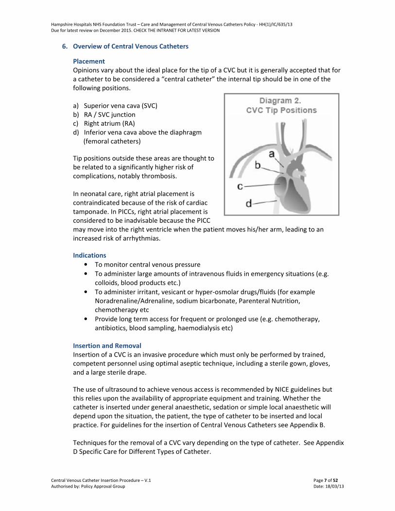
Placement
Opinions vary about the ideal place for the tip of a CVC but it is generally accepted that for
a catheter to be considered a “central catheter” the internal tip should be in one of the
following positions.
a) Superior vena cava (SVC)
b) RA / SVC junction
c) Right atrium (RA)
d) Inferior vena cava above the diaphragm
(femoral catheters)
Tip positions outside these areas are thought to
be related to a significantly higher risk of
complications, notably thrombosis.
In neonatal care, right atrial placement is
contraindicated because of the risk of cardiac
tamponade. In PICCs, right atrial placement is
considered to be inadvisable because the PICC
may move into the right ventricle when the patient moves his/her arm, leading to an
increased risk of arrhythmias.
Indications
• To monitor central venous pressure
• To administer large amounts of intravenous fluids in emergency situations (e.g.
colloids, blood products etc.)
• To administer irritant, vesicant or hyper-osmolar drugs/fluids (for example
Noradrenaline/Adrenaline, sodium bicarbonate, Parenteral Nutrition,
chemotherapy etc
• Provide long term access for frequent or prolonged use (e.g. chemotherapy,
antibiotics, blood sampling, haemodialysis etc)
Insertion and Removal
Insertion of a CVC is an invasive procedure which must only be performed by trained,
competent personnel using optimal aseptic technique, including a sterile gown, gloves,
and a large sterile drape.
The use of ultrasound to achieve venous access is recommended by NICE guidelines but
this relies upon the availability of appropriate equipment and training. Whether the
catheter is inserted under general anaesthetic, sedation or simple local anaesthetic will
depend upon the situation, the patient, the type of catheter to be inserted and local
practice. For guidelines for the insertion of Central Venous Catheters see Appendix B.
Techniques for the removal of a CVC vary depending on the type of catheter. See Appendix
D Specific Care for Different Types of Catheter.

Hampshire Hospitals NHS Foundation Trust – Care and Management of Central Venous Catheters Policy - HH(1)/IC/635/13
Due for latest review on December 2015. CHECK THE INTRANET FOR LATEST VERSION
Page 8 of 52
Choice of Catheter
The choice of device will depend chiefly on the purpose for which it is intended, though
patient preference may be a key factor with long-term catheters. As a general principle the
lumen diameter and the number of lumens should be kept to a minimum, since larger bore
catheters and multiple lumens are associated with higher infection and thrombosis risks.
Clearly there are many other factors to be weighed against these risks – e.g. in high
dependency settings large bore catheters and multiple lumens tend to be used as they are
essential for management of the acutely ill patient.
Where Parenteral Nutrition is to be administered, ideally a single-lumen catheter should be
used. If multiple lumens are essential, then one lumen should be dedicated “exclusively for
that purpose” (except in Neonates see Appendix D).
7. Clinical Practice
Central Venous Catheter Insertion Procedure
The procedure to be followed when inserting a Central Venous Catheter (CVC) is detailed in
Appendix B. It must be documented using the VAD Insertion and Management Form (see
Appendix H) to meet legal and patient care requirements/facilitate audit.
Principles of Care
The principles of ongoing care for a Central Venous Catheters (CVC) is detailed in Appendix
C.
Specific Care for Different Types of Catheter
Specific care for different types of Central Venous Catheters is detailed in Appendix D.
Management of Complications
Information on how to manage complications arising from Central Venous Catheters is
detailed in Appendix E.
Using Thrombolytics
When and how to use thrombolytics is detailed in Appendix F.
Glossary of Complications
A detailed explanation of complications that can occur can be found in Appendix G.
8. Stakeholders Engaged During Consultation
Stakeholder Date of Consultation
Infection Prevention and Control (Lead Infection Prevention &
Control Nurse)
15 February 2013
Health and Safety (Health and Safety Advisor) 15 February 2013
Safeguarding (Trust Safeguarding Lead) 15 February 2013
Information Governance (Information Governance Manager) 15 February 2013

Hampshire Hospitals NHS Foundation Trust – Care and Management of Central Venous Catheters Policy - HH(1)/IC/635/13
Due for latest review on December 2015. CHECK THE INTRANET FOR LATEST VERSION
Page 9 of 52
9. Dissemination and Implementation Plan
The policy will be disseminated in the following ways:
Action(s) Owner
Publicise detail of new document via Intranet and
Midweek message
IPCT and Communication Team
Communication to all Senior Managers to advise
publication of policy
BNN Healthcare Library
The policy will be available on the intranet and
web site
BNHH Healthcare Library and
Communication Team
10. Training
Trained nursing staff will attend an IV Therapy Study Day. Clinical Educators, Practice
Development Nurses and Clinical Nurse Specialists will support the learning, gaining and
maintaining of competencies. Additional training can be offered by the IV Nurse Specialist.
Individuals in the Trust should receive annual infection prevention and control training to
ensure they are aware of their responsibilities. Education and Training will be provided in
accordance with the Trust Training Needs Analysis (Learning and Development Policy).
11. Monitoring Compliance with the Document
Compliance with the policy will be monitored in the following ways:
Minimum
requirements Requirement
Reviewed by Method of
Monitoring Frequency
of Review Monitoring
Committee
A. Effectiveness
of policy
Infection
Prevention
and Control
Team
Quality control
audits to
ensure
continued
standards and
adherence of
policy during
care and
management
of Central
Venous
Catheter
Monthly Infection Prevention
and Control
Committee/Divisional
Governance Boards
B. Clinical
Practice
Supervisors Supervised
practice
Ongoing N/A
Assistant Risk and Compliance Manager (Risk and Compliance) 15 February 2013
Divisional Directors and Divisional Directors (Operational) 15 February 2013
Equality and Diversity Lead (Equality & Diversity) 15 February 2013
Infection Prevention and Control Committee 15 February 2013
Consultant Microbiologists 15 February 2013
Clinical Service Managers/Leads 15 February 2013
Operational Service Managers 15 February 2013

Hampshire Hospitals NHS Foundation Trust – Care and Management of Central Venous Catheters Policy - HH(1)/IC/635/13
Due for latest review on December 2015. CHECK THE INTRANET FOR LATEST VERSION
Page 10 of 52
12. References
Department of Health (2001) The epic Project: Developing National Evidence – based
Policies for Preventing healthcare associated Infections Journal of Hospital Infection (2001)
47 (supplement)
Donaldson I. (1999) Intravenous therapy in critically ill adults: developing a clinically and
cost effective approach Intensive and Critical Care Nursing No 15, 338-345
Dougherty L, Mallett J (2001) The Royal Marsden Hospital Manual of Clinical Nursing
Procedures. Eighth edition Blackwell Science
Fletcher SJ; Bodenham A (1999) Catheter related sepsis: an overview – Part 1 British
Journal of Intensive Care. March/April
Infection Control Nurses Association (2001) Policies for preventing intravascular catheter
related infection NICE (2003) (No. 4)
Care of patients with central venous catheters Clinical policy 2 – Infection control, June
2003 Polderman KH; Girbes AR (2002) Central venous catheter use. Part 2: infectious
complications Intensive Care medicine 2002, Jan; 28(1): 18-28
RCN (March 2004) Essential practice in infection control – Guidance for nursing staff
RCN (Jan 2010) Standards for infusion therapy
Guidelines on the insertion and management of central venous access devices in adults
L. Bishop, L. Dougherty, A. Bodenham, J. Mansi, P. Crowe, C. Kibbler, M. Shannon, J.
Treleaven Int. Jnl. Lab. Hem. 2007, 29, 261–278
Pratt RJ et al (2007) “epic2: National Evidence-Based Guidelines for Preventing Healthcare-
Associated Infections in NHS Hospitals in England”.
http://www.epic.tvu.ac.uk/PDF%20Files/epic2/epic2-final.pdf
National Institute for Clinical Excellence (September 2002) “Guidance on the Use of
Ultrasound Locating Devices for Placing Central Venous Catheters.” NICE Technology
Appraisal No 49. London: National Institute for Clinical Excellence. www.nice.org.uk
An Intervention to Decrease Catheter-Related Bloodstream Infections in the ICU
Peter Pronovost, M.D., Ph.D., Dale Needham, M.D., Ph.D., Sean Berenholtz, M.D., David
Sinopoli, M.P.H., M.B.A., Haitao Chu, M.D., Ph.D., Sara Cosgrove, M.D., Bryan Sexton, Ph.D.,
Robert Hyzy, M.D., Robert Welsh, M.D., Gary Roth, M.D., Joseph Bander, M.D., John
Kepros, M.D., and Christine Goeschel, R.N., M.P.A. N Engl J Med 2006; 355:2725-32.
East Kent Hospitals NHS Trust vascular access guidelines section 7: CVC lines. May 2007
Mayo DJ “Administering Urokinase: Clearing the Way” Nursing98 December
Vesely, T. “Central Venous Catheter Tip Position: A Continuing Controversy” Journal of
Vascular and Interventional Radiology, Volume 14(5) May 2003, pp 527-534

Hampshire Hospitals NHS Foundation Trust – Care and Management of Central Venous Catheters Policy - HH(1)/IC/635/13
Due for latest review on December 2015. CHECK THE INTRANET FOR LATEST VERSION
Page 11 of 52
Racadio, JM, Doellman DA, Johnson ND, Bean JA, Jacobs BR. “Pediatric Peripherally Inserted
Central Catheters: Complication Rates Related To Catheter Tip Location.” Pediatrics.
107(2):E28,
2001 Feb
Puel V et al 1993. “Superior vena cava thrombosis related to catheter malposition in cancer
chemotherapy given through implanted ports”. Cancer. 72(7):2248-52, 1993 Oct 1
Eastridge BJ and Lefor, AT. “Complications of indwelling venous access devices in cancer
patients”. Journal of Clinical Oncology. 13(1):233-8, 1995 Jan.
Schuster M, et al. “The carina as a landmark in central venous catheter placement”. British
Journal of Anaesthesia 2000; 85: 192–4
Fletcher S, Bodenham AR. “Safe placement of central venous catheters: where should the
tip of the catheter lie?” British Journal of Anaesthesia 2000; 85: 188–91.
Department of Health “Review of Four Neonatal Deaths due to Cardiac Tamponade
associated with the Presence of a Central Venous Catheter: Recommendations and
Department of Health Response.” June 2001.
Bivins MH and Callahan MJ “Position-Dependent Ventricular Tachycardia Related To A
Peripherally Inserted Central Catheter” Mayo Clinic Proceedings, 75 (4): 414-6, 2000 Apr
NAVAN (National Association of Vascular Access Networks) “Tip Location Of Peripherally
Inserted Central Catheters” Journal of Vascular Access Devices, Summer 1998
Grove, Jay R and Pevec, William C “Venous Thrombosis Related to Peripherally Inserted
Central Catheters” Journal of Vascular and Interventional Radiology Volume 11(7)
July/August 2000 pp 837- 840
Wickham R et al "Long-term CVCs - Issues for Care" Seminars in Oncology Nursing Vol 8 No
2 May 1992 pp 133-147
Wilson J A "Preventing Infection During IV Therapy" Professional Nurse March 1994 pp 338-
392
Todd J "Peripherally inserted central catheters" Professional Nurse Vol 13 No 5 Feb 1998 pp
297-302
Todd, J “Peripherally Inserted Central Catheters and their Use in IV Therapy” British Journal
of Nursing Vol 8 No 3 1999 pp 140-48
Camp-Sorrell D "Implantable Ports - Everything you Always Wanted to Know" Journal of
Intravenous Nursing Vol 15 No 5 Sep/Oct 1992 pp 262 – 272
Krzywda, E et al “Catheter Infections: Diagnosis, Etiology, Treatment, and Prevention”
Hampshire Hospitals NHS Foundation Trust – Care and Management of Central Venous Catheters Policy - HH(1)/IC/635/13
Due for latest review on December 2015. CHECK THE INTRANET FOR LATEST VERSION
Page 12 of 52
Nutrition in Clinical Practice Vol 14 No 4 August 1999 pp178 – 90
Schulmeister L "Needle Dislodgement from Implanted Venous Access Devices; Inpatient
and Outpatient Experiences" Journal of Intravenous Nursing Vol 12 No 2 March/April 1989
pp 90-92
National Kidney Foundation “Kidney Disease Outcomes Quality Initiative Guidelines 2000”.
2001 National Kidney Foundation
Haller L and Rush K "CVC infection: a review" Journal of Clinical Nursing Vol 1 1992 pp 61-
66
Rowley, S “Aseptic Non Touch Technique (ANTT)” Nursing Times Feb 15th Vol 97 No 7 2001:
Infection Control Supplement V1-V111
Cornock M "Making Sense of CVCs" Nursing Times Vol 92 No 49 Dec 4th 1996 pp 30-31
Young, A “WARP - A multicentre prospective randomised controlled trial (RCT) of
thrombosis prophylaxis with warfarin in cancer patients with central venous catheters
(CVCs)” 2005 ASCO Annual Meeting
RCN IV Therapy Forum “Standards for Infusion Therapy” Royal College of Nursing October
2006
Hadaway L “Major Thrombotic and Nonthrombotic Complications: Loss of Patency” Journal
of Intravenous Nursing Vol 21 No 5S September/October 1998
Gabriel J "Care and management of peripherally inserted central catheters" British Journal
of Nursing Vol 5 No 10 1996 pp 594-599
Maki D G et al "Prospective Randomised Trial of Povidone Iodine, Alcohol and Chlorhexidine
for Prevention of infection Associated with Central Venous and Arterial Catheters” Lancet
383 1991 pp339-343
Krzywda, E “Predisposing Factors, Prevention and Management of Central Venous Catheter
Occlusions” Journal of Intravenous Nursing Vol 22 No 6S November/December 1999 pp S11
– S17
Olson, K et al “Evaluation of a No-dressing Intervention for Tunneled Central Venous
Catheter Exit Sites” Journal Of Infusion Nursing Volume 27(1) January/February 2004 pp
37-44
Oliver L "Wound Cleansing" Nursing Standard Vol 11 No 20 Feb 5th 1997 pp 47-51
Drewett, S “Central venous catheter removal: Procedures and rationale” British Journal of
Nursing. London: Dec 8, 2000-Jan 10, 2001.Vol.9, Iss. 22; pg. 2304

Hampshire Hospitals NHS Foundation Trust – Care and Management of Central Venous Catheters Policy - HH(1)/IC/635/13
Due for latest review on December 2015. CHECK THE INTRANET FOR LATEST VERSION
Page 13 of 52
Krzywda, Elizabeth A “Central Venous Catheter Infections: Clinical Aspects of Microbial
Etiology and Pathogenesis”. Journal of Infusion Nursing Volume 25(1) January/February
2002 pp 29-35
Oncu, S and Sakarya, S. “Central venous catheter-related infections: an overview with
special emphasis on diagnosis, prevention and management”. Internet Journal of
Anesthesiology, 2003, vol.7, no. 1
Citton-R et al. “Old and new tools in the diagnosis of central venous catheter-related
bloodstream infections: Is there a role for brushing?” Journal of Vascular Access 2004
Volume 5 Issue 1 Pg 10-12
Hall, K and Farr, B. “Diagnosis and Management of Long-term Central Venous Catheter
Infections”. J Vasc Interv Radiol 2004; 15:327–334
Marinella, Mark A et al “Spectrum of upper-extremity deep venous thrombosis in a
community teaching hospital” Heart and Lung: The Journal of Acute and Critical Care
Volume 29(2) March/April 2000 pp 113-117
Lee, Agnes Y and Levine, Mark N. “Management of Venous Thromboembolism in Cancer
Patients” Vol 14, no 3, (March 2000)
Balestreri-L et al “Central venous catheter-related thrombosis in clinically asymptomatic
oncologic patients: A phlebographic study.” European Journal of Radiology {EUR-J-RADIOL},
1995, Vol/Iss/Pg. 20/2 (108-111).
Allen, Anthony W et al. “Venous Thrombosis Associated with the Placement of Peripherally
Inserted Central Catheters”. Journal of Vascular and Interventional Radiology Volume
11(10) November/December 2000 pp 1309-1314
Jacobs P et al “Chest Pain As The Presenting Symptom In Catheter-Associated Thrombosis
Of The Superior Vena-Cava” S Afr Medical Journal 88(10) 1998 pp 1284-5
Kayley, J "Skin-Tunnelled Cuffed Catheters" Community Nurse June 1997 pp 21-22
Mazzola JR, Schott-Baer D, Addy L. “Clinical Factors Associated With The Development Of
Phlebitis After Insertion Of A Peripherally Inserted Central Catheter”. Journal of Intravenous
Nursing 1999 Mar-Apr:22(2):60
Teichgräber, UK et al “Central Venous Access Catheters: Radiological Management of
Complications” Cardiovascular and Interventional Radiology (2003) 26:321-333
Gabriel J "Fibrin sheaths in vascular access devices" Nursing Times Vol 93 No 10 March 5
1997
Hampshire Hospitals NHS Foundation Trust – Care and Management of Central Venous Catheters Policy - HH(1)/IC/635/13
Due for latest review on December 2015. CHECK THE INTRANET FOR LATEST VERSION
Page 14 of 52
Mayo DJ and Pearson DC “Chemotherapy Extravasation: A Consequence of Fibrin Sheath
Formation Around Venous Access Devices” Oncology Nurse Forum Volume 22 No 4 May
1995, 675-680
Mehall, JR, Saltzman DA, Jackson RJ and Smith SD “Fibrin Sheath Enhances Central Venous
Catheter Infection” Critical Care Medicine Volume 30 (4) April 2002, 908-912
Aitkin DR and Minton JP "The 'Pinch-off Sign;' A Warning of Impending Problems with
Permanent Subclavian Catheters" American Journal of Surgery Vol 148 Nov 1984 pp 633-
636
Jones GR “A Practical Guide to Evaluation and Treatment of Infections in Patients with
Central Venous Catheters” Journal of Intravenous Nursing Vol21 No 5S September/October
1998 pp S134 – S 142
Banks N “Positive Outcome after Looped Peripherally Inserted Central Catheter
Malposition” Journal of Intravenous Nursing Vol 22 No 1 January/February 1999 pp 14 – 18
Rastogi S et al “Spontaneous Correction Of The Malpositioned Percutaneous Central Venous
Line In Infants” Pediatric Radiology. 28(9): 694-6, 1998 Sep
Moore C et al "Nursing Care and Management of Venous Access Ports" Oncology Nursing
Forum Vol 13 No 3 May/June 1986 pp 35-39
Legislation and Guidance from other organisations
DoH (July 2006) Winning ways high impact working together to reduce healthcare
associated infection in England-intervention 2 Department of Health.
The Health and Social Care Act 2008: Code of Practice on the prevention and control of
infections and related guidance. Department of Health, London, 14 Dec 2010.
Available at
http://www.dh.gov.uk/en/Publicationsandstatistics/Publications/PublicationsPolicyAnd
Guidance/DH_122604
13. Associated Documentation
Aseptic Technique Policy
Learning and Development Policy
14. Contributors
Contributor Job Title Contributor Name
IV Nurse Specialist Sandy Kirk

Hampshire Hospitals NHS Foundation Trust – Care and Management of Central Venous Catheters Policy - HH(1)/IC/635/13
Due for latest review on December 2015. CHECK THE INTRANET FOR LATEST VERSION
Page 15 of 52
Appendix A – Equality Impact Assessment
PART 1
To be completed by the document owner
Document Title: Care and Management of Central Venous Catheters Policy
Yes/No Comments
1. Could the application of this document have a
detrimental equality impact on individuals with any of
the following protected characteristics? (See Note 1)
Age No
Disability No
Gender reassignment No
Race No
Religion or belief No
Sex No
Sexual orientation No
Marriage & civil partnership No
Pregnancy and maternity No
2. If you have identified any potential detrimental impact,
do you consider this to be valid, justifiable and lawful? If
so, please explain your reasoning.
N/A
3. If you have answered ‘no’ to question 2, has the policy
been amended to remove or reduce any potential
detriment?
• If you answer ‘yes’, please summarise the changes
made
• If you answer ‘no’. please explain why not
N/A
4. Based on the answers to questions 1 – 3 do you consider
that a detailed equality analysis is needed?
No
NAME: Sandy Kirk
JOB TITLE: IV Nurse Specialist
DATE: 8 February 2013
PART 2

Hampshire Hospitals NHS Foundation Trust – Care and Management of Central Venous Catheters Policy - HH(1)/IC/635/13
Due for latest review on December 2015. CHECK THE INTRANET FOR LATEST VERSION
Page 16 of 52
To be completed by the Trust’s Equality and Diversity Lead
Brief Summary of potential impact of this document and whether sufficient consideration
has been given to the Equality Duty
The application of this policy for the care and management of central venous catheters is
completely clinically based and ensuring appropriate approach would be the priority,
however the Trust would endeavour to continue to meet patients and employees individual
needs as far as is practicable.
Yes/No Comments
1. Is this document recommended for publication without
amendment?
Yes
2. Is this document recommended for publication but with
recommended amendments? Please specify.
Na
3. Is this document not recommended for publication
without amendments being made? Please specify?
Na
4. Is it recommended that this document requires a more
detailed equality analysis to be undertaken prior to
publication?
No
5. Specify with which, if any, individuals and groups you
have consulted in reaching your decision.
None
NAME: Nicky Smith
JOB TITLE: Equality and Diversity Lead
DATE: 18 March 2013
Note 1
Under the terms of the Equality Act 2010’s public sector Equality Duty, the Trust has a legal responsibility to think
about the following three aims of the Equality Duty as part of our decision making and policy development.
• Eliminate unlawful discrimination, harassment and victimisation;
• Advance equality of opportunity between people who share a protected characteristic and people who do
not share it; and
• Foster good relations between people who share a protected characteristic and people who do not share it.

Hampshire Hospitals NHS Foundation Trust – Care and Management of Central Venous Catheters Policy - HH(1)/IC/635/13
Due for latest review on December 2015. CHECK THE INTRANET FOR LATEST VERSION
Central Venous Catheter Insertion Procedure – V.1 Page 17 of 52
Authorised by: Policy Approval Group Date: 18/02/13
Appendix B - Central Venous Catheter Insertion Procedure
Health Care personnel caring for a patient with a central venous catheter should be trained
and assessed as competent in using and consistently adhering to the infection prevention
practices described in this guideline.
Patients should receive clear and comprehensive information explaining the risks, benefits and
care of the catheter. Signed consent should be obtained prior to catheter insertion (if the
patient is able to do so).
Choice and site of catheter
Nontunnelled catheters are indicated for short-term use when peripheral venous access is
impractical.
Tunnelled central venous catheters are indicated for the repeated administration of
chemotherapy, antibiotics, parenteral feeding and blood products, and for frequent blood
sampling. They are recommended for patients in whom long-term (>30 days) central venous
access is anticipated.
Fully implanted catheters (ports) are more suitable for children and for less frequent
accessing but long-term use, whereas skin-tunnelled catheters are recommended for intensive
access. They should be avoided for inpatient therapy because of limited catheter longevity
and increased incidence of thrombosis. They are more suited to ambulatory or outpatient-
based therapy.
Polyurethane Peripherally inserted central catheters (PICC) allow easier infusion of blood
products as greater flow rates are achieved because the thinner walls provide a larger internal
diameter of the catheter. The decision to use polyurethane catheters should be balanced
against the higher risk of thrombosis with these catheters compared with silicone catheters.
The number of lumina and diameter of catheters should be kept to the minimum.
Insertion
It is strongly recommended that CVCs should be inserted in designated clean areas, e.g.
treatment rooms, radiology, critical care units, operating theatres. Insertion should be
performed by trained and competent staff regardless of specialty - this reduces the mechanical
and infection risks associated with insertion.
Ultrasound guided insertion is recommended for all routes of central venous catheterization.
The use of ultrasound is also recommended for the insertion of PICC when the peripheral veins
are not visible or palpable.
Hands should be decontaminated using alcohol hand rub on visibly clean hands (apply 1 shot,
cover all surfaces, rub hands together until dry). Alternatively, using an antimicrobial liquid
soap e.g. Hibiscrub, Povidone iodine, hands should be thoroughly washed, using a technique,
which aims to cover all surfaces of the hands. Hands should be rinsed in running water before
and after applying the cleansing agent and dried well -this reduces the risk of cross infection
from the operators’ hands during the procedure.

Hampshire Hospitals NHS Foundation Trust – Care and Management of Central Venous Catheters Policy - HH(1)/IC/635/13
Due for latest review on December 2015. CHECK THE INTRANET FOR LATEST VERSION
Central Venous Catheter Insertion Procedure – V.1 Page 18 of 52
Authorised by: Policy Approval Group Date: 18/02/13
Use optimum aseptic technique, including a sterile gown, gloves, hat and mask, and a large
sterile drape (dedicated CVP insertion packs should be used where available) -evidence has
identified that using maximal barrier precautions reduces the risk of subsequent CVC related
infection.
Effective skin preparation will remove bacteria from both hair and skin, avoiding the need for
shaving, which can result in microscopic damage and thus microbial colonisation. If hair
removal is considered necessary, clipping is the preferred option using a disposable clipper
head.
Using Chlorhexidine 2% in 70% alcohol (1-2 applicators of Chloraprep 3mls) applying gentle
friction, disinfect the skin insertion site for 30 seconds. Allow the antiseptic to dry before
inserting the catheter. Use an alcoholic povidone-iodine solution for patients with a history of
chlorhexidine sensitivity. Skin cleansing/antisepsis of the insertion site is one of the most
important measures for preventing catheter related infection. EPIC (2006) recommends an
alcoholic solution of chlorhexidine gluconate 2% as this combines the benefits of rapid action
and excellent residual (ongoing) activity.
Antibiotic/antimicrobial impregnated catheters, for example, chlorhexidine and silver
sulfadiazine impregnated catheters should be considered for appropriate risk groups of
patients to minimize infection risk.
Routine antibiotic prophylaxis is not recommended.
Routine replacement, for example, weekly change, of short-term catheters as a means to
reduce infection rates is not recommended.
Guidewire-assisted catheter exchange to replace a malfunctioning catheter is acceptable if
there is no evidence of infection. However, if infection is suspected the existing catheter
should be removed and a new catheter inserted at a different site. This technique is generally
impractical for cuffed tunnelled catheters or ports when it may be technically easier and safer
to insert a new catheter into a clean site. It is usually preferable to insert a new catheter into a
clean site.
The CVC should be firmly anchored to prevent movement using a mono filament suture -CVCs
readily become colonised and carry micro-organisms from the skin into the insertion tract.
Use a sterile, transparent, semi permeable polyurethane CVC dressing i.e. IV 3000 -this allows
for continuous inspection of the site.
If total parenteral nutrition is being administered use one central venous catheter or lumen
exclusively for that purpose.
The procedure must be documented using the VAD Insertion and Management Form (see
Appendix H) to meet legal and patient care requirements/facilitate audit.

Hampshire Hospitals NHS Foundation Trust – Care and Management of Central Venous Catheters Policy - HH(1)/IC/635/13
Due for latest review on December 2015. CHECK THE INTRANET FOR LATEST VERSION
Central Venous Catheter Insertion Procedure – V.1 Page 19 of 52
Authorised by: Policy Approval Group Date: 18/02/13
Radiological confirmation of the position of the catheter tip must be undertaken once inserted
- to confirm precise location of the catheter tip and exclude immediate complications such as
pneumothorax.
Hampshire Hospitals NHS Foundation Trust – Care and Management of Central Venous Catheters Policy - HH(1)/IC/635/13
Due for latest review on December 2015. CHECK THE INTRANET FOR LATEST VERSION
Principles of Care – V.1 Page 20 of 52
Authorised by: Policy Approval Group Date: 18/02/13
Appendix C - Principles of Care
General Principles
Use an aseptic technique following the Trust Asepsis Policy whenever the CVC is accessed
and during procedures involving exit sites to prevent infection. A strong correlation exists
between bacteraemia and the presence of a CVC.
Wear sterile gloves when carrying out dressing changes and when accessing the catheter.
Gloves should be worn to prevent de-scaling of bacteria onto key parts.
Monitor temperature, pulse, blood pressure, respiratory rate and oxygen saturations at
least a minimum of 12 hourly to detect infection.
Do not allow air to enter the catheter. All syringes and intravenous administration sets
must be carefully primed to prevent air embolism. The negative pressure within the chest
may suck air into the catheter during inspiration especially if the patient is sitting up.
Cap off the catheter with a needle-free access device when not in use (except Neonates).
This will minimise interruptions to the closed system. Unless manufacturer’s instructions
vary, this should be changed every 7 days or every 200 uses, whichever is the sooner. In
adult inpatients with long-term vascular access devices the bungs should be changed on a
set day (e.g. Sunday) to ensure continuity within and between units. The risk of
contamination increases with every interruption to the closed system.
Whenever the bung/access device is removed from the catheter then it must be replaced
with a new, needleless access device/bung to prevent infection.
If the catheter possesses an integral clamp, keep it closed whenever the cap is removed
and at all other times except when administering or withdrawing fluids. Clamping should
always take place at the designated area and never at the thickened area near the hub
(except tunnelled CVCs). The clamp will prevent air entry and bleeding should the luer lock
cap become unattached. Repeated clamping away from the specially reinforced area may
result in damage to the catheter.
Always take signs of systemic or local infection seriously and refer to a member of the
medical staff. Infection continues to be one of the most frequent and most serious
complications associated with CVC Catheters.
The practice of administering prophylactic antibiotics at the time of CVC insertion should
not be routinely followed. The Department of Health’s Epic2 Guidelines on the prevention
of infection in Central Venous Catheters specifically states that this practice is not
supported by research and may encourage resistant organisms.
The practice of administering prophylactic mini-dose Warfarin to patients with CVCs should
not be followed. Mini-dose Warfarin has recently been shown to be ineffective in the
prevention of thrombosis in cancer patients with CVCs. (NB: dose adjusted Warfarin did
show some efficacy but with an increased risk of serious bleeding).

Hampshire Hospitals NHS Foundation Trust – Care and Management of Central Venous Catheters Policy - HH(1)/IC/635/13
Due for latest review on December 2015. CHECK THE INTRANET FOR LATEST VERSION
Principles of Care – V.1 Page 21 of 52
Authorised by: Policy Approval Group Date: 18/02/13
Should the catheter fracture or be accidentally cut, clamp it without delay proximal to the
break. Specialist advice should be sought immediately to consider removal or repair of the
catheter to prevent haemorrhage, air embolism and infection.
Always secure the catheter firmly to the skin away from the exit site with tape or with a
dedicated device such as 'Statlock' for patient's comfort, to prevent tension or accidental
dislodgement, and to reduce 'to and fro' motion which increases the risk of catheter
related sepsis
Accessing the Catheter
Before it is used for administering therapeutic drugs or fluids, the patency and correct
functioning of the catheter should be established (except Neonates when this should only
be done immediately following catheter insertion). Signs of catheter occlusion, whether
partial or complete, should be taken seriously and action should be taken earlier rather
than later to restore full patency. Ignoring the early signs may lead to the development of
more serious problems which cannot then be easily rectified – e.g. complete blockage or
thrombosis.
Nurses using CVCs can be confident of access if all three of the following apply:
o The catheter can be flushed with ease.
o Blood can be withdrawn from the catheter (not Neonates).
o The patient experiences no discomfort during flushing/infusion and there are no
other complications
If any of these criteria are not met you should refer to Appendix F- Management of
Complications.
Ways of assessing these three criteria will vary with the setting. Here are some points to
note:
o A proper assessment of the catheter involves observing the exit site and the
area around as this may reveal any signs of thrombosis, leakage, infection etc.
While this is not necessarily appropriate every time the catheter is used it
should be a regular part of your practice.
o Assessing CVCs in neonates and in patients requiring blood processing (e.g.
haemodialysis / apheresis) requires specialist knowledge: refer to Appendix E
Overview and Specific Care for Different Types of Catheter for care of these
patients.
o In adults and children over 1 year who are due to receive intravenous fluids, a
useful technique is to attach an infusion of 0.9% saline, open the clamp on the
giving set fully and observe for free-flow. You will soon learn to recognise what
is a normal free-flow for a particular type of CVC (for example the flow on a
Non-tunnelled CVC will be much faster than you would expect from a PICC
which is a much longer thinner catheter.) Dropping the bag of fluid briefly below
the patient’s heart with any clamps open will allow you to check for flashback of
blood without interrupting the closed system. As soon as blood is seen in the
tubing, the bag can be replaced on the drip stand and prescribed infusion

Hampshire Hospitals NHS Foundation Trust – Care and Management of Central Venous Catheters Policy - HH(1)/IC/635/13
Due for latest review on December 2015. CHECK THE INTRANET FOR LATEST VERSION
Principles of Care – V.1 Page 22 of 52
Authorised by: Policy Approval Group Date: 18/02/13
started. (NB this technique for checking flashback does not always work with
valved catheters). Ensure to stop the free flow to ensure no unnecessary bolus
of fluid.
Checking for flashback of blood does not necessarily mean you have to discard blood. For
example, attach a syringe containing 10ml 0.9% sodium chloride to the catheter, flush a
couple of ml into the line and then withdraw. As soon as you see a trace of blood in the
catheter or syringe just flush the rest of the sodium chloride into the line using the push-
pause technique as described below.
Flushing After and Between Uses (except Neonates)
Flushing Technique
Where possible, do not use syringes smaller than 10 ml for infusion into the catheter to
prevent excessive pressure being exerted on the lumen which might cause it to rupture.
Smaller syringes exert greater pressure but please note that syringe size alone is not
sufficient to prevent rupture. When resistance is felt, if more pressure applied to overcome
it, catheter fracture could result regardless of the syringe size.
Use a brisk 'push-pause' flushing technique routinely when flushing the catheter - i.e. flush
briskly, pausing briefly after approximately each ml of fluid. The 'push-pause' technique
causes turbulence within the catheter, which helps to flush away any debris and prevent
occlusion of the lumen.
If the catheter possesses a clamp, clamp the line while the final ml of the flush is being
injected. If there is no clamp you can achieve a “positive pressure finish” by removing the
syringe from the needle free bung) while injecting the last ml: but note that to avoid any
spray from the syringe you should hold sterile gauze around the connector while doing
this. Maintaining positive pressure helps prevent blood entering the catheter after flushing,
which might lead to occlusion or thrombus formation.
Do not routinely withdraw and discard blood from the catheter before flushing (except
Renal Dialysis Catheters) in an attempt to avoid flushing bacteria and clots into the patient.
There is no evidence that withdrawing prior to flushing reduces infection or embolism. But
note that if the catheter is to be used for administering drugs or fluids, checking for
“flashback” should be a routine part of catheter assessment: see Accessing the Catheter
above
Frequency of flushing and flushing solutions
This varies depending on the device. See Appendix D - Specific Care for Different Types of
Catheter.
Please note that Hepsal and Heparinised Saline must be prescribed.
Care of the Exit Site (Except Neonates)
Dressings Immediately post insertion

Hampshire Hospitals NHS Foundation Trust – Care and Management of Central Venous Catheters Policy - HH(1)/IC/635/13
Due for latest review on December 2015. CHECK THE INTRANET FOR LATEST VERSION
Principles of Care – V.1 Page 23 of 52
Authorised by: Policy Approval Group Date: 18/02/13
As with any surgical wound, the exit site should ideally be left undisturbed for 1-2 days.
Routine taking down of the dressing post-insertion to inspect the site merely exposes the
patient to increased risk of infection. On the other hand most exit sites bleed to some
extent following insertion. If this leads to “strike-through” on a dry dressing, (i.e.
exudate/blood/serous fluid observed on the outside of a dry dressing) it should be changed
immediately since a wet surface provides “a liquid pathway for bacteria to travel” to the
wound.
The ideal dressing immediately post-insertion is a dry dressing covered and sealed with a
transparent dressing (IV 3000). In most cases this will absorb any oozing but not
necessitate changing the dressing. Ideally this dressing should be left undisturbed for at 1-2
days. If there is excessive bleeding and the gauze becomes soggy the dressing should be
changed.
If allergic to IV 3000 and a dry dressing alone is used post-insertion, it should again ideally
be left undisturbed for 1-2 days but should always be changed as soon as any “strike-
through” occurs using an aseptic technique.
If bleeding is excessive the dressing should be changed every time strike-through occurs
and replaced with a more absorbent or thicker dressing. Pressure should then be applied
to the site and the patient encouraged to lie fairly still until the bleeding settles. It is not
acceptable to add more dressings on top of blood-soaked dressings which have been in
contact with a moist outer surface, because of the infection risk.
On-going Dressing Regimes after the first 1-2 days
As a general principle, where a dressing is used it should be inspected regularly and
renewed immediately should it become soiled, wet or detached. A moist environment is
one in which bacteria readily multiply.
If the exit site is reddened, painful, exudating or infected, swab the site and increase the
frequency of dressing change depending on the amount of exudate.
The most suitable dressing will depend on the setting, the type of CVC and the individual
patient’s needs. See Appendix E Overview and Specific Care for Different Types of Catheter
for recommendations. The main options for dressings are:
6. IV-dedicated occlusive transparent dressing, changed every 7 days except patients
on dialysis and neonates. Some researchers have found IV dedicated transparent
dressings to be associated with a lower risk of infection than other transparent
dressings.
7. Sterile dry dressing taped in situ, changed at least twice a week.
8. No dressing. This may be suitable for some patients with Tunnelled CVCs from
21days post insertion once the tissues have fibrosed around the cuff and in the
absence of exudate or signs of infection.
Cleaning of Exit Site
At dressing changes, the exit site should be cleaned using Chloraprep using a criss cross
motion to avoid transferring bacteria to the exit site.

Hampshire Hospitals NHS Foundation Trust – Care and Management of Central Venous Catheters Policy - HH(1)/IC/635/13
Due for latest review on December 2015. CHECK THE INTRANET FOR LATEST VERSION
Principles of Care – V.1 Page 24 of 52
Authorised by: Policy Approval Group Date: 18/02/13
Cleaning should be carried out using an aseptic technique.
Loose blood, exudate or other debris which might provide a focus or infection or might
impair inspection of the wound may be gently removed by cleaning in the above manner
with sterile 0.9% sodium chloride prior to cleaning with Chloraprep.
Removal
If a short-term CVC has not been used for >24 hours consideration should be given to its
removal. Some CVCs are simple and relatively safe to remove. With others, there is high
risk of air embolism and so removal requires a higher level of training and skill. See
Appendix D Specific Care for Different Types of Catheter for guidelines on removal.

Hampshire Hospitals NHS Foundation Trust – Care and Management of Central Venous Catheters Policy - HH(1)/IC/635/13
Due for latest review on December 2015. CHECK THE INTRANET FOR LATEST VERSION
Specific Care for Different Types of Catheter – V.1 Page 25 of 52
Authorised by: Policy Approval Group Date: 18/02/13
Appendix D - Specific Care for Different Types of Catheter
Care of Centrally-Inserted, Non-tunnelled CVCs
Often called Central Lines /Neck Lines /CVP lines.
Centrally Inserted Non-tunnelled CVCs are most
commonly found in acute settings. They are not
suitable for long-term use because they rarely
remain free of infection for longer than 7 – 10
days, and also because they are relatively
uncomfortable and unsightly.
The catheter is usually inserted via the
subclavian, jugular or femoral veins with the tip
positioned in the Right Atrium, the Superior or
Inferior Vena Cava. It is attached to the patient’s
skin using non-dissoluble sutures.
Non-tunnelled CVCs may have single or multiple
lumens. Each lumen provides independent
access to the venous circulation so that incompatible
drugs/fluids may be administered simultaneously.
Each lumen is equipped with an integral clamp to seal the catheter and guard against air entry,
haemorrhage and infection.
Flushing
Before flushing
o If there are infusional vasoactive drugs in the lumen, withdraw prior to flushing to
avoid bolus dose.
Technique
o Brisk push-pause technique with positive pressure finish
What to flush with
o 0.9% sodium chloride between incompatible drugs / infusions and after blood sampling
(if sodium chloride 0.9% incompatible use suitable alternative).
o Lock with 10ml 0.9% sodium chloride if catheter is to be accessed again within 1 day.
o Lock with 5ml Hepsal 10 U/ml if catheter not to be used again within 1 day.
Frequency of flushing
o Flush unused lumens at least once a week (10ml 0.9% sodium chloride then lock with 5
ml Hepsal 10 U/ml).
Exit site Care
Securement
o Lines are sutured in place, alternatives such as a Statlock can be used.
Sutures
o Leave in place as long as the catheter is in situ.
Cleaning
o Clean exit site at dressing changes using Chloraprep using a criss cross method.

Hampshire Hospitals NHS Foundation Trust – Care and Management of Central Venous Catheters Policy - HH(1)/IC/635/13
Due for latest review on December 2015. CHECK THE INTRANET FOR LATEST VERSION
Specific Care for Different Types of Catheter – V.1 Page 26 of 52
Authorised by: Policy Approval Group Date: 18/02/13
Dressings
o Post-insertion: gauze under transparent dressing for 24 hrs.
o After 24 hrs: Transparent dressing recommended. Change every 7 days unless soiled or loose in
these cases change when required.
Bathing & showering
o The exit site must not be allowed to get wet.
Removal
Who can remove Non-tunnelled CVCs?
o Any qualified nurse who has been assessed as competent and who follows these
guidelines.
Procedure
o You will need assistance during this procedure: do not attempt it alone.
o Check patient’s coagulation status. If there is an increased risk of bleeding discuss with
medical team before proceeding. If platelets are < 50, platelets should be administered
immediately prior to the procedure. If the patient is anticoagulated, this should be
managed as for surgery.
o The risk of air embolism increases if patient is dehydrated, is unable to lie flat, or has an
uncontrolled cough. Assess for these risks. Only proceed if satisfied that it is safe to do
so.
o Use aseptic technique throughout.
o Lie the patient flat and tip the head of the bed downward to reduce the risk of air
embolism (except femoral catheters).
o Remove the dressing. If there is any sign of infection, take a swab of the exit site.
o Remove any stitches.
o Ask patient to perform Valsalva’s manoeuvre (ie take a deep breath, hold it, and bear
down). If patient unable to do this, remove the catheter during expiration and never
when the patient is breathing in, as this will increase the risk of air being sucked into
the venous system.
o Gently and swiftly pull out the catheter and immediately apply pressure to the site
using sterile gauze. The patient can now breathe normally and the bed can be returned
to the flat position.
o Continue applying pressure to the exit site for three minutes (or longer in cases of
deranged clotting).
o If systemic infection is suspected, use sterile scissors to cut off the tip of the catheter
and without contaminating it drop it into a dry sterile specimen pot. Send it to
microbiology for culture (ITU all tips sent for culture).
o Apply a sterile occlusive dressing to prevent air from entering the venous system.
o Advise the patient to lie flat for 30 minutes.
o During this time observe patient for signs of haematoma (ie, swelling, pain, altered
voice, airway obstruction).
o The wound should be kept dry for 5 to 7 days and the wound monitored until healed

Hampshire Hospitals NHS Foundation Trust – Care and Management of Central Venous Catheters Policy - HH(1)/IC/635/13
Due for latest review on December 2015. CHECK THE INTRANET FOR LATEST VERSION
Specific Care for Different Types of Catheter – V.1 Page 27 of 52
Authorised by: Policy Approval Group Date: 18/02/13
Care of Tunnelled CVCs often called Hickman lines
Tunnelled CVCs are intended for longer-term use in patients who require multiple infusions of
fluids, blood products, drugs or Parenteral Nutrition. They also provide easy access for routine
blood sampling. They are more comfortable and discreet than the non-tunnelled CVCs described
above, and can last for much longer.
The Tunnelled CVC is inserted via the subclavian, jugular or femoral veins. The catheter is
tunnelled subcutaneously and exits at a convenient site (usually on the chest wall) where it
is secured with sutures. There is a ‘cuff’ within the tunnel to allow for the adherence of fibrous
tissue which helps to prevent accidental dislodgement after the removal of the sutures and acts as
a mechanical barrier to ascending bacteria.
Single, double and triple lumen catheters are
available. Each lumen provides independent
access to the venous circulation, so that
incompatible drugs/fluids may be
administered simultaneously.
Each lumen of the catheter is equipped
either with an integral clamp, or a 3-way
valve. Valved catheters vary in design: the
valve may be at the internal or external end of
each lumen (e.g. Groshong catheters have a
valve at the internal end, whereas PASV
catheters contain a valve at the external end).
The clamp (or valve) serves to seal the
catheter and guard against air entry,
haemorrhage and infection.
Patients with tunnelled CVCs may be discharged home with the catheter in situ. In these cases
patient education regarding the recognition and reporting of complications is of great importance.
Where possible, care in hospital should be aimed at the promotion of independence in caring for
the Tunnelled CVC, but liaison with the primary health-care team remains vital.
Flushing
Technique
o Brisk push-pause technique with positive pressure finish
What to flush with
o 0.9% sodium chloride between incompatible drugs / infusions and after blood sampling (if
sodium chloride 0.9% incompatible use suitable alternative).
o Lock with 10ml 0.9% sodium chloride if catheter to be used again within 1 day.
o Lock with 5ml Hepsal 10 U/ml if catheter not to be used within 1 day.
o Paediatrics – 5ml Hepsal 10u/ml flush if not to be used within 8 hours.
Frequency of flushing
o Flush unused lumens once a week with 5ml Hepsal 10 U/ml.

Hampshire Hospitals NHS Foundation Trust – Care and Management of Central Venous Catheters Policy - HH(1)/IC/635/13
Due for latest review on December 2015. CHECK THE INTRANET FOR LATEST VERSION
Specific Care for Different Types of Catheter – V.1 Page 28 of 52
Authorised by: Policy Approval Group Date: 18/02/13
Exit Site Care
Securement
o When stitches removed no further securement required – Paediatrics tape lines to patient.
Sutures
o Exit site: remove at 21 days
o Venepuncture site: Remove stitches/Steristrips at 7 days (unless dissolvable)
Cleaning
• Clean exit site at dressing changes using Chloraprep using a criss cross method
• Dressings:
o Exit site:
� Post-insertion: gauze under transparent dressing for 24 hrs
� After 24 hrs choose between
• Transparent dressing (changed every 7 days)
• OR dry dressing (changed at least every 7 days)
� After 21 days: choose between
• transparent dressing (change every 7 days)
• OR dry dressing (change at least twice a week)
• OR no dressing
• A chlorhexidine gel disc may be placed around the exit site to reduce
microbial contamination but the site needs to be visible
o Venepuncture Site:
� Dry dressing and/or transparent dressing until sutures removed / dissolve.
Bathing, showering & swimming
o Bathing: Patient should not submerge exit site in bathwater. For clean water jugged from
tap see “showering” below.
o Showering: If transparent dressing is intact patient can shower. If patient has dry
dressing or no dressing, s/he can shower after 21 days as follows:
o Remove dry dressing (if any) immediately before or after showering
o Dry exit site after shower using sterile gauze and non-touch technique.
o Clean exit site as usual & apply new dressing (if any).
o Swimming: not advised – Paediatrics liaise with Clinical Nurse Specialists.
Patient Education
If patient is discharged with catheter in situ
o Ideally, teach patient / carer to care for their own catheter
o Refer to Community Nursing Staff if necessary
o Provide two weeks’ dressing and flushing supplies unless there are local arrangements with
Community teams. Provide emergency clamp kits for paediatric patients.
o Ensure patient is aware of care required
o Ensure patient is aware of the importance of reporting complications and has a contact
number for this purpose
Removal
Do not remove Tunnelled CVCs unless you have been specifically trained to do so.

Hampshire Hospitals NHS Foundation Trust – Care and Management of Central Venous Catheters Policy - HH(1)/IC/635/13
Due for latest review on December 2015. CHECK THE INTRANET FOR LATEST VERSION
Specific Care for Different Types of Catheter – V.1 Page 29 of 52
Authorised by: Policy Approval Group Date: 18/02/13
Care of PICCs
PICCs (Peripherally Inserted Central Catheters), like Tunnelled CVCs, are intended for mid to long-
term use (up to 6 months, sometimes longer) in patients who require multiple infusions of fluids,
blood products (not neonates), drugs or Parenteral Nutrition. They may also provide access for
routine blood sampling. PICCs are a common choice for central access in Neonatal care.
A PICC is a fine bore CVC inserted into a
peripheral vein – usually the basilic or cephalic
vein – and threaded upwards towards the heart.
Tip position is verified by Chest X-ray following
insertion (unless the tip has been screened
during insertion using Fluoroscopy).
Unlike Tunnelled CVCs, PICCs do not posses a
“cuff” to secure the catheter. There is nothing to
keep the PICC in place unless it is secured to the
skin of the patient’s arm using sutures,
Steristrips or a dedicated fixing device. Checking
the external length of the PICC should be a
routine part of care before administering drugs
or fluids.
PICCs can be single or double lumen. Each lumen
provides independent access to the venous
circulation, so that incompatible drugs/fluids
may be administered simultaneously
Each lumen of a PICC is equipped either with an
integral clamp, or a 3-way valve. Valved PICCs vary in design: the valve may be at the internal tip of
each lumen (e.g. the Groshong PICC). The clamp (or valve) serves to seal the catheter and guard
against air entry, haemorrhage, backtracking of blood and infection.
Patients may return home with a PICC in situ, and therefore patient education regarding the
recognition and reporting of complications is of great importance. The PICC usually exits onto the
patient’s arm and so it is not always practical for the patient to care for the catheter him/herself.
Liaison with the IV Nurse Specialist is vital.
Placement is contraindicated following axillary node dissection or irradiation, or in the case of
lymphoedema of the arm, axillary node disease or skin infection at the insertion site.
A PICC should not be confused with a “midline catheter” which is usually 20cm in length, with the
tip terminating in the region of the axillary vein, and is designed for short-term peripheral drug
delivery. A midline catheter is not a Central Venous Catheter.

Hampshire Hospitals NHS Foundation Trust – Care and Management of Central Venous Catheters Policy - HH(1)/IC/635/13
Due for latest review on December 2015. CHECK THE INTRANET FOR LATEST VERSION
Specific Care for Different Types of Catheter – V.1 Page 30 of 52
Authorised by: Policy Approval Group Date: 18/02/13
General points
• Assess external length of PICC before use: if it has increased by more than 2cm see Appendix F
Management of Complications.
• Take care at all times not to pull PICC out. Unless there are sutures remember there’s nothing to
keep the PICC in apart from the dressing and Statlock.
• Avoid compression to vein containing the PICC. Do not use blood pressure cuff. Any bandage/
tubular dressing must be loose.
• Use volumetric pump with a filtered giving set when infusing blood products to avoid blockage.
Never use PICC for administering contrast medium as this will cause the PICC to split.
Flushing
Technique
o Brisk push-pause technique with positive pressure finish
What to flush with
Bard Groshong valved
o 0.9% sodium chloride between incompatible drugs / infusions or after blood sampling
(if sodium chloride 0.9% incompatible use suitable alternative).
o Lock with 10ml 0.9% sodium chloride
Cook / Kimal open-ended
o 0.9% sodium chloride between / after incompatible drugs / infusions or after blood
sampling (if sodium chloride 0.9% incompatible use suitable alternative).
o Lock with 5ml Hepsal once a day.
Frequency of flushing
Bard Groshong valved
o Flush unused lumens a weekly with 10ml 0.9% sodium chloride
Cook / Kimal open-ended
o Flush unused lumens daily with 5ml Hepsal 10U/ml.
o Do not disconnect continuous infusions to give daily Hepsal 10U/ml.
Exit Site Care
Securement
o Always fix catheter firmly to patient’s skin (e.g. using Steristrips or dedicated device e.g.
Statlock.)
Sutures (if any):
o Leave in situ as long as the PICC is in situ.
Cleaning
o Clean exit site at dressing changes with Chloraprep using a criss cross method.
Dressings
o Post-insertion: gauze under transparent dressing for 24 hrs
o After 24 hrs: transparent dressing (change every 7 days together with Steristrips and
any dedicated fixing device (e.g. Statlock dressing)
Bathing, showering & swimming
o Bathing & Showering: Patient should not get the dressing wet as bath/shower water
can reach the exit site where the PICC protrudes from the dressing. If possible provide a
waterproof covering for bathing and showering (e.g. Bathguard or similar).
o Swimming: not advised.

Hampshire Hospitals NHS Foundation Trust – Care and Management of Central Venous Catheters Policy - HH(1)/IC/635/13
Due for latest review on December 2015. CHECK THE INTRANET FOR LATEST VERSION
Specific Care for Different Types of Catheter – V.1 Page 31 of 52
Authorised by: Policy Approval Group Date: 18/02/13
Patient Education
• For PICCs placed in the inner elbow, advise patient to keep upper arm warm.
• If patient is discharged with catheter in situ
o Refer to Community Nursing Staff if necessary for ongoing care
o Provide two weeks’ dressing and flushing supplies.
o Ensure patient is aware of care required
o Ensure patient is aware of the importance of reporting complications and has a contact
number for this purpose
o If appropriate, teach a carer / member of the patient’s family to care for the PICC
Removal
Who can remove PICCs?
o Any qualified nurse who follows these guidelines.
Procedure
o Patient should be sitting/lying with the PICC exit site below the level of the heart (this
will help prevent air embolism)
o Remove the dressing & any stitches. (Take swab if signs of infection)
o Pull PICC out slowly and gently an inch or two at a time. As each inch goes by, change
the position of your hand so that your fingers are close to the exit site. This will reduce
the likelihood of the catheter breaking.
o If you meet resistance, STOP. Resistance may be due to venospasm. If this happens,
apply warm packs to the patient’s arm for about 5 minutes before resuming.
o Once PICC is out, apply pressure to exit site with sterile gauze for 3 minutes.
o If systemic infection is suspected, use sterile scissors to cut off the tip of the catheter
and without contaminating it drop it into a dry sterile specimen pot. Send it to
microbiology for culture.
o Apply sterile occlusive dressing to prevent air from entering the venous system.
Keep exit site wound dry for 1 to 2 days or until healed

Hampshire Hospitals NHS Foundation Trust – Care and Management of Central Venous Catheters Policy - HH(1)/IC/635/13
Due for latest review on December 2015. CHECK THE INTRANET FOR LATEST VERSION
Specific Care for Different Types of Catheter – V.1 Page 32 of 52
Authorised by: Policy Approval Group Date: 18/02/13
Care of Implantable Ports (TIVAD / Portacaths)
A Totally Implantable Venous Access Device (TIVAD) is similar to a Tunnelled CVC but instead of
protruding from the patient’s chest, the catheter terminates in a self-sealing injection port which
is implanted under the skin. There are therefore no external parts. The port is accessed through
the skin using a dedicated non-coring needle.
Some patients find an Implantable Port more discreet and less intrusive than a Tunnelled CVC.
Ports require less maintenance when not in use than other types of catheter. They may also offer
a lower risk of infection when not in use.
Implantable Ports are suitable for patients who require long-term frequent and intermittent
venous access. Arguably they are less than ideal for long-running continuous infusions because of
the risk of needle dislodgement. The patient may return home with the port in situ, and therefore
patient education regarding the recognition and reporting of complications is of great importance,
as is liaison with the primary health-care team.
Dual lumen devices are available. These are equipped with two access ports side-by-side which
can be accessed separately using two different needles. Each lumen provides independent access
to the venous circulation, so that incompatible drugs/fluids may be administered simultaneously.
Ports may also be used as an alternative to subcutaneous administration of long-term
maintenance therapies when the subcutaneous route has become unacceptable to the patient or
unreliable – e.g. due to subcutaneous nodule formation.
Placement is not recommended in obese or cachexic patients, before or after chest irradiation, or
at mastectomy sites.

Hampshire Hospitals NHS Foundation Trust – Care and Management of Central Venous Catheters Policy - HH(1)/IC/635/13
Due for latest review on December 2015. CHECK THE INTRANET FOR LATEST VERSION
Specific Care for Different Types of Catheter – V.1 Page 33 of 52
Authorised by: Policy Approval Group Date: 18/02/13
General Points
• Only access port using a dedicated non-coring needle with integral extension set with clamp
/stopcock.
• Following insertion there may be oedema and tenderness around port. This may make accessing
port painful and more difficult than usual. Ideally port should be accessed while patient is in
theatre if it is to be used immediately afterwards. A longer needle may need to be used due to
swelling.
If patient undergoes MRI scan, inform scanning personnel about the port.
If patient requires defibrillation do not place paddles directly over the port.
Never use port for administering contrast medium as this may cause the catheter to split.
• Sometimes it is not possible to bleed back ports despite easy flushing.
Inserting the Non-coring Needle
• Which needle?
o Style: For infusions, boluses, blood-taking and flushing a 900 non-coring needle with
extension set should be used.
o Gauge: A 22-gauge needle will suffice for most uses. Use a 20-gauge needle for blood
administration and withdrawal.
o Length: Where a 900 needle is used, the length will depend on the amount of
subcutaneous tissue between the skin surface and the port. The external part of the
needle should not exert pressure on the skin but equally it should not stand too proud.
Hint: a 3/4" needle is suitable for most adult patients. Deeper or more superficial ports
will require longer or shorter needles.
Technique
o Use Aseptic Non-touch Technique.
o Numb skin over the port if required using topical anaesthetic before skin prep (min. 60
minutes before) or subcutaneous Lidocaine 1% (after skin prep).
o Prepare skin over the port using Chloraprep using a criss cross method and allow to dry.
o Prime needle and/or giving set with 0.9% sodium chloride.
o Put on sterile gloves if you need to palpate the port to ensure you are confident of its
position.
o Hold port firmly with thumb and two fingers and stretch skin taut during insertion of
the needle to prevent the port sliding out of the way of the needle, and to reduce the
risk of the port becoming dislodged within the subcutaneous pocket.
o Insert needle firmly until it is felt to contact the back of the port.
o Verify correct position by flushing with 20 ml 0.9% sodium chloride and checking for
aspiration of blood.
o If there is any local discomfort and/or oedema in the tissues around or over the port
this indicates incorrect position of the needle. In this case needle should be removed
(see below for technique) and a fresh attempt made. The skin will need re-cleaning
after 3 minutes if not successful.
o If unsuccessful after 2 attempts please refer to IV Nurse Specialist.
o If the port flushes easily without any local discomfort/oedema but there is no flashback
of blood, this suggests that needle position is correct but that the catheter itself is not
fully functional. Refer to Appendix F Management of Complications.

Hampshire Hospitals NHS Foundation Trust – Care and Management of Central Venous Catheters Policy - HH(1)/IC/635/13
Due for latest review on December 2015. CHECK THE INTRANET FOR LATEST VERSION
Specific Care for Different Types of Catheter – V.1 Page 34 of 52
Authorised by: Policy Approval Group Date: 18/02/13
Flushing
Non-accessed ports
o Flush at least every four to six weeks with 20ml 0.9% sodium chloride and lock with 4-5
ml Heparinised saline 100 U/ml (not 10U/ml)
Accessed ports
o Brisk push-pause technique
o What to flush with:
� 0.9% sodium chloride 5ml between incompatible drugs / infusions or after blood
sampling (if sodium chloride 0.9% incompatible use suitable alternative).
� If needle to be removed: lock with 5 ml Heparinised saline 100 U/ml
� If needle to remain in situ and port to be used within 1 day: lock with 10ml 0.9%
Sodium chloride and follow with 5ml Hepsal 10U/ml.
� If needle to remain in situ and port not to be used within 1 day: lock with 10ml 0.9%
Sodium chloride and follow with 5ml Hepsal 10U/ml.
Removing the Needle
Technique
o Lock port with 5ml heparinised saline 100 U/ml.
o Stabilise the port with one hand during needle withdrawal to avoid trauma to tissues.
Take care to avoid a needle-stick injury.
o Apply gentle pressure to needle site with sterile gauze until minor bleeding has ceased.
A plaster may be applied if necessary / desired.
Exit Site Care
Sutures
o To side of port: remove at 7-10 days (unless dissolvable)
o Venepuncture site: Remove at 7 - 10 days (unless dissolvable)
Frequency of needle change
o If port in constant use for more than a week, change needle weekly using different
puncture site.
o Needles are changed every 14 days in paediatric Cystic Fibrosis patients.
Dressings
o Non-accessed ports:
� No dressing or exit site care required (except immediately following insertion of the
port when wound should be kept covered until stitches removed.)
o Accessed ports:
� Pad needle with sterile gauze if necessary and cover with transparent iv dedicated
dressing. Needle site should be visible for inspection.
� Tape tubing firmly to skin to prevent pulling on the needle.
� Inspect needle entry site at least daily.
� Advise patient to report any discomfort or swelling at the puncture site
immediately
Hampshire Hospitals NHS Foundation Trust – Care and Management of Central Venous Catheters Policy - HH(1)/IC/635/13
Due for latest review on December 2015. CHECK THE INTRANET FOR LATEST VERSION
Specific Care for Different Types of Catheter – V.1 Page 35 of 52
Authorised by: Policy Approval Group Date: 18/02/13
Bathing, showering & swimming
o Non-accessed ports:
� Patient may bath, shower or swim freely once wound has healed.
o Accessed ports:
� Bathing: Patient should not submerge exit site in bathwater
� Showering: Patient may shower if needle site is completely covered with an
occlusive dressing, taking care not to dislodge needle – confirm with the IV Nurse
Specialist.
� Swimming: not advised while needle is in situ.
Patient Education
If patient is discharged with port in situ:
o Ideally, teach patient to care for their own port
o Refer to Community Nursing Staff if necessary. If community staff need training in use
of the port, contact CNS for IV Therapy.
o Provide access needles and flushing supplies for the first month.
o Ensure patient is aware of care required
o Ensure patient is aware of the importance of reporting complications and has a contact
number for this purpose
Removal
Do not remove ports unless you have been specifically trained to do so.
Hampshire Hospitals NHS Foundation Trust – Care and Management of Central Venous Catheters Policy - HH(1)/IC/635/13
Due for latest review on December 2015. CHECK THE INTRANET FOR LATEST VERSION
Specific Care for Different Types of Catheter – V.1 Page 36 of 52
Authorised by: Policy Approval Group Date: 18/02/13
Care of CVCs used for Blood Processing (eg Haemodialysis, Apheresis etc)
Often called Permacaths/Vascaths
CVCs used for blood processing – e.g. Haemodialysis and Apheresis - are very similar to the
catheters described in a) and b) above. They can be non-tunnelled (e.g. Vascaths) or
tunnelled (e.g. Permacaths).
Patients needing haemodialysis often require central venous access repeatedly and for long
periods of time, and so insertion via the jugular vein is preferred to the subclavian approach
because of the high risk of stenosis with a subclavian approach.
These catheters differ from other CVCs in the following respects:
o Larger lumen size compared to other CVCs.
o The internal tip of the catheter is designed differently so as to allow blood to be
withdrawn freely via one lumen and returned via the other lumen downstream of the
blood being withdrawn (thus avoiding recirculation of the treated blood). Confusingly,
the lumens are often colour-coded red and blue and referred to as the “arterial” and
“venous” lumens. In fact both lumens lead into a vein and not an artery.
o In all settings these catheters are locked between uses with an exact volume of solution,
usually Taurolock or rarely concentrated heparin solution to minimise the risk of
occlusion and line colonisation. This varies depending on the patient’s clinical status and
local guidelines. If a lock is used a red bung must be used to signify that the lock must be
withdrawn from the catheter before use otherwise the patient will receive an unwanted
dose of the locking solution or emboli of clotted blood.
Accessing the Catheter
Locking solutions will vary according to local guidelines and practices.
If Taurolock or a concentrated solution of heparin is used to lock the catheter, always remove
indwelling solution by withdrawing and discarding at least the volume of the lumen before
accessing the catheter.
If withdrawal is not possible or there are other patency problems. See Appendix F Management of
Complications.
Flushing
Technique
o Brisk push-pause technique with positive pressure finish
Flushing between incompatible drugs / infusions
o Flush with 0.9% sodium chloride (if sodium chloride 0.9% incompatible use suitable
alternative).
Flushing after use
o Flush both lumens with 10ml 0.9% sodium chloride in 10ml syringes using a push-pause
technique, then lock according to local guidelines and practices.
Unused lumens
o Flush at least weekly: withdraw and discard if necessary (as above), then flush with
10ml 0.9% sodium chloride and lock according to local guidelines and practices.

Hampshire Hospitals NHS Foundation Trust – Care and Management of Central Venous Catheters Policy - HH(1)/IC/635/13
Due for latest review on December 2015. CHECK THE INTRANET FOR LATEST VERSION
Specific Care for Different Types of Catheter – V.1 Page 37 of 52
Authorised by: Policy Approval Group Date: 18/02/13
Exit Site Care
Securement
o Following removal of sutures for tunnelled lines no securement device is needed.
Sutures
o Tunnelled catheters:
� Exit site: remove at 21 days
� Venepuncture site: Remove sutures or Steristrips at 7 days (unless dissolvable)
o Non-tunnelled catheters:
� Leave in place as long as the catheter is in situ
Cleaning
o Clean exit site at dressing changes with Chloraprep using a criss cross method.
Dressings
o Dressings as for Non-tunnelled or Tunnelled CVCs, whichever applies.
Bathing, showering & swimming
o As for Non-tunnelled or Tunnelled CVCs, whichever applies.
Patient Education
If patient is discharged with catheter in situ
o Ideally, teach patient to care for their own catheter
o Refer to Community Nursing Staff for ongoing care if necessary
o Provide two weeks’ dressing and flushing supplies
o Ensure patient is aware of care required
o Ensure patient knows to report any complications and has contact number for this
purpose.
Removal
As for tunnelled or Non-tunnelled catheters, whichever applies

Hampshire Hospitals NHS Foundation Trust – Care and Management of Central Venous Catheters Policy - HH(1)/IC/635/13
Due for latest review on December 2015. CHECK THE INTRANET FOR LATEST VERSION
Specific Care for Different Types of Catheter – V.1 Page 38 of 52
Authorised by: Policy Approval Group Date: 18/02/13
Care in Neonates
General Points
There are two main types of CVC used in neonatal care:
o PICCs (also known as long lines) and
o Tunnelled CVCs (also known as Hickman lines).
Do not use CVCs for blood sampling (except blood cultures): use peripheral or arterial access
instead.
Do not use flashback of blood to assess patency of CVC except immediately following insertion
Flushing
0.5-1ml 0.9% / 0.45% sodium chloride between incompatible drugs / infusions (if sodium chloride
0.9% / 0.45% incompatible use suitable alternative).
After blood sampling flush catheter straight away with 1-2ml 0.9% / 0.45% sodium chloride.
Exit Site Care
Sutures (if any)
o Leave in situ as long as the catheter is needed.
Dressings
o Leave dressings undisturbed in order to avoid trauma to the baby’s skin.
Cleaning
o If any cleaning is deemed necessary, use only sterile gauze and sterile 0.9% sodium
chloride using gentle outward "single-swipe" motion to avoid transferring bacteria to
the exit site. Redress exit site.
Removal
Who can remove neonatal CVCs?
o Tunnelled CVCs (ie Hickman lines): Removed by Medical Staff.
o PICCs: Must only be removed by competent Medical Staff. Procedure:
� Check the baby’s coagulation status. If there is an increased risk of bleeding
discuss with medical team before proceeding.
� Be aware that the risk of air embolism increases if the baby is dehydrated or has a
cough.
� Arrange baby so the PICC exit site below the level of the heart (this will help
prevent air embolism)
� Remove the dressing & any stitches.
� Pull PICC out slowly and gently an inch or two at a time. As each inch goes by
change the position of your hand so that your fingers are close to the exit site. This
will reduce the likelihood of the catheter breaking.
� If you meet resistance, STOP. Resistance may be due to venospasm. If this happens,
wait 5 minutes before resuming.
� Once PICC is out, apply gentle pressure to exit site with sterile gauze until bleeding
stops.
� Check the length of the internal portion of the catheter and compare to the
recorded length. If you suspect there is a portion of the catheter left in the baby,
inform senior medical staff immediately.
� Send tip of line to Microbiology: use sterile scissors to cut off the tip of the catheter
and without contaminating it drop it into a dry sterile specimen pot.
Apply gauze dressing.
Hampshire Hospitals NHS Foundation Trust – Care and Management of Central Venous Catheters Policy - HH(1)/IC/635/13
Due for latest review on December 2015. CHECK THE INTRANET FOR LATEST VERSION
Management of Complications – V.1 Page 39 of 52
Authorised by: Policy Approval Group Date: 18/02/13
Appendix E - Management of Complications
Pyrexia plus or minus: rigor after flushing, sore throat, generally feeling unwell, hypotension,
tachycardia, shock, exit site / tunnel infection
Possible cause:
o Catheter Related Blood Stream Infection
Management:
o Refer to IV Nurse Specialist / medical staff. May be treatable without catheter removal
depending on patient’s clinical status and colonising organism.
o Take blood cultures from each lumen and peripherally. Follow HHFT Standards in
phlebotomy procedures when taking blood cultures. (Neonates and Paediatrics: only take
peripheral blood cultures if requested by Microbiology/Medical Team).
o TPR & BP. Frequency will depend on patient’s clinical status.
o If there are signs of exit site infection see below.
Inflammation and tenderness at the exit site / skin tunnel / port pocket plus or minus exudate
Possible cause:
o Infection
Management:
o Take a swab
o Refer to IV Nurse Specialist / medical staff. May resolve with antibiotics, especially in
tunnelled catheters and PICCs. (But NB infections involving the skin tunnel above the cuff
or a port pocket are very difficult to treat – Do not access in Paediatrics.)
o In Neonates, CVC will probably need to be removed.
o Increase frequency of dressing change & cleaning depending on amount of exudate.
o 4 hourly TPR & BP if patient in hospital.
No flashback of blood at every use but catheter flushes well without pain. (Not Neonates)
Possible causes:
o Clotted blood in catheter
o Fibrin sheath
o Malpositioned catheter
o Build up of lipids (Parenteral Nutrition)
o Drug Precipitation
Management:
o Try asking patient to take deep breath and try different positions. Flush briskly using 20ml
sodium chloride. In a recently inserted line, check the position of the line on X-ray to
ensure the end is not against a heart valve or has not moved. If position satisfactory, the
problem may be due to a very small clot at the end of the line acting as a ball valve.
o If this fails to restore flashback use a thrombolytic eg Urokinase 5000 units in 2ml per
lumen, see Using Thrombolytics section – except Paediatrics, Neonates & Dialysis
patients: follow local guidelines.
o If you have no time to wait for thrombolytic to work you can still use the catheter, but
not if you are giving vesicants / irritant drugs. First test the catheter with 250ml 0.9%
sodium chloride over 15 minutes (50–100ml in Paediatric patients). Arrange thrombolytic
as soon as practicable.
o If thrombolytic fails, see Appendix F Using Thrombolytics

Hampshire Hospitals NHS Foundation Trust – Care and Management of Central Venous Catheters Policy - HH(1)/IC/635/13
Due for latest review on December 2015. CHECK THE INTRANET FOR LATEST VERSION
Management of Complications – V.1 Page 40 of 52
Authorised by: Policy Approval Group Date: 18/02/13
Catheter is sluggish or there is only intermittent free flow of fluids
Possible causes:
o Clotted blood in catheter
o Malpositioned catheter
o Build up of lipids (Parenteral Nutrition)
o Drug Precipitation
o ‘Pinch off’ syndome
o NB: In Implantable Ports needle may be incorrectly positioned: check before taking any
other action.
Management:
o Try asking patient to take deep breath and try different positions. Flush briskly using 20ml
sodium chloride. If this fails to restore function flashback use a thrombolytic eg Urokinase
5000 units in 2ml per lumen, see Appendix F Using Thrombolytics – except Paediatrics,
Neonates & Dialysis patients: follow local guidelines.
o If thrombolytic fails, see Appendix F Using Thrombolytics
Catheter is completely blocked
Possible causes:
o Clotted blood in catheter
o Build up of lipids (Parenteral Nutrition)
o Drug Precipitation
o NB: In Implantable Ports needle may be incorrectly positioned: check before taking any
other action. Consider using new needle.
Management:
o Consider changing bung/needle-free device
o Use 3-way tap technique to instil thrombolytic into catheter, see Appendix F Using
Thrombolytics except Paediatrics, Neonates & Dialysis patients: follow local guidelines.
o If lipids / drug precipitation suspected consult pharmacy advice for suitable agent to
dissolve occlusion. Use 3-way tap technique to instil into catheter, see Appendix F Using
Thrombolytics
Pain or visible swelling when catheter is used or fluid leaks from exit site when catheter is
flushed
Possible causes:
o Malposition of catheter
o Internal catheter fracture
o Fibrin Sheath
o Separation of port and catheter (Implantable ports)
o NB: In Implantable Ports needle may be incorrectly positioned: check before taking any
other action.
Management:
o Refer to IV Nurse Specialist / medical staff: a malpositioned catheter should usually be
removed. Internal fracture cannot be repaired. If there is a fibrin sheath severe enough to
cause leakage the catheter will usually be removed.
o Neonates: refer to Plastic Surgeon if extravasation occurs.
o Chemotherapy: follow Guidelines for the prevention and management of extravasation of
Cytotoxic chemotherapy if extravasation occurs.
o If catheter is fractured or faulty submit Datix report and retain the catheter to send to IV

Hampshire Hospitals NHS Foundation Trust – Care and Management of Central Venous Catheters Policy - HH(1)/IC/635/13
Due for latest review on December 2015. CHECK THE INTRANET FOR LATEST VERSION
Management of Complications – V.1 Page 41 of 52
Authorised by: Policy Approval Group Date: 18/02/13
Leakage from external portion of catheter when flushed
Possible cause:
o External catheter fracture/ damage to external switch mechanism.
Management:
o Clamp catheter above leak to prevent air entry.
o Paediatrics follow Local Guidelines regarding prophylactic antibiotics.
o Catheter must be repaired or removed as soon as possible, contact the IV Nurse Specialist /
medical team. Some catheters can be repaired if equipment & expertise available. The
advisability of repair will depend on the patient’s clinical status as it carries a risk of
infection.
o Submit Datix report and retain the catheter if removed to send to IV Nurse Specialist for
return to manufacturer.
Cuff protrudes from exit site (tunnelled catheters)
Possible cause:
o Tissues within tunnel have failed to adhere to cuff & catheter has migrated out.
Management:
o Stop any infusions
o Tape catheter firmly to skin at exit site
o Refer to medical staff for catheter removal.
Increase in external length of a PICC
Possible cause:
o PICC has migrated out
Management:
o Do NOT push the catheter back in
o Neonates: discuss action with medical team.
o Other patients:
� If PICC has come out by less than 2cm, no action needed.
� If PICC has come out by more than 2cm, refer to specialist team who inserted the
PICC. Examination of the post-insertion CXR may reveal whether or not the tip is
likely to still be in an acceptable place. Otherwise a CXR will need to be carried out
to check tip position.
� If PICC has come out by more than 10cm the PICC should be secured and not used
and the IV Nurse Specialist contacted to replace it over a guidewire at earliest
opportunity.
Swelling of shoulder, neck, arm or face, with or without pain, inflammation, distension of neck
veins/peripheral vessels
Possible cause:
o Thrombosis.
o Surgical (subcutaneous) Emphysema.
Management:
o Refer to IV Nurse Specialist /medical staff for investigation of suspected thrombosis or
surgical emphysema. It may or may not be possible to treat thrombosis without catheter
removal. Thrombosis and infection often occur together so blood cultures may be
necessary.
Nurse Specialist for return to manufacturer.

Hampshire Hospitals NHS Foundation Trust – Care and Management of Central Venous Catheters Policy - HH(1)/IC/635/13
Due for latest review on December 2015. CHECK THE INTRANET FOR LATEST VERSION
Management of Complications – V.1 Page 42 of 52
Authorised by: Policy Approval Group Date: 18/02/13
Patient with PICC develops pain, warmth, hardness and redness confined to path of vein
Possible cause:
o Mechanical (or Infective) Phlebitis
o Thrombosis
Management:
o Refer to IV Nurse Specialist or medical staff for investigation of suspected thrombosis
(and/or infection). It may be possible to avoid catheter removal.
o In meantime, it may be worth trying heat packs, gentle arms exercises, NSAIDs (i.e.
ibuprofen, diclofenac) and elevation of the arm. These sometimes resolve symptoms
within 24 hrs.
o For a heat pack use 250ml bag of 0.9% sodium chloride that has been removed from
outer packaging and heated for short (15 second) bursts (60 seconds maximum) in a
microwave until warm but not too hot to place on tender skin – test on own forearm.
Get patient to hold over affected area until bag cooled. Repeat TDS using a new
unopened bag each time to prevent risk of infection.
Cardiopulmonary symptoms including any of the following: respiratory distress / failure
apnoea, reduced oxygen saturation levels, tachycardia, bradycardia, hypotension, pallor,
cyanosis, anxiety, chest pain, loss of consciousness
Possible causes:
o Pneumothorax
o Air or catheter embolism
o Pulmonary embolism
o Cardiac tamponade / pericardial effusion
Management:
o Call for medical assistance / Outreach / resuscitation team
o Administer O2
o Monitor vital signs
Palpitations / Abnormal ECG immediately post line placement
Possible causes:
o Cardiac arrhythmias related to CVC tip placement
Management:
o Call for urgent medical assessment
o Monitor vital signs
o PICCs: Pulling PICC out by 2cm may resolve the problem immediately

Hampshire Hospitals NHS Foundation Trust – Care and Management of Central Venous Catheters Policy - HH(1)/IC/635/13
Due for latest review on December 2015. CHECK THE INTRANET FOR LATEST VERSION
Using Thrombolytics – V.1 Page 43 of 52
Authorised by: Policy Approval Group Date: 18/02/13
Appendix F - Using Thrombolytics
What is a thrombolytic?
• A thrombolytic is a drug capable of breaking up a thrombus (clot).
• Urokinase is the most common thrombolytic used for unblocking CVCs is: 5000iu in 2ml per
lumen.
• A thrombolytic must always be prescribed.
• Heparin and Hepsal are NOT thrombolytics: they are capable only of inhibiting thrombus
formation.
When should you use a thrombolytic?
• Use a thrombolytic to improve patency in the following situations:
o flashback of blood is absent
o free-flow of fluids is sluggish or intermittent
o resistance is felt when flushing
o the catheter/lumen is completely blocked
• Outside of specialist areas or if you have not used a thrombolytic before please contact the IV
Nurse Specialist
What if the thrombolytic fails to restore function?
• If failure to bleed back is the ONLY problem then you can use the catheter but a Chest X-Ray
should be carried out as soon as practicable to check the position of the line. However, if you
are giving irritant / vesicant medication, you should test the catheter first by infusing 250ml
0.9% sodium chloride over 15 minutes (50 – 100ml in Paediatrics) and check position on Chest
X-Ray. If the patient experiences no discomfort during this time and there are no other
complications, you can proceed.
• If free-flow of fluids is still sluggish or intermittent or if resistance is still felt when flushing
despite use of a thrombolytic a Chest X-Ray +/- contrast should be carried out to check for
malposition or kinking of the catheter.
• Line may need replacing.
How to use a thrombolytic
a) Arrange prescription. (Caution if patient’s clotting is severely deranged or if high doses of an
anticoagulant are being given concurrently.)
b) Draw up the thrombolytic as per manufacturer’s instruction eg for Urokinase: reconstitute
25,000 unit vial with 2ml water for injection and dilute further to 10 ml. Use 2ml (5000 units)
per lumen.
c) Instil the thrombolytic into the catheter and wait 1-2 hours. But note that if the lumen is
completely blocked do NOT force the thrombolytic into the catheter: see Using a
Thrombolytic in a Completely Blocked Catheter below
d) Assess the catheter again. NB if the thrombolytic cannot be withdrawn do not worry: this
very small dose can be flushed into the patient without danger unless the patient has
severely deranged clotting or is on high doses of an anticoagulant.
e) If full function has not returned instil the thrombolytic again and leave in for longer – several
hours or overnight if possible.
f) If the procedure fails to restore function consider whether lipids / drug precipitation could be
causing a blockage. If not, refer to medical staff: Chest X-Ray may reveal malpositioned or
catheter.

Hampshire Hospitals NHS Foundation Trust – Care and Management of Central Venous Catheters Policy - HH(1)/IC/635/13
Due for latest review on December 2015. CHECK THE INTRANET FOR LATEST VERSION
Using Thrombolytics – V.1 Page
44 of 52
Authorised by: Policy Approval Group Date:
18/02/13
Using a Thrombolytic in a Completely Blocked Catheter
(a) Attach 3-way-tap & syringes Diagram 1
see right. (available from ITU) Three-way Tap Technique
(b) Open clamp (if there is one).
(c) Open stopcock to the empty
syringe and the blocked catheter.
(d) Pull back on the plunger of
the empty syringe to create a vacuum
in the catheter. You will need to pull
quite forcibly.
(e) Maintain suction with one
hand and with the other hand turn
stopcock so it is closed to the empty
syringe and open to the syringe
containing thrombolytic, which will
be sucked into the catheter. Do not
worry if it seems that very little
thrombolytic is sucked in: even a tiny
volume will reach several cm into the
catheter.
(f) Leave for 1-2 hours. DO NOT CLAMP CATHETER as this will prevent the thrombolytic from
penetrating into the line.
(g) After this time, attempt withdrawal of blood. If this is not possible, attempt to flush the
catheter using 0.9% Sodium chloride in a 10ml syringe. Do not use excessive force.
(h) This procedure often needs to be repeated several times before it works:
sometimes leaving the thrombolytic in overnight seems to help. Don’t worry about overdosing
the patient: if the catheter is blocked they won’t actually have received any of the drug.
(i) If the procedure fails despite repeated attempts consult IV Nurse Specialist or medical team
with a view to removing the catheter.

Hampshire Hospitals NHS Foundation Trust – Care and Management of Central Venous Catheters Policy - HH(1)/IC/635/13
Due for latest review on December 2015. CHECK THE INTRANET FOR LATEST VERSION
Glossary if Complications – V.1 Page 45 of 52
Authorised by: Policy Approval Group Date: 18/02/13
Appendix G - Glossary of Complications
Pneumothorax
A pneumothorax is the presence of air in the pleural space between the lungs and the
chest wall. It can occur during the insertion of a CVC when a needle used to access the
subclavian or jugular veins inadvertently punctures the lung. The person inserting the
catheter is not always aware that this has happened, so it is essential to screen for
pneumothorax by carrying out a routine CXR four hours after insertion.
A pneumothorax may be clinically silent and only noticed on the routine X-Ray, or
may lead to a life-threatening emergency situation with respiratory distress, reduced
oxygen saturation levels, tachycardia or hypotension. A small pneumothorax may
resolve spontaneously. In severe cases a chest drain may be necessary.
Infection
Infection is the most common complication associated with central venous access
and one of the most serious35 with estimated mortality rates ranging from 1 – 35%.
Contamination can occur during insertion of the CVC or at a later stage via the hands
of healthcare workers, or transferred from the patient’s skin or other anatomical
sites. Infection may be relatively minor or may be life-threatening.
Bacteria can colonise a CVC either on its exterior or interior surface: i.e. colonisation
is either extraluminal or intraluminal. Extraluminal infections usually begin at the exit
site and may remain confined to that area or may track along the catheter into the
bloodstream. Intraluminal infections are caused by contamination via the hub of the
catheter.
Exit site infections can often be treated successfully with antibiotics, especially in
tunnelled CVCs where the vein and the exit site are separated by the tunnel. In non-
tunnelled centrally inserted CVCs, however, treatment is less likely to be successful,
as there is less distance between the exit site and the blood stream10. By the same
token, infections in tunnelled CVCs involving the skin tunnel itself above the cuff are
notoriously difficult to treat and the same applies in implantable ports where there is
infection of the port pocket.
The risk of infection can be reduced by strict adherence to Aseptic Technique.
Intravenous tubing and stopcocks should be changed according the Intravenous
Therapy Guidelines. If Parenteral Nutrition is to be given, one lumen should be used
exclusively for this purpose (except Neonates).
Thrombosis
Thrombosis occurs when a clot develops within the vein around the catheter. Unless
the clot is at the internal tip of the catheter, it will not usually affect the patency of
the catheter. Thrombosis formation is a natural response to vascular injury. Damage
to the vessel wall can occur during catheter insertion, or may be due to mechanical

Hampshire Hospitals NHS Foundation Trust – Care and Management of Central Venous Catheters Policy - HH(1)/IC/635/13
Due for latest review on December 2015. CHECK THE INTRANET FOR LATEST VERSION
Glossary if Complications – V.1 Page 46 of 52
Authorised by: Policy Approval Group Date: 18/02/13
or chemical irritation in an incorrectly placed catheter e.g. where the tip of the
catheter is in too small a vein, or rubbing against the vein wall instead of floating
parallel to it.
The risk of thrombosis is increased in patients who are pregnant or immobile or who
have diabetes or cancer. Surgery, chemotherapy, hormonal agents, haemodialysis
and CVC-related infection are all thought to be risk factors. It used to be thought that
minidose Warfarin might reduce the risk of thrombosis in Cancer patients, but this
has recently been disproved.
Patients who develop thrombosis are at increased risk of pulmonary embolism and
infection.
A large proportion of patients with CVCs have thromboses which are never detected.
When a thrombosis does become symptomatic, it will usually cause swelling of the
arm, neck and / or face. There may be associated pain, tingling or numbness,
distended neck or peripheral veins. The presence of a thrombosis can usually be
confirmed by use of Doppler ultrasound.
Unless the catheter is incorrectly positioned, it is often possible to treat a thrombosis
using anticoagulants without removing the catheter. This is probably the best course
of action for a patient who still requires a CVC, because taking the catheter out will
expose him/her to the added risks of another catheter insertion, including, of course,
thrombosis.
Mechanical Phlebitis (PICCs)
In PICC patients’ so-called “mechanical phlebitis” is a well-known complication in the
first 10 days following insertion, particularly in PICCs placed in the crease of the
elbow. Experience in this Trust suggests that it seems to be much less likely in PICCs
placed above the elbow.
Mechanical phlebitis is probably caused by damage to the vein during insertion and
movement of the catheter within the vein. The patient develops “pain, redness,
warmth, venous cord (a hard, palpable, thrombosed vein), induration and swelling”
along the path of the vein, usually within 14 days of PICC insertion. “Mechanical
phlebitis” probably represents the first stages of thrombosis development but with
careful management using heat to dilate the vein, gentle exercises and elevation of
the arm may resolve the problem before a thrombosis occurs. It is possible that
applying heat to the upper arm and ensuring adequate hydration during the first 14
days following PICC insertion may reduce the risk of mechanical phlebitis. This does
not seem to be necessary with PICCs placed above the elbow.
Air Embolism
An air embolism is a potentially fatal complication. It can happen at any stage if air is
allowed to enter the catheter – e.g. if a catheter is left unclamped when the cap is
removed – but is most likely to occur during the insertion or removal of the catheter.

Hampshire Hospitals NHS Foundation Trust – Care and Management of Central Venous Catheters Policy - HH(1)/IC/635/13
Due for latest review on December 2015. CHECK THE INTRANET FOR LATEST VERSION
Glossary if Complications – V.1 Page 47 of 52
Authorised by: Policy Approval Group Date: 18/02/13
The risk is increased if the patient is dehydrated, is unable to lie flat, or has an
uncontrolled cough at the time of insertion or removal.
As with pneumothorax, air embolism may be clinically silent or may be accompanied
by any or all of the following: anxiety, cyanosis, dyspnoea, tachycardia, hypotension,
chest pain, loss of consciousness and death.
Cardiac Arrhythmias
Atrial or ventricular arrhythmias can occur when the tip of the CVC is placed within
the heart. In practice, CVC tips correctly placed in the right atrium rarely cause
arrhythmias. PICCs are probably most likely to cause problems because the PICC can
move further into the heart as the patient moves his / her arm. Arrhythmias caused
in this way will usually resolve when the catheter is pulled back by a few
centimetres. Any patient experiencing palpitations or arrhythmias should be
assessed by a medical team as soon as possible.
Cardiac Tamponade
This is a rare complication of CVCs, seen mainly in neonates. Cardiac tamponade
arises when fluid (in this case blood) accumulates in the pericardial space around the
heart and impairs cardiac function. This is a catastrophic, often fatal event. The
patient is likely to exhibit a sudden onset of severe cardiorespiratory symptoms.
Cardiac tamponade can arise in a patient with a CVC if the heart is punctured either
during insertion or subsequently by a malpositioned catheter.
Patency Impairment
Patency should be considered to be impaired in any of the following situations:
The catheter is completely blocked and cannot be flushed at all. The catheter can be
flushed using a syringe but there is sluggish, absent or intermittent free-flow when
infusion of fluids by gravity is attempted.
The catheter flushes easily but aspiration of blood is sluggish or absent. Patency
problems should be taken seriously. Ignoring the early signs may lead to the
development of more serious problems with cannot then be easily rectified – e.g.
complete blockage or thrombosis. The causes of patency problems include:
Clotted blood within the catheter
This can be avoided by good flushing techniques as described in these guidelines.
When problems do arise, they can usually be solved relatively easily by use of a
thrombolytic such as Urokinase. See Appendix F Using Thrombolytics.
Fibrin Sheath
Fibrin sheaths are thought to occur in most CVCs left in place for over 7 days. A fibrin
sheath is a kind of sleeve made of a fibrous collagen substance which can form
around the catheter within the blood stream. It may extend to form a kind of “sock”
protruding beyond the tip of the catheter, and if this happens it may impair the
patency of the catheter: most commonly it will prevent blood from being withdrawn

Hampshire Hospitals NHS Foundation Trust – Care and Management of Central Venous Catheters Policy - HH(1)/IC/635/13
Due for latest review on December 2015. CHECK THE INTRANET FOR LATEST VERSION
Glossary if Complications – V.1 Page 48 of 52
Authorised by: Policy Approval Group Date: 18/02/13
from the catheter because the fibrin sheath is sucked against the tip of the catheter.
In severe cases a fibrin sheath may also lead to backtracking of infused fluids
between the fibrin sheath and the catheter, causing leakage of those fluids into the
tissues. Fibrin sheaths are associated with an increased risk of infection as they
provide an ideal medium for the proliferation of bacteria.
Mechanical obstruction
A mechanical obstruction can occur internally or externally. Internal obstruction may
be due the catheter being incorrectly positioned: e.g. there is an internal kink or the
tip of the catheter is resting against a vessel wall rather than floating free within the
bloodstream (see Incorrect Position below). This might be because of poor insertion
technique, or it might be that the catheter was put in correctly but has subsequently
become dislodged. A simple Chest X-Ray will often reveal an incorrectly positioned
catheter. External kinking of the catheter can also cause patency problems: its’
worth checking for a bra-strap or an over-tight stitch before looking for a more
complicated cause!
Build up of lipids from parenteral nutrition or drug precipitation within the catheter
caused by too high a concentration or incompatibility of drugs: If this appears to be a
likely cause of occlusion, consult Medical/Pharmacy advice for a suitable agent to
dissolve occlusion.
Incorrect Position
A CVC should be considered to be in an incorrect position when any of the following
apply:
• The tip is not in the Right Atrium, the Superior Vena Cava or the
Inferior Vena Cava.
• The tip of the catheter is not floating freely parallel to the vein wall.
• The catheter is kinked within the body or pinched between internal
structures.
Incorrect position may be the result of poor insertion technique or may occur
spontaneously in a previously well-positioned catheter. It is not unknown for a CVC
to “migrate” within the venous system for no apparent reason. Hadaway reports
that “Changes in intrathoracic pressure, coughing, sneezing, Valsalva manoeuvre
such as during heavy lifting, vigorous extremity use, forceful flushing, or congestive
heart failure could lead to migration of the tip”. In addition the catheter may
become dislodged if it is not correctly secured in place, or is accidentally pulled.
If a CVC is incorrectly positioned there is a high risk of thrombosis and patency
impairment. If it is kinked internally there is also the risk that the catheter may split,
leading to extravasation of drugs /fluids and in serious cases, embolisation of the
catheter itself.
You should suspect incorrect position if there are patency problems despite the use
of a thrombolytic, if the patient complains of pain on flushing, if the external length

Hampshire Hospitals NHS Foundation Trust – Care and Management of Central Venous Catheters Policy - HH(1)/IC/635/13
Due for latest review on December 2015. CHECK THE INTRANET FOR LATEST VERSION
Glossary if Complications – V.1 Page 49 of 52
Authorised by: Policy Approval Group Date: 18/02/13
of the catheter increases, if the patient develops a thrombosis, or if the cuff of a
tunnelled CVC protrudes from the exit site.
A malpositioned, kinked or pinched catheter should be repositioned, replaced or
removed as soon as practicable (except PICCs and in Neonates in certain situations
discussed below). Leaving it in place for any length of time represents a high risk of
thrombosis and/or catheter fracture / embolism.
Immediately following insertion, PICCs are sometimes found on X-ray to have fed up
into the jugular vein, across into the opposite subclavian, or back down an arm vein.
In these cases it may be worth leaving the PICC in overnight or flushing briskly with
20ml 0.9% sodium chloride and then repeating the X-ray as the PICC will often move
into the Superior Vena Cava. Discuss with the person inserting the PICC and patient’s
medical team.
NB In Neonatal care if a PICC has become displaced it may sometimes be appropriate
to leave the catheter in situ and use as a peripheral catheter. Discuss with the baby’s
medical team.
Extravasation of Fluids / Drugs due to Incorrect Needle Position or Needle
Dislodgement (in Implantable Ports)
The non-coring needle should be correctly placed into the port. If the needle is not
inserted far enough into the port or if the needle misses the port altogether
fluids/drugs may be infused into the subcutaneous tissues.
The needle may become dislodged if it is inadequately secured with dressing tape, if
there is tension on the extension tubing or if the needle used is of insufficient length,
causing the patient's normal movements to loosen the needle. The problem will
usually be noticed when there is discomfort and/or oedema at the entry site
combined with lack of free-flow of fluids.
If extravasation has occurred or is suspected, the needle should be removed and a
fresh needle used to access the port correctly. If vesicant or irritant solutions (e.g.
chemotherapy) are extravasated, seek medical / pharmacy advice and refer to the
HHFT Guidelines for the prevention and management of extravasation of Cytotoxic
chemotherapy.
Catheter Fracture
This may occur externally or internally and may result from over-forceful flushing,
trauma to the catheter or incorrect position (e.g. kinking leading to wear-and-tear).
An external fracture will result in leakage of blood or fluids from the catheter.
Sometimes there is an obvious fracture. The line must be clamped or folded over on
itself immediately to prevent air embolism. Sometimes the catheter can be repaired
or replaced over a guidewire but the advisability of this will depend on the patient’s
clinical status. In addition, unless the correct equipment and expertise are available
Hampshire Hospitals NHS Foundation Trust – Care and Management of Central Venous Catheters Policy - HH(1)/IC/635/13
Due for latest review on December 2015. CHECK THE INTRANET FOR LATEST VERSION
Glossary if Complications – V.1 Page 50 of 52
Authorised by: Policy Approval Group Date: 18/02/13
for a repair to be carried out, the catheter should be removed immediately, as there
is a high risk of infection and air embolism.
Internal fracture will usually result in patency impairment and / or pain, redness and
swelling when the catheter is flushed. There is a risk that the catheter itself will
embolise. If this occurs there may be no symptoms at all or there may be signs of
pulmonary embolism. i.e. acute onset of any or all of the following - anxiety, pallor,
cyanosis, shortness of breath, rapid weak pulse, hypotension, chest pain, loss of
consciousness.
Separation of port and catheter (in Implantable Ports)
This is rare but should always be considered when problems arise with patency of
the port or there is Extravasation with associated discomfort and oedema despite
proper position of needle. As with catheter fracture (see (xi) above) there is a risk
that the catheter may embolise. Surgical removal or repair of the port and catheter
is essential if separation is confirmed.
Surgical (Subcutaneous) Emphysema
If air enters the tissues of the body, particularly in the loose cellular tissue
immediately under the skin, its presence is detected by a crackling sensation as the
skin surface is palpated. The area of surgical emphysema may spread with alarming
rapidity beneath the skin over the chest, extending well up into the neck and down
onto the abdominal wall.
Surgical emphysema usually occurs after an invasive procedure and is a rare but
distressing complication of CVC placement.

Hampshire Hospitals NHS Foundation Trust – Care and Management of Central Venous Catheters Policy - HH(1)/IC/635/13
Due for latest review on December 2015. CHECK THE INTRANET FOR LATEST VERSION
Vascular Access Device Insertion and Management Form – V.1 Page 51 of 52
Authorised by: Policy Approval Group Date: 18/02/13
Appendix H - Vascular Access Device Insertion and Management Form

Hampshire Hospitals NHS Foundation Trust – Care and Management of Central Venous Catheters Policy - HH(1)/IC/635/13
Due for latest review on December 2015. CHECK THE INTRANET FOR LATEST VERSION
Vascular Access Device Insertion and Management Form – V.1 Page 52 of 52
Authorised by: Policy Approval Group Date: 18/02/13