A new classification of congenital abnormalities of UPVS ...
Upload
meducationdotnetCategory
view
401download
1
Embryology of the Heart

Why bother?
• Genetic Evolution• Insight into post-natal anatomy• Understand congenital disease• Treating adult diseases with embryonic
processes

Getting from this

To this

Gastrulation
• Mass Movement and invagination of the blastula (ball of cells) to form three layers– Ectoderm– Mesoderm– Endoderm

Each layer forms:
• Ectoderm– Skin, nervous system, neural crest (coronary
arteries and cardiac outflow)• Mesoderm– All muscle types, most systems (including CV),
blood and kidneys• Endoderm– GI Tract (liver and pancreas) endocrine organs

Cardiovascular Embryology
• Mesoderm– Smooth muscle, blood, heart, endothelium
• Ectoderm– Neural crest cells
• Question: When does it start?

Day 15
• First heart field is RED• The Second heart field
is YELLOW
• The first HF will become the left ventricle
• The second will become the outflow tract, future right ventricle and atria

Over time…
• Day 21 – Tube• Day 28 – 4 Chambers• Day 50 – Fully functioning

Boring transcription factors
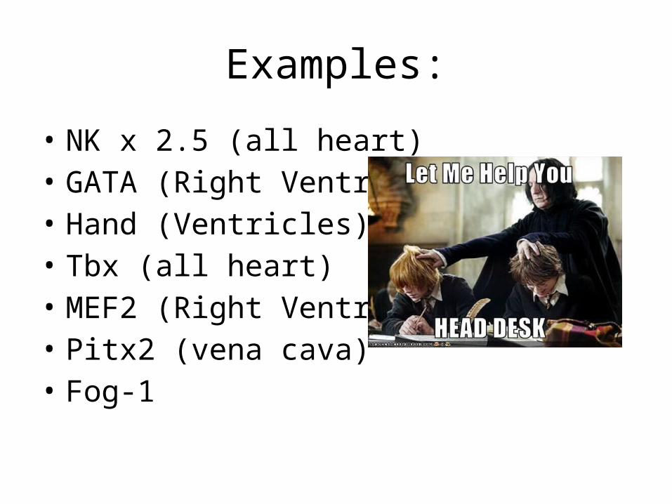
Examples:
• NK x 2.5 (all heart)• GATA (Right Ventricle)• Hand (Ventricles)• Tbx (all heart)• MEF2 (Right Ventricle)• Pitx2 (vena cava)• Fog-1

3 stages of cardiac formation
1. Formation of the primitive heart tube
2. Cardiac looping
3. Cardiac Septation

Forming the heart tube
• 3rd week of development the heart is formed from the cardiogenic region (look like a horse shoe)

Day 19
• 2 endocardial tubes form
• They then fuse to form a single primitive heart tube (GATA)

Endocardial tube
• Becomes surrounded by myocardium and the visceral layer of the serious pericardium
• Cephalic end = Arterial end• Causal end = Venous end• Arterial end is beyond the pericardium with a
large vessel – Aortic sac• Heart beats at day 22• Nkx2.5 increases heart size
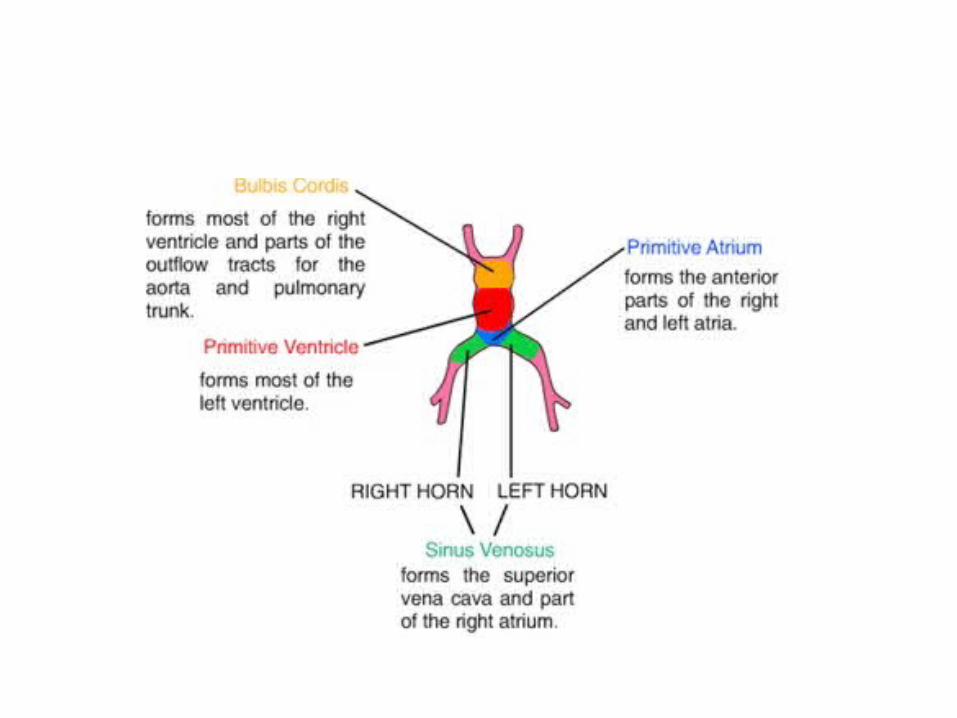

Cardiac Looping – Day 23
• Bulbis Cordis moves inferiorly, anteriorly and to the embryos right
• (like down, in front and right)• ORANGE Cha Cha slide

Continued
• The primitive ventricle moves to the embryos left side
• (RED Box to the left)

Continued
• Primitive atrium and sinus venousus move superiorly and posterior
• (Green and blue Love lifts us up where we belong)
• The sinus venousus is posterior to the primative atrium

So far
• Cha Cha slide• Box to the left• Love lifts us up

Atrioventricular canal
• There is only one common atrium and one ventricle at the moment
• Connected by atrioventricular canal

Endocardial cushions!
• There are two, the superior (on top) and the inferior (bottom)
• They grow together whilst the canal is repositioning itself to the right side of the heart
• They fuse to form two separate openings, left and right canals.

Side view

Cardiac Septation
• Week 4 (this is going on at the same time as the growth etc)
• Septum primum (crescent-shaped tissue) grows towards the endocardial tissues
• The opening between the septum primum and the endocardial cushion is called the foramen primum. Shunting
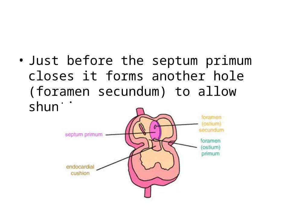
• Just before the septum primum closes it forms another hole (foramen secundum) to allow shunting.

• The second bit of tissue grows towards the endocardial cushions = Septum secundum
• Thick muscular compared to thin first one

End of week 6
• Septum secundum finishes growing• Permanent opening on posterior-inferior
surface• Foramen Ovale

• The septum primum gradually degenerates• The lower part remains as the valve of the
foramen ovale• It closes when
baby is born• Fossa ovalis

Embryology of Circulation

Formation of blood islands
“aggregations of mesenchymal cells in the angioblast of the early embryo, developing into vascular endothelium and blood cells.”

Vascularisation
• Day 17-21• Vascularisation of yolk sac, chorionic villus
(absorbs oxygen) and stalk• Angioblasts from mesoderm
join together to form angioblastic cords through the embryonic disc

Vasculogenesis/Angiogenesis
• Day 18 onwards• Driven by growth factors• Proliferation and sprouting • Mesodermal cells are recruited

Growth factors
• Make things grow• Angiogenic growth factors– vascular endothelial growth factor,– angiopoietin 1 & 2
• Repulsive signals– Plexin / semaphorin signalling, – ephrin / Eph interactions

Aortic arches: 1st and 2nd
Become minor head vessels
1st – small part of maxillary
2nd - artery to stapedius

3rd Aortic arch
• Become common carotid arteries, and proximal internal carotid arteries
• Distal internal carotids come from extension of dorsal aortae

Right dorsal Aortae and Right 4th• R dorsal aorta looses
connections with midline aorta and 6th arch, remaining connected to R 4th arch
• Acquires branch 7th cervical intersegmental artery, which grows into R upper limb
• Right subclavian artery is derived from right 4th arch, right dorsal aorta, and right 7th intersegmental artery

Left dorsal Aortae and left 4th• Left dorsal aorta continues
into trunk• Left 7th cervical
intersegmental artery, which grows into left subclavian artery
• Right subclavian artery is derived from right 4th arch, right dorsal aorta, and right 7th intersegmental artery

5th arches
There are none. HA

6th arches
• Right arch may form part of pulmonary trunk
• Left arch forms ductus arteriosus – communication between pulmonary artery and aorta

This is what it looks like:

Congenital abnormalities

Odd one out?

Ventricular Septal Defect
• Abnormal connection between the two ventricles
• 20% of all heart defects• Many close on their own in childhood

VSD
• High pressure in the left ventricle• Low pressure in the right ventricle• Blood flows from high to low• Some oxygenated blood gets into RV• Increased blood flow to the lungs

VSD- Size of holes
• Large• High pulmonary flow in
infancy• Breathlessness• Poor feeding• Need it fixed (PA band/
repair)• Eisenmenger’s
Syndrome
• Small• Small increase in
pulmonary flow• Risk of endocarditis• No intervention needed

Clinical signs
• Large• Small skinny breathless• Increased resp. rate• Tachycardia• Big heart• Murmur varies
• Small• Loud systolic murmur• Thrill (buzzing)• Well grown• Normal heart rate and
size

Eisenmenger's syndrome
• High pressure pulmonary blood flow• Damage to pulmonary vasculature• Resistance in lungs increase• Right ventricle pressure increases• Blood flow reverses • Shunt• BLUE

Atrial Septal Defects
• Abnormal connection between two atria• Septum primum/secundum or sinus venousus• Common• Present in adulthood mostly• Slightly higher pressure
in the left atrium than the right atrium
• Left to right shunt• NOT BLUE

ASD- Size of holes
• Large• Increased flow to the
right heart and lungs• Right heart dilation• Increased chest
infections• SOBOE• Should be closed if its
stretching the heart
• Small• Small increase in flow• No right heart dilation• No symptoms• Leave it alone!

Clinical signs of ASD’s
• Pulmonary flow murmur• Fixed split second heart sound (delayed
closure of pulmonary valve)• BIG pulmonary arties on X ray• BIG heart• Close:– Surgical– Percutaneous (umbrella)

Atrioventricular Septal Defects
• 2 in 10000 live births (common in downs)• Hole at very centre of heart• Involves Atrial septum, ventricular septum,
mitral and tricuspid valves• Complete or partial• Just one massive malformed
valve• Leaky

AVSD

AVSD Types
• Complete• Breathless as a neonate• Poor weight gain• Poor feeding• Torrential pulmonary
blood flow• Repair/PA band in
infancy• Surgically challenging
• Partial • Present in late
childhood• Presents as a small
ASD/VSD• Leave it alone if not
right heart dilation

Patent Ductus Arteriosus
• Aortic arch joined to the pulmonary artery

PDA- Size of holes
• Large• Torrential flow from
aorta to pulmonary artery
• Breathlessness• Poor feeding• Common in premature
babies• Surgically close
• Small• Little flow from aorta to
pulmonary artery• Murmur• Risk of endocarditis
Clinical Signs and Treatment
• Signs• Continuous machinery
murmur• Big heart and breathless• Eisenmenger’s
syndrome• Blue• Clubbing
• Treatments• Surgical or
Percutaneous• Local anaesthetics• Venous approach (AV
loop)• Low risk operation

Coarctation of the Aorta
• Narrowing of the aorta at the side of insertion of the Ductus Arteriosus

Type of CA
• Severe• Obstruction to aortic
flow• Collapse• Heart failure• Needs urgent repair
• Mild• Presents with
hypertension• Incidental murmur• Repair to prevent long
term problems

Clinical Signs and Long Term Problems
• Signs• Right arm hypertension• Bruits (buzzes) over the
scapulae and back from collateral vessels
• Murmurs
• Problems• Hypertension (stroke,
CAD, sub arachnoid haemorrhage)
• Recoarctation • Aneurysm at site of
repair

Coarctation repair
• Surgical vs Percutaneous• Subclavian flap• End to end repair• Coarctation angioplasty

Bicuspid Atrioventricular Valves
• Normally they have 3• This time there’s only 2• Severely stenotic• Degenerate quicker• Regurgitate earlier• Association with coarctation and dilation of
ascending aorta

Tetralogy of Fallot
1. Ventricular Septal Defect2. Pulmonary Stenosis3. Hypertrophy of Right Ventricle4. Overriding Aorta• Shunt from RV to LV• BlueCause 10% of all heart defects

Clinical Signs and Repair
• Signs• Cyanosed• Clubbed• Harsh Systolic murmur
from the right ventricle outflow tract obstruction (RVOT)
• Repair• By the age of 2• Close the VSD• Resect the RVOT
obstruction• They may get
pulmonary valve obstruction in adult life

Pulmonary Stenosis
• Narrowing of the right ventricle outflow• Can be valvar, sub valvar, supra valvar, branch• 8-12% of all defects

Type of PS
• Severe• Right ventricle collapse
as a neonate• Poor pulmonary blood
flow• Right ventricular
hypertrophy• Tricuspid regurgitation
• Moderate/mild• Well tolerated• Right ventricular
hypertrophy

Treatment
• Balloon valvuloplasty• Open valvostomy• Open trans-annular patch• Shunt to bypass the blockage

Thanks for staying with me!