
Cardiovascular Effects of Mechanical Ventilation
12
Cardiovascular Effects of Mechanical Ventilation G. J. DUKE Intensive Care Department, The Northern Hospital, Epping, VICTORIA ABSTRACT Objective: To review the cardiovascular effects of spontaneous breathing and mechanical ventilation in healthy and pathological states. Data sources: A review of articles published in peer-reviewed journals from 1966 to 1998 and identified through a MEDLINE search on cardiopulmonary interaction. Summary of review: Respiration has a hydraulic influence upon cardiovascular function. Pulmonary and cardiac pathology alter this interaction. Spontaneous inspiration increases right ventricular (RV) preload and left ventricular (LV) afterload. Mechanical ventilation with positive pressure (MV) reduces LV preload and afterload. The influence of MV upon the cardiovascular system (CVS), particularly in critically ill patients, depends upon the mode of ventilation and the pre-existing cardiac and respiratory status. The influence of these factors is reviewed. Consideration of these parameters will enable the clinician to predict the likely effect of MV and develop strategies to minimise adverse events. Conclusions: Mechanical ventilation has an adverse effect upon the CVS in healthy subjects and in patients with pulmonary pathology, particularly in the presence of preload-dependent LV dysfunction or afterload-induced RV dysfunction. Mechanical ventilation may benefit cardiac function in patients with respiratory failure and afterload-dependent or exercise-induced LV dysfunction. (Critical Care and Resuscitation 1999; 1: 388-399) Key Words: Mechanical ventilation, cardiovascular physiology, cardiopulmonary interaction, acute respiratory distress syndrome Respiration and circulation are complementary physiological processes that interact with each other during spontaneous breathing. 1,2 The introduction of mechanical ventilation (MV), or the presence of pulmonary and cardiac disease, increases the complex- ity of this interaction. Research in this area is daunting and interpretation of the data is difficult because these and other interrelated variables must be considered. What is the predominant mechanism underlying this interaction? Explanations have included mechanical (hydraulic), 1 neural, 3 and humoral mechanisms. 4 Phasic variation in cardiac function during respiration is closely linked in time and magnitude to changes in intrathoracic pressure and occurs more rapidly than most neural or humoral processes. Therefore current data support the hydraulic effect of intrathoracic (pleural) pressure (P pl ) on cardiac transmural pressure (P tm ) as the predominant mechanism. 1-3,5 To appreciate the complex cardiovascular effects of MV, especially in critically ill patients, it is prudent first to identify the cardio-respiratory interactions of spontaneous breathing in healthy subjects and in those with respiratory and/or cardiac disease, and then consider the influence of MV in each situation. Consideration of cardiovascular and respiratory changes over time is also important. An understanding of these interactions should enable the clinician to predict the likely cardiovascular effect of MV in a given clinical situation. Thoracic anatomy and transmural pressure Respiration induces phasic swings in cardiac trans- Correspondence to: Dr. G. J. Duke, Intensive Care Department, The Northern Hospital, Epping, Victoria 3076 (e-mail: [email protected]) 388
Transcript of Cardiovascular Effects of Mechanical Ventilation

Cardiovascular Effects of Mechanical Ventilation G. J. DUKE Intensive Care Department, The Northern Hospital, Epping, VICTORIA
ABSTRACT Objective: To review the cardiovascular effects of spontaneous breathing and mechanical ventilation in healthy and pathological states. Data sources: A review of articles published in peer-reviewed journals from 1966 to 1998 and identified through a MEDLINE search on cardiopulmonary interaction. Summary of review: Respiration has a hydraulic influence upon cardiovascular function. Pulmonary and cardiac pathology alter this interaction. Spontaneous inspiration increases right ventricular (RV) preload and left ventricular (LV) afterload. Mechanical ventilation with positive pressure (MV) reduces LV preload and afterload. The influence of MV upon the cardiovascular system (CVS), particularly in critically ill patients, depends upon the mode of ventilation and the pre-existing cardiac and respiratory status. The influence of these factors is reviewed. Consideration of these parameters will enable the clinician to predict the likely effect of MV and develop strategies to minimise adverse events. Conclusions: Mechanical ventilation has an adverse effect upon the CVS in healthy subjects and in patients with pulmonary pathology, particularly in the presence of preload-dependent LV dysfunction or afterload-induced RV dysfunction. Mechanical ventilation may benefit cardiac function in patients with respiratory failure and afterload-dependent or exercise-induced LV dysfunction. (Critical Care and Resuscitation 1999; 1: 388-399)
Key Words: Mechanical ventilation, cardiovascular physiology, cardiopulmonary interaction, acute respiratory distress syndrome
Respiration and circulation are complementary physiological processes that interact with each other during spontaneous breathing.1,2 The introduction of mechanical ventilation (MV), or the presence of pulmonary and cardiac disease, increases the complex-ity of this interaction. Research in this area is daunting and interpretation of the data is difficult because these and other interrelated variables must be considered. What is the predominant mechanism underlying this interaction? Explanations have included mechanical (hydraulic),1 neural,3 and humoral mechanisms.4 Phasic variation in cardiac function during respiration is closely linked in time and magnitude to changes in intrathoracic pressure and occurs more rapidly than most neural or humoral processes. Therefore current data support the hydraulic effect of intrathoracic (pleural) pressure (Ppl)
on cardiac transmural pressure (Ptm) as the predominant mechanism.1-3,5 To appreciate the complex cardiovascular effects of MV, especially in critically ill patients, it is prudent first to identify the cardio-respiratory interactions of spontaneous breathing in healthy subjects and in those with respiratory and/or cardiac disease, and then consider the influence of MV in each situation. Consideration of cardiovascular and respiratory changes over time is also important. An understanding of these interactions should enable the clinician to predict the likely cardiovascular effect of MV in a given clinical situation. Thoracic anatomy and transmural pressure Respiration induces phasic swings in cardiac trans-
Correspondence to: Dr. G. J. Duke, Intensive Care Department, The Northern Hospital, Epping, Victoria 3076 (e-mail: [email protected])
388
Critical Care and Resuscitation 1999; 1: 388-399 G. J. DUKE
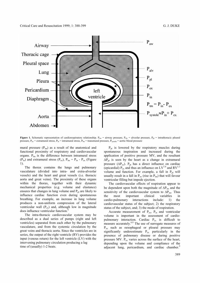
Figure 1. Schematic representation of cardiorespiratory relationship. Paw = airway pressure, Palv = alveolar pressure, Ppl = intrathoracic pleural pressure, Pex = extramural stress, Pin = intramural stress, Ptm = transmural pressure, Psystole = aortic blood pressure mural pressure (Ptm) as a result of the anatomical and functional proximity of respiratory and cardiovascular organs. Ptm is the difference between intramural stress (Pin) and extramural stress (Pex), Ptm = Pin - Pex (Figure 1). The thorax contains the lungs and pulmonary vasculature (divided into intra- and extra-alveolar vessels) and the heart and great vessels (i.e. thoracic aorta and great veins). The proximity of these organs within the thorax, together with their dynamic mechanical properties (e.g. volume and elastance) ensures that changes in lung volume and Ppl are likely to influence cardiac function even during spontaneous breathing. For example, an increase in lung volume produces a non-uniform compression of the lateral ventricular wall (Pex) and, although low in magnitude does influence ventricular function.6 The intra-thoracic cardiovascular system may be described as a dual series of pumps (right and left ventricles) separated from each other by the pulmonary vasculature, and from the systemic circulation by the great veins and thoracic aorta. Since the ventricles are in series, the output of the right ventricle (RV) provides the input (venous return) for the left ventricle (LV) with the intervening pulmonary circulation producing a lag time of (usually) 1-2 beats.
Ppl is lowered by the respiratory muscles during spontaneous inspiration and increased during the application of positive pressure MV, and the resultant ∆Ppl is seen by the heart as a change in extramural pressure (∆Pex). Ppl has a direct influence on cardiac (epicardial) Pex and thus an influence on LV7,8 and RV1,3 volume and function. For example, a fall in Ppl will usually result in a fall in Pex (rise in Ptm) that will favour ventricular filling but impede ejection. The cardiovascular effects of respiration appear to be dependent upon both the magnitude of ∆Ptm and the sensitivity of the cardiovascular system to ∆Ptm. Thus the most important clinical variables in cardio-pulmonary interactions include: 1) the cardiovascular status of the subject; 2) the respiratory status of the subject, and; 3) the mode of respiration. Accurate measurement of Pex, Pin and ventricular volume is important in the assessment of cardio-pulmonary interaction. Cardiac Pex is difficult to measure accurately.6,9 The use of surrogate measures of Pex such as oesophageal or pleural pressure may significantly underestimate Pex particularly in the presence of pulmonary disease or during positive pressure MV. Pex varies across the surface of the heart, depending upon the volume and compliance of the adjacent lung, pericardium, and cardiac chamber.9
389

G. J. DUKE Critical Care and Resuscitation 1999; 1: 388-399
Indirect measurement of cardiac Pin (e.g. estimation of left ventricular end-diastolic Pin by pulmonary artery catheter) may also be misleading.10-13 More direct measurements of Pex ,Pin, and chamber volume are preferred but usually restricted to animal models. Ventricular interdependence (VI) is another important physiological concept, anatomically based on the adjacent LV and RV sharing a common pericardial sac (with limited volume and compliance) and a common interventricular septum. VI is commonly used to imply that the pressure/volume characteristics of the RV influence those of the LV.7,14-16 Cardiovascular effects of spontaneous breathing To understand the cardiovascular changes resulting from MV it is important to first understand cardio-pulmonary interactions during spontaneous breathing. This topic has been extensively reviewed elsewhere2,3 and whilst much of the data comes from animal (usually dog) models there is a substantial agreement with available human data.17
Figure 2. Schematic representation of cardiorespiratory interaction during spontaneous inspiration and cardiac systole. Arrows depict direction of change. Spontaneous inspiration increases Ptm and reduces Psystole. Paw = airway pressure, Palv = alveolar pressure, Ppl = intrathoracic pleural pressure, Pex = extramural stress, Pin = intramural stress, Ptm = transmural pressure, Psystole = aortic blood pressure, LV = left ventricle
Spontaneous inspiration in healthy subjects is usually associated with a small fall in systolic blood pressure (< l0 mmHg). Explanations for this observation include: 1) an increase in LV afterload, 2) a decrease in venous return, 3) the influence of VI, and, 4) transmission of reduced intra-thoracic aortic Pex to extrathoracic vessels. Both animal and human data indicate that the first explanation is probably the most important mechanism.1-
3,7
Under certain pathological states this inspiratory blood pressure fall is exaggerated and referred to as ‘pulsus paradoxus’.3,20,21 This may be found where there is: 1) a greater inspiratory effort and a greater ∆Ppl and ∆Ptm (e.g. acute asthma22,23 or pulmonary oedema24), or: 2) an increased sensitivity to ∆Ptm (e.g. hypovolaemia,25 tamponade,3,21 or congestive cardiac failure26). Inspiration occurs as a result of the reduction in Ppl
which is transmitted to the intra-thoracic organs. Whilst the primary purpose of this pressure fall is to expand the lungs it is also results in a fall in cardiac Pex (and a rise in Ptm). The increase in Ptm facilitates right ventricular diastolic filling - the ‘thoracic pump’ mechanism - until the closing pressure of the extra-thoracic veins is reached1,3 and the resultant increase in RV end-diastolic volume (RVEDV) increases stroke volume via the Frank-Starling mechanism.7,16,18 The subsequent rise in pulmonary flow increases left ventricular end diastolic volume (LVEDV) after a delay of 1-2 beats3,19 - a duration similar to the inspiratory phase of the respiratory cycle. This may partially account for the rise in blood pressure observed during expiration.
During exhalation systolic blood pressure rises as a result of the return of left ventricular afterload to baseline and aided by the augmentation of LV preload from the (delayed effect of the) inspiratory rise in RV output. There is little evidence to suggest other mechanisms have a significant influence. The role of VI is equivocal.1 Although there is little doubt that increasing RV volume can induce leftward septal shift and increase LVEDP, there is little data to support a significant influence during spontaneous breathing14,15 and disagreement as to its clinical significance even in pathological states. Spontaneous breathing has little direct effect on the vasculature. Healthy subjects have a low pulmonary vascular resistance (PVR) which does not appreciably alter during tidal breathing.2,27 There is also little evidence to suggest intra-pulmonary pooling of blood during inspiration. Although the extra-alveolar pulmonary blood volume increases, this is opposed by a fall in intra-alveolar blood volume.1,28 Whilst inspiratory changes in aortic Ptm are transmitted to the extra-
Spontaneous inspiration induces an increase in LV afterload that accounts for the observed inspiratory fall in stroke volume and systolic blood pressure. During inspiration left ventricular Pex, falls and Ptm, rises. Since this ∆Ptm must be ‘overcome’ during systole it creates, by definition, an increase LV afterload. As a result, LV stroke volume and systolic blood pressure are observed to fall and LV end-systolic volume rises.1-3,5,6,16 (Figure 2)
390

Critical Care and Resuscitation 1999; 1: 388-399 G. J. DUKE
thoracic vessels these are inadequate to explain the observed respiratory swing in systolic blood pressure. In summary, there are two predominant cardiovascular effects of spontaneous inspiration: 1) a rise in LV afterload and, 2) a rise in RV preload. An inspiratory fall in systemic blood pressure is produced by the former and limited by the latter. Factors that will potentially accentuate blood pressure fluctuation during spontaneous breathing include: 1) a fall in preload (e.g. hypovolaemia, venodilation),
or; 2) a rise in left ventricular afterload as a result of
increased inspiratory effort (e.g. bronchospasm3 or spontaneous breathing through an endotracheal tube29).
The cardiovascular effects of mechanical ventilation in healthy subjects Like spontaneous breathing, MV is associated with an inspiratory fall in aortic flow and systolic blood pressure, but the mechanism is quite different.2,30 Unlike spontaneous breathing, MV produces a positive ∆Ppl during inspiration whose cardiovascular effects are the opposite to those seen with the negative ∆Ppl of spontaneous breathing. Proposed mechanisms for the blood pressure fall with MV include: 1) reduced LV preload, 2) reduced RV preload, 3) increased PVR and RV impedance, and 4) ventricular interdependence. Most of these mechanisms are present in varying degrees but the predominant effect is a reduction in LV preload.5,31,32 With the commencement of MV, the positive airway pressure results in a fall in right ventricular Ptm and a fall in venous return (or loss of the ‘thoracic pump’), causing a reduction in RVEDV (preload).18,32,33. Positive airway pressure (Paw) is also transmitted to the pulmonary vasculature inducing a (small) rise in pulmonary artery pressure (Ppa) and RV impedance (afterload), and so with time RVEDV returns to the baseline.32 Although Ppa rises, PVR does not - unless intra-thoracic pressure or volume are unusually high.34,35 Thus MV induces a fall in transpulmonary flow and venous return to the LV, reducing LV preload (Figure 3). In contrast to the transient changes in RVEDV, MV produces a sustained reduction in LVEDV because both afterload36,37 and preload38 are reduced. Both aortic pressure and cardiac output fall progressively during inspiration and can be (partially or wholly) reversed by volume loading,18,32 suggesting the fall in preload is more influential. Compensatory increases in sympathetic
drive induce tachycardia, vasoconstriction, oliguria and retention of sodium and water.
Figure 3. Schematic representation of cardiorespiratory interaction during positive pressure inspiration and diastole. Arrows depict direction of change. Inspiration decreases venous return and RVEDV. Paw = airway pressure, Palv = alveolar pressure, Ppl = intrathoracic pleural pressure, Pex = extramural stress, Pin = intramural stress, Ptm = transmural pressure, CVP = central venous pressure, RA = right atrium, RV = right ventricle, IVC = inferior vena cava There is little evidence to support a major influence from ventricular interdependence (VI).9 While a reduction in the LV septal-lateral wall dimension during MV has been reported by early investigators,14,39 recent investigators have either not demonstrated the presence of VI during MV,32,34 or found the reduction in LV dimensions were symmetrical, and no greater in the septal-free wall axis.9 Some even suggest that asymmetrical reduction in the septal-free wall axis would also be consistent with increased lateral wall Pex from the inflation of the adjacent lung and not from VI.40,41 Finally, a number of researchers have demonstrated preservation of LV pressure-volume relationships during MV implying that any effect from VI is clinically insignificant.9,42 By maintaining some spontaneous inspiratory effort, assisted modes of ventilation such as CPAP, PSV, BiPAP and even SIMV, will tend to produce a lower mean Paw and Ppl compared with controlled modes of MV for a similar level of minute volume. These modes have been shown to reduce the adverse cardiovascular effects of MV.18,43 However, assisted modes of respiratory support are unsuitable if the patient has severe lung disease or an inadequate respiratory drive (e.g. from sedation, anaesthesia, or coma) or is at risk from respiratory muscle fatigue (e.g. a high respiratory rate).
391

G. J. DUKE Critical Care and Resuscitation 1999; 1: 388-399
Mechanical or extrinsic PEEP (PEEPe) is commonly used to recruit alveoli, defend end-expiratory lung volume (EELV), and improve oxygenation during MV. PEEP increases mean Paw and (usually) Ppl and thus the haemodynamic effects are similar to MV. Some data support the potential for humoral and neural mediated effects of PEEP on cardiac function in addition to its hydraulic effects.4,9 The concept of ‘best PEEP’ is based upon the balance between the respiratory benefits of PEEP and its adverse cardiovascular (and respiratory) effects.44 Factors that may accentuate the haemodynamic effects of MV in healthy subjects include: 1) a decrease in venous return25,38 (e.g. hypovolaemia,
venodilation), 2) an increase in mean intra-thoracic pressure (e.g. the
use of large tidal volume or high PEEPe), or 3) blunting of compensatory sympathetic reflexes (e.g.
anaesthesia, sedatives). Strategies to minimise these effects include: 1) the use of volume expansion to restore LV preload, 2) the use of assisted modes of ventilation to reduce
∆Ppl, and 3) the avoidance of high mean intra-thoracic pressure
(Ppl) that may occur with a high minute volume, high inspiratory flow or PEEPe.
Cardio-pulmonary interaction in the presence of pulmonary disease Pulmonary disease alters lung mechanics in a number of ways which alter the cardio-pulmonary interactions observed during spontaneous breathing. The important changes in lung mechanics include those affecting lung volume, airflow resistance, minute volume (Ve), work of breathing (WB), and RV impedance. Reduced lung volume Many pulmonary diseases are associated with a reduction in lung compliance and volume. This may occur as a result of 1) bronchial obstruction (e.g. inflammation, secretions), 2) an increase in closing volume (e.g. lung disease, elderly), 3) a reduction in functional residual capacity (FRC, e.g. anaesthesia, supine posture, abdominal and thoracic trauma), or 4) an increase in lung elastance (eg. pulmonary oedema, pneumonia, ARDS). A decrease (or an increase) in lung volume will increase PVR and RV impedance (i.e. afterload).2,27,30 A pathological increase in lung volume most often occurs in the presence of airflow limitation (see later). A reduction in lung volume increases the resistance in extra-alveolar pulmonary vessels1,27 due to hypoxic vasoconstriction,35,45 structural distortion,1,45 and in
some cases vasoconstrictor mediators (e.g. thromboxanes).30,45,46 PVR has an inverse hyperbolic relationship with the lung volume (FRC)2 so that in severe cases, a marked rise PVR may induce RV systolic dysfunction.45 Acute respiratory failure resulting from extensive alveolar collapse (e.g. post-operative respiratory failure) may necessitate the use of MV for respiratory support. In this setting MV may increase both Ppa and PVR through hyperinflation of healthy lung units.9,30,34 The use of PEEPe to recruit collapsed lung units may exacerbate these effects.47-49 The adverse haemodynamic effects of MV seen in healthy subjects are thus accentuated in these patients and strategies to recruit and defend EELV tend to oppose those strategies which minimise the adverse cardiovascular effects of MV.2,9 MV strategies to defend cardiac output in the face of reduced lung elastance and volume include: 1) attempts to minimise any unnecessary elevation in
Ppl that may occur with high tidal volume (Vt), Ve, and inspiratory flow rates, and the judicious use of PEEPe,
2) alveolar recruitment and reversal of hypoxia and acidosis in an effort to reduce PVR,
3) careful use of volume expansion to improve LV preload but avoid pulmonary oedema, and,
4) use of inotropic agents to support cardiac output. In theory, the use of assisted non-invasive modes of ventilation (e.g. CPAP, BiPAP, PSV) to recruit alveoli and provide respiratory support whilst minimising the adverse cardiovascular effects of MV is attractive.50 However, there is little data to support a successful therapeutic role for this modality in these patients.43 Airflow limitation and dynamic hyperinflation Airflow limitation (e.g. asthma, COPD) prolongs the expiratory time and opposes alveolar deflation resulting in an increase in EELV (i.e. dynamic hyperinflation) and the creation of intrinsic PEEP (PEEPi). Expiratory Ppl and Pex will rise as a result of PEEP. These mechanical effects reduce LV (and RV) preload.22,51 Dynamic hyperinflation and inspiratory airflow limitation both increase the inspiratory effort to maintain alveolar ventilation. The elevated lung elastance of high lung volume, together with high inspiratory flow resistance, increases work and the ∆Ppl required to maintain alveolar ventilation. Thus the LV afterload effect (of inspiratory ∆Ppl) is exaggerated in the presence of airflow limitation, producing a greater fall in systolic blood pressure and ‘pulsus paradox-us’.22,51 A number of other factors may reduce RV output and trans-pulmonary flow in the presence of airflow limitation. Although heightened inspiratory effort will tend to increase RV preload, venous return is limited by
392

Critical Care and Resuscitation 1999; 1: 388-399 G. J. DUKE
the collapse of the extra-thoracic veins.2,3 PEEPi will further decrease venous return. Over-inflation of alveoli during inspiration compresses intra-alveolar vessels and (along with other changes in pulmonary vasculature51) increases PVR and RV impedance.2,23,35 With severe airflow obstruction the reduction in Ppl and Pex may be so severe as to cause pulmonary23 and aortic flow to fall dramatically, or hydrostatic pulmonary oedema to occur.52 Alternatively, dynamic hyperinflation and airflow limitation may increase WB
51 to the extent that respiratory fatigue leads to hypercarbia and a respiratory arrest. These mechanisms may account for unexplained sudden death in some asthmatics.53 Great care must be taken when instituting MV in the presence of severe airflow limitation as MV may further increase dynamic hyperinflation and Ppl.54 Even in the presence of mild airflow limitation, the use of MV parameters which inadvertently shorten expiratory time (e.g. large Vt, high frequency, prolonged inspiratory time) may produce dynamic hyperinflation. Monitoring dynamic hyperinflation during MV is not difficult. With appropriate volume-measuring devices, the trapped volume (Vei) of dynamic hyperinflation can be measured during a prolonged expiratory pause (30-60 seconds). Data from Tuxen and Lane54 suggests that a Vei < 20 mL/kg should minimise the adverse haemodynamic effects of MV in the presence of severe airflow limitation. Alternatively, alveolar pressure (Palv) may be measured. In the presence of airflow limitation, Palv cannot be inferred from (upstream) Paw since the later will be higher than Palv during inspiration, and lower during exhalation.55 By temporarily adding an inspiratory or expiratory pause (0.5-1.0 seconds) Paw will more accurately indicate peak inspiratory Palv and end-expiratory Palv (or PEEPi) respectively. Strategies to minimise the haemodynamic effects of MV in patients with mild airflow limitation include the use of, 1) assisted modes of ventilation to reduce threshold
work, inspiratory effort and improve minute volume,43,56
2) MV parameters which reduce the inspiratory to expiratory time (I:E) ratio, and
3) avoidance of PEEPe in excess of PEEPi.57,58 However, in the presence of severe airflow limitat- ion it is often necessary to monitor trapped lung volume (Vei) and PEEPi, and to use a very low I:E ratio with controlled hypoventilation.54,59,60 Increased work of breathing (WB) Most pulmonary diseases are associated with an increase in minute volume (Ve) and/or an increase in respiratory effort per breath (WB).30,61 High Ve demand
may result from an elevation in metabolic rate or deterioration in gas exchange. Increased WB may result from increased lung elastance or airflow limitation.62 The metabolic (e.g. oxygen) cost of breathing - normally only 1-2% of total body oxygen consumption - may rise to as much as 20% in acute respiratory failure.61,65 Respiration may thus be viewed as a form of (cardiac and respiratory) exercise stress.30 A high level of WB may produce respiratory muscle fatigue and cause acute respiratory failure.66-68 Respiratory muscle blood flow may be significantly limited in the presence of cardiac failure, further exacerbating respiratory muscle fatigue, lactic acidosis and respiratory failure.69 In addition, the increased metabolic demand of respiration may induce myocardial ischaemia.70,71 In this setting, MV and muscle relaxation may reduce some of the adverse metabolic and cardiovascular stresses of spontaneous breathing and may improve outcome. 65,69,72-75 Increased right ventricular impedance Pulmonary hypertension (PHT) is a frequent complication of acute and chronic pulmonary disease. Particular mention is warranted here because severe PHT (mean Ppa > 30mmHg) may precipitate acute right heart failure.2,45,76,77 This may arise in such diverse conditions as pulmonary embolism, acute exacerbation of chronic lung disease, or the acute respiratory distress syndrome (ARDS). The compliant, thin-walled RV and pulmonary circulation usually operate as a low-pressure flow generator. PHT increases RV impedance (i.e. afterload), induces acute RV dilatation and, when severe, RV failure.76 This may significantly reduce pulmonary flow (i.e. LV preload) and precipitate systemic hypotension. The initial effects of MV will exacerbate this situation.49 The transmission of positive Palv via the intra-alveolar vessels will increase mean Ppa whilst compression of intra-alveolar vessels will increase PVR. In the long-term, however, MV may beneficially reduce PVR through the recruitment of collapsed lung units and reversal of acidotic and hypoxic pulmonary vasoconstriction.30,45,77 The combination of systemic hypotension and pulm- onary hypertension has an adverse effect on RV myocardial oxygen supply and demand.78,79 RV myocardial perfusion pressure (PRVM) is determined by the balance of the driving pressure (i.e. mean arterial pressure) and the opposing mean ventricular pressure (Pin,RV). The critical level for PRVM is around 25-40mmHg, below which right ventricular ischaemia and acute right heart failure may occur.79 PRVM = Pin,RV - MAP
393

G. J. DUKE Critical Care and Resuscitation 1999; 1: 388-399
Where Pin,RV = 1/3 (Ppa,sys - CVP) + CVP Ppa,sys = systolic pulmonary artery pressure MAP = mean arterial pressure CVP = central venous pressure. Strategies to avoid RV ischaemia include defence of RV preload (with volume loading), defence of MAP (with pressor agents)79,80 and monitoring of PRVM (e.g. via a pulmonary artery catheter). Unfortunately most inotropic agents also have some pulmonary vasoconstrictor properties, and those with significant vasodilatory properties (e.g. isoprenaline, dobutamine and milrinone) may be best avoided. Noradrenaline appears to have the best profile for the RV by producing less pulmonary vasoconstriction for similar levels of inotropic effect and support of myocardial perfusion.80,81 Acute lung injury (ALI), ARDS and MV Special mention of MV in acute lung injury (ALI) and ARDS is warranted because the complexity of the cardio-respiratory interaction of MV with lung disease is epitomised in a patient with ARDS. The dynamic interaction of pathological processes and pulmonary mechanics demonstrates that care should be taken to identify these variables when interpreting clinical and research data. The pathological changes in pulmonary and cardiovascular function with ALI and ARDS are documented elsewhere.46,82 In summary, all the mechanical factors discussed previously are seen in various degrees with ALI and ARDS. For example, increased elastance, alveolar collapse, loss of EELV, airflow limitation, pulmonary hypertension, and elevated metabolic rate and WB. Humoral inflammatory mediators produce pulmonary vasoconstriction, myocardial depression, and systemic hypotension. Control of oxygen flux, organ blood flow and cellular respiration may also be deranged. A number of different modes of MV may be utilised depending upon the stage and severity of ALI. These vary from non-invasive modes (e.g. face-mask PSV and CPAP) for ALI, through to assisted modes (e.g. SIMV and PSV) and to the extremes of (pressure or volume) controlled modes with significant levels of PEEPe being required for ARDS. ARDS is characterised by markedly increased lung elastance so that during MV, a greater inspiratory airway pressure (Paw,i) is required to maintain adequate alveolar ventilation, and high levels of PEEPe are often utilised to prevent airway collapse and aid recruitment of alveoli. Healthy lung units exposed to high Paw,i will be subject to overdistension. To the extent that Paw is transmitted to Ppl, MV will decrease Ptm and impair
cardiac function (see previously). Counteracting this is the fact that non-distensible (i.e. collapsed or consolidated) lung segments will ‘protect’ Ppl and cardiac Ptm from the elevation in Paw.8,34,83 Right ventricular dysfunction during MV is un-avoidable due to the combined effects of positive Paw and Ppl and the presence of PHT (which is often severe) and an elevated PVR.46 Not surprisingly, the cardiovascular effects of MV for ARDS are predominantly borne by the RV.82,84 RV dysfunction, elevated PVR and VI (if present), will all reduce LV preload. The effects of ARDS on LV function are determined by the combined effect of reductions in preload, afterload and contractility.85 Contractility may be impaired by humoral myocardial depressant factors and myocardial ischaemia.46 Peripheral vascular failure (systemic vasodilatation) reduces LV afterload and favours a hyperdynamic circulation so that the clinical observation of a warm periphery and a bounding pulse may mask the presence of significant LV dysfunction. The addition of MV has little cardiovascular benefit in ARDS.86 Strategies to minimise the haemodynamic effects of MV tend to oppose many of the ventilatory strategies used to overcome the pulmonary dysfunction of ARDS. Where possible MV parameters which minimise mean Paw and total PEEP (PEEPe plus PEEPi) will favour cardiac function as well as limiting ventilator-induced lung injury. Maintenance of RV preload (volume loading), RV contractility (inotropic agents) and systemic blood pressure (pressor agents), together with frequent clinical and haemodynamic assessment (e.g. pulmonary artery catheter, echocardiography, etc.) are essential. Clinical goals will often need to be guided by response to these therapeutic challenges and will differ for each patient depending on the severity and progression of the disease. The elusive search for an ideal MV mode for ARDS patients - which supports ventilation without adverse cardiac (and respiratory) side effects - has included the use of high frequency ventilation,87 inverse ratio ventilation,89 extra-corporeal oxygenation,90 partial liquid ventilation,91 inhaled pulmonary vasodilators (e.g. nitric oxide92 and prostaglandins93), prone ventilation, phasic abdomino-thoracic pressure support,74 and extra-thoracic negative-pressure ventilators.94,95 Many of these options are worthy of consideration in the presence of severe cardiovascular dysfunction in response to MV but evidence of improved outcome is lacking. Cardio-pulmonary interaction in the presence of cardiac disease Cardiovascular pathology is a common finding in patients who require MV. Some of these factors have
394

Critical Care and Resuscitation 1999; 1: 388-399 G. J. DUKE
MV may be beneficial in these patients by reducing metabolic demand and WB
65 and by favourably altering LV mechanics (e.g. decrease the afterload). Consistent with this hypothesis is the observation that patients with stress-induced ischaemia may experience difficulty in weaning from supported (MV) to spontaneous breathing.71,72,99,100 Even long-term nocturnal CPAP in severe cardiac failure patients (e.g. New York Heart Association class III & IV) has been shown to produce a sustained improvement in LV and respiratory reserve.101
already been mentioned. The effect of MV is particularly dependent upon the type and severity of cardiac dysfunction. When ventricular dysfunction is preload-dependent (e.g. hypovolaemia, ischaemia, restrictive cardiomyopathy, tamponade, and valvular stenoses), MV will generally cause a further reduction in cardiac output. Poor ventricular compliance (resulting from myocardial ischaemia, hypertrophy or fibrosis) is a particular situation that often causes a rise in Pin and Ptm but not preload (LVEDV). General measures to prevent hypotension include the use of MV parameters that minimise mean Paw together with volume loading and, when necessary, the use of inotropic agents.
In subjects with afterload-induced RV dysfunction (e.g. severe pulmonary hypertension, acute PE, COPD, or RV infarct) MV may also adversely effect the balance of RV oxygen supply and demand. As discussed above, treatment of reversible pulmonary vasoconstriction (e.g. from hypoxia or acidosis) and defence of coronary perfusion pressure with pressor agents may be beneficial.
When left ventricular dysfunction is afterload-dependent, MV may improve cardiac output. Two important groups are worthy of mention: 1) cardiac-induced acute respiratory failure (e.g. cardiogenic pulmonary oedema), and, 2) respiratory-induced acute cardiac failure in the presence of severe systolic dysfunction.
Acute cardiogenic pulmonary oedema most commonly occurs as a result of ischaemic LV diastolic dysfunction leading to a hydraulic shift of fluid from the intravascular compartment into the lung parenchyma.96 Increased inspiratory effort (WB) produces a large negative Ppl that increases LV afterload70 and may lead to respiratory muscle fatigue68,75 and respiratory acidosis. Myocardial ischaemia and afterload impair LV systolic function and cardiac output, initiating a compensatory sympathetic response. RV dysfunction may also occur as a result of adverse changes in afterload (e.g. hypoxic pulmonary vasoconstriction), preload (e.g. hypovolaemia), and myocardial oxygen supply (e.g. coronary hypoperfusion). MV is useful in cardiogenic pulmonary oedema because it reverses hypoxia, reduces metabolic demand and improves afterload72 (Figure 4). Many subjects with acute cardiogenic pulmonary oedema are hypovolaemic and due consideration must be given to the adverse preload effects of MV.97 Noninvasive modes of assisted breathing (e.g. face-mask CPAP) provide high inspired oxygen, reduce WB and improve LV afterload and have been shown to be equally effective as MV, with lower a rate of intubation, compared with pharmacological therapy alone.98
Figure 4. Schematic representation of the effect of positive pressure inspiration in the presence of systolic dysfunction. Arrows depict direction of change. Inspiration decreases afterload (Ptm) and increases aortic pressure (Psystole). Paw = airway pressure, Palv = alveolar pressure, Ppl = intrathoracic pleural pressure, Pex = extramural stress, Pin = intramural stress, Ptm = transmural pressure, LV = left ventricle Summary MV has significant haemodynamic side-effects, the nature of which depend upon the cardiac and respiratory status of the subject, the mode of ventilatory support and the ventilatory parameters chosen. In general, MV reduces RV and LV preload and improves LV afterload. MV will increase the risk of adverse cardiovascular effects in the presence of acute or chronic pulmonary disease, especially in association with preload-dependent LV function, or afterload-induced RV
Respiratory-induced acute cardiac failure occurs in the presence of severe systolic dysfunction (e.g. severe cardiomyopathy or ischaemic heart disease). Since spontaneous breathing is a form of exercise30 an increase in WB during respiratory failure will increase the metabolic (oxygen) demand of the respiratory muscles by as much as ten-fold.68,69,75 If myocardial reserve is insufficient to meet this demand acute myocardial ischaemia or systolic failure may occur.
395

G. J. DUKE Critical Care and Resuscitation 1999; 1: 388-399
15. Maughan WL, Kallman CH, Shoukas A. The effect of right ventricular filling on the pressure-volume relationship of the ejecting canine left ventricle. Circ Res 1981;49:382-388.
dysfunction. Optimal MV modes and parameters to minimise this will depend upon the pulmonary pathophysiology present, and may change over time in the same subject. Conversely, MV may benefit cardiac function in patients with respiratory failure associated with afterload-dependent or exercise-induced LV dysfunction.
16. Robotham JL, Lixfeld W, Holland L, MacGregor D, Bryan AC, Rabson J. Effects of respiration on cardiac performance. J Appl Physiol 1978;44:703-709.
17. Scharf SM, Brown R, Tow DE, Parisi AF. Cardiac effects of increased lung volume and decreased pleural pressure in man. J Appl Physiol 1979;47:257-262.
Received 22.2.99 18. Pinsky MR. Determinants of pulmonary arterial blood
flow variation during respiration. J Appl Physiol 1984;56:1237-1245.
Accepted 18.10.99 REFERENCES 19. Guntheroth WG, Gould R, Butler J, Kinnen E. Pulsatile
flow in pulmonary artery, capillary and vein in the dog. Cardiovasc Res 1974;8:330-337.
1. Permutt S. Wise RA. Handbook of Physiology - The Respiratory System III. Chapter 36 Mechanical interaction of respiration and circulation. Oxford University Press; p647-656.
20. Shabetai R. Fowler NO, Gueron M. The effects of respiration on aortic pressure and flow. Am Heart J 1963;65:525-533. 2. Pinsky MR. Heart Lung Interactions. Ch 29.
Pathophysiologic Foundations of Critical Care. 1993. Editors: Pinsky MR, Dhainaut J-F. Publisher: Williams & Wilkins.
21. Shabetai R, Fowler NO, Fenton JC, Masangkay M. Pulsus paradoxus. J Clin Invest 1965;44:1882-1898.
22. Jardin F, Farcot JC, Boisante L, Prost JF, Gueret P, Bourdarias JP. Mechanism of paradoxical pulse in bronchial asthma. Circulation 1982;66:887-894.
3. Wise RA, Robotham JL, Summer WR. Effects of spontaneous ventilation on the circulation. Lung 1981;159:175-186. 23. Jardin F, Dubourg O, Margairaz A, Bourdarias JP.
Inspiratory impairment in right ventricular performance during asthma. Chest 1987;92:789-795.
4. Grindlinger GA, Manny J, Justice R, Dunham B, Shepro D, Hechtman HB. Presence of negative inotropic agents in canine plasma during positive end-expiratory pressure. Circ Res 1979;45:460-467.
24. Sharp JT, Griffith GT, Bunnell IL et al. Ventilatory mechanics in pulmonary edema in man. J Clin Invest 1957;35:111-117. 5. Scharf SM, Brown R, Saunders N, Green LH.
Hemodynamic effects of positive pressure inflation. J Appl Physiol 1980;49:124-131.
25. Perel A, Pizov R, Cotev S. Systolic blood pressure variation is a sensitive indicator of hypovolaemia in ventilated dogs subject to graded haemorrhage. Anesthesiology 1987;67:498-502.
6. Lloyd TC. Respiratory system compliance as seen from the cardiac fossa. J Appl Physiol 1982;53:57-62.
7. Summer WR, Permutt S, Sagawa K, Shoukas AA, Bromberger-Barnea B. Effects of spontaneous respiration on canine left ventricular function. Circ Res 1979;45:719-728.
26. Naughton MT, Rahman MA, Hara K, Floras JS, Bradley TD. Effect of continuous positive airway pressure on intrathoracic and left ventricular transmural pressures in patients with congestive heart failure. Circulation 1995;91:1725-1731. 8. Robotham JL, Mitzner W. A model of the effects of
respiration on left ventricular performance. J Appl Physiol 1979;46:411-418.
27. Hughes JM, Glazier JB, Maloney JE, West JB. Effect of extra-alveolar vessels on distribution of blood flow in the dog lung. J Appl Physiol 1968;25:701-712. 9. Smith PK, Tyson GS, Hammon JW, et al.
Cardiovascular effects of ventilation with positive airway pressure. Ann Surg 1982;195:121130.
28. Brower R, Wise R, Hassapoyannes C, et al. The effect of lung inflation on pulmonary venous return. Federation Proc 1983;42:594. 10. Raper RF, Sibbald WJ. Misled by the Wedge? The
Swan-Ganz Catheter and Left Ventricular Preload. Chest 1986;89:427-435.
29. Bersten AD, Rutten AJ, Vedig AE, Skowronski GA. Additional work of breathing imposed by endotracheal tubes, breathing circuits, and intensive care ventilators. Crit Care Med. 1989;17:671-677.
11. Calvin JE, Driedger AA, Sibbald WJ. The hemodynamic effect of rapid fluid infusion in critically ill patients. Surgery 1981;90:61-76. 30. Pinsky MR. Clinical applications of cardiopulmonary
1997;48:587-603. 12. O'Quin R. Marini JJ. Pulmonary artery occlusion pressure: Clinical physiology, measurement and interpretation. Am Rev Resp Dis 1983;128:319-326.
31. Morgan BC, Martin WE, Hornbein TF, Crawford EW, Guntheroth WG. Hemodynamic effects of intermittent positive pressure respiration. Anesthesiology 1966;27:584-590.
13. Pinsky MR, Vincent J-L, De Smet J-M. Estimating left ventricular filling pressure during positive end-expiratory pressure in humans. Am Rev Respir Dis 1991;143:25-31.
32. Rankin JS, Olsen CO, Arentzen CE, et al. The effects of airway pressure on cardiac function in intact dogs and man. Circulation 1982;66:108-120. 14. Taylor RR, Covell JW, Sonnenblick EH, Ross J Jr.
Dependence of ventricular distensibility on filling of the opposite ventricle. Am J Physiol 1967;213:711-718.
396

Critical Care and Resuscitation 1999; 1: 388-399 G. J. DUKE
33. Cournand A, Motley HL, Werko L, et al. Physiological studies of the effect of breathing on cardiac output in man. Am J Physiol 1947;149:162-174.
34. Culver BH, Marini JJ, Butler J. Lung volume and pleural pressure effects on ventricular function. J Appl Physiol 1981;50:630-635.
35. Hakim TS, Chang HK, Michel RP. Pressure flow relationships in the pulmonary circulation during lung inflation. Federation Proc 1983;42:594.
36. Buda AJ, Pinsky MR, Ingels NB Jr, Daughters GT 2d, Stinson EB, Alderman EL. The effect of intrathoracic pressure on left ventricular performance. N Engl J Med 1979;301:453-459.
37. Fessler HE, Brower RG, Wise RA, Permutt S. Mechanism of reduced LV afterload by systolic and diastolic pleural pressure. J Appl Physiol 1988 65:1244-1250.
38. Beaussier M, Coriat P. Perel A, et al. Determinants of systolic pressure variation in patients ventilated after vascular surgery. J Cardiothor Vasc Anesth 1995;9:547-551.
39. Jardin F, Farcot JC, Boisante L, Curien N, Margairaz A, Bourdarias JP. Influence of positive end-expiratory pressure on left ventricular performance. N Engl J Med 1981;304:387-392.
40. Cassidy SS, Mitchell JH, Johnson RL Jr. Dimensional analysis of right and left ventricles during positive pressure ventilation in dogs. Am J Physiol 1982;242:H549-H556.
41. Cassidy SS, Ramanathan, M. Dimensional analysis of the left ventricle during PEEP: relative septal and lateral wall displacements. Am J Physiol 1984;246:H792-H805.
42. Fewell JE, Abendschein DR, Carlson CJ, Rapaport E, Murray JF. Continuous positive-pressure ventilation does not alter ventricular pressure-volume relationship. Am J Physiol 1981;241:H821-H826.
43. Duke GJ, Bersten AD. Non-invasive ventilation in adult acute respiratory failure. Part I. Critical Care & Resuscitation. 1999;1:187-198.
44. Grace MP, Greenbaum DM. Cardiac performance in response to PEEP in patients with cardiac dysfunction. Crit Care Med 1982;10:358-360.
45. Rattes M. Calvin JE. Acute Pulmonary Hypertension. Ch 19. Pathophysiologic Foundations of Critical Care. 1993. Editors: Pinsky MR, Dhainaut J-F. Publisher: Williams & Willkins.
46. Bersten AD, Sibbald WJ. Acute lung injury in septic shock. Critical Care Clinics, 1989;5:49-79.
47. Raper RF, Sibbald WJ. Increased right ventricular compliance in response to continuous positive airway pressure. Am Rev Respir Dis 1992;145:771-775.
48. Imai T, Uchiyama M, Maruyama N, Yoshikawa D, Fujita T. Influence of constant sustained positive airway pressure on right ventricular performance. Intensive Care Med 1993;19:8-12.
49. Zwissler B. Forst H. Messmer K. Local and global function of the right ventricle in a canine model of pulmonary microembolism and oleic acid edema:
influence of ventilation with PEEP. Anesthesiology 1990;73:964-975.
50. Bradley TD, Holloway RM, McLaugh. Cardiac output response to continuous positive airway pressure in congestive heart failure. Am Rev Respir Dis 1992;145:377-382.
51. Jardin F. Bourdarias J-P. Acute Asthma. Ch30. Pathophysiologic Foundations of Critical Care. 1993. Editors: Pinsky MR, Dhainaut J-F. Publisher: Williams & Wilkins.
52. Stalcup SA, Mellins RB. Mechanical forces producing pulmonary edema in acute asthma. New Engl J Med 1977;297:592-596.
53. Benatar SR. Fatal asthma. New Engl J Med. 1986;314:423-429.
54. Tuxen DV, Lane S. The effects of ventilatory pattern on hyperinflation, airway pressures, and circulation in mechanical ventilation of patients with severe airflow obstruction. Am Rev Repir Dis 1987;136:872-879.
55. Valta P, Corbeil C, Chasse M, Braidy J, Milic-Emili J. Mean airway pressure as an index of mean alveolar pressure. Effect of expiratory flow limitation. Am J Respir Crit Care Med 1996;153:1825-1830.
56. Keenan SP, Kernerman PD, Cook DJ, Martin CM, McCormack D, Sibbald WJ. Effect of noninvasive positive pressure ventilation on mortality in patients admitted with acute respiratory failure: a meta-analysis. Crit Care Med 1997;25:1685-1692.
57. Marini JJ. Should PEEP be used in airflow obstruction. Am Rev Respir Dis 1989;140:1-3
58. Tobin MJ, Lodato. PEEP, auto-PEEP, and waterfalls. Chest 1989;96:449-451.
59. Dariloi R. Peret C. Mechanical controlled hypoventilation in status asthmaticus. Am Rev Respir Dis 1984;129:385-387.
60. Tuxen DV. Detrimental effects of positive end-expiratory pressure during controlled mechanical ventilation of patients with severe airflow obstruction. Am Rev Respir Dis 1989;140:5-9.
61. Field S. Kelly SM, Macklem PT. The oxygen cost of breathing cardiorespiratory disease. Am Rev Respir Dis 1982;126:9-13.
62. Robertson CH, Foster GH, Johnson RL. The relationship of respiratory failure to the oxygen consumption of, lactate production by, and distribution of blood flow among respiratory muscles during increased inspiratory resistance. J Clin Invest 1977;59:31-42.
63. Viires N, Sillye G, Aubier M, Rassidakis A, Roussos C. Regional blood flow distribution in dog during induced hypotension and low cardiac output. J Clin Invest 1983;72:935-947.
64. Kanak R. Fahey PJ, Vanderwarf. Oxygen cost of breathing. Changes dependent upon mode of mechanical ventilation. Chest 1985;87:126-127.
65. Manthous CA, Hall JB, Kushner R, Schmidt GA, Russo G, Wood LD. The effect of mechanical ventilation on oxygen consumption in critically ill patients. Am J Respir Crit Care Med 1995;151:210-214.
397

G. J. DUKE Critical Care and Resuscitation 1999; 1: 388-399
82. Cook DJ, Raffin TA. Acute hypoxemic respiratory failure. Ch 25 Pathophysiologic Foundations of Critical Care. 1993. Editors: Pinsky MR, Dhainaut J-F. Publisher: Williams & Wilkins.
66. Moxam J. Respiratory muscle fatigue: mechanisms, evaluation and therapy. BJA 1990;65:43-53.
67. Cohen CA, Zagelbaum G, Gross D, Roussos C, Macklem PT. Clinical manifestations of inspiratory muscle fatigue. Am J Med. 1982;73:308-316. 83. Romand J-A, Shi W. Pinsky MR. Cardiopulmonary
effects of positive pressure ventilation during acute lung injury. Chest 1995;108:1041-1048.
68. Mancini DM, Henson D, LaManca J, Levine S. Evidence of reduced respiratory muscle endurance in patients with heart failure. J Am Coll Cardiol 1994;24:972-981.
84. Sibbald WJ, Driedger AA, Myers ML, Short AI, Wells GA. Biventricular function in the adult respiratory distress syndrome. Chest 1983;84:126-134. 69. Aubier M, Lecocguic Y, Murciano D, Pariente R.
Function of the respiratory muscles in acute cardiac decompensation. Schweiz Med Wochenschr 1985;115:190-193.
85. Dhainaut JF, Devaux JY, Monsallier JF, Brunet F, Villemant D, Huyghebaert MF. Mechanisms of decreased left ventricular preload during continuous positive pressure ventilation in ARDS. Chest 1986;90:74-80.
70. Scharf SM, Bianco JA, Tow DE, Brown R. The effects of large negative intrathoracic pressure on left ventricular function in patients with coronary artery disease. Circulation 1981;63:871-875.
86. Artigas A, Bernard GR, Carlet J. et al. The American-European Conference on ARDS, Part 2. Intensive Care Med 1998; 24:378-398. 71. Rasanen J. Nikki P. Heikkila J. Acute myocardial
infarction complicated by respiratory failure. Chest 1984;85:21-28
87. Standiford TJ, Morganroth ML. High-frequency ventilation. Chest 1989;96:1380-1389
88. Abraham E, Yoshihara G. Cardio-respiratory effects of pressure controlled inverse ratio ventilation in severe respiratory failure. Chest 1989;96:1356-1359.
72. Mathru M, Rao TL, El-Etr AA, Pifarre R. Hemodynamic response to changes in ventilatory patterns in patients with normal and poor ventricular reserve. Crit Care Med 1982;10:423-426. 89. Lessard MR, Guerot E, Lorino H, Lemaire F, Brochard
L. Effects of pressure controlled with different ratios versus volume controleed ventilation on respiratory mechanics, gas exchange, and hemodynamics in patients with adult respiratory distress syndrome. Anesthesiology 1994;80:983-991.
73. Mathru M. Editorial. Mechanical breath: non-pharmacological support for the failing heart. Chest 1984;85:1.
74. Pinsky MR, Summer WR. Cardiac augmentation by phasic intrathoracic pressure support in man. Chest 1983;84:370-375. 90. Gattinoni L, Agostini A, Damia G. et al. Hemodynamics
and renal function during low frequency positive pressure ventilation with extracorporeal CO2 removal. Intensive Care Med 1980;6:155-161.
75. Aubier M, Viires N, Syllie G, Mozes R, Roussos C. Respiratory muscle contribution to lactic acidosis in low cardiac output. Am Rev Respir Dis 1982;126:648-652.
91. Hirschl RB, Pranikoff T. Wise C, et al. Initial experience with partial liquid ventilation in adult patients with acute respiratory distress syndrome. JAMA 1996;275:383-390.
76. Squara P. Dhainaut J-F, Brunet F. Acute right ventricular failure. Ch 18. Pathophysiologic Foundations of Critical Care. 1993. Editors: Pinsky MR, Dhainaut J-F. Publisher: Williams & Wilkins.
92. Rossaint R, Falke KJ, Lopez F, Slama K, Pison U, Zapol WM. Inhaled nitric oxide for the adult respiratory distress syndrome. N Engl J Med 1993;328:399-405.
77. Pinsky MR. Determinants of Right Ventricular Performance. Ch 17. Pathophysiologic Foundations of Critical Care. 1993. Editors: Pinsky MR, Dhainaut J-F. Publisher: Williams & Wilkins. 93. Bone RC, Slotman G. Maunder R. et al. Randomised
double-blind multicentre study of prostaglandin E1 in patients with adult respiratory distress syndrome. Chest 1989;96:114-119.
78. Brooks H, Kirk ES, Vokonas PS, Urschel CW, Sonnenblick EH. Performance of the right ventricle under stress: relation to right coronary flow. J Clin Invest 1971;50:2176-2183. 94. Borelli M, Benini A, Denkewitz T, Acciaro C, Foti G,
Pesenti A. Effects of continuous negative extrathoracic pressure versus positive end-expiratory pressure in acute lung injury patients. Crit Care Med 1998;26:1025-1031.
79. Vlahakes GJ, Turley K, Hoffman JIE. The pathophysiology of failure in acute right ventricular hypertension: hemodynamic and biochemical correlations. Circulation 1981;63:87-95 95. Peters J, Hecker B, Neuser D, Schaden W. Regional
blood volume distribution during positive and negative airway pressure breathing in supine humans. J Appl Physiol 1993;75:1740-1747.
80. Ghiogne M, Girling L, Prewitt RM. Volume expansion versus norepinephrine in treatment of a low cardiac output complicating an acute increase in right ventricular afterload in dogs. Anesthesiology 1984;60:132-135.
96. Bersten AD, Holt AW. Acute cardiogenic pulmonary edema. Curr Opinion Crit Care 1995;1:410-419.
97. Fellahi JL, Valtier B, Beauchet A, Bourdarias JP, Jardin F. Does positive end-expiratory pressure ventilation improve left ventricular function? Chest 1998;114:556-562.
81. Ducas J, Duval D, Dasilva H, Boiteau P, Prewitt RM. Treatment of canine pulmonary hypertension: effects of norepinephrine and isoproterenol on pulmonary vascular pressure flow characteristics. Circulation 1987;75:235-242. 98. Bersten AD, Holt AW, Vedig AE, Skowronski GA,
Baggoley CJ. Treatment of severe cardiogenic
398

Critical Care and Resuscitation 1999; 1: 388-399 G. J. DUKE
pulmonary edema with continuous positive airway pressure delivered by face mask. New Engl J Med. 1991;325:1825-1830.
99. Richard C, Teboul JL, Archambaud F, Hebert JL, Michaut P, Auzepy P. Left ventricular function during weaning of patients with chronic obstructive pulmonary disease. Intensive Care Med 1994;20:181-186.
100. Lemaire F, Teboul J-L, Cinotti L, et al. Acute left ventricular dysfunction during unsuccessful weaning
from mechanical ventilation. Anesthesiology 1988;69:171-179.
101. Naughton MT, Liu PP, Bernard DC, Goldstein RS, Bradley TD. Treatment of congestive heart failure and Cheynes-Stokes respiration during sleep by continuous positive airway pressure. Am J Respir Crit Care Med 1995;151:92-97.
399


![Mechanical ventilation[1]](https://static.fdocuments.in/doc/165x107/55a516ae1a28abe77f8b46a8/mechanical-ventilation1.jpg)