Cardiopulmonary Resuscitation: Update, Controversies and ... · Cardiopulmonary Resuscitation:...
12
375 Hospital São Lucas da Pontifícia Universidade Católica do Rio Grande do Sul - Porto Alegre. Comissão de Ressuscitação Cardiorrespiratória - Cerebral. Mailing adress: Alexandre C. Zago - Rua Hilário Ribeiro, 144/201 - 90510-040 - Porto Alegre, RS. Alexandre C. Zago, Cristine E. Nunes, Viviane R. da Cunha, Euler Manenti, Luís Carlos Bodanese Porto Alegre, RS - Brazil Cardiopulmonary Resuscitation: Update, Controversies and New Advances Update Cardiopulmonary arrest is a medical emergency in which the lapse of time between event onset and the ini- tiation of measures of basic and advanced support, as well as the correct care based on specific protocols for each clinical situation, constitute decisive factors for a success- ful therapy. Cardiopulmonary arrest care cannot be restricted to the hospital setting because of its fulminant nature. This necessitates the creation of new concepts, strategies and structures, such as the concept of life chain, cardio- pulmonary resuscitation courses for professionals who work in emergency medical services, the automated external defibrillator, the implantable cardioverter- defibrillator, and mobile intensive care units, among others. New concepts, strategies and structures motivated by new advances have also modified the treatment and im- proved the results of cardiopulmonary resuscitation in the hospital setting. Among them, we can cite the concept of cerebral resuscitation, the application of the life chain, the creation of the universal life support algorithm, the adjust-ment of drug doses, new techniques – measure of the end-tidal carbon dioxide levels and of the coronary perfusion pressure - and new drugs under research. Cardiopulmonary resuscitation (CPR) was developed in a model similar to the current basic life support (BLS), in 1865, by a young medical officer of the U.S. Army – Charles Augustus Leale – but the reports about resuscitation measures date back to Biblical times. Since then, studies about the improvement of the method have been carried out and, in 1948, a meeting about CPR was held by the National Academy of Sciences—National Research Council. Sporadic meetings for the discussion of new techniques were held until 1961, when the American Heart Association (AHA) created the Committee on Cardiopulmonary Resus- citation, which began to coordinate research, training programs and standardization of CPR throughout the world. In 1973, the American Heart Association and the National Academy of Sciences—National Research Council held a new meeting in which the first steps of cardiopulmonary arrest (CPA) management were standardized, creating the protocols, also known as guidelines 1,2 . This article aims to examine the new concepts, physio- logical mechanisms and therapeutical measures involving cardiopulmonary resuscitation; to point out and discuss relevant controversies, based on available studies; and to analyze the new advances documented in experimental studies in humans and animals. The last guideline published by the AHA in JAMA 3 is the primary landmark of the aims cited above. In 1992, the AHA and the American College of Cardio- logy (ACC) established a classification of the therapeutical measures according to their relative values. These measures were slightly modified by the International Liaison Com- mittee on Resuscitation (ILCOR), created in 1992 and composed of organizations from several countries, aiming to bring the CPR measures into uniformity throughout the world 3,4 . The new classification will be used in this article to grade the usefulness of the therapeutical measures and is organized as follows: class I, definitely useful; class IIa, probably useful; class IIb, possibly useful; class III, inconclusive, possibly harmful. The recently created concept of life chain is defined by a set of subsequent and strongly interrelated measures that, once implemented, provide better results than CPR alone. The links of the life chain consist of the early recognition of the CPA, early beginning of the BLS, precocious defibrillation, when indicated, and precocious implementation of the advanced cardiac life support (ACLS). This concept was initially proposed for out-of- hospital care of CPA, but was later extended to in-hospital resuscitation 5,6 . In this study, each link of the life chain will be discussed in detail. Basic cardiopulmonary resuscitation (CPR) Basic CPR or BLS should be started right after any health care professional inside a hospital or any professio- nally qualified first aid person verifies the unconsciousness of the patient. These people should be properly trained to
Transcript of Cardiopulmonary Resuscitation: Update, Controversies and ... · Cardiopulmonary Resuscitation:...
Arq Bras Cardiolvolume 72, (nº 3), 1999
Zago et alCardiopulmonary resuscitation. Controversies and new advances
375
Hospital São Lucas da Pontifícia Universidade Católica do Rio Grande do Sul -Porto Alegre.Comissão de Ressuscitação Cardiorrespiratória - Cerebral.Mailing adress: Alexandre C. Zago - Rua Hilário Ribeiro, 144/201 - 90510-040 -Porto Alegre, RS.
Alexandre C. Zago, Cristine E. Nunes, Viviane R. da Cunha, Euler Manenti, Luís Carlos Bodanese
Porto Alegre, RS - Brazil
Cardiopulmonary Resuscitation:Update, Controversies and New Advances
Update
Cardiopulmonary arrest is a medical emergency inwhich the lapse of time between event onset and the ini-tiation of measures of basic and advanced support, as wellas the correct care based on specific protocols for eachclinical situation, constitute decisive factors for a success-ful therapy.
Cardiopulmonary arrest care cannot be restricted tothe hospital setting because of its fulminant nature. Thisnecessitates the creation of new concepts, strategies andstructures, such as the concept of life chain, cardio-pulmonary resuscitation courses for professionals whowork in emergency medical services, the automatedexternal defibrillator, the implantable cardioverter-defibrillator, and mobile intensive care units, amongothers.
New concepts, strategies and structures motivated bynew advances have also modified the treatment and im-proved the results of cardiopulmonary resuscitation in thehospital setting. Among them, we can cite the concept ofcerebral resuscitation, the application of the life chain,the creation of the universal life support algorithm, theadjust-ment of drug doses, new techniques – measure of theend-tidal carbon dioxide levels and of the coronaryperfusion pressure - and new drugs under research.
Cardiopulmonary resuscitation (CPR) was developedin a model similar to the current basic life support (BLS), in1865, by a young medical officer of the U.S. Army – CharlesAugustus Leale – but the reports about resuscitationmeasures date back to Biblical times. Since then, studiesabout the improvement of the method have been carried outand, in 1948, a meeting about CPR was held by the NationalAcademy of Sciences—National Research Council.Sporadic meetings for the discussion of new techniqueswere held until 1961, when the American Heart Association(AHA) created the Committee on Cardiopulmonary Resus-citation, which began to coordinate research, trainingprograms and standardization of CPR throughout the world.In 1973, the American Heart Association and the National
Academy of Sciences—National Research Council held anew meeting in which the first steps of cardiopulmonaryarrest (CPA) management were standardized, creating theprotocols, also known as guidelines 1,2.
This article aims to examine the new concepts, physio-logical mechanisms and therapeutical measures involvingcardiopulmonary resuscitation; to point out and discussrelevant controversies, based on available studies; and toanalyze the new advances documented in experimentalstudies in humans and animals.
The last guideline published by the AHA in JAMA 3
is the primary landmark of the aims cited above.In 1992, the AHA and the American College of Cardio-
logy (ACC) established a classification of the therapeuticalmeasures according to their relative values. These measureswere slightly modified by the International Liaison Com-mittee on Resuscitation (ILCOR), created in 1992 andcomposed of organizations from several countries, aiming tobring the CPR measures into uniformity throughout theworld 3,4. The new classification will be used in this article tograde the usefulness of the therapeutical measures and isorganized as follows: class I, definitely useful; class IIa,probably useful; class IIb, possibly useful; class III,inconclusive, possibly harmful.
The recently created concept of life chain is definedby a set of subsequent and strongly interrelated measuresthat, once implemented, provide better results than CPRalone. The links of the life chain consist of the earlyrecognition of the CPA, early beginning of the BLS,precocious defibrillation, when indicated, and precociousimplementation of the advanced cardiac life support(ACLS). This concept was initially proposed for out-of-hospital care of CPA, but was later extended to in-hospitalresuscitation 5,6. In this study, each link of the life chain willbe discussed in detail.
Basic cardiopulmonary resuscitation (CPR)
Basic CPR or BLS should be started right after anyhealth care professional inside a hospital or any professio-nally qualified first aid person verifies the unconsciousnessof the patient. These people should be properly trained to
376
Zago et alCardiopulmonary resuscitation. Controversies and new advances
Arq Bras Cardiolvolume 72, (nº 3), 1999
provide the first aid of life support, which aims to maintainadequate cerebral and heart oxygen supply until theinstallation of the ACLS and reversal of the cardiac arrhy-thmia causing CPA with electrical shocks, when indicated.
Basic CPR comprises not only the cardiopulmonarysupport measures because it begins with the verification ofthe unconsciousness of the patient. Once this is done, thenext step is to activate the ACLS service; to put the patient inthe correct position; to check the known ABCs of CPR: A(airway) – to clear airway; B (breathing) – to check breathingand the need for ventilation support; C (circulation) - to checkthe central pulse, if absent, begin circulatory support 7; and,more recently, D (defibrillation) - used in cases of ventricularfibrillation (VF) and ventricular tachycardia (VT) without pul-se. These four measures form the ABCDs of the BLS 8.
Inclusion of defibrillation in the BLS and the activationof the ACLS (second step of the basic CPR measures) resultfrom the observation that 80% to 90% of the cases of CPAhave VF as the initial cardiac rhythm and its treatmentrequires either immediate or the most precocious possibledefibrillation.
Precocious defibrillation in BLS can be performedeither by means of a conventional defibrillator, whichrequires special training of the operator for recognition ofcardiac arrhythmias, or by means of an automated externaldefibrillator, which has the ability to recognize cardiacarrhythmias. This allows its use by first aid personnel with abasic training level, which is discussed in detail in thesection “Perspectives and New Advances”. Defibrillationshould be performed only in cases of VF or VT without pul-se with up to three consecutive electrical shocks ofprogressively higher voltage. At this stage, BLS is comple-ted and ACLS begins 8.
In case of trauma or drowning affecting children oradults, in which pulmonary arrest precedes cardiac arrestand early ventilation is decisive for the prognosis, the basicmeasures of CPR should be established one minute beforeasking for advanced support. Recent data suggest that VFis a cardiac rhythm rarely found in individuals younger than30 years, and, therefore, the same conduct described aboveshould be followed for this age range, in the absence ofclinical and circumstantial information 9.
If more than one first aid person is available, thediscussion about the activation of the ACLS is solvedthrough the division of tasks: while one first aid person asksfor the ACLS, the others begin the BLS.
In case of suspected cervical damage, the patientshould be very carefully positioned. If in doubt, act as ifthere were cervical damage, i.e., hold the patient’s neck andoccipital region with your hands while the victim is beingmoved, thus avoiding medullar compression and itsharmful consequences.
The ABCDs of CPR, when correctly performed, canprovide through mouth-to-mouth ventilation air with 16% to17% of oxygen, causing an alveolar tension of 80mmHg.This is considered adequate for the victim, in opposition tothe former conception that the air expired by the first aidperson would be poor in oxygen. AMBU (airway manual
breathing unit) connected to an oxygen source of 10 to 15L/min allows the obtainment of inspiratory oxygen concen-tration of up to 90%, when coupled with a facial mask, andup to 100%, when coupled with a tracheal tube.
External cardiac massage by chest compression,performed according to the conventional orientation,produces systolic pressure (SP) of 60 to 80mmHg anddiastolic pressure (DP) of 15 to 35mmHg. The mean carotidpressure, however, rarely exceeds 40mmHg. Cardiac outputresulting from external chest compressions ranges fromone forth to one third of the normal; additional studies arenecessary for its better assessment 7,10.
In experimental studies, external chest compressioninterposed with abdominal compression revealed increasein SP and DP in the aorta, of the coronary perfusionalpressure (CPP) and of the venous return to the heart,resulting in the elevation of cardiac output. The success ofCPR, reported in a study in pigs, was encouraging 11, butresults of CPR studies in humans were inconclusive 12,13.
External chest compression associated with simul-taneous ventilation aims at an additional increase of theintrathoracic pressure and, therefore, of the carotid flow,when compared with isolated external chest compression.Experimental studies in animals showed an increase of thecarotid flow with this technique. Clinical studies, however,showed unsatisfactory results 12-15.
Need for ventilatory support during the initial phase ofthe CPR measures was recently questioned by an experi-mental study in pigs 16, stimulated by previous studies inanimals. The results obtained showed similarities betweenthe groups – ventilatory + circulatory support and isolatedcirculatory support, both initiated 2 minutes after CPA – inregard to survival in 24 and 48 hours and to neurologicalprognosis. These results, however, should not be inter-preted as definitive. They do not reduce the importance ofventilatory support in the context of cardiopulmonaryresuscitation, but they emphasize the importance of thereestablishment of the coronary blood flow and of thecardiac output through the measures of circulatorysupport. Additional studies should be carried out todefinitely clarify this question; data currently available,however, suggest that the use of isolated circulatorysupport is better than indifference, resulting either fromignorance of the BLS measures or from the resistance ofthe first aid personnel in performing mouth-to-mouthventilation until the arrival of ACLS.
Precordial thump
Precordial thump has mechanical energy that is con-verted into a stimulus estimated between 1 to 10 J 17, whichcan be strong enough to reverse a cardiac arrhythmia, suchas VT without pulse in 11% to 25% of the cases and VF inapproximately 2% 3,18. Precordial thump, however, canaccelerate the VT or convert it into VF, asystolia or electro-mechanical dissociation, worsening the clinical conditionsof the patient 3,18.
Arq Bras Cardiolvolume 72, (nº 3), 1999
Zago et alCardiopulmonary resuscitation. Controversies and new advances
377
Precordial thump is considered a class I therapeuticalmeasure by ILCOR in cardiac arrest assisted by cardiacmonitoring in the cases of VF or VT without pulse, and isconsidered class IIb in nonassisted cardiac arrest, but theAHA considers the precordial thump an optional techniquein the cardiac arrest assisted with available defibrillator andclass IIb when the defibrillator is not immediately available 19.
Defibrillation
In the presence of VF or VT without pulse, defibrilla-tion should be performed as soon as possible because thistherapeutical measure proved to be the most effectivetreatment for reversing the cited arrhythmias.
The energy levels recommended establish 200 J forthe first electric shock, 200 to 300 J for the second, and 360 Jfor the third and subsequent therapeutical electric shocks.In case the arrhythmia is arrested with an energy level infe-rior to 360 J during the sequence of determined shocks, thereappearance of the arrhythmia implies the use of the lastcharge of energy that caused its reversion 3,5,19.
Tracheal intubation
Tracheal intubation is a considered class I therapeu-tical measure by the ILCOR and AHA. It should be empha-sized, however, that its performance must strictly followthe order according to the algorithm determined by thearrhythmia 3,19.
The fact of giving priority to this procedure instead ofthe therapeutical electric shock in cases of VF or VT withoutpulse is contraindicated because of the delay in the appli-cation of the electric shock and, consequently, the decreasein success due to evolution of the arrhythmia with time.
Vias of drug infusion
The establishment of a via of access for drug infusion- class I therapeutical measure according to ILCOR - and theadministration of drugs constitute secondary measures 3,19.Therefore they should only be performed after the onset ofthe basic maneuvers of resuscitation, the cardiac defi-brillation, when indicated, and the adequate obtainment andhandling of the patent airway, which are considered primarymeasures 3. Even though playing a secondary role, medici-nal therapy is quite important for the success of thetreatment of patients in CPA.
There are four vias of access for drug administration:venous, tracheal, intracardiac, and intraosteal.
The venous via of access is the first choice and can beobtained by catheterization of a peripheral or central vein.The peripheral vein should be located preferably in the su-perior limbs and the medication infused in the form of boluswith subsequent injection of 20 ml of fluids and elevation ofthe corresponding limb. The central vein catheterized canbe either the internal jugular or subclavian vein and theinfusion of additional fluid and postural maneuvers are not
necessary. The venous access should be kept through theinfusion of solutions – saline solution or Ringer’s solution –providing a flow not inferior to 10 ml/min 20.
In emergencies, when the venous access is mandatory,a peripheral or central vein can be catheterized withoutobserving the recommended techniques for asepsis.However, with clinical stabilization of the patient, this viashould be replaced by another, and the new punctureshould be made observing all the asepsis guidelines 20.
The tracheal access via represents an alternativewhen the venous access is impossible. The dose ofmedication administered by tracheal via should be 2 to 2.5times greater than the recommended intravenous dose andshould also be diluted in 10 ml of saline solution or distilledwater. The use of a long catheter that surpasses the distalextremity of the tracheal tube is advisable, as are subse-quent pulmonary insufflations to transform the solutioninto aerosol, making its absorption easier 3.
The intracardiac access via is used almost exclusivelyin CPA with open chest (surgery), when the venous accessis impossible, and the heart is easily accessible under directsight 21,22.
The intraosteal access via is also an alternative viaalthough restricted to children, preferably under six yearsof age 3,20.
Pharmacology
Oxygen - Oxygen should be used as soon as possiblein all patients in CPA, in its highest concentration, i.e.,preferably 100% (class I). This way, the arterial tension ofoxygen is increased and, consequently, also the hemo-globin saturation, improving tissular oxygenation in thepresence of cardiac output 3.
Fluids - Intravenous infusion of liquids is not recom-mended routinely in the management of ACLS, because itcan reduce cerebral and coronary blood flow by hemodi-lution. In cases of CPA with volume depletion, however,fluids should be infused – crystalloid and colloidalsolutions or blood –as needed 3.
Adrenaline or epinephrine - It is indicated in all casesof CPA at the dose of 1 mg (average of 0.014mg/kg)intravenously every 3 minutes (class I) 3,19,23. The use ofhigh doses (0.2mg/kg) did not show benefits in relation tothe conventional dose, in regard to return of the spon-taneous circulation and postresuscitation survival 24-27.However, higher doses (0.1mg/kg) can be used in cases notresponding to the conventional dose (class IIb) 3. Analternative schema is the use of intravenous escalating do-ses of adrenaline, beginning with 1 mg and increasing thedose every 3 minutes (1mg, 3mg, 5mg, etc) until completingthe total of 0.2mg/kg 3,23. Using this therapeutical schema,elevated doses are avoided in patients who need low doses
378
Zago et alCardiopulmonary resuscitation. Controversies and new advances
Arq Bras Cardiolvolume 72, (nº 3), 1999
and, on the other hand, adequate doses can be used in thecases responding to high doses.
In a recent controlled study with placebo 28, adrenalinein conventional and high doses did not show benefits inregard to the success of CPR measures when compared withplacebo. A possible explanation could be the beta-adre-nergic stimulus through the increase of myocardial oxygenconsumption, with a consequent increase in lactateproduction and a decrease in the concentration of adeno-sine triphosphate (ATP) 29. Additional studies, however,should be carried out to clarify this question.
Atropine - Atropine is recommended in cases ofasystolia, electric activity without pulse and symptomaticsinus bradycardia (class I), and in atrioventricular block,when accompanied by low output or ventricular arrhythmia(class IIa). The initial dose for asystolia and electric activitywithout pulse is 1 mg given intravenously, which can berepeated every 3 to 5 minutes in the absence of initialresponse. In symptomatic sinus bradycardia, the recom-mended initial dose is 0.5 to 1 mg given intravenously,which can be repeated every 3 to 5 minutes until the totaldose of 3 mg (0.04mg/kg), with which the complete vagalblockade can be obtained. The possible parasympa-thomimetic and depressing effect on heart rate should beemphasized when it is administered in doses less than0.5mg, as well as the possibility of triggering tachy-arrhy-thmias and consequently increasing the myocardial oxygendemand, when used in the dose of complete vagal blockade.This suggests that this dose should be restricted to the ca-ses of asystolia 3.
Lidocaine – Lidocaine is indicated in the cases of VFand VT without pulse resistant to treatment with electricdefibrillation and administration of adrenaline (class IIb).The initial recommended dose is 1 to 1.5mg/kg in bolus,with additional doses, if necessary, of 0.5 to 1.5mg/kg inbolus and administered every 5 to 10 minutes until the totaldose of 3mg/kg. In refractory VF, the recommended initialdose is 1.5mg/kg in bolus, followed by 1.5mg/kg in bolusevery 3 to 5 minutes, totaling 3mg/kg. Once completed themaximum dose, additional doses of 0.5mg/kg in bolus can beadministered every 8 to 10 minutes, which should berespected because smaller intervals determine high poten-tial of toxicity. After reversal of the cardiac arrest andreestablishing spontaneous blood circulation, a conti-nuous infusion of 2 to 4mg/min should be initiated prefe-rably in cases of refractory VF. In patients with low cardiacoutput, older than 70 years and with impairment of thehepatic function, the dose should be reduced to half. After24 hours of continuous infusion in any patient, the doseshould also be reduced or the serum levels of the drugshould be monitored 3.
Procainamide - Procainamide is indicated in thetreatment of recurrent VT and of the premature ventricularcomplexes, when the use of lidocaine is contraindicated ordoes not have satisfactory results (class IIb). The recom-
mended dose is 20mg/min until arrhythmia suppression isobtained, as well as arterial hypotension, enlargement of 50%of the QRS complex or total dose of 17mg/kg. In an emergency,a dose superior to 30mg/min adding up to a total dose of17mg/kg can be used. The maintenance dose is 1 to 4mg/min,which should be reduced in the presence of renal failure 3.
Bretylium – Bretylium is used in the treatment ofpersistent or recurrent VF without response to treatmentwith defibrillation, adrenaline and lidocaine; of VT with pul-se that can not be controlled with lidocaine and procai-namide; and of VT with enlarged complexes withoutresponse to lidocaine and adenosine. In VF, it is recom-mended at the dose of 5mg/kg intravenously in bolus,followed by defibrillation in 30 to 60 seconds. In theabsence of a satisfactory response, the dose can beincreased to 10mg/kg and repeated every 5 minutes until themaximum dose of 30 to 35 mg/kg has been administered. Inrecurrent VT, a dose of 5 to 10mg/kg diluted in 50ml of 5%saline solution is used every 8 to 10 minutes, followed by amaintenance dose of 1 to 2 mg/min in continuous infusion 3.
Calcium – Although an important element in cardio-vascular physiology, calcium did not prove to have anybeneficial effects in retrospective and prospective studiescarried out in patients in CPA. Therefore, the use of calciumsalts is only recommended in cases of hyperkalemia,hypocalcemia, and intoxication due to calcium channelblockers (class IIa) 3,30.
Calcium should be administered in bolus of 2 to 4mg/kgof a solution of 10% calcium chloride (1.36mEq Ca+2/100mgof salt/mL) or 5 to 8 mL of 10% calcium gluconate (0.46mEqCa+2/ 100mg of salt/mL), repeated at intervals of 10 minutes,if necessary 3,31.
Sodium bicarbonate – In 1962, sodium bicarbonatewas incorporated into the routine pharmacological treat-ment measures for CPA, because of its benefits thensuggested 32. In further clinical and experimental studies,not only absence of the desired effects but also harmfuleffects with its use were evidenced, as well. Currently, it isknown that sodium bicarbonate: 1) does not alter thesuccess indices of defibrillation, restoration of spon-taneous circulation and short-term survival; 2) does shiftthe curve of saturation of oxyhemoglobin, inhibiting therelease of oxygen; 3) does induce hyperosmolarity andhypernatremia; 4) does cause paradoxical intracellularacidosis through the formation of carbon dioxide resultingfrom the splitting of the molecule of NaHCO
3, which is easily
diffused to the interior of the myocardial and cerebral cells,aggravating the already existing ischemic process; 5) doesinactivate the catecholamines simultaneously administered;and 6) can cause extracellular alkalosis, making possible theappearance of refractory arrhythmias 3.
Based on the considerations cited above, the indi-cations for the use of sodium bicarbonate were reassessed
Arq Bras Cardiolvolume 72, (nº 3), 1999
Zago et alCardiopulmonary resuscitation. Controversies and new advances
379
and the drug has been restricted to the following situa-tions: hyperkalemia (class I); preexisting metabolic acidosisand intoxication due to tricyclic antidepressant andphenobarbital (class IIa); and prolonged CPR (class IIb).Therefore, contrary to what was formerly believed, sodiumbicarbonate should not be used in the hypoxic lacticacidosis (class III), where, in addition to being ineffective, itcan also be harmful 3.
The initial recommended dose of sodium bicarbonateis 1mEq/kg, followed by 0.5mEq/kg every 10 minutes in theabsence of analysis of blood gases 3. When this analysis isavailable, however, the dose can be calculated according tothe following formula 33:
HCO3 deficit (mEq) = 0.6 X weight (kg) X (desired - measured HCO3)
Note: If serum HCO3 is inferior to 10 mEq/L, the constant 0.7
should be used instead of 0.6.
In order to reduce the risk of alkalosis induced by theadministration of sodium bicarbonate, the completecorrection of the base deficit should be avoided. However,some authors do not accept the isolated analysis of bloodgases as representing the tissular perfusion in CPA. Theysuggest a more adequate evaluation with the interpretationof the mixed arterial and venous analysis of blood gases,arteriovenous difference of pH and of partial pressure ofCO
2, and capnography 32,34-39.As already cited, sodium bicarbonate remains an
important drug for cerebral CPR. However, the significance ofalveolar ventilation and blood circulation, when promptly andadequately installed, should be emphasized in the reesta-blishment of the acid-base balance, avoiding many times theuse of alkaline solutions 3,32,34,35.
Magnesium sulfate – Magnesium sulfate is indicated incases of hypomagnesemia associated with VT or refractoryor recurrent VF and also torsade de pointes (class IIb). Therecommended dose is 1 to 2 g diluted in 100 ml of 5% salinesolution, administered in 1 to 2 minutes 3.
Cardiac pacemaker
A cardiac pacemaker is indicated in the presence ofdysfunction in the formation or conduction of the electricimpulse, causing bradycardia with hemodynamic repercus-sion (systolic pressure <80mmHg, alteration of conscious-ness, myocardial ischemia and/or pulmonary edema), orsupraventricular/ventricular tachyarrhythmias refractory topharmacological treatment or electric cardioversion.Bradycardia with hemodynamic repercussion may originatefrom second and third degree atrioventricular blocks,bradycardia induced by drugs (digoxin, beta-blockers,calcium channel blockers, procainamide), idioventricularbradycardia, refractory bradycardia secondary to hypo-volemic shock, and atrial fibrillation or flutter with a highdegree of atrioventricular block 3.
A cardiac pacemaker is not indicated for VF or VT, butcan be used for the treatment of asystolia (class IIb) andelectric activity without pulse, although generally ineffec-tive. This occurs because these rhythms are usually triggeredby prolonged myocardial hypoperfusion, showing, conse-quently, restricted myocardial response to electric stimulus.Therefore, the emergency pacemaker should be used assoon as possible in these cases, when indicated, forsatisfactory results, because of their close relation with theperiod of myocardial ischemia 3.
The main indication for the emergency cardiac pace-maker comprises the arrhythmias potentially causing CPA,acting, thus, as an important prophylactic and modesttherapeutical measure.
There are two types of pacemakers that can be used inthese cases: transthoracic and pervenous. The placement ofthe transthoracic pacemaker is simple because it does notrequire vascular puncture. This makes it the first choice incases requiring immediate treatment and in patients recei-ving or who may receive thrombolytic agents. Analgesiawith narcotics or sedation with benzodiazepine compoundsare recommended because of the discomfort resulting frommuscle contraction. The placement of the pervenouspacemaker requires puncture of a central vein for theintroduction of an electrode that will be fixed to the rightventricle apex. The choice of the type of pacemaker de-pends on the situation. The transthoracic pacemaker maybe temporarily used aiming at hemodynamic stabilizationuntil the pervenous one can be placed 3,22,40.
Universal algorithm – ILCOR
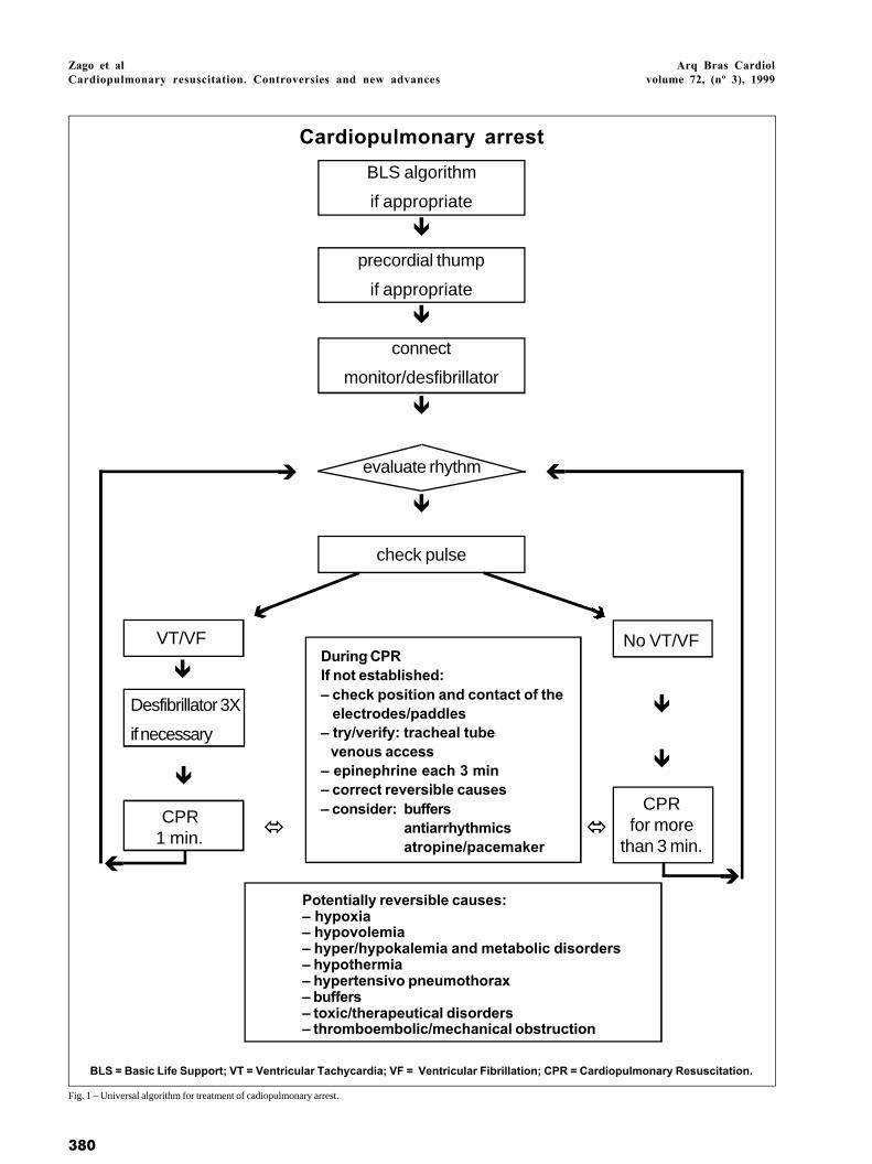
In 1992, the AHA published an extensive detailed andupdated review on CPR, comprising nine algorithms fortreatment of CPA and severe clinical situations that maylead to CPA.
In that same year (1992), ILCOR was formed, com-prising the AHA, the European Resuscitation Council(ERC), the Heart and Stroke Foundation of Canada(HSFC), the Australian Resuscitation Council (ARC), theResus-citation Councils of Southern Africa (RCSA) andthe recent Council of Latin America for Resuscitation(CLAR), aiming at standardizing and spreading thescientific knowledge on CPR 4.
In 1997, ILCOR published a universal algorithm toguide the treatment of different situations of CPA (fig. 1) 19.
This algorithm is markedly simple resulting, therefore,in easy understanding, immediate acceptance, and practicalmanagement. On the other hand, the main negative point issuperficiality, which restrains the isolated use of thealgorithm for the treatment of CPA, because it does notprovide medication doses and alternative therapeuticalroutes to increase the chances of success of the basic andadvanced support measures. Another negative point is thelack of agreement among the ILCOR members in regard toeffectiveness and indication of the precordial thump.
Considering the positive and negative points of the
380
Zago et alCardiopulmonary resuscitation. Controversies and new advances
Arq Bras Cardiolvolume 72, (nº 3), 1999
BLS algorithm
if appropriate
precordial thump
if appropriate
connect
monitor/desfibrillator
evaluate rhythm
check pulse
VT/VF
Desfibrillator 3X
if necessary
CPR 1 min.
BLS = Basic Life Support; VT = Ventricular Tachycardia; VF = Ventricular Fibrillation; CPR = Cardiopulmonary Resuscitation.
ê
ê
ê
ê
CPRfor more
than 3 min.
No VT/VF
ê
ç
è
è
ç
óó óó
Potentially reversible causes:– hypoxia– hypovolemia– hyper/hypokalemia and metabolic disorders– hypothermia– hypertensivo pneumothorax– buffers– toxic/therapeutical disorders– thromboembolic/mechanical obstruction
ê
ê
ê
í
Cardiopulmonary arrest
Fig. 1 – Universal algorithm for treatment of cadiopulmonary arrest.
During CPRIf not established:– check position and contact of the electrodes/paddles– try/verify: tracheal tube venous access– epinephrine each 3 min– correct reversible causes– consider: buffers
antiarrhythmicsatropine/pacemaker
Arq Bras Cardiolvolume 72, (nº 3), 1999
Zago et alCardiopulmonary resuscitation. Controversies and new advances
381
simplified algorithm, its use is justified in the orientation ofCPR teams in regard to which steps to follow, along with theexperience of the leading physician of the CPR team and theeventual alternative source of consultation.
Cerebral resuscitation
The brain is the most sensitive organ of the humanbody to fluctuations in the oxygen supply, because it usesonly aerobic metabolism to obtain energy and it cannotstore glucose and oxygen. Its oxygen and glucose contentsare depleted in about 5 minutes of metabolism, with subse-quent neuronal damage and death 41,42.
Therefore, the central nervous system is currentlyreceiving more attention in the context of CPR. The conceptof cerebral resuscitation has arisen corresponding to themeasures taken after the reestablishment of spontaneouscirculation aiming to arrest or minimize the anoxic-ischemiccerebral lesion, directly related to the functional incapacityand poor prognosis following CPR 41,42.
A neurological lesion is determined by tissularischemia during the absence of blood flow and can beprolonged for hours or days after the reestablishment of thespontaneous circulation by the so-called postresuscitationcerebral syndrome, which occurs after 10 to 20 minutes ofabsence of spontaneous cerebral blood flow in normo-thermic conditions. This syndrome consists of fourcomponents: 1) failure of cerebral perfusion with decreaseof oxygen supply to the brain; 2) cerebral reoxygenation,which causes damage through the activation of a cascadeof chemical events, i.e., intracellular influx of calcium,tissular lactic acidosis and increase in the concentration offree fatty acids, free radicals and excitatory amino acids,such as glutamate and aspartate. This cascade has beendocumented in vitro and in extracerebral organs, but not yetin the brain in vivo; 3) extracerebral abnormalities, such ashyperthermia, hypoxemia, hypercapnia, disorders of theacid-base metabolism, renal and hepatic failure, andbacterial translocation from the intestinal wall impaired byischemia; and 4) blood abnormalities secondary to stasis,such as aggregates of polymorphonuclear leukocytes andmacrophages that can release free radicals, damage thevascular endothelium and obstruct blood capillariesresulting in microinfarctions 41-44.
Based on the postresuscitation neuronal viability, i.e.,the possibility of reversing the process of neurologicallesions that spreads after the reestablishment of spon-taneous cerebral circulation, and on the pathophysio-logical mechanisms of the postresuscitation neuronallesion, therapeutical measures were established, consistingin the cerebral resuscitation presented below.
Cardiovascular measures - Among the cardio-vascular measures, the increase of the perfusion pressurethrough the use of adrenaline during the CPR maneuvers,should be implemented as soon as possible. The adminis-tration of high doses of adrenaline during CPR is potentiallybeneficial to cerebral perfusion.
Another measure is the induction of hypertension inthe phase of spontaneous cerebral reperfusion – SPbetween 150 and 200 mmHg – during 1 to 5 minutes 45-49,which can occur secondarily to the use of adrenaline duringCPR or directly with the administration of noradrenalinewith the reestablishment of the spontaneous circulation.Increased pressure levels in this phase are necessary tosurpass the no-reflow phenomenon – multifocal andtransitory absence of cerebral perfusion 50,51 – and theincrease of the intracranial pressure, which providesimportant resistance to blood flow. However, severeinduced hypertension should not be prolonged, because ofthe secondary myocardial ischemia and the worsening ofthe cerebral vasogenic edema 52,53.
Elevation of the head of the bed at 30° facilitates cere-bral venous drainage, reducing the intracranial pressure,which is usually increased 20 to 30 minutes after spon-taneous circulation resumes. Knowing that the cerebralperfusion pressure is equivalent to the difference betweenthe systemic mean blood pressure and the intracranialpressure, the use of the combination transitory hyper-tension and elevation of the head of the bed is justified andprovides better results 3,22,41,45.
After the period of transitory hypertension, normo-tension or mild hypertension are recommended, which canbe attained with the infusion of fluids isolated or associatedwith dopamine, dobutamine and other drugs.
Hemodynamic monitoring with the Swan-Ganzcatheter, when possible, is indicated for adequate hemo-dynamic management.
Pulmonary measures - Respiratory support can rangefrom oxygen use through a nasal catheter to mechanicalventilation with intermittent positive pressure, dependingon the level of consciousness and on the parameters of theblood gases.
In cases of coma following CPA, controlled mecha-nical ventilation for at least 12 hours is recommended toavoid cardiopulmonary failure in addition to providingoxygen during the period of the highest oxygen extraction,i.e., 2 to 12 hours after spontaneous cerebral reperfusion 41.Positive end-expiratory pressure (PEEP) should be avoidedor used in its lowest value possible in order to minimize theincrease of the intrathoracic pressure, which causesreduction of the venous return to the heart.
Water and electrolyte measures - Blood volume,diuresis and electrolytes should be evaluated to keep serumosmolarity between 280 and 330mOsm/L and normal levelsof electrolytes, correcting electrolyte disorders andreplacing fluids, whenever needed 41. Infusion of dextrose(class III) should be avoided because of the potential risk ofincreasing the production of lactic acid, which promotescellular damage 3.
Hematological measures – Hematocrit levels shouldbe kept between 30 and 35% 41.
382
Zago et alCardiopulmonary resuscitation. Controversies and new advances
Arq Bras Cardiolvolume 72, (nº 3), 1999
Metabolic measures – Hypo and hyperglycemiashould be corrected to keep serum levels of glucosebetween 100 and 200 mg/dL 41.
Normothermia must be maintained and convulsionscontrolled with phenobarbital, phenytoin or diazepam,because hyperthermia and convulsions increase oxygenconsumption by the brain. Even mild hyperthermia shouldbe corrected. Experimental studies in animals showedbenefits with mild hypothermia (34°C) that, in addition toexerting a cerebral protective effect, does not inducearrhythmia as occurs in moderate hypothermia (30°C).Conclusive clinical studies in humans, however, are notavailable 41.
Calcium channel blockers (nimodipine and lidoflazine),barbiturates, corticosteroids and anti-free radicals arebeing studied. The results obtained so far, however, do notshow concrete benefits that justify their use in cerebralresuscitation 41,42.
In regard to neurological prognosis, clinical studiesproved it to be poor with rare cases of neurological reco-very without functional incapacity after three days of coma.The results were even worse after seven days of coma; in thisgroup, no case of neurological recovery without functionalincapacity was evidenced. Analyzing patients in coma for 30days, provided discouraging results, because there was norecord that consciousness had been regained 54,55.
These prognostic factors of neurological recoveryonly emphasize the importance of the adequate mana-gement of the patient in CPA and the adoption of measuresof cerebral resuscitation, in order to avoid or minimizeneuronal lesion, providing better neurological results.
Ethical aspects. Identification of a final eventand suspension of life support measures
Measures of basic and advanced life support aim toprevent premature death with the reestablishment of the vi-tal functions and of the functions of interpersonal andenvironmental relations. This does not imply prolonging thesuffering of patients with end-stage chronic disease.
CPA can be subdivided into the potentially reversibleand the irreversible. In the first situation, once the event isreversed, the patient regains conditions that allow thereturn to normal life if there are no neurological sequelae, orthe patient can resume his/her normal activities withvariable degrees of adaptation, depending on the resultingneurological damage. The second situation corresponds tothe final event or death, in which the establishment of themeasures of life support either does not reverse the cardio-pulmonary collapse or enables only temporary success,because in a short period of time a new CPA will occur,determined by the base disease that cannot be treated orcontrolled, leading the patient invariably to death. In thislast situation, the reversal of the CPA does not save a life butprolongs the patient’s suffering; therefore, the use of CPR inpatients with end-stage chronic diseases is not justified 56.
Determining the adequate moment to suspend CPR
measures, in the absence of reversion of the arrhythmiatriggering the cardiopulmonary collapse, constitutes adifficult clinical decision, which, however, may be ine-vitable 57. Aiming to help in this decision and based onclinical studies, four criteria were determined, which whenfulfilled, allow the suspension of the life support measures:1) CPA in adults not associated with trauma, hypothermia,drug intoxication and nonrespiratory etiology; 2) ACLS formore than 25 minutes; 3) failure in reestablishing spon-taneous circulation, i.e., heart rate >60bpm for at least 5minutes; and 4) exclusion of refractory or recurrent VF/VTwithout pulse or of neurological activity (spontaneousrespiration, eye opening or motor response) 58.
Perspectives and new advances
Techniques - End-tidal carbon dioxide (CO2) levels
constitute an indirect measure of cardiac output during CPR.Several studies have shown its importance as a prognosticfactor in patients in CPA with cardiac electrical activitywithout palpable arterial pulse 59-62. The results obtainedevidenced that end-tidal CO
2 levels ≤10mmHg measured 20
minutes after ACLS onset are associated with a poorimmediate prognosis, i.e., failure of the CPR measures inpatients with cardiac electrical activity without palpable ar-terial pulse 59,63-65. However, there was no correlationbetween the end-tidal CO
2 levels measured before ACLS
onset and immediate prognosis, as well as there was nocorrelation between the end-tidal CO
2 levels and survival
after the success of CPR 9. Another important function ofthe end-tidal CO
2 levels would be the documentation of
the efficacy of the CPR measures, through the indirectmeasurement of cardiac output provided by therapeu-tical managements.
Coronary perfusional pressure (CPP) constitutes thepressure gradient between the aortic pressure and the rightatrial pressure during the diastole of a normal cardiac cycleor during the relaxing phase in CPR. Experimental studies inanimals and humans showed CPP’s capacity as a prog-nostic factor in patients in CPA, especially after a longperiod of time, a situation for which data on humans areavailable 66-71. Results of a study 66 showed that initial CPP<0mmHg and maximum CPP <15mmHg are associated withabsence of return to spontaneous circulation, but maximumCPP >15mmHg and 25mmHg is associated with 57% and79% of return to spontaneous circulation, respectively.Another important function of CPP measure is monitoring inreal time, which allows therapeutical decisions based ondata obtained from the patient instead of decisions relatedto protocols of cardiac rhythms, such as currently occurs.
Drugs - Vasopressin or antidiuretic hormone (ADH) isproduced by the posterior lobe of hypophysis (neuro-hypophysis) and released into blood flow in conditions ofserum hyperosmolarity or depletion of extracellular volume.However, experimental studies in animals showed a potenteffect of vasoconstriction even in the presence of hypoxia
Arq Bras Cardiolvolume 72, (nº 3), 1999
Zago et alCardiopulmonary resuscitation. Controversies and new advances
383
and acidosis, superior to and longer lasting than that ofadrenaline. Therefore, vasopressin improves myocardialperfusion when compared with adrenaline72-74. Anotherimportant characteristic is that vasopressin does notincrease myocardial consumption of oxygen (MVO
2) and,
consequently, does not increase the production of lactateduring CPA, unlike adrenaline 72. Results of a study onhumans showed higher efficiency of vasopressin at theinitial dose of 40U in bolus than adrenaline at the conven-tional dose (protocol recommended by AHA) in therestoration of spontaneous circulation in patients with VF insevere hypoxemia and acidosis 72. Angiotensin II is anonadrenergic drug with a potent vasopressor actionthrough the receptors of angiotensin. Experimental studiesin animals showed its efficacy in increasing CPP, myocardialblood flow and myocardial oxygen supply (MDO
2) during
CPR; however, there was also an increase in oxygen con-sumption (MVO
2). With the resumption of spontaneous
circulation, myocardial blood flow and MDO2 and MVO
2
remained elevated compared with the physiologicalparameters (preinduction of CPA). CPP, however, which hadan improvement with angiotensin II during CPR, showedsignificant reduction compared with the physiologicalparameters 75,76.
Endothelin-1, a peptide secreted by the endothelialcells, has a potent vasoconstrictive effect in vivo and invitro, ten times stronger than angiotensin II and 1000 timesstronger than noradrenaline. It acts through the nona-drenergic via, increasing the concentration of calcium insmooth muscle cells of the vascular wall 77-79. It also haspositive chronotropic and inotropic activities in heartmodels in vitro 80-82. Results obtained in a study of dogsshowed a significant increase in CPP with the use ofendothelin-1 combined with the conventional dose ofadrenaline. This was not observed with the isolated use ofendothelin-1 and adrenaline at the conventional dose 77.Additional studies in animals, however, analyzing thedifferent experimental models and additional clinicalvariables are necessary to justify clinical studies in humans.
Carbicarb® is a buffer solution consisting of the mixtureof sodium bicarbonate (NaHCO
3 ) and sodium carbonate
(Na2HCO
3) in the concentration of 1:1. It has less bicarbonate,
resulting in smaller formation of CO2 than sodium bicarbonate
alone. This makes Carbicarb® a more effective buffer,according to results of experimental studies 32,83-85.
Equipment - The automated external defibrillator hasbeen gaining increasing importance with the inclusion ofprecocious defibrillation in basic CPR, due to its simplicity,accuracy, and safety, enabling its handling by first-aidpersonnel with limited training 86. This increases thenumber of people capable of performing defibrillation,increasing the chances of precocious defibrillation and itssignificant benefits, as already discussed.
The automated external defibrillator is connected tothe patient by two adhesive paddles - one at the right supe-rior sternum border and the other upon the apex of the heart
– responsible for capturing and transmitting the cardiacrhythm from the patient to the device and for applying theelectric shock. The fully automated defibrillator requiresonly placement of the adhesive paddles and pressing thebutton that starts the device. If the cardiac rhythm is VF orVT, the device automatically charges and applies theelectric shock 87. In the semiautomated or shock advisorydefibrillator, the first aid person pushes the button“analyze” to analyze the cardiac rhythm and the button“shock” to apply the electric shock. Theoretically, thesemiautomated defibrillator would be safer than the fullyautomated because in the first one the shock is applied at themoment the first aid person makes the determination. Clinicalexperience, however, shows that both are equally safe 88.
The automated external defibrillator should be placedclose to the left ear of the patient in a supine position,because of easy access to the controls and placement of theadhesive paddles. Furthermore, this position allows asecond person to perform simultaneous basic CPR 87.
The main difference between the automated and theconventional defibrillator is that the first one analyzes andinterprets the cardiac rhythm, not requiring electrocar-diographic knowledge from its operator.
This device is fundamental for mobile emergencyservices not constituted by physicians, because of easytraining and maintenance, quick operation and cost viability.Other places that may require a defibrillator are medicaloffices, odontology clinics, maxillofacial surgical suites, orany place with high risk for sudden CPA. Places with difficultaccess are also potential candidates, such as aircrafts andlong-distance ships for passenger transportation, and placeswith high concentrations of people 86.
The implantable cardioverter-defibrillator is a smalldevice with six basic components - one battery, one sensorialamplifier, one control circuit (microprocessor), one highvoltage charger circuit, one capacitor for holding the powerfor defibrillation, and one high voltage circuit breaker -capable of identifying potentially hazardous arrhythmias andautomatically performing defibrillation or cardioversion 89.
Indications for the use of this device were consen-sually established by ACC, AHA and the North AmericanSociety of Pacing and Electrophysiology in 1991, accordingto the following therapeutical classes: class I or acceptable:survival after cardiac arrest; VF or VT in patients whoseelectrophysiological test or Holter monitoring cannot beused as predictors of therapeutical efficacy; recurrent VF orVT despite pharmacological treatment guided by an elec-trophysiological test or Holter monitoring; VF or VT in pa-tients in whom the medications are not tolerated or are inap-propriate; and persistently inducible VF or VT despite ade-quate medicamentous treatment or ablation; class II or arbi-trary: syncope of indefinite cause in patients with sustainedVF or VT inducible in electrophysiological test; class III orusually not justified: VF or VT of irreversible cause (acuteischemia, recent acute myocardial infarction (AMI), toxic ormetabolic disorders); recurrent syncope of indefinite causeand noninducible arrhythmia; incessant VF or VT; VF
384
Zago et alCardiopulmonary resuscitation. Controversies and new advances
Arq Bras Cardiolvolume 72, (nº 3), 1999
secondary to the syndrome of Wolf-Parkinson-White; andcoexistent medical, surgical, or psychiatric contraindica-tions 89. After the Cardiac Arrhythmia Suppres-sion Trial(CAST) and Multicenter Automatic Defibrillator ImplantationTrial (MADIT) studies 90,91, the indications then restricted tosecondary prevention were extended for use in primaryprevention of cardiac arrest in patients with a history of AMIassociated with left ventricular ejection fraction ≤35%,documented episode of nonsustained VT and/or tachyar-rhythmias inducible in electrophysiological testing.
The current devices have been programmed withpredefined power of defibrillation between 18 and 25 J, butthere is not yet a consensus in regard to the determinationof the value of the predefined power of defibrillation basedon the threshold of defibrillation, i.e., the smallest powerapplied necessary to arrest the cardiac arrhythmia. Howe-ver, recent data suggest that the severity of the left ventri-cular dysfunction and the use of some antiarrhythmicagents can increase the threshold of defibrillation tounacceptable values 92.
In regard to efficacy, the Cardiac Arrest StudyHamburg (CASH) 93 carried out in individuals survivingsudden cardiac arrest showed that the implantable cardio-verter-defibrillator is superior to propafenone consideringtotal mortality in one year and is associated with a signi-ficant reduction of two-year mortality, when compared withamiodarone and metoprolol, without any relevant differencebetween these two drugs (total mortality in two years:12.1% and 19.6%, respectively, with p=0.047). The Cana-dian Implantable Defibrillator Study (CIDS) study 94
compared the implantable cardioverter-defibrillator withamiodarone in patients surviving cardiac arrest or sympto-matic sustained VF/VT and showed a modest, although notsignificant, reduction in total mortality in four years in thegroup of the implantable cardioverter-defibrillator (total
mortality: 27% and 33%, respectively, with p=0.07).However, it should be observed that 30-day mortality washigher for the devices implanted with thoracotomy than forthe nonthoracotomy lead system approach (3.3% and0.36%, respectively).
Significant considerations about the implantablecardioverter-defibrillator should be emphasized. The firstaid person in contact with the patient at the moment of thedischarge can feel the shock, but it does not offer risks. Theelectrodes of the device usually cover a part of the epi-cardium surface, and can reduce the transcardiac current ofthe transthoracic shock. Therefore, if there is no success withthe defibrillation with 360 J, the position of the electrodes ofthe paddles of the external defibrillator should be changed toanteroposterior and the shock repeated. This change inposition of the electrodes can increase the transcardiaccurrent and, therefore, make the defibrillation easier 87.
Course on learning and systematization ofCPA management
The standardization of management of cerebral CPRdeveloped by the AHA aims at providing complete assis-tance to patients based on state-of-the-art technology andevidence-based medicine, offering the best results possible.Therefore, the importance of the courses on learning andsystematization of CPA management developed by theAHA should be emphasized, such as the courses on BasicLife Support (BLS), Advanced Cardiology Life Support(ACLS), and Pediatric Advanced Life Support (PALS).
Acknowledgements
The authors thank Dr. Timerman for his significantcontribution.
1. Standards for cardiopulmonary resuscitation (CPR) and emergency cardiac care(ECC). JAMA 1974; 227: 833-68.
2. Bianchi e Silva CH, Mallmann Neto C. Reanimação Cardiorrespiratória Básica.In: Vieira SRR (coord.). Porto Alegre: Manual de Emergências Médicas: Editora.da Universidade/UFRGS, 1995: 15-43.
3. Adult Advanced Cardiac Life Support. JAMA 1992; 268: 2199-241.4. Cummins RO, Chamberlain DA. Advisory Statements of the International L
Committee on Resuscitation. Circulation 1997; 95: 2172-3.5. Kloeck WK, Cummins RO, Chamberlain D, et al. Early Defibrillation. Cir-
culation 1997; 95: 2183-4.6. Cummins RO, Sanders A, Mancini E, Hazinski MF. In-Hospital Resuscitation.
Circulation 1997; 95: 2211-2.7. Adult Basic Life Support. JAMA 1992; 268: 2184-98.8. Advanced Cardiac Life Support. American Heart Association.1997; 1: 1-77.9. Handley AJ, Becker LB, Allen MA, et al. Single-rescuer adult basic life support.
Circulation 1997; 95: 2174-9.10. Paradis NA, Martin GB, Goetting MG, et al. Simultaneous aortic, jugular bulb,
and right atrial pressures during cardiopulmonary resuscitation in humans.Circulation 1989; 80: 361-8.
11. Lindner KH, Ahnefeld FW, Bowdler IM. Cardiopulmonary resuscitation with
interposed abdominal compression after asphyxial or fibrillatory cardiac arrestin pigs. Anesthesiology 1990; 72: 675-81.
12. American Heart Association. Adjuncts for artificial circulation. Textbook ofAdvanced Cardiac Life Support 1994; 11: 1-8.
13. Paiva EF. Equipamentos acessórios para controle da via aérea e técnicas especiaisde ressuscitação cardiopulmonar. Rev Soc Cardiol Est SP 1997; 1: 33-41.
14. Chandra N, Weisfeldt ML, Tsitlik J, et al. Augmentation of carotid flow duringcardiopulmonary resuscitation by ventilation at high airway pressuresimultaneous with chest compression. Am J Cardiol 1981; 48: 1053-63.
15. Krischer JP, Fine EG, Weisfeldt ML, et al. Comparison of prehospital conven-tional and simultaneous compression-ventilation cardiopulmonary resus-citation. Crit Care Med 1989; 17: 1263-9.
16. Berg RA, Wilcoxson D, Hilwig RW, et al. The need for ventilatory supportduring bystander CPR. Ann Emerg Med 1995; 26: 342-50.
17. Pennington JE, Taylor J, Lown B. Chest thump for reverting ventricular tachy-cardia. N Engl J Med 1970; 283: 1192-5.
18. Kerber RE, Robertson CE. Transthoracic defibrillation. In: Paradis NA - CardiacArrest: the Science and Practice of Resuscitation Medicine. Baltimore: Williams& Wilkins, 1996: 370-81.
19. Kloeck WK, Cummins RO, Chamberlain D, et al. The universal advanced lifesupport algorithm. Circulation 1997; 95: 2180-2.
Referências
Arq Bras Cardiolvolume 72, (nº 3), 1999
Zago et alCardiopulmonary resuscitation. Controversies and new advances
385
20. Stefanini E. Estabelecimento e manutenção das vias de infusão de medicamentos.Rev Soc Cardiol Est SP 1997; 1: 65-70.
21. Albarran-Sotelo R. Farmacologia. In: Lane JC, Albarran-Sotelo R (eds.) -Reanimação Cardiorrespiratória Cerebral. Rio de Janeiro: Medsi, 1993: 247.
22. Zago AC, Verri JM. Reanimação cardiorrespiratória avançada. In: Vieira SRR(coord.) - Manual de Emergências Médicas. Porto Alegre: Editora da Univer-sidade/UFRGS, 1995: 79-114.
23. Araújo S. Farmacologia no suporte avançado de vida. Rev Soc Cardiol Est SP1997; 7: 71-8.
24. Ornato JP. Use of adrenergic agonists during CPR in adults. Part 2. Ann EmergMed 1993; 22: 411-6.
25. Brown CG, Martin DR, Pepe PE, et al. A comparison of standard-dose and high-dose epinephrine in cardiac arrest outside the hospital. N Engl J Med 1992; 327:1051-5.
26. Stiell IG, Hebert PC, Weitzman BN, et al. High-dose epinephrine in adult cardiacarrest. N Engl J Med 1992; 327: 1045-50.
27. Abramson NS, Safar P, Sutton-Tyrrell K, et al. A randomized clinical trial ofescalating doses of high dose epinephrine during cardiac resuscitation. CritCare Med 1995; 23: A178.
28. Woodhouse SP, Cox S, Boyd P, et al. High dose and standard dose adrenaline donot alter survival compared with placebo, in cardiac arrest. Resuscitation 1995;20: 243-9.
29. Ditchey RV, Lindenfeld J. Failure of epinephrine to improve the balance betweenmyocardial oxygen supply and demand during closed chest resuscitation indogs. Circulation 1988; 78: 382-9.
30. Thompson BM, Steuven HS, Tonsfeldt DJ, et al. Calcium: limited indications,some danger. Circulation 1986;74(suppl IV): IV90-IV3.
31. Marino P - Calcium and phosphorus. In: Marino P - The ICU Book. Baltimore:Williams & Wilkins, 1998: 673-88.
32. Máttar JA. Bicarbonato de sódio na parada cardiorrespiratória. Rev Soc CardiolEst SP 1997; 7: 79-83.
33. Marino P. The organic acidoses. In: Marino P - The ICU Book. Baltimore:Williams & Wilkins, 1998: 592-607.
34. Timerman A. Monitorização hemodinâmica e hemogasométrica na paradacardiorrespiratória. In: Terzi RGG, Araújo S (eds.) - Monitorização Hemo-dinâmica e Suporte Cardiocirculatório do Paciente Crítico. São Paulo: Atheneu,1995: 269-77.
35. Máttar JA. Distúrbios ácido-básicos do adulto. In: Felix VN, Carvalho WB,Auler Jr JOC (eds.) - Terapia Intensiva Adulto. Pediatria/RN. São Paulo: Savier,1997: 136-43.
36. Weil M, Rackow E, Trevino R, et al. Differences in acid-base state betweenvenous and arterial blood during cardiopulmonary resuscitation. N Engl J Med1986; 315: 153-6.
37. Gazmuri RJ, Von Plant M, Weil MH, et al. Arterial PCO2 as an indicator of systemic
perfusion during cardiopulmonary resuscitation. Crit Care Med 1989; 17: 237.38. Kette F, Weil MH, Von Plant M, et al. Buffer agents do not reverse intramyocardial
acidosis during cardiac resuscitation. Circulation 1990; 81: 1660-6.39. Weil MH, Grundler W, Yamaguchi M, et al. Arterial blood gases fail to reflect
acid-base status during cardiopulmonary resuscitation: a preliminary report.Crit Care Med 1985; 13: 884-5.
40. Mateos JCP. Marcapasso externo - indicações e procedimentos básicos. Rev SocCardiol Est SP 1997;7:58-64.
41. Safar P. Cerebral resuscitation after cardiac arrest: research initiatives and futuredirections. (pt 2). Ann Emerg Med 1993; 22: 324-49.
42. Knobel E, Ferraz AC, Neto AC. Ressuscitação cerebral. Rev Soc Cardiol Est SP1997; 7: 92-8.
43. Safar P. Cerebral resuscitation after cardiac arrest: A review. Circulation 1986;74(suppl IV): IV138-IV53.
44. Kochanek PM, Hallenbeck JM. Polymorphonuclear leukocytes and monocytes-macrophages in the pathogenenesis of cerebral ischemia and stroke: A review.Stroke 1992; 23: 1367-75.
45. Canto R, Ames A, DiGancinto G, et al. Hypotension: A major factor limitingrecovery from cerebral ischemia. J Surg Res 1969; 9: 525-9.
46. Fischer EG, Ames A. Studies on mechanisms of impairment of cerebral circulationfollowing ischemia. Effect of hemodilution and perfusion pressure. Stroke 1972;3: 538-42.
47. Harrison MJG, Sedal L, Arnold J, et al. No-reflow phenomenon in the cerebralcirculation of the gerbil. J Neurol Neurosurg Psychiatry 1975; 38: 1190-3.
48. Hossmann KA, Lechtape-Gruter H, Hossmann V. The role of cerebral blood flowfor the recovery of the brain after prolonged ischemia. J Neurol 1973; 204: 281-99.
49. Safar P, Stezoski W, Nemoto EM. Amelioration of brain damage after 12 minutes ofcardiac arrest in dogs. Arch Neurol 1976; 33: 91-5.
50. Leonov Y, Sterz F, Safar P, et al. Hypertension with hemodilution prevents mul-tifocal cerebral hypoperfusion after cardiac arrest in dogs. Stroke 1992; 23: 45-53.
51. Sterz F, Leonov Y, Safar P, et al. Multifocal cerebral blood flow by Xe-CT and globalcerebral metabolism after prolonged cardiac arrest in dogs. Reperfusion with open-chest CPR or cardiopulmonary bypass. Resuscitation 1992; 24: 27-47.
52. Bleyaert AL, Sands PA, Safar P, et al. Augmentation of postischemic braindamage by severe intermittent hypertension. Crit Care Med 1980; 8: 41-7.
53. Klatzo I. Brain edema following brain ischemia and the influence of therapy. Br JAnaesth 1985; 57: 18-22.
54. Levy DE, Caronna JJ, Singer BH, et al. Predicting Outcome From Hypoxic-Ischemic Coma. JAMA 1985; 253: 1420-6.
55. Bell JA, Hodgson HJF. Coma after cardiac arrest. Brain 1974; 97: 361-72.56. Abdalla LA. Aspectos éticos e médico-legais da ressuscitação cardiopulmonar
- ordens de não ressuscitar. Rev Soc Cardiol Est SP 1997;7: 175-82.57. Kellermann AL, Hackman BB, Somes G. Predicting the outcome of unsuccessful
prehospital advanced cardiac life support. JAMA 1993; 270: 1433-6.58. Bonnin MJ, Pepe PE, Kimball KT, Clark OS. Distinct Criteria for Termination of
Resuscitation in the Out-of-Hospital Setting. JAMA 1993; 270: 1457-62.59. Levine RL, Wayne MA, Miller CC. End-Tidal Carbon Dioxide and Outcome of
Out-of-Hospital Cardiac Arrest. N Engl J Med 1997; 337: 301-6.60. Falk JL, Rackow EC, Weil MH. End-tidal carbon dioxide concentration during
cardiopulmonary resuscitation. N Engl J Med 1988; 318: 607-11.61. Asplin BR, White RD. Prognostic value of end-tidal carbon dioxide pressures
during out-of-hospital cardiac arrest. Ann Emerg Med 1995; 25: 756-61.62. Cantineau JP, Lambert Y, Merckx P, et al. End-tidal carbon dioxide during
cardiopulmonary resuscitation in humans presenting mostly with asystole: apredictor of outcome. Crit Care Med 1996; 24: 791-6.
63. Sanders AB, Kern KB, Otto CW, et al. End-tidal carbon dioxide monitoringduring cardiopulmonary resuscitation: a prognostic indicator for survival.JAMA 1989; 262: 1347-51.
64. Kern KB, Sanders AB, Raife J, et al. A study of chest compression rates duringcardiopulmonary resuscitation in humans: the importance of rate-directed chestcompressions. Arch Intern Med 1992; 152: 145-9.
65. Cantineau JP, Merckx P, Lambert Y, et al. Effect of epinephrine on end-tidalcarbon dioxide pressure during prehospital cardiopulmonary resuscitation. AmJ Emerg Med 1994; 12: 267-70.
66. Paradis NA, Martin GB, Rivers EP, et al. Coronary perfusion pressure and thereturn of spontaneous circulation in human cardiopulmonary resuscitation.JAMA 1990; 263: 1106-13.
67. Kern KB, Ewy GA, Voorhees WD, et al. Myocardial perfusion pressure: apredictor of 24-hour survival during prolonged cardiac arrest in dogs.Resuscitation 1988; 16: 241-50.
68. Redding JS, Pearson JW. Resuscitation from ventricular fibrillation. JAMA1968; 203: 255-60.
69. Sanders AB, Ewy GA, Taft TV. Prognostic and therapeutic importance of theaortic diastolic pressure in resuscitation from cardiac arrest. Crit Care Med 1984;12: 871-3.
70. Niemann JT. Differences in cerebral and myocardial perfusion during closed-chestresuscitation. Ann Emerg Med 1984; 13: 849-53.
71. Niemann JT, Criley JM, Rosborough JP, et al. Predictive indices of successfulcardiac resuscitation after prolonged arrest and experimental cardiopulmonaryresuscitation. Ann Emerg Med 1985; 14: 521-8.
72. Lindner KH, Prengel AW, Brinkmann A, et al. Vasopressin Administration inRefractory Cardiac Arrest. Ann Intern Med 1996; 124: 1061-4.
73. Lindner KH, Brinkmann A, Pfenninger EG, et al. Effect of vasopressin onhemodynamic variables, organ blood flow, and acid-base status in a pig model ofcardiopulmonary resuscitation. Anesth Analg 1993; 77: 427-35.
74. Lindner KH, Prengel AW, Pfenninger EG, et al. Vasopressin improves vitalorgan blood flow during closed-chest cardiopulmonary resuscitation in pigs.Circulation 1996; 91: 215-21.
75. Little CM, Brown CG. Angiotensin II improves myocardial blood flow in cardiacarrest. Resuscitation 1993; 26: 203-10.
76. Little CM, Hobson JL, Brown CG. Angiotensin II effects in a swine model ofcardiac arrest. Ann Emerg Med 1993; 22: 244-7.
77. DeBehnke DJ, Spreng D, Wickman LL, et al. The effects of endothelin-1 oncoronary perfusion pressure during cardiopulmonary resuscitation in a caninemodel. Acad Emerg Med 1996; 3: 137-41.
78. Yanagisawa M, Kurihara H, Kimura S, et al. A novel potent vasoconstrictorpeptide produced by vascular endothelial cells. Nature 1988; 332: 411-5.
79. Weitzberg E. Circulatory responses to endothelin-1 and nitric oxide withspecial reference to endotoxin shock and nitric oxide inhalation. Acta PhysiolScand 1993; 611:2-72.
80. Ishikawa T, Yanagisawa M, Kimura S, et al. Positive inotropic action of novelvasoconstrictor peptide endothelin on guinea pig atria. Am J Psysiol 1988;255(suppl): H970-H3.
81. Moravec CS, Reynolds EE, Stewart RW, et al. Endothelin is a positive inotropicagent in human and rat heart in vitro. Biomed Biophys Res Commun 1989; 159:14-8.
386
Zago et alCardiopulmonary resuscitation. Controversies and new advances
Arq Bras Cardiolvolume 72, (nº 3), 1999
82. Ishikawa T, Yanagisawa M, Kimura S, et al. Positive chronotropic effects ofendothelin, a novel endothelium derived vasoconstrictor peptide. Eur J Physiol1988; 413: 108-10.
83. Bersin RM, Arieff AI. Improved hemodynamic function during hypoxia withCarbicarb®: a new agent for the management of acidosis. Circulation 1988; 77:227-33.
84. Kucera R, Shapiro J, Whalen M, et al. Brain pH effects of NaHCO3 and Carbicarb®
in lactic acidosis. Crit Care Med 1989; 17: 1320-3.85. Leung JM, Landow L, Franks M, et al. Safety and efficacy of intravenous Carbicarb®
in patients undergoing surgery: comparison with sodium bicarbonate in thetreatment of mild metabolic acidosis. Crit Care Med 1994; 22: 1540-9.
86. Cummins RO. From concept to standard-of-care? review of the clinical experiencewith automated external defibrillators. Ann Emerg Med 1989; 18: 1269-75.
87. Advanced Cardiac Life Support. American Heart Association, 1997; 4: 1-22.88. Stults KR, Cummins RO. Fully automatic vs shock advisory defibrillators: what
are the issues? J Emerg Med Serv 1987; 12: 71-3.
89. Wolfe DA, Kosinski D, Grubb BP. Update on implantable cardioverter-defibrillators - new, safer devices have led to changes in indications. Post-graduate Medicine 1998; 103: 115-30.
90. Cardiac Arrhytmia Suppression Trial (CAST) Investigators. Preliminary report:effect of encainide and flecainide on mortality in a randomized trial of arrhytmiasuppression after myocardial infarction. N Engl J Med 1989; 321: 406-12.
91. Multicenter Automatic Defibrillator Implantation Trial (MADIT). Am J Cardiol1997; 79(suppl 6A): 16-7.
92. Yee R, Klein GJ, Thakur RK. Clinical predictors of successful implantablecardioverter defibrillator implantation (part 2). Am Heart J 1994; 127: 1068-72.
93. Cardiac Arrest Study Hamburg (CASH). Meeting Highlights - 47th AnnualScientific Sessions of the American College of Cardiology. Circulation 1998; 97:2377-81.
94. Canadian Implantable Defibrillator Study (CIDS). Meeting Highlights - 47thAnnual Scientific Sessions of the American College of Cardiology. Circulation1998; 97: 2377-81.