cardiomyopathiesPostgraduate MedicalJournal(December 1972) 48, 703-713. Clinical definitions and...
10
Postgraduate Medical Journal (December 1972) 48, 703-713. Clinical definitions and classification of cardiomyopathies C. M. OAKLEY CARDIOMYOPATHY is simply heart muscle disease, but it is surprisingly difficult to find a common language whereby we know that we are talking about the same thing. The study and recognition of cardio- myopathies has been delayed because of the seman- tic difficulties which we have encountered. The first working classification to be generally adopted (Goodwin et al., 1961) was a clinical classification into congestive, constrictive and hypertrophic obstructive types. This clinical classification antedated our present understanding of the haemodynamic de- rangements and it lacks pathological specificity. Congestive cardiomyopathy simply means that the patient has left ventricular pump failure which may result from many different pathologies involving the left ventricle. We need something new if we are not to struggle with three concurrent classifications, one clinical, one functional and one pathological. Although many different disease processes can give rise to left ventricular dysfunction, in the vast majority of patients with heart muscle disease no specific aetiology can be determined by either clin- ician or pathologist. Before deciding that a patient has a primary cardiomyopathy it is necessary to consider carefully and to exclude all recognized or possible causes of the myocardial problem. From the viewpoint of haemodynamic function we can recognize the difference between a ventricle which is dilated because it fails to empty at each beat and one which is hypertrophied and not dilated but is stiff during diastolic filling. Turning to aeti- ology, we can classify cardiomyopathies according to whether there is a known cause or whether there is a disease elsewhere in the body. In view of the magnitude of the problem presented by the common heart failures of unknown origin it appears important TABLE 1. Aetiological classification of heart muscle disorder I. Primary cardiomyopathy Facio-scapulo-humoral (Landouzy-Dejerine) Systolic pump failure Congestive cardiomyopathy Dystrophia myotonica (COCM) (b) Neuronal dystrophies Endocardial fibroelastosis Friedreich's ataxia Diastolic compliance Hypertrophic cardiomyopathy ( failure (HOCM) Associated with general system dseases Obliterative Endomyocardial fibrosis Systemic lupus erythematosus (EMF) Polyarteritis nodosa fflers disease Rheumatoid arthritis ('Endocarditis parietalis Dermatomyositis and polymyositis Scleroderma fibroplastica') Sclroderma Sarcoidosis II. Heart muscle disease of known cause or association Leukaemia (1) Infective Post-myocarditis (? viral) Protozoal (Trypanosoma Cruzi, (5) Sensitivity phenomena Chagas' Disease) Serum sickness (2) Metabolic Post vaccinal (2) Metabolic Sulphonamides Pregnancy Peripartal and puerperal Endocrine Thyrotoxic (6) Toxic (due to specific myocardial poisons) Hypothyroid Heavy metals Acromegalic Antimony (Stilbamidine) Familial Haemochromatosis Arsenic Glycogen storage disease (Pompe's Cobalt disease) Emetine Hurler's syndrome Alcohol Deprivation Beri-beri (thiamine deficiency) Phenothiazines and tricyclic antidepressants Amyloid 'Primary' cardiac Anaesthetics Associated with myelomatosis Chloroform Dysproteinaemias Cyclopropane Familial Mediterranean fever Halothane Chronic sepsis (7) Due to physical agents (3) Associated with neuromuscular disorders Radiation fibrosis (a) Muscular dystrophies Heatstroke Pseudohypertrophic (Duchenne's) Limb girdle (Erb's) (8) Senile by copyright. on June 3, 2020 by guest. Protected http://pmj.bmj.com/ Postgrad Med J: first published as 10.1136/pgmj.48.566.703 on 1 December 1972. Downloaded from
Transcript of cardiomyopathiesPostgraduate MedicalJournal(December 1972) 48, 703-713. Clinical definitions and...
Postgraduate Medical Journal (December 1972) 48, 703-713.
Clinical definitions and classification of cardiomyopathiesC. M. OAKLEY
CARDIOMYOPATHY is simply heart muscle disease, butit is surprisingly difficult to find a common languagewhereby we know that we are talking about thesame thing. The study and recognition of cardio-myopathies has been delayed because of the seman-tic difficulties which we have encountered. The firstworking classification to be generally adopted(Goodwin et al., 1961) was a clinical classification intocongestive, constrictive and hypertrophic obstructivetypes. This clinical classification antedated ourpresent understanding of the haemodynamic de-rangements and it lacks pathological specificity.Congestive cardiomyopathy simply means that thepatient has left ventricular pump failure which mayresult from many different pathologies involving theleft ventricle. We need something new if we are notto struggle with three concurrent classifications, oneclinical, one functional and one pathological.
Although many different disease processes can giverise to left ventricular dysfunction, in the vastmajority of patients with heart muscle disease nospecific aetiology can be determined by either clin-ician or pathologist. Before deciding that a patienthas a primary cardiomyopathy it is necessary toconsider carefully and to exclude all recognized orpossible causes of the myocardial problem.From the viewpoint of haemodynamic function
we can recognize the difference between a ventriclewhich is dilated because it fails to empty at eachbeat and one which is hypertrophied and not dilatedbut is stiff during diastolic filling. Turning to aeti-ology, we can classify cardiomyopathies accordingto whether there is a known cause or whether thereis a disease elsewhere in the body. In view of themagnitude of the problem presented by the commonheart failures of unknown origin it appears important
TABLE 1. Aetiological classification of heart muscle disorder
I. Primary cardiomyopathy Facio-scapulo-humoral (Landouzy-Dejerine)Systolic pump failure Congestive cardiomyopathy Dystrophia myotonica
(COCM) (b) Neuronal dystrophiesEndocardial fibroelastosis Friedreich's ataxia
Diastolic compliance Hypertrophic cardiomyopathy (failure (HOCM) Associated with general system dseases
Obliterative Endomyocardial fibrosis Systemic lupus erythematosus(EMF) Polyarteritis nodosafflers disease Rheumatoid arthritis('Endocarditis parietalis Dermatomyositis and polymyositis
Sclerodermafibroplastica') SclrodermaSarcoidosisII. Heart muscle disease of known cause or association Leukaemia
(1) Infective Post-myocarditis (? viral)Protozoal (Trypanosoma Cruzi, (5) Sensitivity phenomena
Chagas' Disease) Serum sickness(2) Metabolic Post vaccinal(2) Metabolic SulphonamidesPregnancy Peripartal and puerperal
Endocrine Thyrotoxic (6) Toxic (due to specific myocardial poisons)Hypothyroid Heavy metalsAcromegalic Antimony (Stilbamidine)
Familial Haemochromatosis ArsenicGlycogen storage disease (Pompe's Cobalt
disease) EmetineHurler's syndrome Alcohol
Deprivation Beri-beri (thiamine deficiency) Phenothiazines and tricyclic antidepressantsAmyloid 'Primary' cardiac Anaesthetics
Associated with myelomatosis ChloroformDysproteinaemias CyclopropaneFamilial Mediterranean fever HalothaneChronic sepsis (7) Due to physical agents(3) Associated with neuromuscular disorders Radiation fibrosis
(a) Muscular dystrophies HeatstrokePseudohypertrophic (Duchenne's)Limb girdle (Erb's) (8) Senile
by copyright. on June 3, 2020 by guest. P
rotectedhttp://pm
j.bmj.com
/P
ostgrad Med J: first published as 10.1136/pgm
j.48.566.703 on 1 Decem
ber 1972. Dow
nloaded from
C. M. Oakley
to polarize our research endeavours and advisethat the term cardiomyopathy should be reservedfor 'heart muscle disorder of unknown cause orassociation' and that heart muscle disease of knowncause or association should be so described, forexample, sarcoid heart disease, amyloid heartdisease, etc. If we accept this concept, we cannarrow the problem down and concentrate first onfunctional differences between the cardiomyopathies.There is the primary pump failure with a dilated
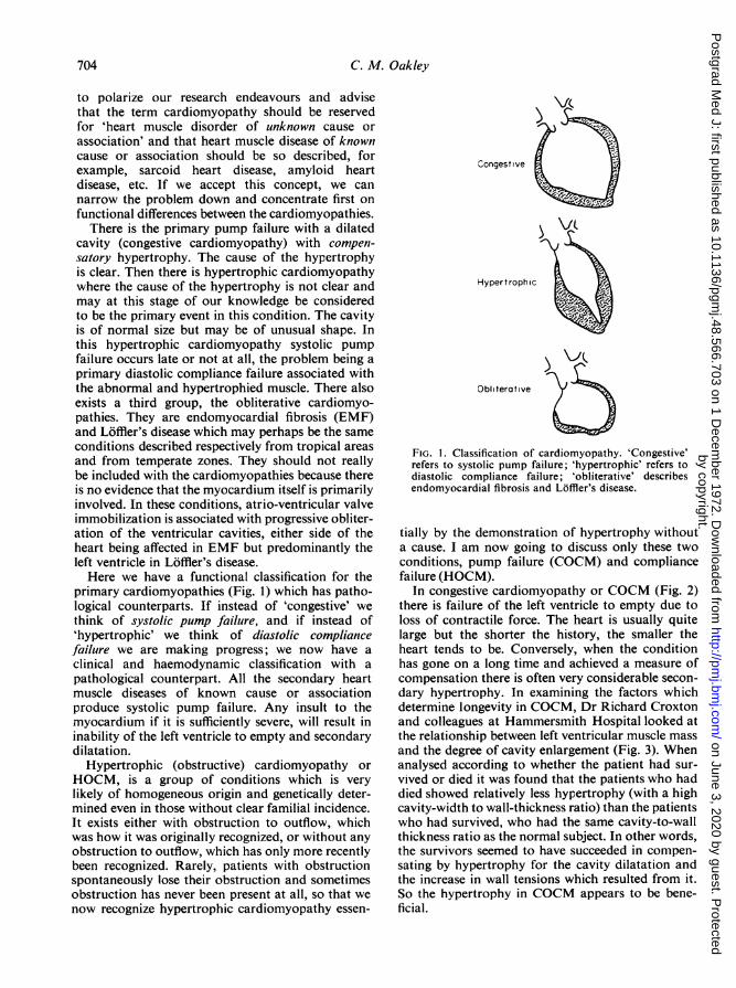
cavity (congestive cardiomyopathy) with compen-satory hypertrophy. The cause of the hypertrophyis clear. Then there is hypertrophic cardiomyopathywhere the cause of the hypertrophy is not clear andmay at this stage of our knowledge be consideredto be the primary event in this condition. The cavityis of normal size but may be of unusual shape. Inthis hypertrophic cardiomyopathy systolic pumpfailure occurs late or not at all, the problem being aprimary diastolic compliance failure associated withthe abnormal and hypertrophied muscle. There alsoexists a third group, the obliterative cardiomyo-pathies. They are endomyocardial fibrosis (EMF)and Loffler's disease which may perhaps be the sameconditions described respectively from tropical areasand from temperate zones. They should not reallybe included with the cardiomyopathies because thereis no evidence that the myocardium itself is primarilyinvolved. In these conditions, atrio-ventricular valveimmobilization is associated with progressive obliter-ation of the ventricular cavities, either side of theheart being affected in EMF but predominantly theleft ventricle in Loffler's disease.Here we have a functional classification for the
primary cardiomyopathies (Fig. 1) which has patho-logical counterparts. If instead of 'congestive' wethink of systolic pump failure, and if instead of'hypertrophic' we think of diastolic compliancefailure we are making progress; we now have aclinical and haemodynamic classification with apathological counterpart. All the secondary heartmuscle diseases of known cause or associationproduce systolic pump failure. Any insult to themyocardium if it is sufficiently severe, will result ininability of the left ventricle to empty and secondarydilatation.
Hypertrophic (obstructive) cardiomyopathy orHOCM, is a group of conditions which is verylikely of homogeneous origin and genetically deter-mined even in those without clear familial incidence.It exists either with obstruction to outflow, whichwas how it was originally recognized, or without anyobstruction to outflow, which has only more recentlybeen recognized. Rarely, patients with obstructionspontaneously lose their obstruction and sometimesobstruction has never been present at all, so that wenow recognize hypertrophic cardiomyopathy essen-
Congestive
Hypertrophic
Obliterative
FIG. 1. Classification of cardiomyopathy. 'Congestive'refers to systolic pump failure; 'hypertrophic' refers todiastolic compliance failure; 'obliterative' describesendomyocardial fibrosis and Loffler's disease.
tially by the demonstration of hypertrophy withouta cause. I am now going to discuss only these twoconditions, pump failure (COCM) and compliancefailure (HOCM).
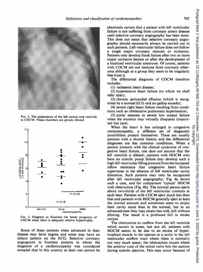
In congestive cardiomyopathy or COCM (Fig. 2)there is failure of the left ventricle to empty due toloss of contractile force. The heart is usually quitelarge but the shorter the history, the smaller theheart tends to be. Conversely, when the conditionhas gone on a long time and achieved a measure ofcompensation there is often very considerable secon-dary hypertrophy. In examining the factors whichdetermine longevity in COCM, Dr Richard Croxtonand colleagues at Hammersmith Hospital looked atthe relationship between left ventricular muscle massand the degree of cavity enlargement (Fig. 3). Whenanalysed according to whether the patient had sur-vived or died it was found that the patients who haddied showed relatively less hypertrophy (with a highcavity-width to wall-thickness ratio) than the patientswho had survived, who had the same cavity-to-wallthickness ratio as the normal subject. In other words,the survivors seemed to have succeeded in compen-sating by hypertrophy for the cavity dilatation andthe increase in wall tensions which resulted from it.So the hypertrophy in COCM appears to be bene-ficial.
704
by copyright. on June 3, 2020 by guest. P
rotectedhttp://pm
j.bmj.com
/P
ostgrad Med J: first published as 10.1136/pgm
j.48.566.703 on 1 Decem
ber 1972. Dow
nloaded from
Definitions and classification of cardiomyopathies
FIG. 2. The appearance of the left atrium and ventriclein COCM. These chambers are grossly dilated.
12r-
10
8On
£,
x u
=-600o-i
4
2
0
I-JL
0
0
*-
0
-:--0@
- -0--0
0
0
*
0
P=0 02
P=O 18
Normal Alive DeadCardiomyopat hy
FIG. 3. Diagram to illustrate the better prognosis ofCOCM when there is adequate relative hypertrophy.
Some of these patients when advanced in theirdisease may have angina and some may have aninfarct pattern on the ECG. Selective coronaryangiograms in fourteen patients in whom thediagnosis of a cardiomyopathy was consideredrevealed that in this country at least one cannot be
absolutely certain that a patient with left ventricularfailure is not suffering from coronary artery diseaseuntil selective coronary angiography has been done.This does not mean that selective coronary angio-graphy should necessarily always be carried out insuch patients. Left ventricular failure does not followa single major coronary stenosis or occlusion.Patients only develop frank failure after two or moremajor occlusive lesions or after the development ofa localized ventricular aneurysm. Of course, patientswith COCM are not immune from coronary ather-oma although as a group they seem to be singularlyfree from it.The differential diagnosis of COCM therefore
includes:(1) ischaemic heart disease;(2) hypertensive heart failure (to which we shall
refer later);(3) chronic pericardial effusion (which is recog-
nized by a normal ECG and no gallop sounds);(4) severe right heart failure resulting from condi-
tions such as obliterative pulmonary hypertension;(5) aortic stenosis in severe low output failure
when the murmur may virtually disappear (import-ant but rare).When the heart is less enlarged in congestive
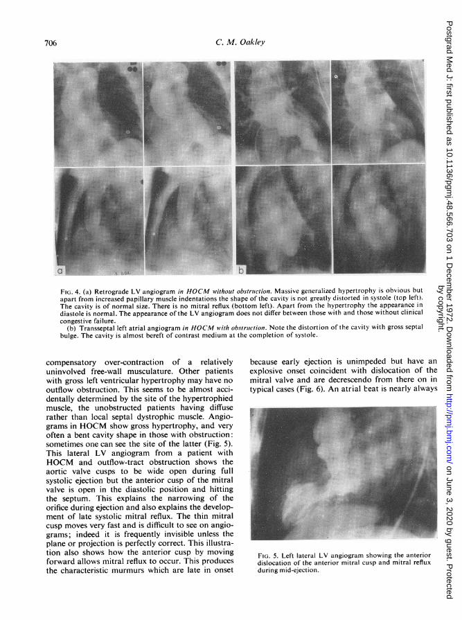
cardiomyopathy, a different set of diagnosticpossibilities present themselves. These are usuallypatients with a shorter history and the differentialdiagnoses are less common conditions. When apatient presents with the clinical syndrome of con-gestive heart failure, one dare not assume that theleft ventricle is dilated; patients with HOCM whohave no systolic pump failure may develop such ahigh left ventricular filling pressure from the increasedinflow resistance that congestive heart failuresupervenes in the absence of left ventricular cavitydilatation. Such patients may only be recognizedafter left ventricular angiography; Fig. 4a showssuch a case, and for comparison 'typical' HOCMwith obstruction (Fig. 4b). The normal person ejectsabout two-thirds of the left ventricular contents ateach beat. Patients with COCM eject much less thanthat and patients with HOCM generally eject at leastthe normal amount and sometimes seem to emptytheir cavity more than in the normal, but in anadvanced state they may empty less well, still withoutdilating. The result is a profound fall in strokeoutput.The obstruction to outflow from the left ventricle
which occurs in some, but not all, patients withHOCM seems to be due to an excess of hyper-trophied muscle in the septum at a point in the leftventricular outflow tract where there is normallynot very much space; the obstruction occurs wherethe anterior cusp of the mitral valve hits the septumduring systolic ejection. This may occur because of
Of, --
705
by copyright. on June 3, 2020 by guest. P
rotectedhttp://pm
j.bmj.com
/P
ostgrad Med J: first published as 10.1136/pgm
j.48.566.703 on 1 Decem
ber 1972. Dow
nloaded from
706 C. M. Oakley
.lii.-.....;......
.... .....: .' ,-
ae*:°i'ii......i....i:......
UsZ...
........ .....Y~~i~y~iiiii:, Ii
FIG. 4. (a) Retrograde LV angiogram in HOCM without obstruction. Massive generalized hypertrophy is obvious butapart from increased papillary muscle indentations the shape of the cavity is not greatly distorted in systole (top left).The cavity is of normal size. There is no mitral reflux (bottom left). Apart from the hypertrophy the appearance indiastole is normal. The appearance of the LV angiogram does not differ between those with and those without clinicalcongestive failure.
(b) Transseptal left atrial angiogram in HOCM with obstruction. Note the distortion of the cavity with gross septalbulge. The cavity is almost bereft of contrast medium at the completion of systole.
compensatory over-contraction of a relativelyuninvolved free-wall musculature. Other patientswith gross left ventricular hypertrophy may have nooutflow obstruction. This seems to be almost acci-dentally determined by the site of the hypertrophiedmuscle, the unobstructed patients having diffuserather than local septal dystrophic muscle. Angio-grams in HOCM show gross hypertrophy, and veryoften a bent cavity shape in those with obstruction:sometimes one can see the site of the latter (Fig. 5).This lateral LV angiogram from a patient withHOCM and outflow-tract obstruction shows theaortic valve cusps to be wide open during fullsystolic ejection but the anterior cusp of the mitralvalve is open in the diastolic position and hittingthe septum. This explains the narrowing of theorifice during ejection and also explains the develop-ment of late systolic mitral reflux. The thin mitralcusp moves very fast and is difficult to see on angio-grams; indeed it is frequently invisible unless theplane or projection is perfectly correct. This illustra-tion also shows how the anterior cusp by movingforward allows mitral reflux to occur. This producesthe characteristic murmurs which are late in onset
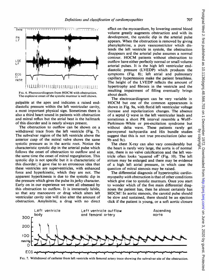
because early ejection is unimpeded but have anexplosive onset coincident with dislocation of themitral valve and are decrescendo from there on intypical cases (Fig. 6). An atrial beat is nearly always
C··;:·
,;·'"s. ':·"'i:.·· :·.:: ':':.
.iii. d:i ':::i·:::·
·::··i...i..·:·
i' i· :.::i·iii.
FIG. 5. Left lateral LV angiogram showing the anteriordislocation of the anterior mitral cusp and mitral refluxduring mid-ejection.
by copyright. on June 3, 2020 by guest. P
rotectedhttp://pm
j.bmj.com
/P
ostgrad Med J: first published as 10.1136/pgm
j.48.566.703 on 1 Decem
ber 1972. Dow
nloaded from
Definitions and classification of cardiomyopathies 707
Date 1 I i I 1r I , r i
PALF
LSELF
ECG
INSP
1 LJLL!:ll I II 1 LJ. IIJ. Ill J LI l l1..1_1. .1FIG. 6. Phonocardiogram from HOCM with obstruction.The explosive onset of the systolic murmur is well shown.
palpable at the apex and indicates a raised end-diastolic pressure within the left ventricular cavity,a most important physical sign. Sometimes there isalso a third heart sound in patients with obstructionand mitral reflux but the atrial beat is the hallmarkof this disorder and is nearly always present.The obstruction to outflow can be shown on a
withdrawal trace from the left ventricle (Fig. 7).The subvalvar region of the left ventricle above theanterior cusp of the mitral valve shows the samesystolic pressure as in the aortic root. Notice thecharacteristic systolic dip in the arterial pulse whichfollows the onset of obstruction to outflow and atthe same time the onset of mitral regurgitation. Thissystolic dip is not specific but it is characteristic ofthis disorder; it gave rise to an erroneous idea thatthese ventricles are superventricles, with enhancedforce and hyperkinetic, which they are not. Theapparent hyperkinesia is due to the systolic dip inthe pressure which gives the pulse its jerky character.Early on in our experience we were all obsessed bythis obstruction to outflow. It is immensely labile,so that any manoeuvre or drug which alters leftventricular cavity size will also alter the amount ofobstruction. Amylnitrite, a drug with no direct
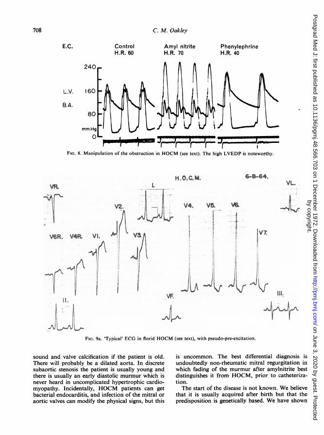
effect on the myocardium, by lowering central bloodvolume greatly augments obstruction and with itsdevelopment, the systolic dip in the arterial pulseappears. When the obstruction is removed by givingphenylephrine, a pure vasoconstrictor which dis-tends the left ventricle in systole, the obstructiondisappears ard the arterial pulse assumes a normalcontour. HOCM patients without obstruction tooutflow have either perfectly normal or small volumearterial pulses. It is the high left ventricular end-diastolic pressure (LVEDP) which produces thesymptoms (Fig. 8); left atrial and pulmonarycapillary hypertension make the patient breathless.The height of the LVEDP reflects the amount ofhypertrophy and fibrosis in the ventricle and theresulting impairment of filling eventually bringsabout death.The electrocardiogram can be very variable in
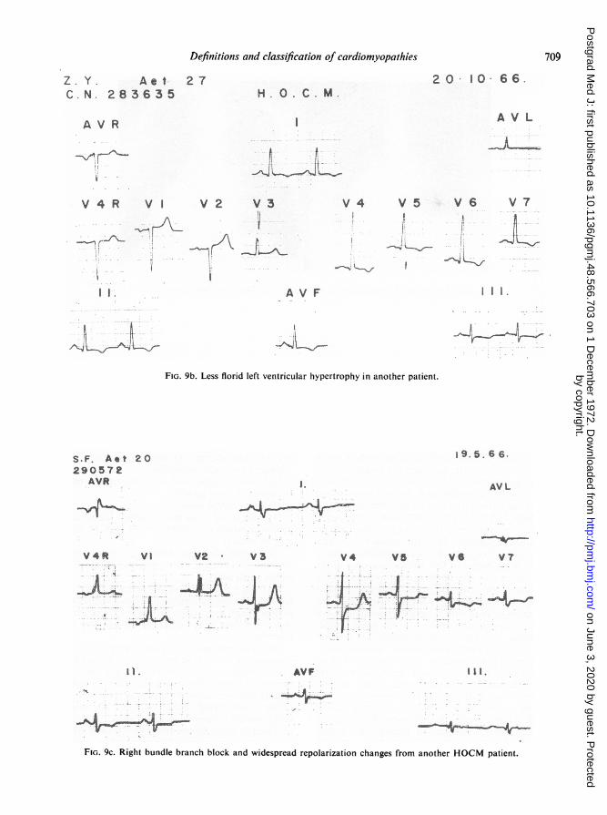
HOCM but one of the common appearances isshown in Fig. 9a, with florid left ventricular voltageincrease and repolarization changes. The absenceof a septal Q wave in the left ventricular leads andsometimes a short PR interval resemble a Wolff-Parkinson--White or pre-excitation syndrome butwithout delta wave. These patients rarely getparoxysmal tachycardia and His bundle studiessuggest that this is not true pre-excitation (also see9b and 9c).The chest X-ray can also vary considerably but
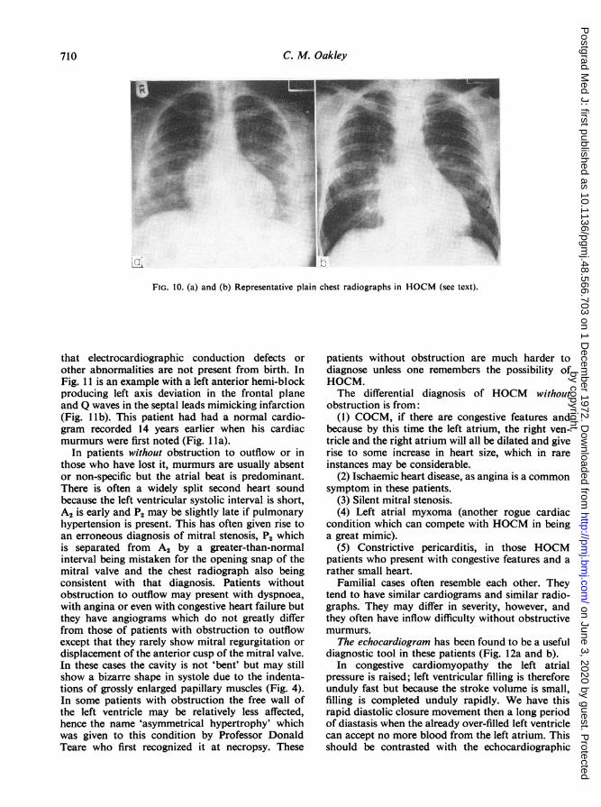
the heart is rarely very large, the aorta is of normalsize, there is no valve calcification and the left ven-tricle often looks 'squared off' (Fig. 10). The leftatrium may be enlarged and there may be evidenceof a high left atrial pressure, in which case thequestion of mitral stenosis may be raised.The differential diagnosis of hypertrophic cardio-
myopathy with obstruction is that of other conditionswhich give rise to systolic murmurs. Once you startto wonder which of the five main differential diag-noses the patient has, then he almost certainly hasHOCM! In aortic stenosis, the carotid pulse shouldbe slow and sustained, there should be an ejectionclick if the patient is young, or a soft aortic closure
Left ventricle Left ventricle outflow Ascendingbody and femoral artery aorta
300
200
100mmHg
.... .
ECG . --; - ,.-...-, ,
FIG. 7. Withdrawal of catheter from left ventricle with femoral artery trace showing the subvalvar site of the obstruction.
by copyright. on June 3, 2020 by guest. P
rotectedhttp://pm
j.bmj.com
/P
ostgrad Med J: first published as 10.1136/pgm
j.48.566.703 on 1 Decem
ber 1972. Dow
nloaded from
708 C. M. Oakley
E.C. Control Amyl nitrite PhenylephrineH.R. 60 H.R. 70 H.R. 40
240
LV. 160
B.A.80
mm Hg
0-. -1'T. .,,,_PI Ir rr -tFIG. 8. Manipulation of the obstruction in HOCM (see text). The high LVEDP is noteworthy.
'.'·~,...:., ir.'?:s.it.--]--,' . .. ... ......- ......--,....
:* * .-:.:...
-. ., .'.':::l-J''. -· : .-^ ^. ...... t1 ;: :.V. ",*'' :*:""?. l..".' ' :' ":,j, .**, e e $ ." -' e to. · ", .*r{;2t| .:'S; "" .; . . . .
·I'I'I'
... -.-JiL LFIG. 9a. 'Typical' ECG in florid HOCM (see text), with pseudo-pre-excitation.
sound and valve calcification if the patient is old.There will probably be a dilated aorta. In discretesubaortic stenosis the patient is usually young andthere is usually an early diastolic murmur which isnever heard in uncomplicated hypertrophic cardio-myopathy. Incidentally, HOCM patients can getbacterial endocarditis, and infection of the mitral oraortic valves can modify the physical signs, but this
is uncommon. The best differential diagnosis isundoubtedly non-rheumatic mitral regurgitation inwhich fading of the murmur after amylnitrite bestdistinguishes it from HOCM, prior to catheteriza-tion.The start of the disease is not known. We believe
that it is usually acquired after birth but that thepredisposition is genetically based. We have shown
by copyright. on June 3, 2020 by guest. P
rotectedhttp://pm
j.bmj.com
/P
ostgrad Med J: first published as 10.1136/pgm
j.48.566.703 on 1 Decem
ber 1972. Dow
nloaded from
Definitions and classification of cardiomyopathies 709
· ~~1..:1....._--.~,, .. i1-4", .;"· ..~ ~.::;...~..'~·~?,·':;'?..:- . . I·
~~ ~ ~ ~~~~·*:...;~[~,~.~1.;,..:...'"'~~f .. ?"';
FIG. 9b. Less florid left ventricular hypertrophy in another patient.
:!.r ^^- I ...-'."..'.:"..'.,,:< , '.:'''.~.':.'. *, .- '.'i;..:::"...* **
..,.'·*--' - ,:....':" ..."'..a » ' t ;' .' !..-*.* *.* -*
< ',-,.' ; . . . ;.-- - .* ..*,
." " *' :\ ':'' "-;'"
· 6sto.
a: ,,-1.{-;: ...-. ".'...'. ...
..:- - st;; i-'..C·
1 ; ** *y * :r ib .1^ /| i* -*\. :..:^l '*';.;; * i--. "i;'
:.* ;^ ^ . ;...; S. '? w
;- s:*\ - :I)L :* y ; / 31 :,, 4 *; :- *.- - -*
* f-.t1- ,-;
-i· ...'.. ... .
* .' -?:.;;'... , ..
. ,; ., ,. ..
* '- *.:.*..-.. ... ^ ..:., .,,... .. _., ,.' ,..... .
- ..
f. .? ..:. .; *1Z~~ ~ ~~~ ~ ~ ~ ~·I*··r· !:* 4**s t -.
'"- - - ---t': '. ;'', -
., ... ... ..,,,.,..3'. ,!: ,.'.>.F4'i..;...... . .....;..:...... ,......, :............
...... ''....::· , . r, '·· :·· ·.·~
:.:'::''~.-" .::*:-..~::.--.."'."'';.'.'.;'..'.. .....%:"···;·,., !
'.*.. :..:·,." :. '... .:
'5 3: ....'"'~··.... ' ~~ . .' ''r: . .- ·~i.' !.··." ·'...:'>,.~..~"'" , '-. ' ¢" ' .."'''...... '":''"'
·-
*, . : , .... ' r. ~F.· ...-~:.~.~.:: ~ ~ ~ ~ ~ ~ ~~~·'"~ :-'·i:'~.:',:',.'-:.-~,:::,::~:,:r-.:...::,',"":~i. ~' ~":.-,':.!~..~''? ," i.'.: .' ".: :'' :".~.....,
-s.-. ..;.;; ~ Pt. ..r .
. ·. i.;. ..-; .·i.'': : r i: ' ' ' ': . :'.~.: ].:.:i ''·''· J".. :, [~'..J
....·: ...:..... ...'. ..,
:.
.. ...·:; . ,.: .. , :.
.-.. . .,: .. . _ ......'-.,." :.',.:'':.." . ..:-'..''' . ''&.l';....",.:''......-,.. . ....,.... . ! t,'. ...:.
~".,._":.::...i."..... . . .~~~~1*~~~~~"'""'·1;.Z'".-' · , ..~..r · ·:'" ..........
:.·k : :,.. · ., .·.. ....·1I .·..,.... ~:.' . ... :,,. ..... ?;.;',FIG. 9c. Right bundle branch block and widespread repolarization changes from another HOCM patient.
by copyright. on June 3, 2020 by guest. P
rotectedhttp://pm
j.bmj.com
/P
ostgrad Med J: first published as 10.1136/pgm
j.48.566.703 on 1 Decem
ber 1972. Dow
nloaded from
710 C. M. Oakley
..··:.
iC.;;[ii·;
i···bi.e.
a:·
;:I::
IEiB:".:.
r*:: :::.!-es .SJL·::ii··:
:i:;.; 'I..BEE;I.Pa.L.S.gl.r. '.Pf .ai.:$; .lll.llll.s.ii' diK·
:·::·::,..·3B.BIAP.~
:iiii.iC"II
Ib
FIG. 10. (a) and (b) Representative plain chest radiographs in HOCM (see text).
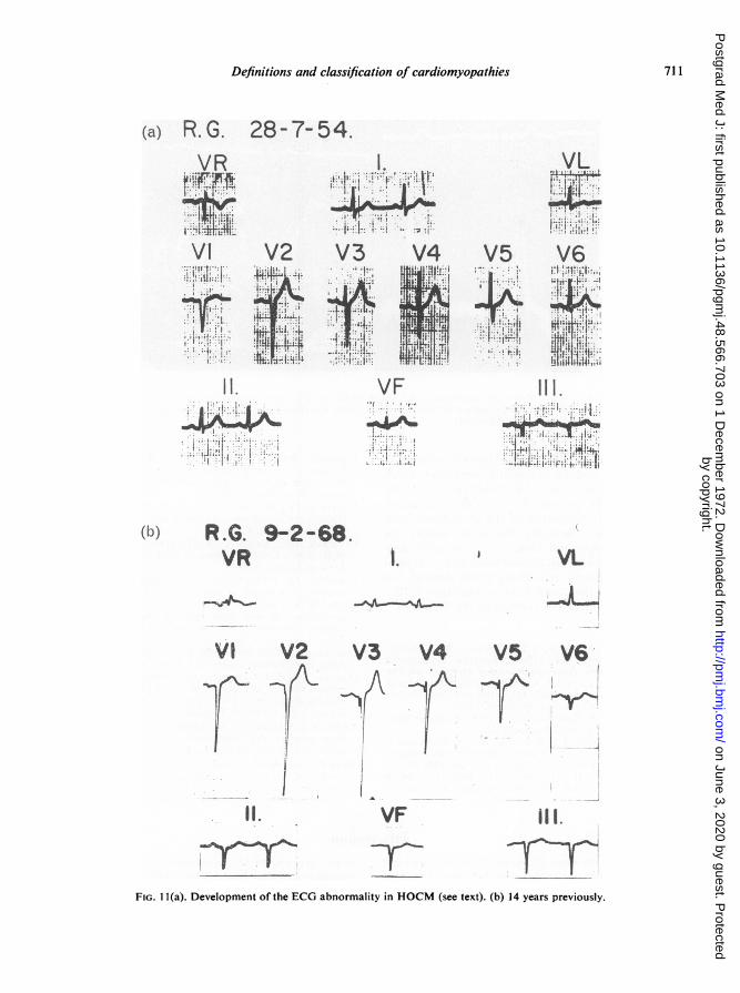
that electrocardiographic conduction defects orother abnormalities are not present from birth. InFig. 11 is an example with a left anterior hemi-blockproducing left axis deviation in the frontal planeand Q waves in the septal leads mimicking infarction(Fig. lb). This patient had had a normal cardio-gram recorded 14 years earlier when his cardiacmurmurs were first noted (Fig. 11 a).
In patients without obstruction to outflow or inthose who have lost it, murmurs are usually absentor non-specific but the atrial beat is predominant.There is often a widely split second heart soundbecause the left ventricular systolic interval is short,A2 is early and P2 may be slightly late if pulmonaryhypertension is present. This has often given rise toan erroneous diagnosis of mitral stenosis, P2 whichis separated from A2 by a greater-than-normalinterval being mistaken for the opening snap of themitral valve and the chest radiograph also beingconsistent with that diagnosis. Patients withoutobstruction to outflow may present with dyspnoea,with angina or even with congestive heart failure butthey have angiograms which do not greatly differfrom those of patients with obstruction to outflowexcept that they rarely show mitral regurgitation ordisplacement of the anterior cusp of the mitral valve.In these cases the cavity is not 'bent' but may stillshow a bizarre shape in systole due to the indenta-tions of grossly enlarged papillary muscles (Fig. 4).In some patients with obstruction the free wall ofthe left ventricle may be relatively less affected,hence the name 'asymmetrical hypertrophy' whichwas given to this condition by Professor DonaldTeare who first recognized it at necropsy. These
patients without obstruction are much harder todiagnose unless one remembers the possibility ofHOCM.The differential diagnosis of HOCM without
obstruction is from:(1) COCM, if there are congestive features and
because by this time the left atrium, the right ven-tricle and the right atrium will all be dilated and giverise to some increase in heart size, which in rareinstances may be considerable.
(2) Ischaemic heart disease, as angina is a commonsymptom in these patients.
(3) Silent mitral stenosis.(4) Left atrial myxoma (another rogue cardiac
condition which can compete with HOCM in beinga great mimic).
(5) Constrictive pericarditis, in those HOCMpatients who present with congestive features and arather small heart.
Familial cases often resemble each other. Theytend to have similar cardiograms and similar radio-graphs. They may differ in severity, however, andthey often have inflow difficulty without obstructivemurmurs.
The echocardiogram has been found to be a usefuldiagnostic tool in these patients (Fig. 12a and b).
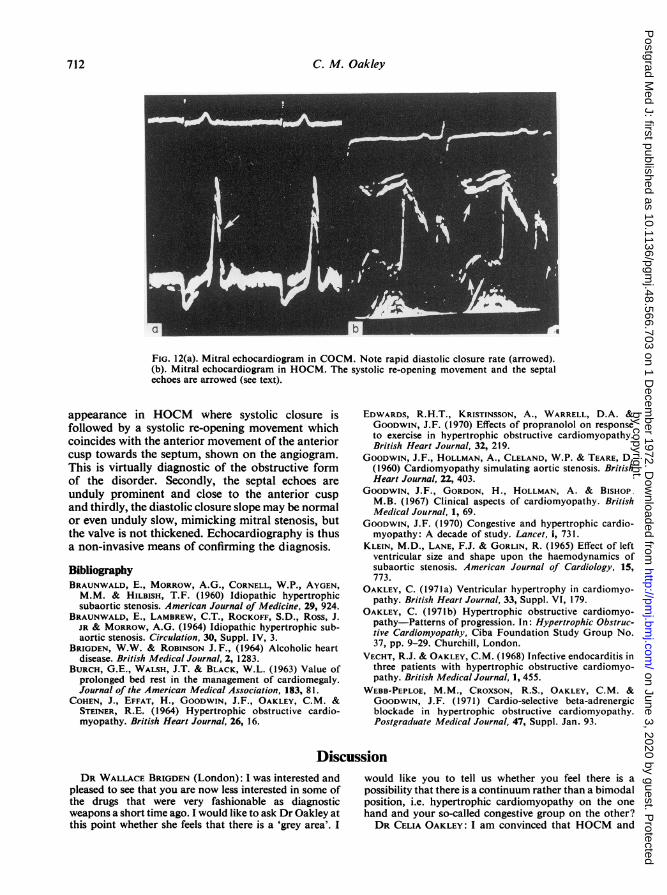
In congestive cardiomyopathy the left atrialpressure is raised; left ventricular filling is thereforeunduly fast but because the stroke volume is small,filling is completed unduly rapidly. We have thisrapid diastolic closure movement then a long periodof diastasis when the already over-filled left ventriclecan accept no more blood from the left atrium. Thisshould be contrasted with the echocardiographic
by copyright. on June 3, 2020 by guest. P
rotectedhttp://pm
j.bmj.com
/P
ostgrad Med J: first published as 10.1136/pgm
j.48.566.703 on 1 Decem
ber 1972. Dow
nloaded from
Definitions and classification of cardiomyopathies 711
(a) R.G.. 28-7-54.
II. VF I
... .
: , ,.''S',' f.'@ ' -' '.-^^^ ''.
* R.6. 2-..-.
V V. .VL
VI V2 V3 V4 V5 VS
.Vt 'f...31.1 F111.
",.:.:.! ."l " '"'"' '.i-.'-3
R.. 9268. I
V·.
VI V2 V3. .V4 VS V6.
... .. . . . ....
*I. VF III.. !
,... ... .:.
tcV2Vr V4 VTnV6'
It.r vr ~·
FIG. 1 l(a). Development of the ECG abnormality in HOCM (see text). (b) 14 years previously.
by copyright. on June 3, 2020 by guest. P
rotectedhttp://pm
j.bmj.com
/P
ostgrad Med J: first published as 10.1136/pgm
j.48.566.703 on 1 Decem
ber 1972. Dow
nloaded from
712 C. M. Oakley
Io
FIG. 12(a). Mitral echocardiogram in COCM. Note rapid diastolic closure rate (arrowed).(b). Mitral echocardiogram in HOCM. The systolic re-opening movement and the septalechoes are arrowed (see text)
appearance in HOCM where systolic closure isfollowed by a systolic re-opening movement whichcoincides with the anterior movement of the anteriorcusp towards the septum, shown on the angiogram.This is virtually diagnostic of the obstructive formof the disorder. Secondly, the septal echoes areunduly prominent and close to the anterior cuspand thirdly, the diastolic closure slope may be normalor even unduly slow, mimicking mitral stenosis, butthe valve is not thickened. Echocardiography is thusa non-invasive means of confirming the diagnosis.BibliographyBRAUNWALD, E., MORROW, A.G., CORNELL, W.P., AYGEN,M.M. & HILBISH, T.F. (1960) Idiopathic hypertrophicsubaortic stenosis. American Journal of Medicine, 29, 924.
BRAUNWALD, E., LAMBREW, C.T., ROCKOFF, S.D., Ross, J.JR & MORROW, A.G. (1964) Idiopathic hypertrophic sub-aortic stenosis. Circulation, 30, Suppl. IV, 3.
BRIGDEN, W.W. & ROBINSON J.F., (1964) Alcoholic heartdisease. British Medical Journal, 2, 1283.
BURCH, G.E., WALSH, J.T. & BLACK, W.L. (1963) Value ofprolonged bed rest in the management of cardiomegaly.Journal of the American Medical Association, 183, 81.
COHEN, J., EFFAT, H., GOODWIN, J.F., OAKLEY, C.M. &STEINER, R.E. (1964) Hypertrophic obstructive cardio-myopathy. British Heart Journal, 26, 16.
EDWARDS, R.H.T., KRISTINSSON, A., WARRELL, D.A. &GOODWIN, J.F. (1970) Effects of propranolol on responseto exercise in hypertrophic obstructive cardiomyopathy.British Heart Journal, 32, 219.
GOODWIN, J.F., HOLLMAN, A., CLELAND, W.P. & TEARE, D.(1960) Cardiomyopathy simulating aortic stenosis. BritishHeart Journal, 22, 403.
GOODWIN, J.F., GORDON, H., HOLLMAN, A. & BISHOP.M.B. (1967) Clinical aspects of cardiomyopathy. BritishMedical Journal, 1, 69.
GOODWIN, J.F. (1970) Congestive and hypertrophic cardio-myopathy: A decade of study. Lancet, i, 731.
KLEIN, M.D., LANE, F.J. & GORLIN, R. (1965) Effect of leftventricular size and shape upon the haemodynamics ofsubaortic stenosis. American Journal of Cardiology, 15,773.
OAKLEY, C. (1971a) Ventricular hypertrophy in cardiomyo-pathy. British Heart Journal, 33, Suppl. VI, 179.
OAKLEY, C. (1971b) Hypertrophic obstructive cardiomyo-pathy-Patterns of progression. In: Hypertrophic Obstruc-tive Cardiomyopathy, Ciba Foundation Study Group No.37, pp. 9-29. Churchill, London.
VECHT, R.J. & OAKLEY, C.M. (1968) Infective endocarditis inthree patients with hypertrophic obstructive cardiomyo-pathy. British Medical Journal, 1, 455.
WEBB-PEPLOE, M.M., CROXSON, R.S., OAKLEY, C.M. &GOODWIN, J.F. (1971) Cardio-selective beta-adrenergicblockade in hypertrophic obstructive cardiomyopathy.Postgraduate Medical Journal, 47, Suppl. Jan. 93.
DiscussionDR WALLACE BRIGDEN (London): I was interested and
pleased to see that you are now less interested in some ofthe drugs that were very fashionable as diagnosticweapons a short time ago. I would like to ask Dr Oakley atthis point whether she feels that there is a 'grey area'. I
would like you to tell us whether you feel there is apossibility that there is a continuum rather than a bimodalposition, i.e. hypertrophic cardiomyopathy on the onehand and your so-called congestive group on the other?DR CELIA OAKLEY: I am convinced that HOCM and
by copyright. on June 3, 2020 by guest. P
rotectedhttp://pm
j.bmj.com
/P
ostgrad Med J: first published as 10.1136/pgm
j.48.566.703 on 1 Decem
ber 1972. Dow
nloaded from