Cardiomyocytes from phorbol myristate acetate- activated ... · activated mesenchymal stem cells...
6
Cardiomyocytes from phorbol myristate acetate- activated mesenchymal stem cells restore electromechanical function in infarcted rat hearts Heesang Song a,1 , Hye Jin Hwang b,1 , Woochul Chang b,1,2 , Byeong-Wook Song b,c , Min-Ji Cha b,c , Il-Kwon Kim b,c , Soyeon Lim b,3 , Eun Ju Choi b,c , Onju Ham b,c , Chang Youn Lee d , Jun-Hee Park d , Se-Yeon Lee b,c , Eunmi Choi b,e , Chungkeun Lee f , Myoungho Lee f , Moon-Hyoung Lee g , Sung-Hou Kim d,h,4 , Yangsoo Jang b,c,e,g,4 , and Ki-Chul Hwang b,c,e,4 a Research Institute of Science for Aging, Yonsei University, Seoul 120-752, Korea; b Cardiovascular Research Institute and c Brain Korea 21 Project for Medical Science, Yonsei University College of Medicine, Seoul 120-752, Korea; d Department of Integrated Omics for Biomedical Sciences, Graduate School, Yonsei University, Seoul 120-749, Korea; e Severance Biomedical Science Institute, Yonsei University College of Medicine, Seoul 120-752, Korea; f Department of Electrical and Electronic Engineering, Yonsei University, Seoul 120-749, Korea; g Cardiology Division, Yonsei University College of Medicine, Seoul 120-752, Korea; and h Department of Chemistry, University of California, Berkeley, CA 94702 Contributed by Sung-Hou Kim, October 26, 2010 (sent for review September 2, 2010) Despite the safety and feasibility of mesenchymal stem cell (MSC) therapy, an optimal cell type has not yet emerged in terms of electromechanical integration in infarcted myocardium. We found that poor to moderate survival benefits of MSC-implanted rats were caused by incomplete electromechanical integration induced by tissue heterogeneity between myocytes and engrafted MSCs in the infarcted myocardium. Here, we report the development of cardio- genic cells from rat MSCs activated by phorbol myristate acetate, a PKC activator, that exhibited high expressions of cardiac-specific markers and Ca 2+ homeostasis-related proteins and showed adren- ergic receptor signaling by norepinephrine. Histological analysis showed high connexin 43 coupling, few inflammatory cells, and low fibrotic markers in myocardium implanted with these phorbol myristate acetate-activated MSCs. Infarct hearts implanted with these cells exhibited restoration of conduction velocity through de- creased tissue heterogeneity and improved myocardial contractility. These findings have major implications for the development of bet- ter cell types for electromechanical integration of cell-based treat- ment for infarcted myocardium. cell therapy | optical mapping | differentiation | heart infarction | arrhythmia A lthough various cell types have been considered for stem cell therapy to treat ischemic hearts (1–4), questions regarding the prevention of postinfarct arrhythmias and survival benefits remain unresolved (5). Previously, we published studies focused on enhancing the survival of mesenchymal stem cells (MSCs) transplanted in the harsh pathologic conditions of an infarcted myocardium (6–8). However, we found that such MSC trans- plantation does not provide a proportional survival benefit compatible with significant improvement in cardiac contractile function. One possible explanation for this discrepancy is that the focal application of MSCs that have not differentiated into electrically functional cardiomyocytes creates fixed heterogeneity among host tissues in the engrafted region, possibly predisposing the heart to ventricular arrhythmia. Because ventricular arrhythmia is a common and lethal com- plication after myocardial infarction (MI) as well as recurrent MIs or cardiac rupture (9, 10), the development of cell types that are able to overcome the arrhythmic and electrophysiological consequences after transplantation is essential. In fact, the in- complete electromechanical integration leading to the equivalent incidence of arrhythmias with nonengrafted infarcted animals has been reported with many cell types, including skeletal myo- blasts, ES cells, and MSCs (11). It seems that the focal appli- cation of MSCs could create fixed heterogeneity that disturbs the myocardial contractility and conduction velocity (CV) (9). Although cardiomyocytes seem to be the most obvious re- source for complete electromechanical integration, the pro- curement of a sufficient amount of cells is not yet achievable. In addition, differentiation of naive MSCs into cardiomyocytes has been observed only in vivo at extremely low rates, and their aptitude for electromechanical coupling is controversial (12). A cell type capable of electromechanically synchronizing with the surrounding myocardium and maintaining long-term electro- mechanical stability needs to be developed for use in clinical settings for infarcted hearts. In our search for small molecules that induce the stem cell fate to specific lineages, we screened 189 chemicals (135 inhibitors and 54 activators of protein kinases) and found several com- pounds that induced differentiation of rat MSCs to myocytes. One of these, phorbol myristate acetate (PMA), a PKC activator, up-regulates cardiogenic properties from MSCs. This chemically activated cardiogenic MSC (ccMSC) prevents sudden death after engraftment into infarcted rats by electromechanically synchro- nizing with the host myocardium. Results MSC Therapy Is Insufficient for Prevention of Sudden Death in Infarcted Animals. MSCs were isolated from mixed culture with hematopoietic cells based on their attachment to the culture plate and further purified by exclusion with magnetic beads targeting the hematopoietic marker CD34. Yield was 3 × 10 6 cells (95% purity) after 2 wk of culture. Consistent with a pre- vious report, the cultured MSCs expressed CD71, CD90, CD105, CD106, and ICAM-1, but not the hematopoietic and macro- phage markers, CD34 and CD14, respectively (7, 8). In our previous attempts to increase the beneficial effects of MSC transplantation (6–8), we found that MSC implants were suboptimal for improving survival, although treatment did reduce the incidence of sudden death in a rat model (31.6% in 19 MSC- injected vs. 55.6% in 27 sham-injected rats; P = 0.14; Fig. 1A). To investigate the poor performance of the MSC implants, we eval- uated the recovery of histophysiology in MSC-engrafted myocar- Author contributions: S.-H.K., Y.J., and K.-C.H. designed research; H.S., H.J.H., W.C., B.-W.S., M.-J.C., I.-K.K., S.L., E.J.C., O.H., C.Y.L., J.-H.P., S.-Y.L., E.C., C.L., M.L., and M.-H.L. performed research; S.-H.K., Y.J., and K.-C.H. contributed new reagents/analytic tools; H.S., H.J.H., W.C., B.-W.S., M.-J.C., I.-K.K., S.L., E.J.C., O.H., C.Y.L., J.-H.P., S.-Y.L., E.C., C.L., M.L., M.-H.L., S.-H.K., Y.J., and K.-C.H. analyzed data; and S.-H.K., Y.J., and K.-C.H. wrote the paper. The authors declare no conflict of interest. Freely available online through the PNAS open access option. 1 H.S., H.J.H., and W.C. contributed equally to this work. 2 Present address: Department of Pharmacology, Yale University School of Medicine, New Haven, CT 06510. 3 Present address: Cardiovascular Research Institute, University of Rochester School of Medicine and Dentistry, Rochester, NY 14642. 4 To whom correspondence may be addressed. E-mail: [email protected], [email protected], or [email protected]. This article contains supporting information online at www.pnas.org/lookup/suppl/doi:10. 1073/pnas.1015873107/-/DCSupplemental. 296–301 | PNAS | January 4, 2011 | vol. 108 | no. 1 www.pnas.org/cgi/doi/10.1073/pnas.1015873107
Transcript of Cardiomyocytes from phorbol myristate acetate- activated ... · activated mesenchymal stem cells...

Cardiomyocytes from phorbol myristate acetate-activated mesenchymal stem cells restoreelectromechanical function in infarcted rat heartsHeesang Songa,1, Hye Jin Hwangb,1, Woochul Changb,1,2, Byeong-Wook Songb,c, Min-Ji Chab,c, Il-Kwon Kimb,c,Soyeon Limb,3, Eun Ju Choib,c, Onju Hamb,c, Chang Youn Leed, Jun-Hee Parkd, Se-Yeon Leeb,c, Eunmi Choib,e,Chungkeun Leef, Myoungho Leef, Moon-Hyoung Leeg, Sung-Hou Kimd,h,4, Yangsoo Jangb,c,e,g,4, and Ki-Chul Hwangb,c,e,4
aResearch Institute of Science for Aging, Yonsei University, Seoul 120-752, Korea; bCardiovascular Research Institute and cBrain Korea 21 Project for MedicalScience, Yonsei University College of Medicine, Seoul 120-752, Korea; dDepartment of Integrated Omics for Biomedical Sciences, Graduate School, YonseiUniversity, Seoul 120-749, Korea; eSeverance Biomedical Science Institute, Yonsei University College of Medicine, Seoul 120-752, Korea; fDepartment ofElectrical and Electronic Engineering, Yonsei University, Seoul 120-749, Korea; gCardiology Division, Yonsei University College of Medicine, Seoul 120-752,Korea; and hDepartment of Chemistry, University of California, Berkeley, CA 94702
Contributed by Sung-Hou Kim, October 26, 2010 (sent for review September 2, 2010)
Despite the safety and feasibility of mesenchymal stem cell (MSC)therapy, an optimal cell type has not yet emerged in terms ofelectromechanical integration in infarcted myocardium. We foundthat poor to moderate survival benefits of MSC-implanted rats werecaused by incomplete electromechanical integration induced bytissue heterogeneity between myocytes and engrafted MSCs in theinfarcted myocardium. Here, we report the development of cardio-genic cells from rat MSCs activated by phorbol myristate acetate,a PKC activator, that exhibited high expressions of cardiac-specificmarkers and Ca2+ homeostasis-related proteins and showed adren-ergic receptor signaling by norepinephrine. Histological analysisshowed high connexin 43 coupling, few inflammatory cells, andlow fibrotic markers in myocardium implanted with these phorbolmyristate acetate-activated MSCs. Infarct hearts implanted withthese cells exhibited restoration of conduction velocity through de-creased tissue heterogeneity and improved myocardial contractility.These findings have major implications for the development of bet-ter cell types for electromechanical integration of cell-based treat-ment for infarcted myocardium.
cell therapy | optical mapping | differentiation | heart infarction |arrhythmia
Although various cell types have been considered for stem celltherapy to treat ischemic hearts (1–4), questions regarding
the prevention of postinfarct arrhythmias and survival benefitsremain unresolved (5). Previously, we published studies focusedon enhancing the survival of mesenchymal stem cells (MSCs)transplanted in the harsh pathologic conditions of an infarctedmyocardium (6–8). However, we found that such MSC trans-plantation does not provide a proportional survival benefitcompatible with significant improvement in cardiac contractilefunction. One possible explanation for this discrepancy is thatthe focal application of MSCs that have not differentiated intoelectrically functional cardiomyocytes creates fixed heterogeneityamong host tissues in the engrafted region, possibly predisposingthe heart to ventricular arrhythmia.Because ventricular arrhythmia is a common and lethal com-
plication after myocardial infarction (MI) as well as recurrentMIs or cardiac rupture (9, 10), the development of cell types thatare able to overcome the arrhythmic and electrophysiologicalconsequences after transplantation is essential. In fact, the in-complete electromechanical integration leading to the equivalentincidence of arrhythmias with nonengrafted infarcted animalshas been reported with many cell types, including skeletal myo-blasts, ES cells, and MSCs (11). It seems that the focal appli-cation of MSCs could create fixed heterogeneity that disturbs themyocardial contractility and conduction velocity (CV) (9).Although cardiomyocytes seem to be the most obvious re-
source for complete electromechanical integration, the pro-curement of a sufficient amount of cells is not yet achievable. In
addition, differentiation of naive MSCs into cardiomyocytes hasbeen observed only in vivo at extremely low rates, and theiraptitude for electromechanical coupling is controversial (12). Acell type capable of electromechanically synchronizing with thesurrounding myocardium and maintaining long-term electro-mechanical stability needs to be developed for use in clinicalsettings for infarcted hearts.In our search for small molecules that induce the stem cell fate
to specific lineages, we screened 189 chemicals (135 inhibitorsand 54 activators of protein kinases) and found several com-pounds that induced differentiation of rat MSCs to myocytes.One of these, phorbol myristate acetate (PMA), a PKC activator,up-regulates cardiogenic properties from MSCs. This chemicallyactivated cardiogenic MSC (ccMSC) prevents sudden death afterengraftment into infarcted rats by electromechanically synchro-nizing with the host myocardium.
ResultsMSC Therapy Is Insufficient for Prevention of Sudden Death inInfarcted Animals. MSCs were isolated from mixed culture withhematopoietic cells based on their attachment to the cultureplate and further purified by exclusion with magnetic beadstargeting the hematopoietic marker CD34. Yield was 3 × 10⁶
cells (95% purity) after 2 wk of culture. Consistent with a pre-vious report, the cultured MSCs expressed CD71, CD90, CD105,CD106, and ICAM-1, but not the hematopoietic and macro-phage markers, CD34 and CD14, respectively (7, 8).In our previous attempts to increase the beneficial effects of
MSC transplantation (6–8), we found that MSC implants weresuboptimal for improving survival, although treatment did reducethe incidence of sudden death in a rat model (31.6% in 19 MSC-injected vs. 55.6% in 27 sham-injected rats; P = 0.14; Fig. 1A). Toinvestigate the poor performance of the MSC implants, we eval-uated the recovery of histophysiology in MSC-engrafted myocar-
Author contributions: S.-H.K., Y.J., and K.-C.H. designed research; H.S., H.J.H., W.C., B.-W.S.,M.-J.C., I.-K.K., S.L., E.J.C., O.H., C.Y.L., J.-H.P., S.-Y.L., E.C., C.L., M.L., and M.-H.L. performedresearch; S.-H.K., Y.J., and K.-C.H. contributed new reagents/analytic tools; H.S., H.J.H., W.C.,B.-W.S., M.-J.C., I.-K.K., S.L., E.J.C., O.H., C.Y.L., J.-H.P., S.-Y.L., E.C., C.L., M.L., M.-H.L., S.-H.K.,Y.J., and K.-C.H. analyzed data; and S.-H.K., Y.J., and K.-C.H. wrote the paper.
The authors declare no conflict of interest.
Freely available online through the PNAS open access option.1H.S., H.J.H., and W.C. contributed equally to this work.2Present address: Department of Pharmacology, Yale University School of Medicine, NewHaven, CT 06510.
3Present address: Cardiovascular Research Institute, University of Rochester School ofMedicine and Dentistry, Rochester, NY 14642.
4To whom correspondence may be addressed. E-mail: [email protected],[email protected], or [email protected].
This article contains supporting information online at www.pnas.org/lookup/suppl/doi:10.1073/pnas.1015873107/-/DCSupplemental.
296–301 | PNAS | January 4, 2011 | vol. 108 | no. 1 www.pnas.org/cgi/doi/10.1073/pnas.1015873107
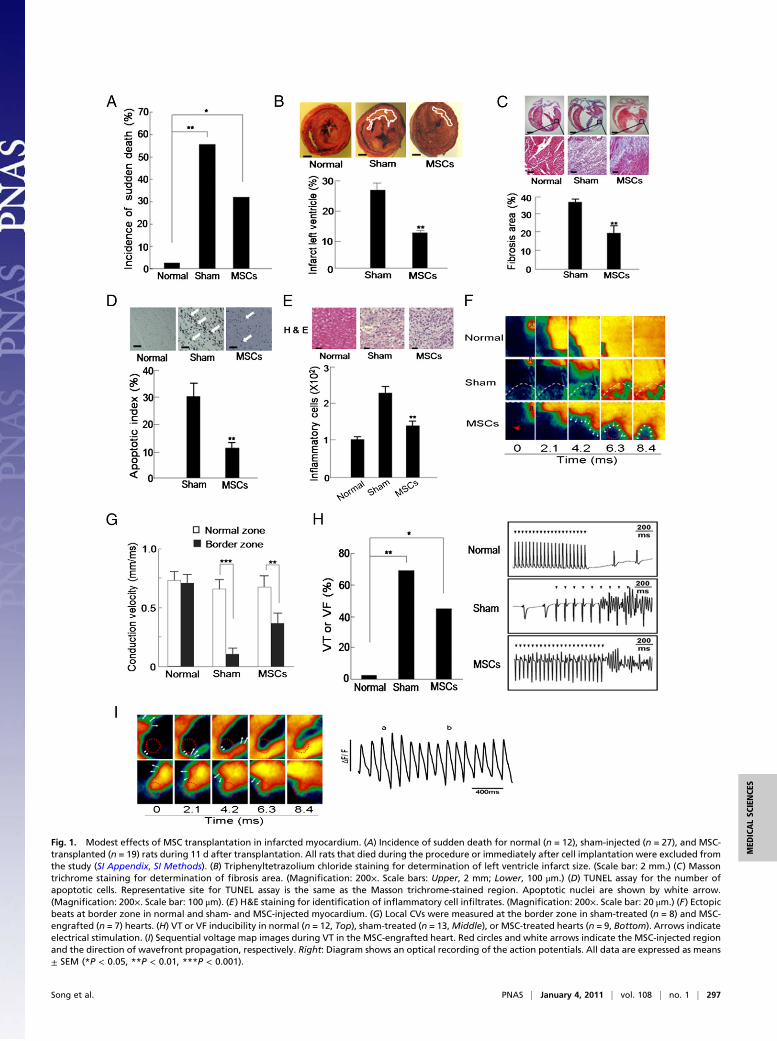
Fig. 1. Modest effects of MSC transplantation in infarcted myocardium. (A) Incidence of sudden death for normal (n = 12), sham-injected (n = 27), and MSC-transplanted (n = 19) rats during 11 d after transplantation. All rats that died during the procedure or immediately after cell implantation were excluded fromthe study (SI Appendix, SI Methods). (B) Triphenyltetrazolium chloride staining for determination of left ventricle infarct size. (Scale bar: 2 mm.) (C) Massontrichrome staining for determination of fibrosis area. (Magnification: 200×. Scale bars: Upper, 2 mm; Lower, 100 μm.) (D) TUNEL assay for the number ofapoptotic cells. Representative site for TUNEL assay is the same as the Masson trichrome-stained region. Apoptotic nuclei are shown by white arrow.(Magnification: 200×. Scale bar: 100 μm). (E) H&E staining for identification of inflammatory cell infiltrates. (Magnification: 200×. Scale bar: 20 μm.) (F) Ectopicbeats at border zone in normal and sham- and MSC-injected myocardium. (G) Local CVs were measured at the border zone in sham-treated (n = 8) and MSC-engrafted (n = 7) hearts. (H) VT or VF inducibility in normal (n = 12, Top), sham-treated (n = 13,Middle), or MSC-treated hearts (n = 9, Bottom). Arrows indicateelectrical stimulation. (I) Sequential voltage map images during VT in the MSC-engrafted heart. Red circles and white arrows indicate the MSC-injected regionand the direction of wavefront propagation, respectively. Right: Diagram shows an optical recording of the action potentials. All data are expressed as means± SEM (*P < 0.05, **P < 0.01, ***P < 0.001).
Song et al. PNAS | January 4, 2011 | vol. 108 | no. 1 | 297
MED
ICALSC
IENCE
S

dium quantitatively. Although histological analysis showed thatinfarct size, fibrosis, and the number of apoptotic cells induced byischemia were significantly decreased in theMSC-engrafted regioncompared with those in the sham-injected region (Fig. 1 B–D),these recoveries did not reach the level of the noninfarcted regionand more than 50% of damages still remained in the regionimplanted with MSCs. In fact, remaining fibrosis may lead tocontinued electrical disturbances (5). We also observed that theMSC-injected group had fewer inflammatory cell infiltrates in theborder region than did the sham controls (Fig. 1E). In addition,cardiac dimensions and systolic performances as measured by leftventricular catheterization were better in MSC-injected rats (SIAppendix, Figs. S1 and S2). Despite histological and functionalimprovements 7 and 11 d after transplantation, respectively, mostof the engrafted MSCs did not express cardiac troponin T (cTnT),
suggesting that they were not yet differentiated into cardio-myocytes, whereby they might induce reentrant arrhythmia byacting as a current sink.
Alleviation of Electrical Vulnerability by MSCs Is Poor to Moderate.We further evaluated the effect of MSCs on the electrical sta-bility of the infarcted heart through optical mapping usingLangendorff perfusion and an electrical vulnerability test innormal, sham-injected, and MSC-engrafted hearts 11 d afterinjury and treatment.In MSC-engrafted hearts, action potentials displayed in-
homogeneous and slow propagation into the infarct zone (Fig.1F). Local CV from the activation time points of the actionpotentials was still depressed in the MSC-engrafted regioncompared with that of the noninfarcted region (Fig. 1G). In an
Fig. 2. Characterization of ccMSCs. (A) Principal component analysis (PCA) for small molecule-induced modification of MSCs. We obtained the coordinates ofchemicals and target cell types by using the first two principal components from PCA and scaled the two sets of coordinates to plot them together in the map.The two largest principal components of PCA analysis are represented as PC1 and PC2. The spheres attached to the longer vectors are more efficient dif-ferentiation inducers for the cell types. The chemicals tested are indicated by green spheres (PMA is shown in blue) and the specific cell lineages are shown asred arrows. Tested molecules are from Calbiochem and are distinguished by numerals as follows: 0, no inhibitor; 1, 1,3-dihydro-1-(1-((4-(6-phenyl-1H-imidazo[4,5-g]quinoxalin-7-yl)phenyl)methyl)-4-piperidinyl)-2H-benzimidazol-2-one; 2, 1L6-hydroxymethyl-chiroinositol-2-(R)-2-O-methyl-3-O-octadecyl-sn-glycer-ocarbonate; 3, SH-5 ([(2R)-2-methoxy-3-octadecoxypropyl] (2,3,4-trihydroxy-6-methoxycyclohexyl) hydrogen phosphate); 4, lavendustin (5-(N-2′,5′-dihydrox-ybenzyl) aminosalicylic acid); 6, PMA (phorbol 12-myristate 13-acetate); 7, DMAT (2-dimethylamino-4,5,6,7-tetrabromo-1H-benzimidazole); 8, D4476 (4-(4-(2,3-dihydrobenzo[1,4]dioxin-6-yl)-5-pyridin-2-yl-1H-imidazol-2-yl)benzamide); 12, NU6102 [6-cyclohexylmethoxy-2-(4′-sulfamoylanilino)purine]; 13, [3-(pyr-idin-2-yl)-4-(4-quinonyl)]-1Hpyrazole; 17, H-89 [N-[2-((p-bromocinnamyl)amino)ethyl]-5-isoquinolinesulfonamide, 2HCl]; 18, SH-6 ([(2R)-2-methoxy-3-octade-coxypropyl] (2,3,4-trihydroxycyclohexyl) hydrogen phosphate); 21, Gö6983 (2-[1-(3-dimethylaminopropyl)-5-methoxyindol-3-yl]-3-(1H-indol-3-yl)) maleimide;22, guanosine 3′,5′-cyclic monophosphorothioate, β-phenyl-1, N2-etheno-8-bromo-, Rp-isomer, sodium salt; 23, compound 56 (4-[(3-bromophenyl)amino]-6,7-diethoxyquinazoline); 24, SU11652 (5-[(Z)-(5-chloro-2-oxo-1,2-dihydro-3H-indol-3-ylidene)methyl]-N-[2-(diethylamino)ethyl]-2,4-dimethyl-1H-pyrrole-3-car-boxamide); 27, N-(4-pyridyl)-N′-(2,4,6-trichlorophenyl) urea; 30, 4,5-dimethoxy-2-nitrobenzaldehyde; 35, SB 202190 [4-(4-fluorophenyl)-2-(4-hydroxyphenyl)-5-(4-pyridyl)1H-imidazole]; and 43, (5-PHENYL-2-ureido)thiophene-3-carboxamide. (B) Immunocytochemical determination for the altered expression of theMSC-specific marker CD90 and cardiac-specific markers cTnT, myosin light chain (MLC), myosin heavy chain (MHC), and Cx43 in control MSCs and the ccMSCs atthe designated days after treatment. (Scale bar: 50 μm. Magnification: 400×.) (C) Immunocytochemical analysis for the homogenic characterization of theccMSCs. Blue indicates nuclei and green is FITC for specific cardiac markers. (Magnification: 100×. Scale bar: 250 μm.) (D) Functional behavior of the ccMSCs byNE stimulus. (E) Blocking of NE-induced hypertrophic signals by prazosin. (F) Altered expression of Ca2+ homeostasis-related proteins sarcoplasmic reticulumCa2+ ATPase (SERCA 2a) and L-type Ca2+ channel (LTCC) in MSCs and the ccMSCs. All data are expressed as means ± SEM (*P < 0.05, **P < 0.01).
298 | www.pnas.org/cgi/doi/10.1073/pnas.1015873107 Song et al.

Fig. 3. ccMSCs overcome postinfarct arrhythmia and mortality through electromechanical incorporation. (A) Immunohistochemical analysis betweeninjected cells and the surrounded cardiomyocytes in the infarcted region. (Magnification: 200×. Scale bar: 100 μm.) (B) Representative examples of CV maps inMSC- and ccMSC-engrafted regions (white circle, 78 μm × 78 μm). (C) Real heart image and CV vector in MSC-engrafted (Left) and ccMSC-engrafted hearts(Right). Red boxes indicate cell-engrafted regions. Black boxes indicate optical view fields (10 mm × 10 mm). (D) Triphenyltetrazolium chloride staining fordetermination of left ventricle infarct size. (Scale bar: 2 mm.) (F) Masson trichrome staining for determination of fibrosis area (Magnification: 200×. Scale bars:Upper, 2 mm; Lower, 100 μm.) (E) TUNEL assay for the number of apoptotic cells. (Magnification: 200×. Scale bar: 100 μm.) (G) Immunostaining for fibroticmarkers collagen I, fibronectin, and α-smooth muscle actin (α-SM actin). (Magnification: 200×. Scale bar: 50 μm.) (H) H&E staining for identification of in-flammatory cell infiltrates. (Magnification: 200×. Scale bar: 20 μm.) (I) Surface ECG recording (lead II) from MSC- and ccMSC-engrafted rats. (J) Representativeleft ventricular pressure volume loops in ccMSC-engrafted rats. End-systolic pressure–volume relationship (ESPVR) during preload reduction is indicated by thedashed line. (K) Kaplan-Meier survival curves for normal (n = 12), sham-injected (n = 27), MSC-engrafted (n = 19), and ccMSC-engrafted rats (n = 12) 11 d aftertransplantation. All rats that died during the procedure or immediately after cell implantation were excluded from the study (SI Appendix, SI Methods). Alldata are expressed as means ± SEM (*P < 0.05, **P < 0.01).
Song et al. PNAS | January 4, 2011 | vol. 108 | no. 1 | 299
MED
ICALSC
IENCE
S

electrical vulnerability test using the burst pacing protocol, ven-tricular tachycardia (VT) or ventricular fibrillation (VF) wereinduced in 69.2% of sham-operated rats (n = 13) and in 44.4%of the MSC-engrafted group (n = 9; P = 0.38; Fig. 1H), sug-gesting only moderate prevention of inducible reentrant ven-tricular arrhythmia by MSC transplantation. The MSC-engraftedsite acts as an electroanatomical substrate for reentrant ar-rhythmia evoked by electrical stimulation (Fig. 1I). Therefore,modification of MSCs to cell types that are able to electrome-chanically synchronize with the surrounding myocardium beforetransplantation might be a better strategy for reducing tissueheterogeneity and to lower the incidence of VT.
PMA-Activated MSCs Exhibit Cardiogenic Properties. In a previousstudy, we showed that small chemical molecules, including pro-tein kinase inhibitors, recognizably change stem cell fate (13).After screening 189 such small chemicals, we found that PMA,a PKC activator, specifically induced the expression of cardio-genic markers as revealed by sandwich ELISA (Fig. 2A). Im-munocytochemical staining showed increased fluorescence ofcardiac-specific markers such as cTnT, myosin light chain, andmyosin heavy chain, but levels of the MSC-specific marker CD90were lower after 9 d in the ccMSCs produced by PMA (Fig. 2B),which were consistent with the results of sandwich ELISA thatthe expression levels were significantly increased with time-dependent manner (SI Appendix, Fig. S3). In addition, we ob-served that the expression of the cardiac-specific transcriptionfactor NkX2.5 was also significantly up-regulated in ccMSCs onsandwich ELISA (SI Appendix, Fig. S3). Interestingly, the fluo-rescence of connexin 43 (Cx43), a gap junction, was markedlyincreased in the ccMSCs (Fig. 2B). We observed that the cardiacphenotypes were similarly up-regulated in the ccMSCs (Fig. 2C),indicating that PMA endows MSCs with more homogeneouscardiogenic properties. In addition, the expressions of adrenergicand muscarinic receptors, which play critical roles in modulatingcardiac functions such as heart rate and myocardial contractility(14–16), were significantly up-regulated in the ccMSCs com-pared with naive MSCs, except for α1D, which is consistent withother results (17) (SI Appendix, Fig. S4 A and C). We furtherobserved that phosphorylated ERK1/2 levels were significantlyincreased by norepinephrine (NE) in a time-dependent mannerin both the ccMSCs and cardiomyocytes, but not in controlMSCs that were blocked by prazosin, an α-adrenoreceptorblocker (Fig. 2 D and E), indicating that the ccMSCs are func-tionally active cells like cardiomyocytes. The expressions of sar-coplasmic reticulum Ca2+ ATPase and L-type Ca2+ channel,which are closely related to excitation–contraction coupling op-erated by Ca2+ influx in cardiomyocytes (18), were also signifi-cantly increased in the ccMSCs, but not in control MSCs, ina time-dependent manner (Fig. 2F). Collectively, these findingssuggest that the ccMSCs are involved in the differentiating stageinto cardiomyocytes or mature cardiomyocytes. This observationsuggests that the activation by PMA triggers a cascade of tran-scriptional activation that regulates the differentiation of MSCsinto cardiomyocytes.
ccMSC Engraftment Enhances Electrical Stability. We investigatedthe expression pattern of Cx43 in MSC- or ccMSC-engraftedregions because the incidence of VT is critically protected by theexpression of Cx43 (19). We observed that Cx43 was expressed ina punctate fashion predominantly throughout the plasma mem-brane in the MSC-engrafted region, whereas Cx43 was detectedmainly as plaques in the ccMSC-engrafted region, which in-dicated that the engrafted ccMSCs were well coupled with hostcardiomyocytes through Cx43 (Fig. 3A). We further examinedthe electrical stability of cells in the ccMSC-engrafted hearts. Onoptical mapping, wave propagation during programmed electricalstimulation from the noninfarct site of the left ventricle base wasmarkedly improved in the ccMSC-engrafted hearts. CV mapsrevealed marked restoration of CV in the ccMSC-engraftedregion with recovery of local CV (0.71 ± 0.12 mm/ms; n = 6),
in contrast to partial restoration of CV in the MSC-engraftedregion (0.38 ± 0.08 mm/ms; n = 7; Fig. 3B). The direction of theCV vector was homogeneous in the ccMSC-engrafted site com-pared with the MSC-engrafted site (Fig. 3C). In an electricalvulnerability test using a burst pacing protocol, VT induction wasprofoundly suppressed in the ccMSC-engrafted group [13% (n =8) and 69.2% (n = 13) for ccMSC- and sham-injected groups,respectively].
ccMSC Engraftment Improves Cardiac Remodeling. As the restora-tion of CV and related arrhythmia protection results from ad-ditional paracrine effects of the ccMSCs on the surroundingtissue, we examined fibrosis, apoptosis, and inflammation in theengrafted region. Histological analysis showed that infarct sizeand interstitial fibrosis were markedly decreased in the ccMSC-injected rats compared with those in MSC-injected rats (Fig. 3 Dand E). In addition, the number of apoptotic cells induced byischemia in the transplanted region was significantly lower in theccMSC-injected animals than in MSC-injected animals (Fig. 3F).The levels of immunostained collagen I and fibronectin de-creased, whereas α-smooth muscle actin increased in the ccMSC-injected region compared with the MSC-injected region (Fig.3G). We also observed that the ccMSC-engrafted region hadfewer inflammatory cell infiltrates than did the MSC region (Fig.3H). Moreover, we observed that the levels of proinflammatorycytokines such as IFN-γ, IL-1a, IL-1b, IL-6, and TNF-α that areincreased by MI were significantly lower in the ccMSC-injectedregion than in the MSC-injected region (SI Appendix, Fig. S5 andTable S1). These results indicate that paracrine effects of theccMSCs in injured myocardium could contribute to the elimi-nation of tissue heterogeneity and, consequently, lead to CVrestoration.
ccMSC Engraftment Prevents Sudden Deaths in Infarcted Rats. Sur-face six-lead ECG showed a shorter QRS duration in the ccMSC-engrafted rats compared with sham-injected and MSC-engraftedrats (Fig. 3I and SI Appendix, Table S2), indicating more homo-geneous ventricular activation of the ccMSC-engrafted myocar-dium. In baseline surface ECG, premature ventricular contractions(PVCs) did not occur in the ccMSC-engrafted rats (0%; n = 12).To assess the electrical stability of the ccMSCs to catecholamin-ergic stimulation, we investigated the incidence of PVCs duringsystemic administration of isoproterenol. Catecholaminergic stim-ulation did not increase the occurrence of PVCs in the ccMSC-engrafted rats (SI Appendix, Fig. S6). The functional effects of theccMSCs on infarcted hearts were evaluated by catheterization 21d after injury and transplantation compared with those of MSC-engrafted hearts. Pressure-volume loop analyses showed less leftventricular dilation in the ccMSC-engrafted group compared withthe MSC-engrafted group (Fig. 3J). The ccMSC transplantationresulted in a better catheterization-determined ejection fractionand a steeper slope of the end-systolic pressure–volume relation-ship, suggesting cardiac regeneration through the ccMSC engraft-ment (Fig. 3J and SI Appendix, Fig. S2). Ultimately, sudden deathswere markedly reduced in the ccMSC-transplanted rats in thisstudy (Fig. 3K).
DiscussionMSCs offer several potential advantages over other types of stemcells for cardiac repair, but they still face several challenges thatneed to be addressed in preclinical studies, such as delivery, sur-vival after transplantation, and electromechanical integration andsafety (5). Although preconditioning of MSCs, including geneticmodification, has increased their therapeutic potency (6–8, 20),the most obvious concern for clinical applications is how engraftedMSCs electromechanically integrate with host tissue (12).In this report, we show that MSC-engrafted regions can func-
tion as electroanatomical substrates for the initiation of re-entrant arrhythmia. However, remaining fibrosis in the MSC-engrafted region may lead to continued electrical disturbances,which is supported by reports that dense fibrosis presents a for-
300 | www.pnas.org/cgi/doi/10.1073/pnas.1015873107 Song et al.

midable physical barrier to cell regeneration (5) and acts as anelectrical barrier that disturbs direct wave propagation, facili-tating reentry (21, 22). Moreover, excessive inflammation in theMSC-engrafted region might also cause inhomogenous conduc-tion and delayed repolarization (23, 24) as well as preventing therecruitment and survival of progenitor cells (25). Inexcitableproperties of undifferentiated MSCs contribute to decreases inCV, increasing the susceptibility to ventricular arrhythmia andleading to sudden death. In fact, the results that action potentialsdisplayed inhomogeneous and slow propagation into the infarctzone in MSC-engrafted hearts indicate that the conditions re-quired to elicit stable reentrant circuit movement were noteliminated (26). The depressed local CV in the MSC-engraftedregion may be attributable to the inexcitable nature of MSCs andtheir ability to act as a current sink (9, 12).To create a cell type that is able to synchronize with sur-
rounding cardiomyocytes electromechanically, we treated MSCswith PMA. This approach was inspired by the results from ourprevious study that small molecules, including protein kinaseinhibitors, can change the fates of stem cells in recognizable ways(13). We found that PMA can induce the expression of cardio-genic markers. We showed that the transplantation of a uniquecell type from MSCs exhibiting cardiogenic properties into theinfarcted heart could help avoid problems caused by heteroge-neity between the implanted cells and the myocardium, leadingto further improvements in contractile function and electricalsafety. Moreover, our result that Cx43 expression was markedlyincreased in the ccMSCs is supported by the result that Cx43-expressing cells prevent postinfarct arrhythmia (19). It is alsoreported that NE influences the contractile properties of theheart and induces a series of changes characteristic of the hy-pertrophic phenotype through α-adrenergic receptors in car-diomyocytes (14).Nevertheless, it has to be further evaluated whether the im-
plantation of ccMSCs may have additional beneficial effects thatprevent other modes of sudden death, such as pump failure orasystole, because recent data showed that sudden death afteracute MI in human patients may be a result of multiple different
causes (10), even though arrhythmic death as a result of VT/VFis a possible cause of sudden death.Ultimately, the application of this cell type may facilitate the
prevention of sudden death caused by naive MSC transplantation,providing new strategies for enhancing electromechanical in-tegration in cell-based therapy for MI. In addition, MSCs can bemodified by PMA into cardiomyocytes that may better integratewith the electromechanisms of host tissue after engraftment, rep-resenting a promising therapeutic strategy for the clinical prepa-ration of MSCs for transplantation to the infarcted myocardium.We recognize that the differentiation of MSCs to cardiomy-
ocytes involves elaborate orchestration of multiple signalingpathways and feedback circuits, and that PMA may, in vivo,modulate activities of molecules other than PKC. We present ourresults here because of their potential importance toward thedevelopment of clinically valuable cell types for cell therapy ofMI. We plan to pursue more detailed studies on the molecularmechanisms of PMA and other chemicals that induce conversionof MSCs to ccMSCs.
MethodsEx Vivo Modification of MSCs. At passage 2, MSCs were seeded in 60-mmplates at 2 × 10⁵ cells/mL and treated with 1 μM PMA (Sigma) at a finalconcentration. The media were replaced with fresh PMA-containing mediaevery 3 d for a maximum of 9 d.
Further detailed information on reagents and antibodies, cell culture,induction of MI and cell transplantation, histological analysis, optical map-ping, catheterization, immunofluorescence, and other methods is given inSI Appendix, SI Methods.
ACKNOWLEDGMENTS. This research was supported by Korea Science andEngineering Foundation Grant M106410200010 6N410200110 funded byMinistry of Education, Science and Technology (MOEST), Republic of Korea;Grant SC-2150 from the Stem Cell Research Center of the 21st CenturyFrontier Research Program funded by MOEST, through the WCU Project(R31-2008-000-10086-0); and Grant A085136 from the Korea Health 21Research and Development Project, Ministry of Health andWelfare, Republicof Korea.
1. Burt RK, et al. (2008) Clinical applications of blood-derived and marrow-derived stemcells for nonmalignant diseases. JAMA 299:925–936.
2. Vulliet PR, Greeley M, Halloran SM, MacDonald KA, Kittleson MD (2004) Intra-coronary arterial injection of mesenchymal stromal cells and microinfarction in dogs.Lancet 363:783–784.
3. Perin EC, et al. (2003) Transendocardial, autologous bone marrow cell transplantationfor severe, chronic ischemic heart failure. Circulation 107:2294–2302.
4. Wollert KC, et al. (2004) Intracoronary autologous bone-marrow cell transfer aftermyocardial infarction: the BOOST randomised controlled clinical trial. Lancet 364:141–148.
5. Segers VF, Lee RT (2008) Stem-cell therapy for cardiac disease. Nature 451:937–942.6. Song H, et al. (2007) Tissue transglutaminase is essential for integrin-mediated
survival of bone marrow-derived mesenchymal stem cells. Stem Cells 25:1431–1438.7. Chang W, et al. (2009) Mesenchymal stem cells pretreated with delivered Hph-1-
Hsp70 protein are protected from hypoxia-mediated cell death and rescue heartfunctions from myocardial injury. Stem Cells 27:2283–2292.
8. Song SW, et al. (2009) Integrin-linked kinase is required in hypoxic mesenchymal stemcells for strengthening cell adhesion to ischemic myocardium. Stem Cells 27:1358–1365.
9. Beeres SL, et al. (2007) Electrophysiological and arrhythmogenic effects of intra-myocardial bone marrow cell injection in patients with chronic ischemic heart disease.Heart Rhythm 4:257–265.
10. Pouleur AC, et al.; VALIANT Investigators (2010) Pathogenesis of sudden unexpecteddeath in a clinical trial of patients with myocardial infarction and left ventriculardysfunction, heart failure, or both. Circulation 122:597–602.
11. Laflamme MA, Murry CE (2005) Regenerating the heart. Nat Biotechnol 23:845–856.12. Chang MG, et al. (2006) Proarrhythmic potential of mesenchymal stem cell
transplantation revealed in an in vitro coculture model. Circulation 113:1832–1841.13. Hwang KC, et al. (2008) Chemicals that modulate stem cell differentiation. Proc Natl
Acad Sci USA 105:7467–7471.
14. Chien KR, Knowlton KU, Zhu H, Chien S (1991) Regulation of cardiac gene expressionduring myocardial growth and hypertrophy: molecular studies of an adaptivephysiologic response. FASEB J 5:3037–3046.
15. Hosey MM (1992) Diversity of structure, signaling and regulation within the family ofmuscarinic cholinergic receptors. FASEB J 6:845–852.
16. Steinberg SF (1999) The molecular basis for distinct beta-adrenergic receptor subtypeactions in cardiomyocytes. Circ Res 85:1101–1111.
17. Hakuno D, et al. (2002) Bone marrow-derived regenerated cardiomyocytes (CMG Cells)express functional adrenergic and muscarinic receptors. Circulation 105:380–386.
18. Shin SY, Choo SM, Woo SH, Cho KH (2008) Cardiac systems biology and parametersensitivity analysis: Intracellular Ca2+ regulatory mechanisms in mouse ventricularmyocytes. Adv Biochem Eng Biotechnol 110:25–45.
19. Roell W, et al. (2007) Engraftment of connexin 43-expressing cells prevents post-infarct arrhythmia. Nature 450:819–824.
20. Mangi AA, et al. (2003) Mesenchymal stem cells modified with Akt preventremodeling and restore performance of infarcted hearts. Nat Med 9:1195–1201.
21. de Bakker JM, et al. (1993) Slow conduction in the infarcted human heart. ‘Zigzag’course of activation. Circulation 88:915–926.
22. Anderson KP, et al. (1993) Myocardial electrical propagation in patients withidiopathic dilated cardiomyopathy. J Clin Invest 92:122–140.
23. Hoffman BF, Feinmark SJ, Guo SD (1997) Electrophysiologic effects of interactionsbetween activated canine neutrophils and cardiac myocytes. J Cardiovasc Electro-physiol 8:679–687.
24. Ishii Y, et al. (2005) Inflammation of atrium after cardiac surgery is associated withinhomogeneity of atrial conduction and atrial fibrillation. Circulation 111:2881–2888.
25. Poss KD, Wilson LG, Keating MT (2002) Heart regeneration in zebrafish. Science 298:2188–2190.
26. Takahashi T, et al. (2004) Optical mapping of the functional reentrant circuit ofventricular tachycardia in acute myocardial infarction. Heart Rhythm 1:451–459.
Song et al. PNAS | January 4, 2011 | vol. 108 | no. 1 | 301
MED
ICALSC
IENCE
S